HAL Id: tel-01343859
https://tel.archives-ouvertes.fr/tel-01343859
Submitted on 10 Jul 2016
HAL is a multi-disciplinary open access archive for the deposit and dissemination of sci-entific research documents, whether they are pub-lished or not. The documents may come from teaching and research institutions in France or abroad, or from public or private research centers.
L’archive ouverte pluridisciplinaire HAL, est destinée au dépôt et à la diffusion de documents scientifiques de niveau recherche, publiés ou non, émanant des établissements d’enseignement et de recherche français ou étrangers, des laboratoires publics ou privés.
Structural studies of microbial rhodopsins and other
membrane proteins by means of X-ray crystallography
and computer modeling
Ivan Yu. Gushchin
To cite this version:
Ivan Yu. Gushchin. Structural studies of microbial rhodopsins and other membrane proteins by means of X-ray crystallography and computer modeling. Biomolecules [q-bio.BM]. Université de Grenoble, 2014. English. �NNT : 2014GRENY075�. �tel-01343859�
Université Joseph Fourier / Université Pierre Mendès France / Université Stendhal / Université de Savoie / Grenoble INP
THÈSE
Pour obtenir le grade de
DOCTEUR DE L’UNIVERSITÉ DE GRENOBLE
Spécialité : Physique pour les sciences du vivant Arrêté ministériel : 7 août 2006
Présentée par
Ivan GUSHCHIN
Thèse dirigée par Prof. Dr. Valentin GORDELIY codirigée par Dr. Sergei GRUDININ
préparée au sein du Laboratoire des Transporteurs Membranaires de l’Institut de Biologie Structurale dans l'École Doctorale de Physique
Études structurales des rhodopsines
microbiennes et des autres
protéines membranaires au moyen
de la cristallographie aux rayons X
et de la modélisation informatique
Thèse soutenue publiquement le 5 Septembre 2014, devant le jury composé de :
Prof. Dr. Valentin GORDELIY Directeur de thèse
Directeur de Recherche CEA, Institut de Biologie Structurale, Grenoble, France
Dr. Sergei GRUDININ Co-encadrant de thèse
Chargé de Recherche CNRS, Inria, Grenoble, France
Prof. Dr. Georg BÜLDT Rapporteur
Head of the Laboratory of Prospective Studies of Membrane Proteins, Moscow Institute of Physics and Technology, Moscow, Russia
Prof. Dr. Dieter WILLBOLD Rapporteur
Director of the Institute of Complex Systems-6, Structural Biochemistry, Research Center Jülich, Jülich, Germany
Prof. Dr. Martin ENGELHARD Examinateur
Research group leader at the Max Planck Institute of Molecular Physiology, Dortmund, Germany
Prof. Dr. Eva PEBAY-PEYROULA President du jury
1
Contents
Contents ... 1 Abbreviations ... 6 Abstract ... 8 Résumé ... 10 1 Introduction ... 12 1.1 Microbial rhodopsins ... 121.1.1 Description of the family ... 12
1.1.2 Bacteriorhodopsin: historical perspective ... 12
1.1.3 Bacteriorhodopsin: structure and function ... 15
1.1.4 Microbial rhodopsins ... 16
1.1.5 Structures and relationship between microbial rhodopsins ... 17
1.1.6 Haloarcula marismortui bacteriorhodopsin I ... 21
1.1.7 Exiguobacterium sibiricum rhodopsin ... 22
1.1.8 Light-driven sodium pump KR2 ... 23
1.2 Photo- and chemotaxis ... 24
1.2.1 Bacterial and archaeal taxis ... 24
1.2.2 Principles of microbial locomotion ... 24
1.2.3 Regulation of flagella by chemo- and photoreceptors... 25
1.2.4 Spatial organization of chemotaxis system ... 27
1.2.5 CheA and CheW proteins ... 27
1.2.6 Architecture of chemoreceptors and sensory rhodopsin-transducer complexes ... 29
1.2.7 Signal generation in chemoreceptors ... 30
1.2.8 Signal generation in the phototaxis system ... 32
2
1.3 CDP-OH transferases and IPCT-DIPPS ... 36
1.3.1 CDP-OH transferases ... 36
1.3.2 Di-myo-inositol phosphate and the IPCT-DIPPS enzymes ... 37
2 Materials and methods ... 40
2.1 Protein expression and purification ... 40
2.1.1 Expression and purification of HmBRI ... 40
2.1.2 Expression and purification of ESR ... 40
2.1.3 Expression and purification of KR2 ... 41
2.1.4 Expression and purification of NpSRII ... 41
2.1.5 Expression and purification of IPCT-DIPPS ... 41
2.2 Crystallization details ... 42
2.3 Data collection and processing ... 44
2.4 Crystallographic structure determination ... 44
2.4.1 HmBRI structure determination ... 44
2.4.2 ESR structure determination ... 45
2.4.3 KR2 structure determination ... 45
2.4.4 NpSRII structure determination ... 45
2.4.5 IPCT-DIPPS structure determination ... 47
2.5 Details of the modeling studies: simulations of the NpHtrII HAMP domain region ... 48
2.5.1 Initial data and models ... 48
2.5.2 Molecular dynamics simulations ... 48
2.5.3 Listing of the simulated systems ... 49
2.5.4 Evaluation of structure stability of the inter-HAMP and HAMP domain regions in MD simulations ... 51
2.5.3 Longitudinal shift calculations ... 55
3
2.5.5 HAMP domain axis calculation ... 55
2.5.6 Protonation state of the ionizable residues ... 55
2.6 Details of the modeling studies: simulations of the NpHtrII first HAMP domain ... 57
2.6.1 Initial model preparation ... 57
2.6.2 Molecular dynamics simulations ... 57
2.6.3 Listing of the simulated systems ... 58
2.6.4 Principal components analysis ... 58
2.7 Details of the IPCT-DIPPS modeling studies ... 59
2.7.1 Modeling of the soluble domain dimerization ... 59
2.7.2 Docking of the IPCT-DIPPS ligands ... 59
3 Results and discussion ... 61
3.1 Crystal structure of Escherichia coli-expressed Haloarcula marismortui bacteriorhodopsin I in the trimeric form ... 61
3.1.1 Crystallization and crystal packing of HmBRI ... 61
3.1.2 Structure of HmBRI ... 63
3.1.3 Novel inter-helical hydrogen bonds in HmBRI ... 65
3.1.4 HmBRI trimers and its D-E loop ... 67
3.1.5 Lipid molecules around HmBRI ... 68
3.1.6 Conclusions ... 69
3.2 Crystal structure of Exiguobacterium sibiricum rhodopsin ... 70
3.2.1 Crystallographic structure of ESR and its characteristic features ... 70
3.2.2 Structure of the retinal binding pocket, proton release and proton uptake groups. . 77
3.2.3 Proposed model of the photocycle. ... 82
3.3 Crystal structure of the light-driven proton pump KR2 ... 84
3.4 Determination of the Natronomonas pharaonis sensory rhodopsin II active state structure ... 86
4
3.4.2 Procedure of the active state trapping ... 88
3.4.3 Spectroscopic characterization of the intermediate state occupancy ... 89
3.4.4 Determination of the intermediate occupancy by crystallographic methods ... 90
3.4.5 Heterogeneity of the NpSRII active state ... 92
3.4.6 Conformational changes in the NpSRII active state ... 92
3.4.7 Functionally important conformational changes in NpSRII ... 96
3.4.8 Implications for NpSRII proton pumping ... 97
3.5 Modeling of the Natronomonas pharaonis HtrII HAMP domain region ... 98
3.5.1 Model of the inter-HAMP region of SR transducers ... 98
3.5.2 MD simulations of Af1503 and NpHtrII HAMP domains ... 102
3.5.3 MD simulations of the NpHtrII HAMP domain region ... 102
3.5.4 Discussion ... 107
3.6 Two Distinct States of the HAMP Domain from Sensory Rhodopsin Transducer Observed in Unbiased Molecular Dynamics Simulations ... 110
3.6.1 MD simulations of NpHtrII HAMP1. ... 110
3.6.2 Two states of the NpHtrII HAMP domain ... 111
3.6.3 Structural analysis of the two observed states ... 115
3.6.4 Conformation of the F124 pair at the HAMP domain core ... 116
3.6.5 Comparison of the NpHtrII HAMP1 resting and signaling states... 118
3.6.6 Conclusions ... 118
3.7 Crystal structure and modeling of the Archaeoglobus fulgidus IPCT-DIPPS bifunctional enzyme ... 120
3.7.1 Crystallographic data ... 120
3.7.2 Description of the A.f. IPCT-DIPPS structure ... 122
3.7.3 DIPPS active site and ligand docking ... 127
Future work ... 131
5
6
Abbreviations
A.f., Archaeoglobus fulgidus E. coli, Escherichia coli H.m., Haloarcula marismortui H.s., Halobacterium salinarum N.p., Natronomonas pharaonisBR, bacteriorhodopsin ChR, channel rhodopsin
ESR, Exiguobacterium sibiricum rhodopsin GR, Gloeobacter rhodopsin HR, halorhodopsin KR2, Krokinobacter rhodopsin 2 PR, proteorhodopsin SR, sensory rhodopsin XR, xanthorhodopsin
HAMP domain, histidine kinases, adenylyl cyclases, methyl-accepting chemotaxis proteins and phosphatases domain
IPCT, inositol-phosphate cytidylyltransferase DIPPS, di-inositol-phosphate-phosphate synthase
MD, molecular dynamics MR, molecular replacement
RMSD, root mean square deviation UV, ultra-violet (light)
7 This page is intentionally left blank
8
Abstract
Every living cell on Earth is surrounded by a lipid membrane. Proteins residing in the membrane perform a variety of functions crucial for the cell’s survival. Among them are active and passive transport in and out of the cell, signaling and reaction catalysis.
One of the largest membrane protein families are microbial rhodopsins, which utilize light energy for their function. Members of this family count among them light-driven proton, cation and anion pumps, light-gated ion channels and photoreceptors. While the basic aspects of their functioning have been known for some time, there is a plenty of unanswered questions. In this dissertation, several structures of microbial rhodopsins (among them the first proteorhodopsin structure and the first light-driven sodium pump structure) are presented and analyzed. The structures open the way for understanding the similarities and differences between the various microbial rhodopsins and for exploiting this understanding to create better microbial rhodopsin-based instruments for biological applications, for example, in the field of optogenetics.
While the first part of this work deals with the novel structures of microbial rhodopsins, the second part presents the simulation approach for understanding the sensory rhodopsin-based signaling in phototaxis. The HAMP domains of the sensory rhodopsin transducer protein are studied by means of molecular dynamics, and it is demonstrated that the simulations may be used for building and validating the atomic structures of signaling domains, as well as for understanding the signaling-associated conformational changes, initiated by light-driven sensory rhodopsin transformations.
The third and the last part describes the work on the Archaeoglobus fulgidus IPCT-DIPPS proteins, an enzyme catalyzing two consecutive steps of di-inositol-phosphate biosynthesis. The determined structure may serve as a model for understanding the catalytic mechanism of CDP-alcohol transferases, a large family of proteins counting thousands of members, among which are five human proteins that catalyze the major steps of lipid biosynthesis. The structure was also used to predict the binding sites of the ligands at the enzyme active site and to propose the mechanism of catalytic action.
To sum up, this dissertation presents the structural studies of various membrane proteins by means of X-ray crystallography and modeling that advance our understanding of fundamental and practical aspects of membrane protein functioning.
9
The text of the manuscript is partially based on the published works1–4:
Section 3.2 is an adaptation of the article “Structural insights into the proton pumping by unusual proteorhodopsin from nonmarine bacteria” (Proceedings of the National Academy of Sciences of the USA, 2013)1.
Section 3.4 is an adaptation of the article “Active State of Sensory Rhodopsin II: Structural Determinants for Signal Transfer and Proton Pumping” (Journal of Molecular Biology, 2011)3.
Section 3.5 is an adaptation of the article “Role of the HAMP Domain Region of Sensory Rhodopsin Transducers in Signal Transduction” (Biochemistry, 2011)4.
Section 3.6 is an adaptation of the article “Two Distinct States of the HAMP Domain from Sensory Rhodopsin Transducer Observed in Unbiased Molecular Dynamics Simulations” (PLoS ONE, 2013)2.
Finally, section 3.7 is partially adapted from the manuscript “X-ray Structure of a CDP-alcohol phosphatidyltransferase membrane enzyme and insights into its catalytic mechanism” (Nature Communications, accepted).
10
Résumé
Chaque cellule vivante sur notre Terre est entourée d'une membrane lipidique. Les protéines résidant dans la membrane exécutent multitude de fonctions essentielles pour la survivance de la cellule. Parmi eux sont le transport actif et passif dans et hors de la cellule, la signalisation et la catalyse des réactions.
Une des plus grandes familles de protéines membranaires sont rhodopsins microbiennes, qui utilisent l'énergie de la lumière pour leur fonction. Les membres de cette famille comptent parmi eux les pompes de protons, cations et anions, entraînée par l'illumination, les canaux ioniques activés par l'illumination et, finalement, photorécepteurs. Bien que les aspects fondamentaux de leur fonctionnement ont été connus depuis un certain temps, il ya une abondance de questions sans réponse. Dans cette thèse, plusieurs structures de rhodopsines microbiennes (y compris la première structure de protéorhodopsine et la première structure de la pompe à sodium) sont présentés et analysés. Les structures ouvrent la voie pour comprendre les similitudes et les différences entre les différents rhodopsines microbiennes et pour exploiter cette connaissance pour créer de meilleurs instruments à base de rhodopsines microbiennes pour des applications biologiques, par exemple, dans le domaine de optogenetics.
Alors que la première partie de ce travail porte sur les nouvelles structures de rhodopsines microbiennes, la deuxième partie présente l'approche de simulation pour comprendre la signalisation en fonction des rhodopsines sensorielles dans phototaxie. Les domaines HAMP des protéines transductrices des signals des rhodopsines sensorielles sont étudiés au moyen de la dynamique moléculaire, et il est démontré que les simulations peuvent être utilisés pour la construction et la validation des structures atomiques des domaines de signalisation, ainsi que pour la compréhension des changements conformationnels associée à signalisation, initié par les transformations des rhodopsine sensorielles.
La troisième et la dernière partie décrit le travail sur la protéine IPCT-DIPPS de
Archaeoglobus fulgidus, une enzyme catalysant deux étapes consécutives de
di-inositol-phosphate biosynthèse. La structure résolue peut servir de modèle pour comprendre le mécanisme catalytique de transférases CDP-alcool, une grande famille de protéines comptant des milliers de membres, parmi lesquels sont cinq protéines humaines, qui catalysent les étapes majeures de la biosynthèse des lipides. La structure a également été utilisé pour prédire
11
les sites de liaison des ligands sur le site actif de l'enzyme et pour proposer le mécanisme d'action catalytique.
Pour résumer, cette thèse présente les études structurales de diverses protéines membranaires par la cristallographie aux rayons X et la modélisation qui font progresser notre compréhension des aspects fondamentaux et pratiques de fonctionnement des protéines membranaires.
12
1 Introduction
1.1 Microbial rhodopsins
1.1.1 Description of the family
Microbial rhodopsins are a large family of photoactive membrane proteins present in bacteria, archaea, eukaryota and viruses5. The proteins are composed of seven transmembrane α-helices, labeled A to G, three intracellular and three extracellular loops (Figure 1.1.1a). Among them, the B-C loop is usually the longest one and its conformation is varied the most among the different microbial rhodopsins. The unifying feature of microbial rhodopsins is the cofactor retinal (Figure 1.1.1b) that is covalently bound via the Schiff base to the side chain of a lysine residue situated in the middle of the helix G. The retinal imparts the microbial rhodopsins the ability to utilize light for their function.
1.1.2 Bacteriorhodopsin: historical perspective
The first member of the microbial rhodopsins family, and the most studied to date is the light-driven proton pump bacteriorhodopsin from Halobacterium salinarum6. It was discovered in 1971 as the major constituent of the H.s. purple membrane7. Due to its high abundance in the
H.s. membrane and ease of purification, BR can be produced in large quantities. This property
made it the model membrane protein, extensively used for the development of numerous biophysical techniques. One of the first applications of bacteriorhodopsin was in providing the evidence for the Mitchell’s “chemiosmotic theory”, according to which the proton gradient across the cell membrane can be utilized for ATP production8. Later, bacteriorhodopsin served as a model system for development of the electron microscopy techniques, which provided the first glimpse into its structure at the resolution of 7 Å9. After this, the resolution was gradually improved to almost-atomic10,11.
At the same time, bacteriorhodopsin was extensively studied using different optical techniques such as UV-Visual12,13, FTIR14 and resonance Raman15 spectroscopy. It was determined that upon absorption of a light photon the visual light absorption spectrum of bacteriorhodopsin undergoes a series of transformations, called photocycle (Figure 1.1.2a). Based on the absorption spectra, presence of several interconverting quasi-stable states, called K, L, M, N and O (Figure 1.1.2b), was postulated. In the M-state the retinal is in the 13-cis state and deprotonated, and, consequently, the M-state spectrum differs the most from the
13
ground state spectrum. Apart from the main photocycle, there are also states P and Q that can be induced by second photon absorption. Finally, in the absence of illumination, thermal isomerization of the retinal from the all-trans to 13-cis 15-syn conformation is possible that gives rise to a so-called “dark” photocycle, which doesn’t result in a proton translocation.
Figure 1.1.1. General features of the microbial rhodopsin structures. a) Microbial rhodopsin
fold, exemplified by Halobacterium salinarum bacteriorhodopsin16. There are 7 transmembrane helices marked A to G. The B-C loop (blue) is elongated and caps the extracellular side of the protein. Membrane borders calculated using the PPM server17 are shown with the black lines. b) Chemical structure of the all-trans retinal covalently linked to a lysine side chain. There are 20 carbon atoms in the retinal. Usually, the isomerization occurs around the C13-C14 and C15-N double
bonds. The Schiff base bears a positive charge when protonated.
Although bacteriorhodopsin did not become the first membrane protein, for which X-ray crystallographic structure could be obtained (it was the Rhodopseudomonas viridis photosynthetic reaction canter in 198518), experiments with it gave rise to a plethora of nontraditional crystallization techniques, where different lipidic phases are utilized as a
14
crystallization media. Among them are crystallization in the lipidic cubic phase19,20, crystallization from bicelles21 and crystallization from vesicles22. Initially, these crystallization methods were considered too difficult for routine application, however, advent of automatization23 and successful determination of first non-rhodopsin GPCR structure24 resulted in widespread adoption of in meso crystallization. Meanwhile crystallization from bicelles is less known, it also was used successfully for proteins other than microbial rhodopsins such as β2 adrenergic G-protein-coupled receptor25 and respiratory complex I26.
Finally, crystallization from vesicles was used to obtain not-twinned crystals of BR22 and crystals of trimeric ar-227.
Figure 1.1.2 Bacteriorhodopsin photocycle (adapted from Birge et al.28). a) The main
bacteriorhodopsin photocycle starting from the light adapted state. Additional transitions are possible upon absorption of a second photon. b) Absorption spectra of the ground and intermediate states.
Other structural techniques have also been applied to bacteriorhodopsin. Magic angle spinning NMR studies of 13C labeled BR were used to get insight into protonation states of important aspartic acid residues29. Neutron and X-ray diffraction experiments on the normal and deuterated BR samples provided the information about the motions of BR helices during the photocycle30,31. Spin label attachment at the specific BR sites allowed probing of the microenvironment by the electron spin resonance technique32. Finally, various atomic force microscopy (AFM) experiments were performed on BR. While simple AFM experiments corroborated the information about the BR packing in the purple membrane33, advanced high-speed AFM allowed visualization of BR motions in the membrane and assembly of the highly-ordered hexagonal lattice34. Combination of AFM and single-molecule force spectroscopy revealed the unfolding pathways of bacteriorhodopsin35.
15
1.1.3 Bacteriorhodopsin: structure and function
The aforementioned studies resulted in extensive characterization of bacteriorhodopsin and its mechanism of function6. The proton translocation pathway consists of three major checkpoints: the proton uptake site, the Schiff base region and the proton release region (Figure 1.1.3).
Figure 1.1.3. Structure of bacteriorhodopsin (PDB ID 1C3W). The internal cavities are
represented by the red surfaces. The crystallographically observed water molecules are shown as the red spheres. Helix F is not shown.
There is a general agreement on the mechanism of the proton pumping by bacteriorhodopsin. In the ground state the proton is stored at the retinal Schiff base that makes a hydrogen bond to a proximal water molecule W402. W402 is also coordinated by the aspartates 85 and 212. The W402 cavity also contains two other water molecules. The cavity is blocked by the retinal at one side and by the Arg-82 side chain at the other.
16
Absorption of the light photon by the retinal leads to isomerization of the retinal from the
all-trans to 13-cis conformation. In this new position, the protonated state of the Schiff base is
energetically unfavorable, and the proton transitions to the proton acceptor residue Asp-85. After that, the proton needs to pass to the proton release group, consisting of the glutamates 194 and 204. However, this is impossible because of the Arg-82 side chain. In the M-state this side chain turns away from the Schiff base.
As a next step the retinal is reprotonated with the proton stored at the proton uptake site on the Asp-96 side chain. Consequently, Asp-96 needs to be reprotonated from the cytoplasm, which can be possible due to a transient water molecule wire36 or unlatching of the cytoplasmic side37. Reprotonated retinal pushes the Asp-85 proton to Glu-194/204, where it displaces another proton occupying that site, which goes in its turn into the bulk solvent.
Although these general points are agreed upon by most of the researchers, the details of the proton translocation mechanism and the associated conformational changes in particular, are often disputed. For example, there are considerable differences in the crystallographic structures of the intermediate states published by different research groups38. These differences might be a consequence of crystal twinning39 or unusual radiation sensitivity and consequent damage of BR active site40. However, not only particular details but also the amplitudes of structural changes are questioned, as the electron microscopy studies, where the protein motions are supposedly less restrained by the environment, provide a picture of the larger-amplitude motions which are not observed in any of the crystallographic structures41. All these discrepancies are yet to be resolved.
1.1.4 Microbial rhodopsins
While Halobacterium salinarum bacteriorhodopsin is the most studied microbial rhodopsin, many other proteins of this family have been identified. Soon after the discovery of BR, a light-driven chloride pump was identified in the membrane of H.s., dubbed halorhodopsin42,43. Although no other ion pumping proteins were detected in H. salinarum, two light sensors with different functions were found: sensory rhodopsin I, responsible for both attractant and repellent responses44–47, and sensory rhodopsin II, responsible for the repellent response and also known as phoborhodopsin48–51. Similar proteins were also found in other haloalkaliphilic archaea52.
Advent of metagenomics and sequencing of environmental samples that contained uncultivated bacteria marked a new era of microbial rhodopsin research. In the year of 2000, a
17
bacterial rhodopsin gene, coding for a functional protein, was discovered in a genomic fragment from a marine γ-proteobacteria, collected in Californian Monterey Bay53. Soon, it was demonstrated that the protein, dubbed proteorhodopsin, is indeed present in oceanic bacteria54. It also appeared that the proteins from this microbial rhodopsin subfamily were spectrally tuned to different habitats – that is, they absorbed light at different wavelengths in accordance with illumination available in the environment54. Since then, the proteorhodopsin genes were found to be distributed among divergent bacterial taxa55,56, even as peculiar as Siberian permafrost bacteria57, and pump not only protons but also the sodium and chloride ions58,59.
Finally, presence of microbial rhodopsins is not limited to bacteria and archaea as they were also found, for example, in algae60 and giant viruses infecting unicellular eukaryotes61. Algal channelrhodopsins, which open a cation channel upon illumination, became of extreme importance in the recently developed field of optogenetics62 and were extensively modified to achieve some useful properties such as anion conduction63–65.
To sum up, microbial rhodopsins performing a large variety of functions were identified. Among them are light-driven proton pumps, such as bacteriorhodopsin from the archaeon
Halobacterium salinarum7 or proteorhodopsins from marine bacteria53,56, anion pumps such as halorhodopsin66,67 or Nonlabens marinus rhodopsin 359, cation pumps58,68, light-gated ion channels (channelrhodopsins)69–71, and, finally, light-regulated sensory proteins, such as archaeal sensory rhodopsins72 that conduct the signal through a chemoreceptor-like transducer protein, bacterial Anabaena sensory rhodopsin that utilizes a soluble second messenger73, or algal histidine kinase rhodopsins74,75.
1.1.5 Structures and relationship between microbial rhodopsins
In the recent years, atomic structures of various microbial rhodopsins have been elucidated. Although the majority of them come from X-ray crystallography, some structures were determined using solution76 or solid state77 NMR. Comparison of the available structures is presented in the Figure 1.1.4 and the corresponding information is summarized in the Table 1.1.1 (the structures presented in this work were not included). In the situations where several structures are available, only the one corresponding to the ground state and the best resolution is shown. The phylogenetic tree showing the relations between the important microbial rhodopsins is presented in the Figure 1.1.5.
18
Table 1.1.1. Known structures of microbial rhodopsins.
Name of the protein Abbreviation PDB ID Resolution, Å Reference
Bacteriorhodopsin BR, HsBR 1C3W 1.55 Luecke et al.16
Archaerhodopsin 1 aR-1 1UAZ 3.4 Enami et al.78
Archaerhodopsin 2 aR-2 2EI4 2.1 Yoshimura and Kouyama27
Deltarhodopsin 3 dR-3 4FBZ 2.7 Zhang et al.79
Halorhodopsin HR, HsHR 1E12 1.8 Kolbe et al.80
Natronomonas pharaonis
halorhodopsin
NpHR 3A7K 2.0 Kouyama et al. 81
Natronomonas pharaonis
sensory rhodopsin II
NpSRII 1H68 2.1 Royant et al.82
Anabaena sensory rhodopsin ASR 1XIO 2.0 Vogeley et al.73
Acetabularia rhodopsin 2 AR2 3AM6 3.2 Wada et al.83
Xanthorhodopsin XR 3DDL 1.9 Luecke et al.84
Channelrhodopsin ChR 3UG9 2.3 Kato et al.85
Proteorhodopsin from Med12 PR 4JQ6 2.3 Ran et al.86
19
Figure 1.1.4 Superposition of the known microbial rhodopsin structures. The retinal is shown
in green. Channelrhodopsin, whose structure differs the most from other proteins, is shown in blue (structure of the soluble parts is not shown). Most variation is observed in the exposed loop regions and, among the transmembrane helices, in the position of the helix A.
20
Figure 1.1.5. Phylogenetic tree of microbial rhodopsins. PDB IDs are shown in parentheses
where available. The residues at the positions of BR’s proton path-forming D85, D212 and D96, and at the positions of ESR’s helix F kink-forming residues W154 and N224 (Section 3.2.1 and Figure 3.2.2) are shown. pop is a proteorhodopsin gene from the recently described uncultured marine Euryarchaeota87. MacR (marine Actinobacteria rhodopsin) is a protein from ultra-small marine Actinobacteria88. GR is Gloeobacter rhodopsin.
21
1.1.6 Haloarcula marismortui bacteriorhodopsin I
Historically, microbial rhodopsins performing four different physiological functions have been described in archaea. These are proton pumping, chloride pumping, and mediation of light-attractant and/or repellent functions. Correspondingly, four or less microbial rhodopsins are observed in a single species – for example, four in Halobacterium salinarum and three in
Natronomonas pharaonis.
Recently, an unusual six-rhodopsin system of Haloarcula marismortui has been described by Fu and coworkers89. The system comprises two proton pumps (called HmBRI and HmBRII and possessing 50% sequence identity), a chloride pump, sensory rhodopsins I and II and an additional sensory rhodopsin of a novel type. Interestingly, absorption maximum of both
HmBRI and HmBRII is at 552 nm. It is not clear, why two proton pumps absorbing at the
same wavelength are needed, however it is possible that the proteins are optimized for functioning at different pH90.
HmBRI could be expressed in Escherichia coli with a high expression level, and the D94N
mutation allowed to raise it to 70 mg per 1 liter of culture91. Such a high yield allowed using
HmBRI as an expression tag for efficient production of other membrane proteins91. As E. coli production allows for easy genetic manipulation and rapid production of mutants, HmBRI could become a convenient model for the proton transport mechanism studies as well as for protein engineering for optogenetics needs62.
Section 3.1 of this work describes the crystal structure of the HmBRI D94N mutant. The structure is interesting as, despite the expression in Escherichia coli, the protein still assembles as a trimer, and the E. coli lipids occupy the positions very similar to that of archaeal H. salinarum lipids in HsBR structures.
22
1.1.7 Exiguobacterium sibiricum rhodopsin
Permafrost soil is a soil that remains at or below the freezing point of water for extended periods of time. High numbers of microorganisms such as fungi, yeast, algae, actinomycetes, and bacteria have been discovered in the Siberian permafrost samples. Recently, one particular member of this unique microbial community, called Exiguobacterium sibiricum, has been isolated from 3 million years-old samples and extensively characterized92. E.s. was found to be adapted to the harsh conditions of the permafrost as it can grow at the temperatures as low as -5º C (the full range of growth temperatures is -5º C to +39º C)93. At the same time, this adaptation appears to be constitutive as little differential gene expression is observed between 4º C and 28º C93.
Interestingly, sequencing of the E.s. genome has revealed presence of potential proteorhodopsin gene93. This protein, called E.s. rhodopsin (ESR), could be expressed in
E. coli, bound the all-trans retinal and displayed an absorption maximum at 534 nm57. ESR, incorporated in vesicles, could efficiently transport the protons across the membrane under light illumination57.
Compared to other microbial rhodopsins, ESR revealed two notable features. First, its proton uptake residue is a lysine, which can only be positively ionized, while all the other proton pumps studied to date possess a negatively ionizable amino acid (Asp or Glu) at that position94. This fact bears qualitative consequences for the proton pump function, as the proton uptake group is neutral in the ground state, thus it is protonated in the classical proton pumps and deprotonated in ESR. Recently, several other proteorhodopsins also having a lysine residue at the proton uptake have been found in marine uncultured Euryarchaeota87 (pop gene, Figure 1.1.5). The second unique quality of ESR is its broad proton pumping pH range - above the pH of 4.595. This feature was ascribed to the retinal-proximal pair of residues Asp-85 and His-5795, which are, however, also observed in xanthorhodopsin and proteorhodopsins.
ESR presented an interesting target for a study as, first, it is an unusual protein from a psychrophilic bacterium, and second, no atomic three-dimensional proteorhodopsin structure has been known at the beginning of the study. Its structure is described in the Section 3.2.
23
1.1.8 Light-driven sodium pump KR2
While microbial rhodopsins acting as anion or proton pumps were known for a long time, the light-driven sodium pumps remained elusive. This elusiveness could even be rationalized by speculating that the positively charged protonated retinal Schiff base displaces a proton during its isomerization, and can displace an anion, pulling it by Coulombic attraction, but cannot move a cation as there is no suitable interaction.
Recently, a novel class of microbial rhodopsins, called NQ-rhodopsins, was described68. The name of the proteins comes from the fact that the retinal counterion position (D85 in bacteriorhodopsin) is occupied by asparagine in this family, and proton uptake (D96 in bacteriorhodopsin) is occupied by glutamine (Figure 1.1.5). Despite these seemingly destructive substitutions the proteins were still able to pump protons68.
Soon thereafter, Inoue and colleagues58 studied the rhodopsins 1 and 2 of the bacterium
Krokinobacter eikastus (Figure 1.1.6a) and showed that, while KR1 is a typical
proton-pumping proteorhodopsin, KR2, a member of the NQ-rhodopsins subfamily, is a light-driven sodium pump, which can pump protons in absence of sodium. In both cases, the photocycle consists of several bacteriorhodopsin-like intermediates (K, L, M and O, Figure 1.1.6b), although there are slight differences probably explained by the fact that KR2 pumps preferably sodium and in its absence the turnover is slower and the pump is less efficient. The protein was also found to be very selective, pumping the Li+ and Na+ ions but not K+, Rb+ or Cs+.
The unique properties of KR2 could potentially be very useful in the field of optogenetics62, and knowledge of its structure could open the way to rationalization of its pumping and generation of, for example, more effective or potassium-selective pumps. Section 3.7 of this work describes the structure of KR2.
Figure 1.1.6 Position of Krokinobacter rhodopsins in the phylogenetic tree (a) and KR2 photocycles in NaCl and KCl (b)58.
24
1.2 Photo- and chemotaxis
1.2.1 Bacterial and archaeal taxis
Taxis is the movement of an organism in response to environmental stimuli such as light or chemical gradients. Some of the examples are aerotaxis (response to oxygen), chemotaxis (to chemicals), phototaxis (to light) and thermotaxis (to temperature).
Bacteria and archaea live in highly variable and often dangerous environments, and their survival and proliferation depends heavily on their ability to move towards energy sources and to avoid hazardous conditions such as UV-illumination. In this chapter, bacterial and archaeal chemotaxis and phototaxis that are related by common mechanisms of action are described.
1.2.2 Principles of microbial locomotion
Microorganisms such as bacteria and archaea move with a help of flagella – lash-like appendages that protrude from the cell body and whose motions are controlled by the organisms (Figure 1.2.1). Although the structures of bacterial96,97 and archaeal98,99 flagella are different, the principles of the flagella-based motion of these organisms are the same. In both cases there is a filament anchored to the membrane (Figure 1.2.1d) that can rotate either clock-wise (CW) or counter-clock-wise (CCW). CCW rotation of the flagellum results in the cell moving straight, while CW rotation leads to tumbling and change of direction of the straight motion.
Rational movements of the bacterium or archaeon in response to environmental stimuli are a consequence of the behavior called biased random walk: during the straight runs the cell measures the changes in the environment; if the changes are positive, the motion continues; if not, the cell stops and starts to tumble – with a high probability, the new orientation will be in the opposite direction to previous motion and probably towards the positive stimuli.
25
Figure 1.2.1. Bacterial flagella. a-c) Electron micrograph images illustrating the different types of
flagellar arrangement in bacteria96. Some bacteria, such as Vibrio cholerae, Pseudomonas
aeruginosa or Idiomarina loihiensis, have a single flagellum and called monotrichous (a). Many
bacteria have numerous flagella and, if these are co-located on the surface of the cell to form a tuft, the bacterium is lophotrichous (b, Vibrio fischeri). For spirochaetes, such as species of Borrelia (c), a specialized set of flagella are located in the periplasmic space, the rotation of which causes the entire bacterium to move forward in a corkscrew-like motion. d) Model of the bacterial flagellum structure and assembly97,100. The bacterial flagellum consists of three parts: the filament, the hook and the complex basal body. The locations and roles of many of its components are known. The proton gradient across the cytoplasmic membrane drives rotation of the rotor and the attached filament. HAP is a hook-associated protein.
1.2.3 Regulation of flagella by chemo- and photoreceptors
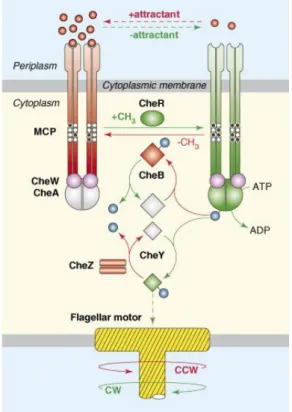
The cell controls the rotation of the flagella by means of its chemotaxis and/or phototaxis system. The systems are very similar, both for phototaxis and chemotaxis, and bacteria and archaea. In most cases, the signal is detected by a transmembrane receptor (although some receptors are soluble). The receptor controls the activity of histidine kinase CheA, attached to it via the adapter protein CheW. Attractant signal results in less intensive phosphorylation of CheY, which in its turn results in CCW rotation of the flagellum (Figure 1.2.3). An increased sensitivity of the chemo- or phototaxis signal is attained by methylation of the receptor by the
26
protein CheR (which sensitizes the receptors) and demethylation by CheB (which has an opposite effect). Finally, the protein CheZ serves to dephosphorylate CheY.
The described proteins, present, for example, in Escherichia coli, form the minimal version of the functioning yet highly sensitive taxis system. In other bacteria, the supplementary proteins such as CheC and CheD can also be present, as well as multiple variants of CheY, CheW and other proteins101.
Figure 1.2.2. The chemotaxis signaling pathway in E. coli (from Hazelbauer et al.102).
Components and reactions in red promote counter clockwise (CCW) flagellar rotation; those in green promote clockwise (CW) flagellar rotation. Components in gray represent inactive forms. Solid lines represent enzymatic reactions; broken lines indicate binding interactions. CheA-derived phosphoryl groups are shown as blue spheres. Receptor modification sites are shown as white (unmethylated) and black (methylated) circles.
27
1.2.4 Spatial organization of chemotaxis system
The large complexes comprising the chemoreceptors and proteins CheA, CheW and CheZ are usually found close to the cell pole of bacteria103,104 and can be easily visualized by electron tomography105–107 (Figure 1.2.3a) or light microscopy104,108. There, the elongated chemoreceptor proteins form hexagonally ordered arrays (Figure 1.2.3b), which are extremely stable in vitro109. The degree of ordering of the chemoreceptors within the arrays appears to depend on the environmental conditions as well as on the presence of attractants or repellents110.
While the nature and stoichiometry of the proteins forming the chemoreceptor arrays were known for a long time102,104,111, their exact arrangement remained unknown. The cryo-electron tomography studies showed that inside the arrays, the inherently dimeric chemoreceptors form trimers (trimers-of-dimers, Figure 1.2.3d-g), with the kinase CheA and accessory protein CheW residing at the receptor tips (Figure 1.2.3d). Soon thereafter, the experiments with nanodisc-reconstituted chemoreceptors demonstrated that two chemoreceptor trimer-of-dimers, together with two CheW molecules and CheA dimer form the core unit of the chemotaxis signaling complexes112.
1.2.5 CheA and CheW proteins
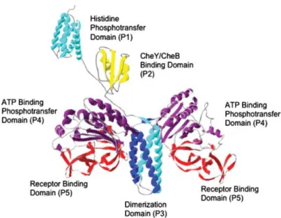
The histidine kinase CheA in its long form (CheAL) consists of five domains of known
structure113: histidine phosphotransfer domain (P1), CheY/CheB binding domain (P2), dimerization domain (P3), ATP-binding phosphotransfer domain (P4) and CheW-like receptor binding domain (P5, Figure 1.2.4). The shorter form CheAS lacks the most of its P1
domain and thus cannot be phosphorylated. The most important for understanding chemoreceptor cluster assembly are the crystallographic structures of P3, P4 and P5 domains114 and of the complex of CheA P4 and P5 domains with CheW115. Besides the crystallographic CheA-bound structure, there are structures of the CheW protein, determined by NMR spectroscopy116,117 and by X-ray crystallography118.
Although the details of the interaction of CheA and CheW with chemoreceptors were not elucidated for a long time, the recently determined crystallographic structure of the chemoreceptor-CheA-CheW complex together with electron-cryotomographic studies provided a clear picture of the chemoreceptor array organization119,120.
28
Figure 1.2.3. Spatial organization of chemoreceptor arrays (Caulobacter crescentus, adapted from Khursigara et al.106). a-b) Direct visualization of Caulobacter chemoreceptor arrays.
a) Tomographic slice of the polar region of an intact Caulobacter swarmer cell demonstrating a continuous surface layer (SL), outer membrane (OM), cytoplasmic membrane (CM), and cytoplasmic filaments (F). The inset shows an expanded view of the chemoreceptors array and signaling scaffold (white arrowhead) and proposed CheR/B interaction sites (black arrowhead). b) In the face-on orientation, chemoreceptor arrays are characterized by a distinct, partially ordered pattern ~32 nm below the cytoplasmic membrane that corresponds to the signaling scaffold. (Inset) The power spectrum of this region clearly demonstrates the ~12-nm hexagonal spacing. The scale bars in panels a and b are 100 nm; the panel a inset scale bar is 50 nm. c-g) Averaged packing arrangement of trimeric receptors in Caulobacter chemoreceptor arrays. c) Averaged density map of a chemoreceptor array demonstrating idealized hexagonal packing, with trimeric chemoreceptors model structures (cyan and magenta) docked into a single hexagonal unit. d) One-half of the hexagonal unit described in panel c, with continuous receptor densities emanating from the signaling scaffold. The gold circles represent the extra density attributed to molecules within the signaling scaffold. The three white dashed lines represent the heights at which cross-sectional cuts were made in the density map to obtain panels e through g, from top to bottom, respectively. e, f) Trimer-of-dimer organization of chemoreceptors. There are multiple interactions between adjacent trimers. g) Cross-sectional view from below the level of chemoreceptor shows the extra density directly below every other receptor trimer (magenta). The scale bars in panels A and B are 20 and 10 nm, respectively, and the scale bars in panels C to E are 5 nm.
29
Figure 1.2.4. Molecular model of the histidine protein kinase CheA113. The histidine
phosphotransfer domain (P1) and the response regulator CheY/CheB-binding domain (P2) are depicted as monomers connected to one another and the remainder of CheA via flexible linkers. The dimerization domain (P3), ATP-binding phosphotransfer domain (P4), and the receptor-binding domain (P5) are all depicted within a CheA dimer.
1.2.6 Architecture of chemoreceptors and sensory
rhodopsin-transducer complexes
Overall, the primary receptors of the chemo- and phototaxis systems have a very similar architecture (Figure 1.2.5). In the chemotaxis system, it is a homodimer of chemoreceptor proteins. In the phototaxis system, the photosensor and signal transducer are separate polypeptide chains that interact via their transmembrane domains and dimerize via transducers.
In the chemotaxis system, the signal is generated by the extracellular domains of chemoreceptors, and then transmitted inside the cell by the transmembrane helices. In the phototaxis system, the signal (illumination) is detected by the photosensor sensory rhodopsin, a member of microbial rhodopsin family. The sensory rhodopsin passes the signal to the transmembrane part of the transducer protein. In the cytoplasm, in both systems the signal passes through the HAMP domain to the kinase control module, which, in its turn, regulates the activity of the kinase CheA.
In the following sections, structures and conformational changes of taxis systems constituents are discussed.
30
Figure 1.2.5. Domain architecture of bacterial chemoreceptors (top), of the phototactic signal transducers HsHtrI and NpHtrII (middle), and of the transducer and chemoreceptor HsHtrII (bottom). Both chemoreceptors and sensory rhodopsin transducers are dimeric, but only
one protomer is shown for clarity. TM1 and TM2 are the transmembrane helices, AS1 and AS2 are the helices of the HAMP domain. The cell membrane is shown in blue.
1.2.7 Signal generation in chemoreceptors
At the moment, structures of several chemoreceptor domains belonging to different families are known. However, for most of them only the apo- or holo- structure is known, which is not sufficient for speculations about the signaling mechanism. The most structurally characterized domain, and the first one for which the atomic structure has been determined, is the aspartate sensing domain of Escherichia coli aspartate receptor Tar. The structures of the domain in apo- and holo- forms121 led to the first model of the signal transduction through the membrane122 (Figure 1.2.6).
Briefly, in the absence of ligand the domain is a symmetric dimer. Each protomer consists of 4 α-helices, where the first one continues right after the first transmembrane helix TM1, and the forth one is followed by the second transmembrane helix TM2 (Figures 1.2.5, 1.2.6a). Binding of the ligand at the dimerization interface of the protomers leads to displacement of the helix α4 of one of the protomers. This displacement is then passed to TM2 and inside the cell. This piston-like motion of TM2122 is also observed in chemoreceptor kinases of two-component signaling systems123.
31
Figure 1.2.6. Structure and aspartate-induced conformational changes in the Tar periplasmic ligand-binding domain. a) Structure of the periplasmic ligand-binding domain (PDB
ID 1VLS124). In the absence of aspartate, the protein is symmetric dimer. The protomers are shown in yellow and blue, each protomer consists of 4 α-helices, marked α1-α4 (prime denotes the second protomer). b) Aspartate-induced conformational changes in the Tar periplasmic ligand-binding domain122. Shown is a superposition of the crystal structures121 for the apo (grey) and aspartate-occupied (black) ligand-binding domain, depicting the single bound aspartate molecule and the periplasmic regions of the four membrane-spanning helices (residues 44-75 of helices αl/TMI and αl'/TM1'; residues 146-175 of helices α4/TM2 and α4'/TM2'). Upon aspartate binding, the α4/TM2 helix of subunit A is observed to translate 1.6 ± 0.2 Å downward (or toward the cytoplasm in the intact receptor) and to tilt 5º, yielding the new position highlighted in red. By contrast, the remaining three transmembrane helices of subunits A (cylindrical ribbon) and B (square ribbon) are relatively stationary, exhibiting aspartate-induced translational and angular displacements less than 0.5 Å and 1º in magnitude, respectively.
32
1.2.8 Signal generation in the phototaxis system
The primary light receptors of the phototaxis system are sensory rhodopsins. While sensory rhodopsin I serves for both attractant response to 565 nm light and repellent to 370 nm light, sensory rhodopsin II (sometimes called phoborhodopsin) generates only repellent signal to illumination with 480 nm light. Much more structural information is known for
Natronomonas pharaonis SRII than for other sensory rhodopsins and, consequently, we will
focus on NpSRII.
In the cellular membrane NpSRII is bound to its cognate transducer NpHtrII in a 2:2 complex125,126 (Figures 1.2.5 and 1.2.7a). Absorption of a light photon and consequent trans-cis isomerisation of the retinylidene chromophore of NpSRII initiates the photocycle. As a consequence the protein passes through several intermediates until it reaches back to the original state in about 1 second. The functionally important conformational step occurs during the M1→M2 transition which leads to the signaling state127,128
. During this transition the signal is transferred through the interface comprising helices F and G to NpHtrII, which results in a rotary motion of transmembrane helix TM2126,129 (Figure 1.2.7b). This activation occurs at the level of the membrane. How this signal is then transmitted to signal domain thereby modulating the activity of the histidine kinase CheA and triggering the cytoplasmic two component signaling cascade is not known yet 102,125,129,130.
Figure 1.2.7. Structure of the sensory rhodopsin-transducer complex and conformational changes during the photocycle. a) Structure of 2:2 NpSRII:NpHtrII complex in the membrane125. The sensory rhodopsin is shown in red and the transducer in green. b) Conformational changes in the NpSRII-NpHtrII complex during the photocycle129.
Published X-ray and NMR structures of NpSRII76,82,131,132 coincide within experimental errors and strongly resemble those obtained for bacteriorhodopsin16. The conformation of the backbone and that of conserved side-chains in the retinal-binding pocket are more or less
33
identical. Moreover, the structure of the receptor76,82,131,132 is almost the same as compared to the NpSRII/NpHtrII complex125.
These observations pose an interesting question. How does nature fine-tune common scaffolds to engender two completely different functions: sensor and ion pump? From previous work it is evident that NpSRII as well as SRI is capable of pumping protons, albeit with poor efficiency133–136. Similarly, bacteriorhodopsin can be converted into a sensor by just three mutations137. On the other hand, despite great efforts it has not been possible to change NpSRII into an efficient proton pump138. Apparently, the requirements for an effective ion pump are much more demanding than those for a functional sensor.
A second observation relates to the inhibition of the proton pump on transducer binding NpHtrII to its cognate receptor NpSRII139,140. It has been discussed that in the 2:2 complex the cytoplasmic channel cannot open sufficiently, thereby altering the proton uptake kinetics such that the reprotonation of the Schiff base is faster from the extracellular side. This kinetic correlation would result in a futile proton cycle (reviewed in Sasaki and Spudich141). Certainly, this proposal can only be verified by a comparison of NpSRII active states with and without bound transducer NpHtrII.
Crystal structures are available for NpSRII ground state82,131,132 and for its K-intermediate, which is formed at room temperature in the nanosecond range142, as well as for the NpSRII/HtrII complex, for which data of the ground state and its K- and M-intermediate (the active state) are available125,129. Despite several attempts the structures of the late (active) states of NpSRII were not obtained yet. The reason for this failure has been related to large conformational changes upon the light-activated M-formation, which lead to severe disturbance of the crystal packing and consequently a substantial decrease in resolution. A similar difficulties arise in experiments with the intermediate states of visual pigment rhodopsin143,144. Section 3.4 of this work describes the process of determining the structure of the NpSRII in the active state and the structure itself.
1.2.9 Signal transduction by the HAMP domain.
HAMP domain is a ubiquitous signaling module found in histidine kinases, adenylyl cyclases, methyl-accepting chemotaxis proteins and phosphatases (recently reviewed by Parkinson145). The domain was first identified as an amphipathic linker between the transmembrane helices and the signal output domain146,147. Much later, the atomic structure of the HAMP domain part of the thermophile Archaeoglobus fulgidus putative protein Af1503 was determined by
34
NMR148 (Figure 1.2.8). The structure revealed that the HAMP domain is organized as a symmetric homodimeric parallel coiled coil. Each protomer has two α-helices, AS1 and AS2, connected by a flexible linker segment. Later, a similar structure was observed in a crystallographic structure of three consecutive HAMP domains from the Aer2 protein of Pseudomonas aeruginosa149. The arrangement was also verified by biochemical and biophysical methods for a number of other proteins – chemoreceptors Tar and Tsr150–153, aerotaxis protein Aer154, phototaxis signal transducer HtrII155–157 and sensory histidine kinases EnvZ and NarX158,159.
Figure 1.2.8. Structure of the HAMP domain and different models of its conformational changes. a) Structure of the Af1503 HAMP domain148. The domain is a symmetrical homodimer/4-helical parallel coiled coil. b) Gear-box model of Hulko et al.148 Schematic representation of complementary x-da packing (left) versus knobs-into-holes packing (right). The two packing modes can be interconverted by rotating adjacent helices by 26 in opposite directions, as illustrated by the cogwheel diagram. c) Model of Airola et al.149, based on the structures of the three HAMP domains from the soluble chemoreceptor Aer2. d) Model of Wang et al.160, based on the cysteine accessibility scanning of the NpHtrII first HAMP domain. Upon illumination, accessibility of the residues at the HAMP domain ends increases, which implies that the helices move in opposite directions.
Currently, there are several models of signal transduction through the HAMP domain145. The gearbox model posits that the HAMP domain helices switch between the orthodox a-d packing and the unusual x-da packing148,161,162 (Figure 1.2.8b). Atomic structures of HAMP domain-DHp phosphotransfer domain fusions show that the rotation of the HAMP domain'
35
helices results in rotation of adjacent helices of DHp161,162. This mechanism explains the signal transduction in receptor histidine kinases, but it is not clear whether it is the case for chemo- and photoreceptors. Alternatively, experimental data reveal that the signal input in chemoreceptors and NarX is a piston-like motion of the transmembrane helix, to which the HAMP domain is connected122,123,163 (Figure 1.2.6b). The HAMP domain itself may switch between two conformations145,149 (Figure 1.2.8c). The output was proposed to be coded by the dynamic properties – looser or tighter packing of the HAMP domain' helices145,150,151,164. As for phototactic signal transducers, it was first proposed that the HAMP domain of NpHtrII transduces the signal via switching between a compact and a highly dynamic states156,157. Later, the fluorescent labeling studies revealed that the helices AS1 and AS2 move in opposite directions during signal transduction160 (Figure 1.2.8d). Molecular modeling and NMR studies have shown that the NpHtrII HAMP domains have the same fold as the HAMP domains for which the structure is known155,165.
Recently, several groups have studied the properties of chemo- and phototaxis proteins by means of modeling. Models of the NpHtrII HAMP1 as well as the HAMP domain region were built by Nishikata et al.165. Nishikata et al. have also studied the dynamics of the NpSRII-NpHtrII complex in the ground and the M states by means of molecular dynamics166. Signal transduction via the transmembrane part of chemoreceptors Tar167,168 and sensor kinase PhoQ169 was studied extensively by different groups. Finally, Hall et al. have generated a model of the entire chemoreceptor Tsr and of the trimer-of-dimers of these chemoreceptors that has shown how the small structural changes may be propagated across the system170. Despite this extensive work on the HAMP domains, little information existed about the inter-HAMP region of sensory rhodopsin transducers (Figure 1.2.5). It was proposed that it might act as a mechanical joint171, however no details were described. The model of this region and its implications are presented in the Section 3.5 of this work. After the model of the HAMP-domain region was established, the question about the conformational changes, accompanying NpHtrII HAMP domain signaling, remained. The Section 3.6 describes the analysis of the NpHtrII HAMP1 motions in molecular dynamics simulations and consequent elucidation of two distinct states, displaying the features similar to those observed in experiments (Figure 1.2.8c-d).
36
1.3 CDP-OH transferases and IPCT-DIPPS
1.3.1 CDP-OH transferases
CDP-alcohol phosphatidyltransferases (CDP-OH transferases) are a large family of membrane proteins that catalyze the displacement of CMP from a CDP-alcohol by a second alcohol with formation of a phosphodiester bond and concomitant breaking of a phosphoride anhydride bond172. Currently (checked on 15/05/2014) there are 11822 CDP-OH transferase sequences in the Pfam database172, 27917 sequences in InterPro173, and 25077 sequences in Prosite174. The majority of the CDP-OH transferase proteins are involved in lipid biosynthesis. For example, there are five such enzymes in mammals, including humans, that participate in the
de novo biosynthesis pathways of all the major glycerophospholipids in mammalian cells175
(Fig. 1.3.1).
Figure 1.3.1. Pathways of glycerophospholipid biosynthesis in mammalian cells175. The
biosynthesis starts either from diacylglycerol (DAG) or phosphatidic acid (PA), and results in production of phosphatidylcholine (PC), phosphatidyletanolamine (PE), phosphatidylserine (PS), phosphatidylinositol (PI) and cardiolipin (CL)175. The intermediate metabolites are CDP-diacylglycerol (CDP-DAG), phosphatidylglycerol phosphate (PGP) and phosphatidylglycerol. The CDP-alcohol phosphatidyltransferase family enzymes in mammals are highlighted, CPT1 is cholinephosphotransferase 1, CEPT1 is choline/ethanolaminephosphotransferase 1, EPT1 is ethanolaminephosphotransferase 1, CDIPT1 is phosphatidylinositolsynthase 1 and CLS1 is cardiolipinsynthase 1.
37
As it can be seen, the ligands in the reactions catalyzed by CDP-OH transferases are often very different. Consequently, there is a high level of diversity among the CDP-OH transferase sequences, which is highlighted in the sequence logo176,177 of the family (Figure 1.3.2). Overall, the consensus motif of the family is D(x)2DG(x)2AR(x)7-12G(x)3D(x)3D.
Figure 1.3.2. Sequence logo176,177 of the CDP-OH transferase domain. The Hidden Markov
Model used in preparation of this figure is based on the alignment of 124 representative Pfam protein family sequences as seeds172.
1.3.2 Di-myo-inositol phosphate and the IPCT-DIPPS enzymes
Di-myo-inositol phosphate (DIP) is one of the major osmoprotecting molecules in a number of hyperthermophilic species of archaea and bacteria178. DIP level is also highly increased in some species at supraoptimal growth temperatures179,180. Based on this, it was proposed that DIP, besides being an osmoprotectant, might also protect the cellular macromolecules against the harmful effects of high temperature.
Recently, it was clarified that DIP is synthesized from glucose-6-phosphate in 4 steps (Figure 1.3.3)181,182. First, glucose-6-phosphate is converted to L-myo-inositol-1-phosphate by NAD+-dependent inositol phosphate synthase (IPS). Then, the inositol-1-phosphate is activated to CDP-inositol via the activity of CTP:inositol-1-phosphate cytidylyltransferase (IPCT). In the next step, CDP-inositol is condensed with inositol-1-phosphate to DIP-phosphate (DIPP) by DIPP synthase (DIPPS). Finally, DIPP is dephosphorylated to DIP by the action of inositol monophosphatase (IMP).
38
Figure 1.3.3. DIP synthesis pathway. Adapted from Rodionov et al.181
Meanwhile two of the enzymes in the DIP synthesis pathway were known for a long time183,184, the two others (IPCT and DIPPS) have only recently been discovered181,185. It was shown that in some organisms they are conducted by two parts of a bifunctional enzyme IPCT-DIPPS, meanwhile in others the same domains are coded in separate genes. The DIPPS enzymes and the DIPPS domains of the bifunctional enzymes are the members of the CDP-OH transferase family (Section 1.3.1).
One of the best studied IPCT-DIPPS proteins is the enzyme from the hyperthermophilic archaeon Archaeoglobus fulgidus185. It was shown that its cytidylyltransferase domain is absolutely specific for CTP and l-myo-inositol-1-phosphate and that the DIPP synthase domain used only l-myo-inositol-1-phosphate as an alcohol acceptor, but could recognize CDP-glycerol, as well as CDP-l-myo-inositol and CDP-d-myo-inositol, as alcohol donors. Crystallographic structure of the IPCT domain was recently determined at a resolution of 1.9 Å186. The protein revealed a Rossman-like fold characteristic for nucleotidyltransferase family enzymes (Figure 1.3.4). The domain has a central mixed sheet with six parallel β-strands and one antiparallel. These are surrounded by six α-helices. The domain could not be co-crystallized with ligands to determine the active site and ligand binding sites. However, based on the structures of homologous enzymes, it was possible to predict the binding sites of CDP and inositol-1-phosphate (Figure 1.3.4).
Structure of the DIPPS domain remained unknown, and its determination was one of the goals of the present work, as well as analysis and prediction of the ligand binding site (Section 3.7).
39
Figure 1.3.4. Active site of the Archaeoglobus fulgidus IPCT-DIPPS IPCT domain186.
a) Superposition of the IPCT (cartoon in blue) with several structures of homologous protein RmlA. RmlA is in the apo form (PDB code 1FZW; ribbon in dark green), and in complex with different ligands (PDB codes 1FXO, 1G0R, 1G2V, 1G1L, 1G23, and 1G3L; ribbons in different shades of green). dTDP-d-glucose (from 1G1L) is shown in sticks. b) IPCT active site pocket with fitted CDP-inositol; residues at the entrance are labeled. The figure is taken from Brito et al.186
40
2 Materials and methods
2.1 Protein expression and purification
2.1.1 Expression and purification of HmBRI
Haloarcula marismortui bacterio-opsin gene (bop, UniProt ID: Q5UXY6) with D94N
mutation was synthesized de novo. The nucleotide sequence was optimized for E. coli expression using the GeneOptimizer™ software (Life Technologies, USA). The gene was introduced into the pSCodon1.2 expression vector (Staby™Codon T7, Eurogentec, Belgium) via NdeI and XhoI restriction sites. Consequently, the expressed construct harbored an additional C-terminal tag with a sequence LEHHHHHH. For protein expression, the E. coli strain SE1 cells (Staby™Codon T7, Eurogentec, Belgium) were transformed with the pSC-oHmBRI-His6 plasmid. The cells were grown at 37º C in shaking baffled flasks in an
auto-inducing medium ZYP-5052187 containing 100 mg/L ampicillin. After the glucose level in the growing bacterial culture dropped below 10 mg/L, the incubation temperature was reduced to 20º C and incubation continued overnight. Collected cells were disrupted using the M-110P Lab Homogenizer (Microfluidics) at 25000 psi in a buffer containing 20 mM Tris-HCl pH 8.0, 5% glycerol and 50 mg/L DNase (Sigma-Aldrich, USA). Membrane fraction of cell lysate was obtained by ultracentrifugation at 90000 g for 1 h at 4º C. The pellets were resuspended in a buffer containing 20 mM Tris-HCl pH 8.0, 0.1 M NaCl and 1% DDM (Anatrace, Affymetrix, USA). All-trans-retinal (Sigma-Aldrich, USA) was added to 10 µM and immediate red shift of the solution color was observed. The mixture was left overnight for solubilization. Insoluble fraction part was removed by ultracentrifugation at 90000 g for 1 h at 4º C. The supernatant was loaded on Ni-NTA column (Qiagen, Germany) and the His-tagged protein was eluted in a buffer containing 20 mM Tris-HCl pH 7.5, 0.1 M NaCl, 50 mM EDTA, 0.01% DDM. The eluate was subjected to size-exclusion chromatography (125 ml Superdex 200 PG, GE Healthcare Life Sciences, USA) in a buffer containing 50 mM NaH2PO4/Na2HPO4 pH 7.5, 0.1 M NaCl, 0.01% DDM. Protein-containing colored fractions
were collected and concentrated to 40 mg/ml for crystallization.
2.1.2 Expression and purification of ESR
ESR with a C-terminal hexahistidine tag was expressed in Escherichia coli strain Rosetta2(DE3)pLysS and purified as described57. ESR was growing in fermenter for 3 days at