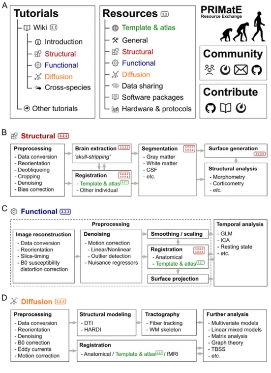
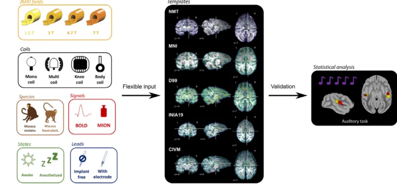
A collaborative resource platform for non-human primate neuroimaging
Texte intégral
Figure


Documents relatifs
Scott Love, Marine Siwiaszczyk, Marie Auge, Christophe Destrieux, Frédéric Andersson, Elodie Chaillou.. To cite
present study defines a method to optimize the design of optode and the choice of stimulation parameters for optogenetics and more generally light delivery to deep and large volumes
Different flavors of BIDS can be designed to support such formats, which would additionally require (1) identification of the metadata fields that should be included in the sidecar
FreeSurfer 6 has recently been released, providing tools for partial volume correction (PVC) and projection of signals onto surfaces. However, these functionalities are
Les figures IV.13, IV.14 et IV.15 montrent l'évolution de la température à l'intérieur d'un canal chauffé asymétriquement en fonction du rapport d'aspect, de la taille et
It represents actually the basic mechanism used for the formation of graphene nanoribbons (GNRs) on Au(111), 151 where the Ullmann coupling between dibromo-bianthryl precursors
LLO dependent translocation of the late endosomal/lysosomal membrane proteins LAMP-1 and LAMP-2 was also characterized by flow cytometry: a marked increase of both proteins at the
Response surface and contour plots for the effect of processing pressure and processing time at constant initial moisture content of peels on pectin yield in % (g
