Annals of Oncology 9: 1015-1022, 1998.
© 1998 Kluwer Academic Publishers. Printed in the Netherlands
Clinical case
Primary cervical malignant teratoma with a rib metastasis in an adult:
Five-year survival after surgery and chemotherapy
A case report with a review of the literature
C. Als,
1'
2H. Laeng,
2T. Cerny,
3J. A. Kinser,
1H. Rosier
1& R. Hausler
4Departments of'Nuclear Medicine, 3 Medical Oncology, "Otorhinolaryngology, Head and Neck Surgery, 2 Institute of Pathology, Inselspital, University of Berne, Berne, Switzerland
Summary
We report a case of a man presenting with a cervical malignant teratoma and a chondrosarcomatous rib metastasis. He was alive and free of recurrence five years and 10 months (= 70 months) after resection of the primary mass, followed by che-motherapy and subsequent resection of the rib tumor. This is the 35th patient reported in the literature and the first descrip-tion in which an 'adjuvant' or primary chemotherapy was used. Previous patients with a cervical malignant teratoma, reported after lethal outcome, had survivals of one to 22 months (me-dian nine months). In all patients with a preoperative clinical
impression of an aggressive, differentiated or undifferentiated malignancy, the definite diagnosis of teratoma could only be made histologically. By analogy to germ cell tumors, the prog-nosis of malignant teratoma might be improved if complete excision is combined with new, adjuvant chemotherapy proto-cols for germ cell tumors. Lessons learned from this case are placed in the context of germ cell tumors in general and of non-gonadal malignant teratomas in particular.
Key words: chemotherapy, cervical, germ cell tumor, prognosis,
surgery, teratoma
Case report
a) History and clinical findings
A white, 33-year-old man was referred because of a mass on the anterior left side of the neck. He had first felt the painless nodule a month previously and was aware that it had grown after this date. There were no symptoms of mechanical obstruction. He had no previous history of any operation of or irradiation to his neck. On clinical
examination, he was in a good general condition,
with-out any distress. A firm, elastic, 6 x 5 c m tumor mass was situated in front of the medial rim of the left sterno-cleidomastoid muscle and extended into the left thoracic aperture. It was fixed to the underlying structures but not to the skin. There were no palpable lymph nodes.
Laryngoscopy was normal. b) Preoperative investigations
A chest X-ray showed a focal swelling of the 10th left rib due to a poorly delineated osteolytic lesion, compatible with an enchondroma. A computerized tomography of
the neck (Figure 1) after contrast injection showed an
inhomogeneous tumor mass ( 8 x 4 cm). This was lateral to the thyroid with compression of the left thyroid lobe, of the left side of the trachea and the left neck vessels and with possible infiltration of the jugular vein. The density
of the mass was 11-73 HU. Routine blood and urine examinations were normal. Thyroid studies revealed euthyroidism with normal values of thyroxine, triiodo-thyronine, thyrotropine. The thyroid uptake (two hours, 123-1, 7.4 MBq) was 9%. The 1:1 thyroid scan (123-1 and 99mTc-Methoxyisobutylisonitrile [4] (MIBI, Cardiolite®
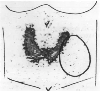
Figure I. Transverse CT section of the neck after intravenous injection
of contrast media: note the large, inhomogeneous tumor mass in a left supraclavicular location, measuring 8 x 4 cm in diameter. The tumor density was 11-73 HU. Infiltration of the left internal jugular vein was suspected. Compression of the left tracheal wall, displacement of the left thyroid lobe, of the left vessels and of the sternocleidomastoid muscle were noted.
Figure 2. Thyroid scan, performed two hours after oral administration
of 7.4 MBq 123-1 (Polyscan), showing a homogeneous activity distri-bution within a slim, 20 g gland. The lateral margin of the left lobe is compressed by a cold nodule of 6 x 5 cm diameter.
Dupont, USA): 185 MBq) showed homogeneous activity distribution in a slim, 20 g gland (Figure 2). The lateral margin of the left lobe was impressed by a cold nodule of 6 x 5 cm diameter. Two-dimensional ultrasonography showed a well delineated nodule of inhomogeneous echostructure lateral to and compressing the left thyroid lobe. Several small cystic areas, a few hyperdense foci, but no calcium deposits were noted. Fine needle
aspira-tion biopsy produced only cystic fluid without cellular
elements suspicious of malignancy. Cytology of a second aspiration showed, besides psammous bodies, some papillary components containing medium sized cells with rough nuclei, clear nucleoli and even some nucleic vacuoles. Based on fine needle biopsy, a papillary carci-noma of the thyroid was initially suspected.
c) Neck surgery
A left cervicotomy with total removal of a large, supra-and infraclavicular tumor measuring 7 x 5 cm in diame-ter, with a clear margin of healthy tissue around it, was performed on 1 July 1992. The tumor capsule was not incised or ruptured. There was no direct link to the thyroid. As the tumor was firmly adherent to the left jugular vein, the vein was resected en bloc with the tumor. The postoperative course was uncomplicated.
d) Histology
Pathologic macroscopic examination revealed a solitary
tumor with a thin, but intact capsule of fibrous tissue. Cross sections revealed multiple haemorrhagic and se-rous cysts (±50% of the volume) being separated by yellow-whitish solid areas. Microscopically, the cystic spaces were lined by columnar epithelia bordering regu-lar serous salivary gland parenchyma (approximately
Figure 3 Histological section: Cyst lined by columnar epithelium
bordering regular serous salivary gland parenchyma with loose lympho-histiocytic infiltrates (H & E, original magnification x 87).
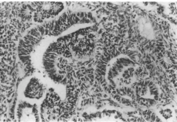
Figure 4. Histological duct structures, which may represent remnants
of mature salivary glands. A solitary concentric focus of squamous epithelial metaplasia is present within abundant irregularly arranged fibrous stroma (H & E, original magnification x 87).
10% of the total volume) with patchy lymphocytic infil-trates (Figure 3). Thyroid parenchyma was not seen. The composition of the remainder of the tissue volume was heterogeneous. Intermingled with mature duct structures which might represent remnants of salivary gland ducts, concentric foci of squamous epithelial metaplasia in irregularly arranged loose fibrous stroma were present (Figure 4). Other mesenchymal counterparts (±30% of the volume) were made up of mature smooth muscle, but there was also hypercellular hyaline cartilage with slight irregularities in cellular size and shape, which suggested a grade 1 chondrosarcoma (Figure 5). In addition, tubu-lar epithelial structures resembling embryonal carcinoma with occasional yolk sack-like projections were frequent. A few true rosettes, typical of neuroepithelial differentia-tion were present [6, 7, 9] (Figure 6). They were growing in an immature mesenchymal stroma, also displaying cytomorphologic atypia and mitotic activity (Figure 6), while additional features of so-called unfavorable histol-ogy such as rhabdomyosarcoma were absent. Scattered haemorrhagic necrosis was present in the malignant
1017
•;•;•.-•: v t o
1
Figure 5. Histological section: Hypercellular mature hyaline cartilage
with variation in cellular size and shape is suggestive of low-grade chondrosarcoma and is a frequent component of the mesenchymal counterpart of the tumor (H & E, original magnification x 175).
/*Jr.V'.tin
us*
y
Figure 6. Histological section: Malignant epithelial counterpart of the
tumor resembling embryonal carcinoma with yolk sack-like structures (arrow) and true rosettes (arrowhead) typical for neuroepithelial differ-entiation. The immature stroma displays mitotic activity (H & E, original magnification x 175).
components. A minimal local invasion of the intact fibrous capsule and of the sinuses of a small neighboring lymph node was seen. The conclusion was that this was a malignant teratoma (or teratocarcinoma) with compo-nents of both germ cell and of differentiated mesenchy-mal tumor. According to the WHO classification, this entity with two tumor components is distinct from an extragonadal germ cell tumor of mixed type, that lacks differentiated (e.g., non-germ cell) tissue.
e) Postoperative investigations
In order to differentiate between a primary or a meta-static teratoma to the neck, further diagnostic procedures were performed. The laboratory findings of:
alphafoeto-protein, lactate dehydrogenase, human chorionic gonado-tropin, human thyreoglobuline were normal. A clinical
examination by an urologist was normal. A
computer-ized tomography of the pelvis and of the abdomen and an ultrasonography of the testicles showed no pathologic
tumor masses. A computerized tomography of the thorax again showed the known swelling of the 10th lateral left rib, compatible with an enchondroma. A bone
scintigra-phy with 99mTc-MDP showed no sign of skeletal
metas-tases and in particular, the scintigraphic findings within the swollen 10th left rib were normal. During the next four months, the patient refused surgical resection of the rib mass. Finally, on 6 November 1992, a partial excision
of the 10th left rib with total tumor resection was
performed. Histology revealed a grade 1-2 chondro-sarcoma, which probably represented a rib metastasis of the known malignant teratoma, although another primary tumor could not be formally excluded.
f) Adjuvant chemotherapy and outcome
Immediately after neck surgery, the medical intention was to treat with adjuvant chemotherapy. This is why, prior to the surgical rib resection, which was initially refused by the patient, two cycles of classical BEP
chemotherapy (bleomycin: absolute doses 30 mg days 1,
8 and 15; etoposide: 100 mg/m2 over one hour from day 1-5 included, and cisplatin: 20 mg/m2 intravenously as a 30 minutes infusion from day 1-5 included [5]) were administered (September to October 1992). The chemo-therapy was intended to be an adjuvant treatment, though the subsequent finding of a rib chondrosarcoma led to the conclusion that chemotherapy had instead been given for metastatic disease. Chemotherapy was well tolerated. Since then, the patient has remained free of symptoms and in good condition for more than five years (five years and 10 months at the time of this report) since resection of the primary neck tumor. Regular clin-ical, radiologic and blood serum tests have not indicated any sign of tumor recurrence.
We have given two adjuvant cycles of chemotherapy according to our policy in patients with testicular stage I germ cell tumors with elements of embryonal histology, where a relapse rate of 30% is to be expected. This policy was shown to reduce the relapse rate from 30% to 5% in this group of patients [44]. Since the rib lesion was scinti-graphically negative and initially judged to be benign, additional chemotherapy was not thought to be neces-sary. Only after excision did it become clear that it was more likely a metastasis, possibly cleared of embryonal elements due to previous chemotherapy. Also the possi-bility of a synchronous multifocal teratocarcinoma can formally not be ruled out.
Discussion
a) Classification
Teratomas are tumors composed of several different tis-sues foreign to the anatomic site of origin in which they arise. They consist of all three germ layers: ectoderm, mesoderm and endoderm. A fine capsule is the rule. Microscopic examination reveals a variety of tissues
Reference 1 Uirje, 1908 [11] 2 Waechter, 1909 [12] 3 Fritzsche, 1920 [12] 4 Buckwalter, 1954(13) 5 Kemp, 1967 [19] 6 Hajdu, 1967 [21] 7 Pilheu, 1969 [22] 8O'Higgins, 1975 [23] 9 Colton, 1978 [24] lOKimler, 1978(25] 11 Dhellemcsl, 1979 [27] 12Dhellemesll, 1979 [27] 13 Murao, 1979 [28] 14 Przystasz, 1979 [2] 15Trotoux, 1979 [29] 16Tobey, 1980 [30] 17Ceriani, 1984 [31] IS Kicr, 1985(32] 19 Buckley, 1986 [33] 20 Mochizuki. 1986 [34] 21 Jordan, 1988(36] 22 Kahle. 1990 [37] 23 Cain. 1991 [38]
24Als, 1998 [this public] Country CH Germany CH USA, w Autralia, w USA, w Argentine Irak USA USA, b France France Japan Poland France USA Italy USA USA, w Japan USA Germany Australia CH Sex F M F F F F M M M F F F F F F M M M M F F F M M Age (y) 52 71 41 23 85 68 27 23 77 37 28 20 19 48 28 51 19 27 27 26 68 45 38 33 Tumor 0(cm) Massive 9 10 9 5 17 14 8 10 10.5 7 Small 5 6 6 6 8 3 20 5 17 5.5 2 7 Location Thyroid ? Thyroid Thyroid Thyroid E-thyroid Thyroid Thyroid Thyroid Neck Thyroid Thyroid Thyroid Thyroid Thyroid Thyroid Neck Thyroid Thyroid Thyroid Neck NR Thyroid St.cl.mast.L Neck Path. Mai Mai Mai Mai Mai Mai Mai Mai Mai Mai Mai Mai Unc Mai Mai Mai Mai Mai Mai Mai Mai Mai Mai Mai Symptom duration 7 7 5 weeks 6 months 50 years 8 months 12 months 24 months 7 months 3 months 3 months 4 months 4 years 4 months 4 months 5 weeks 3 months 12 months 12 months Asymptom. 8 months Immediate 2 weeks 1 month Imaging No No No X . I X Yes NR I X US, X, NB NR NR 1. X X, I NR X, Ba, Tc, IVP X,Tc CT FN, X, CT X, I NR US, FN Ba, CT, US TcMlbl, US, X, CT Surgery No ? Yes Yes Yes Yes Yes Yes 2x Yes Yes Yes Yes Yes Yes Yes 2x Yes Yes Yes Yes 2x Yes Yes Radiation therapy (Gy) No 7 No Neck, med No Recurr. No Yes No Yes Recurr. No Yes 46 Yes No Recurr Yes Yes No Recurr. Yes No No Chemo-therapy No 7 No No No No No Yes No Yes Yes Yes No Yes Yes Cy,C, B, Ac No NR Cy. A, E, C No No Vb, C, B E. C, B C, E, B, adj Local recur-rence Yes 9 Yes Yes (sarcoma) No Yes No Cerv. In Yes Yes Yes Yes No No Yes Cerv. In 3x NR No No Yes Yes Yes, 5 cm No Distant metastases Lu: sarcoma 7 Med, bo, lu Med, lu, pleu No Med, lu, pleu
Lu No No No Lu No No No lu Med, retrocrural Med, lu, pleu, skin NR
Lu, meningia No Med, lu, pleu Lu, li, bo Lu, brain bo (sarcoma) Survival (months) 1 ? 1 9 Alive > 7 9 9 10 ? 6 14 22 Alive > 8 7 11 Alive > 9 9 NR 2 Alive > 16 8 11 3 well > 7 0 Cause of death Metastatic tumor No follow-up Asphyxia Sudden, jug. vein infiltr Incompl. follow-up Sudden asphyxia No follow-up Local recurrence No follow-up Gastric aspiration Carotid rupture NR Incompl. follow-up No follow-up NR Incompl. follow-up Asphyxia No follow-up NR Incompl follow-up Sudden asphyxia Cachexia, cardiac failure Cachexia Probably cured Remarks Diagnosis at autopsy Quoted by [12] No autopsy Autopsy [cf 36] Pregnant, child well Path, descrip-tion incompl. Autopsy Local fistula, thyroid or. Pat unnoticed. thyroid or. Autopsy [cf 21] Strumectomy < 2 years: ben. hCG mediated hyperthyr Adjuvant chemotherapy
Chemotherapy is detailed where known; in all patients (except ours), it was administered in the presence of recurrence or of metastases. Note that the case reported by Hajdu in 1967 [8] was later reported again by Jordan in 1988 [36] Abbreviations: w white; b black; F female; M male; y years, e extra, NR not reported; St.el.mast L left sternocleidomastoid muscle; path pathology, Mai malignant; unc uncertain, ben benign, CT computerized tomography; US ultrasound; Tl 201T1 thyroid scan; MIBI 99mTcmethoxyisobutylisonitrile thyroid scan, Tc 99mTc pertechnetate thyroid scan, I radioiodide thyroid scan; X Xray; Ba barium swallow; NB -needle biopsy; FN fine -needle aspiration cytology; IVP - intravenous pyelography; In - lymph nodes, cerv - cervical; med - mediastinum, lu - lung; bo - bone; pleu - pleura: h - liver; or - origine; recurr - recurrence; adj - adjuvant; A adriamycine; Ac actinomycine D; B - blcomycinc; C - cysplatine. Cy - cyclophosphamide; E - vepesid; V - vincnstine, Vb - vinblastine; incompl - incomplete
1019 00 C O C3 C o ,2 E Q S 2 a 2 U -S
is
D. — CO T3I
1 €
C/3 .C O 3 Z U o os z z g g | 8II
£1
OS 3 Z JS z z a: o z z Z A z z z z z z z z a: X Z Z t-c-. Ss'l
I
"5 & 23 ^ S 2 S J= o r-, a .£II
li
5 £ n o £> csi o Z Z OS o o Z Z Z o o o Z Z Z o o o Z Z Z CQ X'g l
2 £ 'S > > > 3 (3 D . =. £ _ o o := z £ £•5 5 o -c o o o o S £ CU = £ = = s - ^ »— r^ < < O 02 O Z Z Z o ai o z z z z z z o o o z z z - o" i-i- U c -a -a s ri e e c O C O O ,5 E E (N — o M -Tf r-, (N < < « v: n d D D -^ =. i?i » = S C- — 2 - ~. §! o «with a wide range of cellular differentiation and variable degrees of maturation. Immature components corre-sponding most often to germ-cell tumors seem to be of lesser prognostic significance in infants [6] but represent a poor prognosis in adults [7].
Since the first report of a neck teratoma by Hess in 1854 [10], about 230 cases have been reported in all ages, of which 34 were in adults (Tables 1 and 2) [1-3, 11-39]. 'Cervical teratoma', the term used nowadays, generally incorporates lesions arising in the anterior and posterior triangles of the neck. Cervical teratomas are rare and represent only 2%-9% of all reported teratoma cases [40]. The term does not include teratomas of the skull base or along the cervical spine [7]. The rare event of a cervical teratoma in an adult should be viewed in a more general context. By far, most teratomas occur in infants before the age of one year and are benign, many of them are present at birth [6, 8]. In adults, teratomas are very rare; 70% are malignant. Teratomas most often occur in para-axial or mid-line location from the brain to the sacral area as well as in the gonads. Table 3 illustrates that the primary tumor location shows different de-creasing frequencies in children and adults [7, 9, 45].
The former clinical distinction between a thyroid or neck origin of a teratoma, - according to: 1) origin of feeding vessels, 2) direct continuity between tumor and gland, 3) localization of tumor within the gland, 4) entire absence of the gland, - has no practical significance because there are no differences in prognosis or treat-ment [36]. Moreover, these criteria may be misleading because it is often impossible to determine the vascular connections of the tumor. Thyroid parenchyma within the tumor may represent remaining thyroid from which the teratoma arose, or well differentiated elements arising in the teratoma itself, or even thyroid tissue secondarily invaded by the teratoma.
b) Clinical presentation of cervical teratoma in adults
Out of the 35 reported cases (our case included) of cervical teratoma in adults, covering various ethnic origins (Tables 1 and 2), 32.5% were benign and 68.5% (one uncertain case included) were malignant. The tu-mor diameter at presentation varied from 3 cm to
mas-7a6/e i. Decreasing frequencies of the locations of a primary teratoma in infants and children as compared to adults [7, 9,40,48].
Infants and children Adults Sacrococcygeal region
Ovary
Head and neck Retroperitoneum Mediastinum Brain and spinal cord Testes
Liver
Abdominal wall, para-umbilical Scapula (back) 40% 37% 5.5% 5% 4% 3.5% 3% 1% : 1% Ovary Testes Sacrococcygeal region Mediastinum Retroperitoneum Central nervous system Liver
Nasal sinuses Neck
sive tumors of more than 30 cm. The median symptom duration before operation was much longer with benign than with malignant teratomas. Typically, rapid progres-sion started after a long dormant phase. Metastases and local recurrences were often sarcomatous.
The median age of the 35 patients was 32 years (range 19-85 years). In 90% of benign teratomas and 58% of malignant teratomas, the diagnosis was made before the age of 40 years. The higher the age at diagnosis, the more likely was the malignant nature of the tumor: the me-dian age in case of benign disease was 24 years, in case of malignant disease 35 years. Mochizuki [34] had postu-lated that cervical teratoma in adults undergoes a rapid change in biological behavior from benign to malignant above the age of 20 years, as the youngest malignant case was seen in a 19-years-old female [28].
c) Imaging
Because most tumors were clinically suspected to be related to the thyroid, previous imaging of cervical teratomas was reported as thyroid scintigraphy (99m-Tc pertechnetate or radioactive iodine), tumor scintigraphy with 201-T1 or 99mTc-MIBI, ultrasound or CT (Tables 1 and 2). In case of papillary thyroid carcinoma, tumor scintigraphy with 99mTc-MIBI has been reported pos-itive [4, 41, 42]. However, it was negative in our patient.
d) Treatment and prognosis
The prognosis of benign teratoma is generally good (Table 2). But the prognosis of malignant teratoma is sombre (Table 1): none of the patients with prolonged follow-up was cured, despite various treatment protocols. Thus, the maximal survival of the malignant cervical teratoma reported to date was 22 months (median nine months, average eight months, Table 1). External radio-therapy since the 1950's and, since the late 1970's, che-motherapy, relying today mostly on cisplatin-containing regimens [43], have been administered after surgical ex-cision. These treatments were always applied late in the course of the disease, when there were large tumor bur-dens, and their effect was only palliative. For other extra-gonadal teratocarcinomas such as those of the anterior mediastinum, the prognosis is inferior to the one of testicular teratocarcinomas with longterm survivals of 40%-50%, where as such tumors arising from the ova-ries (non-dysgerminomatous germ-cell tumors) have an excellent survival after intensive chemotherapy, compa-rably to testicular teratocarcinoma [46,47].
In contrast, our patient with a cervical malignant teratoma with components of germ cell and of mesen-chymal tumors was given BEP-chemotherapy [5] with adjuvant intent in the postoperative course based on clinical experience with germ cell tumors in general. In a British study of clinical stage I patients with non-seminomatous germ cell tumors of the testicle, the pre-study relapse rate was 30%. This was reduced to 5% by a treatment policy of two courses of adjuvant bleomycin,
etoposide and cisplatin (BEP). This 5% relapse rate com-pared favorably with that of 18.5% in clinical stage I patients treated by surgery alone [44]. Therefore, ad-juvant BEP-chemotherapy in the postoperative course in general is effective in preventing relapse in patients with testicular germ cell tumors in general and, hope-fully with malignant teratoma (with germ-cell compo-nents).
As is the case with chemotherapy, prognosic factors for malignant teratoma are deduced from those of germ cell tumors in general. For testicular germ-cell cancer, the extent of disease and serum tumor-marker concen-trations were identified as independent predictors of prognosis, and previously untreated patients with meta-static disease were subdivided into good-risk and poor-risk groups [50, 51]. This staging system was developed because of the high cure rate and substantial toxicity of combination chemotherapy and a need to identify patients more likely ('good-risk') and less likely ('poor-risk') to be cured by standard chemotherapy. In another British multicenter study on non-seminomatous germ-cell tumors of the testicle, the only significant independ-ent prognostic factors were based on histology and lymphatic invasion [48]. The relapse rates ranged from six out of 50 cases (12%) in teratocarcinoma patients without lymphatic invasion to seven out of nine cases (78%) of embryonal carcinoma patients with lymphatic invasion [49]. By analogy, our patient belonged to the high-risk category for relapse because of embryonal components in the primary tumor with minimal lym-phatic invasion and because of the bone metastasis (excised chondrosarcoma in the 10th left rib). This metas-tasis was already present at the time of the neck oper-ation, albeit not yet pathologically proven because of the patient's initial refusal of rib surgery. Nevertheless, the patient has remained tumor free for nearly six years after neck surgery. The en bloc surgical removal of the tumor without rupture of the capsule, followed by BEP-chemotherapy and complete removal of the solitary metastasis, appear to have eradicated the tumor. Five-year progression-free survival rates between 66% and 78% according to histological findings of postchemo-therapy resections of residual masses from metastatic non-seminomatous testicular germ-cell tumors retro-spectively confirm the Tightness of our therapeutic atti-tude. It especially confirms how essential the primary en bloc surgical removal of the tumor is [46]. This is amplified since, as also shown in mediastinal non-semi-nomatous germ-cell tumors, only patients who achieve disease-free status after cisplatin-based combination chemotherapy are likely to be cured [47].
Of the previously reported patients in the literature (Tables 1 and 2), [1-3, 11-39], our patient was the only one, where there was a decision to treat with adjuvant chemotherapy. This meant that there was no delay in the patient receiving chemotherapy for what turned out to be metastatic disease. Compared to the sombre progno-sis of cervical malignant teratoma in adults (Table 1), the good clinical five-year outcome of our patient suggests
1021 that the use of 'adjuvant' BEP-chemotherapy, - as
classi-cally used with germ cell tumors, - immediately after the neck resection and independently of the pathologic nature of the rib tumor, was appropriate. We suggest that a policy of adjuvant chemotherapy should be exam-ined in other non-gonadal malignant teratoma patients in the future, in order to gain prospective evidence of its possible beneficial influence on prognosis.
Acknowledgements
Mr. Y. Kurokochi, Mrs. M. Listewnik and Mr. F. Garayalde provided or translated [1-3].
References
1. Sawafuji M, Kakizaki T, Yamamoto T et al. A case of cervical teratoma in an adult. Nippon Kyobu Geka Gakkai Zasshi 1993; 41 (11): 2220-3.
2. Przystasz T, Badowski A, Dumansli Z. Zlosliwy potworniak tarczycy u chorej z wolem lymfatycznym Hashimoto. Polski Przeglad Chirurgiczny 1979; 51 (5): 511-5.
3. Marescot IE. Tumores teratoides del cuello. Rev Espan Cir 1945; 2: 382-3.
4. Als C, Listewnik M, Ritter EP et al. Quantitative subtraction scintigraphy with 99mTc-MIBI in cold thyroid nodules. Eur J Nucl Med 1993; 20 (10): 852 (Abstr).
5. Williams SD, Birch R, Einhorn LH et al. Treatment of dissemi-nated germ-cell tumors with cisplatin, bleomycin and either vin-blastine or etoposide. N Engl J Med 1987; 316: 1435-40. 6. Hyams VJ, Batsakis JG, Michaels L. Hamartomas, teratoid
tu-mors and teratomas. In Armed Forces Institute of Pathology (ed): Tumors of the Upper Respiratory Tract and Ear. Atlas of Tumor Pathology, 2nd series, fascicle 25. Washington, DC 1988; 200-6. 7. Gonzalez-Crussi F. Teratomas of the neck. In Armed Forces
Institute of Pathology (ed): Atlas of Tumor Pathology, 2nd series, fascicle 18. Washington, DC 1982; 118-27.
8. Hajdu SI, Faruque AA, Hadjdu EO, Morgan WS. Teratoma of the neck in infants. Am J Dis Child 1966; 111: 412-6.
9. Gonzalez-Crussi F. Teratomas of the sacrococcygeal region. In Armed Forces Institute of Pathology (ed): Atlas of Tumor Pathol-ogy, 2nd series, fascicle 18. Washington, DC 1982; 50-76. 10. Hess W. Beitrag zur Kasuistik der Geschwiilste mit
zeugungsahn-lichem Inhalt. In Merck M, Wetzel C (eds): Inaugural Disser-tation. Germany: Giessen 1854.
11. Lurje M. Uber einTeratom der Schilddruse. In Keynes WM, Bale G F (eds): Inaugural Dissertation. Zurich: J. J. Meier 1908, 1959; 27.
12. Fritzsche R. Ueber ein malignes embryonales Teratom der Schild-drusengegend. Arch Klin Chir 1920; 114 (2): 317-31.
13. Buckwalter JA, Layton JM. Malignant teratoma in the thyroid gland of an adult. Ann Surg 1954; 139 (2): 218-23.
14. Cavallero G. Su di un caso di teratoma della ghiandola tiroide in adulto. Pathology 1954; 46: 213-7.
15. Bale GF. Teratoma of the neck in the region of the thyroid gland. A review of the literature and report of four cases. Am J Pathol 1950; 26: 565-79.
16. Keynes WM. Teratoma of the neck. BrJSurgl959;46: 466-72. 17. Noteboom G, Everts-Suarez EA. Dermoid cyst of thyroid gland
in an adult. US Armed Forces M J 1959; 10: 722-6.
18. Priifer HJ. Teratome der Schilddruse. Zentbl Chir 1965; 90: 2234-9. 19. Kemp DR. Teratoma of the neck in the adult. Aust NZ J Surg
1967; 36: 324-7.
20. Stone HH, Henderson WD, Guidio FA. Teratomas of the neck. Am J Dis Child 1967; 113: 222-4.
21. Hajdu SI, Hajdu EO. Malignant teratoma of the neck. Arch Pathol 1967; 83: 567-70.
22. Pilheu FR, Bracco A, Guruceaga M. Teratoma maligno cervical en el adulto. Prensa Med Argent 1969; 56: 435-7.
23. O'Higgins N, Taylor S. Malignant teratoma in the adult thyroid gland. Br J Clin Pract 1975; 29 (9): 237-8.
24. Colton JJ, Batsakis JG, Work WP. Teratomas of the neck in adults. Arch Otolaryngol 1978; 104: 271-2.
25. Kimler SC. Muth WF. Primary malignant teratoma of the thyroid. Cancer 1978; 42: 311-7.
26. Woods RD, Pearson BW, Weiland LH. Benign cervical cystic teratoma. ORL 1978; 86: 468-72.
27. Dhellemes C, Caillou B, Gerard-Marchant R. Les teratomes malins du corps thyroide. Ann Anat Pathol 1979; 24: 305-11. 28. MuraoT, Nakanishi M,Toda K, Konishi H. Malignant teratoma
of the thyroid gland in an adolescent female. Acta Pathol Jap 1979; 29 (1): 109-17.
29. Trotoux J, Hugon G, Vilde F, Strunski W, Pinel J. Teratome malin primitif de la thyroide. Rapport d'un cas et revue de la litterature. Ann Otolaryngol Chir Cervicofac 1979; 96: 519-29.
30. Tobey DN, Mangham C. Malignant Cervical Teratomas. Oto-laryngol Head Neck Surg 1980; 88: 215-7.
31. Ceriani R, D'Urbano C. Teratoma maligno della tiroide nelF adulto. Min Chir 1984; 39: 437-42.
32. Kier R, Siverman PM, Korobkin M et al. Malignant teratoma of the thyroid in an adult: CT Appearance. J Comp Assist Tomog-raphy 1985; 9: 174-6.
33. Buckley NJ, Warner FR, Burch M, Leight GS. Malignant ter-atoma in the thyroid gland of an adult: A case report and a review of the literature. Surgery 1986; 100 (5): 932-7.
34. Mochizuki Y, Noguchi S, Yokoyama S et al. Cervical teratoma in a fetus and an adult. Two case reports and review of literature. Acta Pathol Jap 1986; 36 (6): 935-43.
35. Cannon CR, Johns ME, Fechner RE. Immature teratoma of the larynx. Otolaryngology - Head Neck Surg 1987; 96 (4): 366-8.
36. Jordan RB, Gauderer MWL. Cervical teratomas: An analysis. Literature review and proposed classification. J Pediatr Surg 1988; 23 (6): 583-91.
37. Kahle M, Filler RD. Das maligne Teratom der Schilddruse. Deutsche Med Wschr 1990; 115 (20): 784-6.
38. Cain HJ, Pannall PR, Kotasek D, Norman RJ. Choriogonado-tropin-mediated thyrotoxicosis in a man. Clin Chem 1991; 37 (6): 1127-31.
39. Kuhel WI, Yagoda M, Peterson P. Benign cervical teratoma in the adult: Report of a rare case with dense fibrosis involving adja-cent vital structures. Otolaryngol Head Neck Surg 1996; 115 (1):
152-5.
40. Ward RF, April M. Teratomas of the head and neck. Otolaryngol Clin North Am 1989; 22: 621-9.
41. Delmon-Moingeon LI, Piwnica-Worms D, van den Abbeele AD, Holman L. Uptake of the cation hexakis(2-methoxy-isobutyl-isonitrile)technetium-99m by human carcinoma cell lines in vitro. Cancer Res 1990; 50: 2198-202.
42. Briele B, Hotze A, Kropp J et al.Vergleich von 201T1 und 99mTc-MIBI in der Nachsorge des differenzierten Schilddriisenkarzi-noms. Nukl Med 1991; 30: 115-24.
43. Newlands ES, Begent RHJ, Kaye SB et al. Chemotherapy of advanced malignant teratomas. Br J Cancer 1980; 42: 378-84. 44. Oliver RTD, Raja MA, Ong J, Gallagher CJ. Pilot study to
evaluate impact of a policy of adjuvant chemotherapy for high-risk stage I malignant teratoma on overall relapse rate of stage I cancer patients. J Urol 1993; 148:1453-6.
45. Tapper D, Lack EE. Teratomas in infancy and childhood. A 54-year experience at the children's hospital medical center. Ann Surg 1983; 198 (3): 398-410.
46. Hartmann JT, Schmoll HJ, Kuczyk A et al. Postchemotherapy resections of residual masses from metastatic non-seminomatous testicular germ cell tumors. Ann Oncol 1997; 8: 531-8.
non-semi-nomatous germ cell tumours (MNSGCT) treated with cisplatin- 51. Bosl GJ, Motzer RJ. Testicular germ-cell cancer. N Engl J Med based combination chemotherapy. Ann Oncol 1997; 8: 555-9. 1997; 337 (4): 242-53.
48. Hoskin P, Dilly S, Easton D et al. Prognostic factors in stage I
non-seminomatous germ cell testicular tumors managed by orchi- Received 3 December 1997; accepted 26 February 1998. ectomy and surveillance. Implications for adjuvant
chemother-apy. J Clin Oncol 1986; 4: 1031-9.
49. Peckham M. Testicular cancer. Reviews in Oncology 1. Acta Correspondence to: Oncol 1988; 27 (4): 439-53. Claudine Als, MD, PhD 50. Mead GM. International consensus prognostic classification for Institute of Pathology
metastatic germ cell tumors treated with platinum based chemo- University of Berne therapy: Final report of the international germ cell cancer collab- CH-3010 Berne orative group (IGCCCG). Proc Am Soc Oncol 1995; 14: 235 Switzerland