Annals of Oncology 13: 802–805, 2002
Clinical case
DOI: 10.1093/annonc/mdf057© 2002 European Society for Medical Oncology
Fusion PET–CT imaging of neurolymphomatosis
A. Trojan
1*, M. Jermann
1, C. Taverna
1& T. F. Hany
2*Correspondence to: Dr A. Trojan, Division of Oncology, Department of Internal Medicine, University Hospital Zürich, Rämistrasse 100, 8091 Zürich, Switzerland. Tel: +41-1-255-22-14; Fax: +41-1-255-45-48; E-mail: [email protected]
1
Division of Oncology, Department of Internal Medicine and 2
Division of Nuclear Medicine, Department of Radiology, University Hospital Zürich, Switzerland Received 29 August 2001; accepted 11 September 2001
In a patient suffering from peripheral neuropathy due to neurolymphomatosis, fused PET–CT imaging, performed on a novel in-line PET–CT system, showed multiple small nodular lesions extending along the peripheral nerves corresponding to an early relapse of a transformed B-cell non-Hodgkin’s lymphoma.
Key words: fusion PET–CT imaging, neurolymphomatosis, non-Hodgkin’s lymphoma, peripheral
neuropathy
Introduction
Non-Hodgkin’s lymphoma involving the peripheral nervous system is a rare cause of peripheral neuropathy. The differen-tial diagnosis comprises herpes zoster infection, vinca alkaloid toxicity, compression or infiltration of nerve roots, lymphoma-associated vasculitis and systemic amyloidosis [1]. In the present case we demonstrate that co-registered imaging with positron-emission tomography (PET) and com-puted tomography (CT), in a novel in-line PET–CT scanner, represents a valuable method to visualize peripheral nerve involvement in an early relapsed aggressive non-Hodgkin’s lymphoma.
Case report
A 65-year-old woman was admitted to the hospital because of progressive motor and sensory impairment in her right arm. Nine years previously, the patient had undergone radiotherapy (36 Gy) for a stage IA follicular B-cell non-Hodgkin’s lymphoma in the left lower pelvis. Thereafter, the patient remained in complete remission. Eight months ago, the woman was admitted to the hospital for abdominal discomfort associated with increasing weakness of the right leg over a course of 3 months. On physical examination, a large mass was noted in the right abdomen and edema of the lower right extremity. Lactate dehydrogenase was 1880 U/l (<540) and no pathological protein was detected in the serum immune electrophoresis. A CT scan of the abdomen and pelvis showed a large abdominal/pelvic mass (18 × 18 × 12 cm); the histo-pathological examination of a biopsy revealed a diffuse large B-cell non-Hodgkin’s lymphoma. No hepatosplenomegaly, lymphadenopathy or bone marrow infiltration were found.
The weakness of the right leg was due to an obturator mono-neuropathy. After six cycles of CHOP chemotherapy (cyclo-phosphamide, doxorubicin, vincristine, prednisone), magnetic resonance imaging (MRI) revealed no residual tumor. How-ever, the weakness of the right leg improved only slightly.
Two weeks after the last systemic chemotherapeutic cycle, the patient complained of painful sensations and progressive weakness in the extensors of the fingers of the right hand. A neurologic examination revealed the beginnings of atrophy in all the small muscles of the right hand. An electromyographic examination showed a small median-nerve compound muscle action-potential with delayed distal motor latency. The ulnar-sensory and motor-nerve conduction signals were patho-logical with high spontaneous activity potentials, consistent with a severe proximal axonal sensomotoric neuropathy of the ulnar and median nerve. The fusion of PET and CT scans showed a number of small lesions along the plexus brachialis and vessel-nerve bundle of the right arm (Figures 1 and 2). Due to the rapid progression of neurologic symptoms, percu-taneous radiotherapy was performed involving the right axil-lary and brachial region (total 40 Gy). However, the patient’s disease progressed rapidly with an extensive increase in 2-[F-18]-fluoro-2-deoxy-D-glucose (FDG) uptake in the right upper arm and both axillary regions (Figures 3 and 4). After two cycles of salvage therapy (rituximab-ifosfamide, cytarabin, etoposide) the lymphoma showed impressive clinical regres-sion. However, no recovery of the associated neuropathy could be observed. After four cycles of salvage chemotherapy the combined PET–CT showed complete regression of all lymphoma manifestations. Unfortunately, the associated neuropathy of the right arm persisted.
Materials and methods
A fusion of PET and CT scans was performed on an in-line PET–CT system to confirm the hypothesis that the early relapse of the lymphoma with extension along the peripheral nerves was underlying the peripheral
803
neuropathy. Imaging and data acquisition was performed on a novel com-bined PET–CT in-line system (Discovery LS; GE Medical Systems, Waukesha, WI, USA), combining the ability to acquire CT images and PET data from the same patient in one session. A GE Advance NXi PET scanner and a multislice helical CT (LightSpeed plus) were integrated in this dedicated system. The axes of both systems were mechanically aligned to coincide perfectly. Sixty to 120 min after i.v. injection of 10 mCi of FDG, emission data were acquired at six incremental table positions, each 146 mm wide, thereby covering 867 mm of table travel. For each position, 35 two-dimensional non-attenuation-corrected scans were obtained simultaneously over a 4 min period. CT data were used for attenuation correction. Therefore, data acquisition was performed within 25 min to cover from the scull-base to the level of the pelvic floor. Viewing of co-registered images was performed with dedicated software (eNTEGRA, ELGEMS, Haifa, Israel).
Discussion
Peripheral nervous system involvement of lymphoma is rare. Experience with lymphoma presenting as a solitary tumor of a peripheral nerve and the dissemination of lymphoma extend-ing along peripheral nerves is limited to few case reports [2, 3]. The clinicopathologic syndrome of neurolymphomato-sis (NL), or lymphomatous infiltration of peripheral nerves, is a relatively rare condition that usually develops in patients with widespread non-Hodgkin’s lymphoma and may be the first manifestation or the sole relapse site [4, 5]. Neuro-lymphomatosis-related painful polyneuropathy has been
described during or immediately after a course of systemic chemotherapy and despite a good response of the systemic disease [5], similar to the clinical presentation in our patient.
Differential diagnoses include reactivation of latent herpes zoster virus, which usually shows a dermatomal distribution associated with severe pain, but which may also lead to sys-temic manifestation [6]. Furthermore vinca alkaloids, which are commonly used drugs in lymphoma treatment, as well as radiation plexopathy, may cause peripheral neuropathy [7]. Lymphoma-associated vasculitis and systemic amyloidosis
Figure 1. Maximum intensity projection PET image showing a
contiguous lesion along the plexus brachialis and the vessel-nerve bundle of the right arm. A second lesion is seen in the left axillary region.
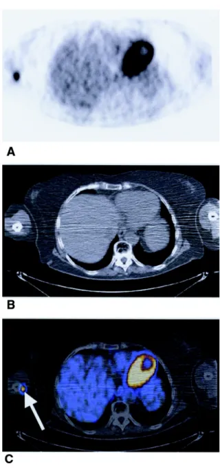
Figure 2. (A) PET image indicating increased FDG uptake in the medial
aspect of the arm and enhancement of the heart. (B) Non-enhanced CT scans at the same level showing no evidence of a tumor. (C) PET–CT fusion shows clear localization of the lesion within the neurovascular bundle of the right arm (arrow).
804
may present with peripheral neuropathy and can be difficult to distinguish from nerve root compression or multifocal neo-plastic infiltration [1, 8–10].
In patients with Morbus Hodgkin and, as recently described, non-Hodgkin’s lymphoma, who develop neurological symp-toms, Guillain–Barre syndrome should also be considered in the differential diagnosis [11]. Diagnosis of NL usually requires histologic demonstration of infiltrating malignant lymphocytes in a peripheral nerve. However, nerve biopsy may fail despite the widespread lymphomatous infiltration of peripheral nerves [5]. While MRI can be useful in identifying affected nerves, it may not be sensitive enough for small lesions [12, 13]. The role of PET in the assessment of non-Hodgkin’s lymphoma and as a prognostic marker is under evaluation. Recent reports show encouraging results, especially in follow-up examinations after chemotherapy [14–18].
During the early years of clinical tumor imaging with PET the potential of multi-modality image fusion has been recog-nized [19]. Meanwhile, several studies have shown that a combination of PET and CT by software co-registration is more accurate than CT alone, especially in staging non-small-cell lung cancer and detection of mediastinal lymph node metastases [20]. Recently, Townsend et al. [21] introduced a combined PET–CT scanner, using a low performance helical CT and a low performance PET scanner, which permits the acquisition of co-registered PET and CT images in the same imaging session. Analysis of the results showed an improve-ment in lesion localization and classification. The technique
used in this case is more advanced since high-end PET and CT scanners were involved, which allowed for rapid data acquisition in <30 min. An advantage lies in the higher con-fidence level of lesion localization that fusion PET–CT may provide. However, this technique is limited by the nature of FDG uptake, since inflammatory lesions may show an increased accumulation of the tracer as well. In this case, inflammatory changes can be excluded by the clinical course of the disease.
In conclusion, causes of peripheral neuropathy may be dif-ficult to distinguish during progressive disease and therapy of aggressive lymphoma. The fusion of PET and CT data may provide a novel tool for imaging NL in relapse of aggressive
Figure 3. Maximum intensity projection PET image from the follow-up
examination, revealing an extensive increase in the tumor burden in the right arm and new lesions in the left side.
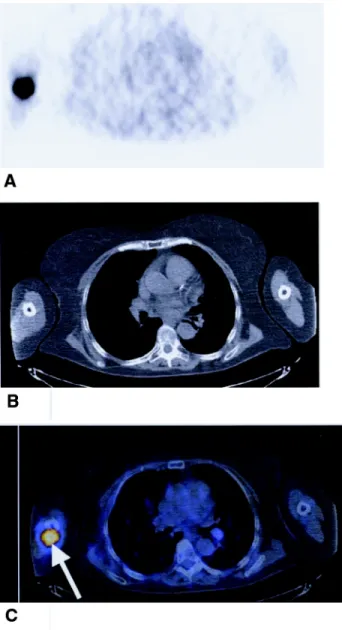
Figure 4. (A) Axial PET image at the level of the right arm showing an
increase in FDG uptake in the right arm. (B) In this CT image, a mass is seen in the neurovascular bundle, which corresponds to the FDG uptake as proven by (C) the fused PET–CT image (arrow).
805
non-Hodgkin’s lymphoma and prompt therapeutic implica-tions without the risk of invasive manoeuvers.
References
1. Hughes RA, Britton T, Richards M. Effects of lymphoma on the peripheral nervous system. J R Soc Med 1994; 87: 526–530. 2. Misdraji J, Ino Y, Louis DN et al. Primary lymphoma of peripheral
nerve: report of four cases. Am J Surg Pathol 2000; 24: 1257–1265. 3. Wulff EA, Simpson DM. Peripheral neuropathy associated with
acquired immunodeficiency syndrome (AIDS)-related Burkitt’s lymphoma. Muscle Nerve 2000; 23: 1764–1766.
4. Pietrangeli A, Milella M, De Marco S et al. Brachial plexus neuro-pathy as unusual onset of diffuse neurolymphomatosis. Neurol Sci 2000; 21: 241–245.
5. van den Bent MJ, de Bruin HG, Bos GM et al. Negative sural nerve biopsy in neurolymphomatosis. J Neurol 1999; 246: 1159–1163. 6. Plotkin SA. Clinical and pathogenic aspects of varicella-zoster.
Post-grad Med J 1985; 61 (Suppl 4): 7–14.
7. Postma TJ, Benard BA, Huijgens PC et al. Long-term effects of vincristine on the peripheral nervous system. J Neurooncol 1993; 15: 23–27.
8. Case records of the Massachusetts General Hospital. Weekly clinico-pathological exercises. Case 9-2001. A 64-year-old woman with peri-pheral neuropathy, paraproteinemia, and lymphadenopathy. N Engl J Med 2001; 344: 917–923.
9. Rogers LR, Borkowski GP, Albers JW et al. Obturator mononeuro-pathy caused by pelvic cancer: six cases. Neurology 1993; 43: 1489– 1492.
10. Liang R, Kay R, Maisey MN. Brachial plexus infiltration by non-Hodgkin’s lymphoma. Br J Radiol 1985; 58: 1125–1127.
11. Re D, Schwenk A, Hegener P et al. Guillain-Barre syndrome in a patient with non-Hodgkin’s lymphoma. Ann Oncol 2000; 11: 217– 220.
12. Quinones-Hinojosa A, Friedlander RM, Boyer PJ et al. Solitary sciatic nerve lymphoma as an initial manifestation of diffuse neuro-lymphomatosis. Case report and review of the literature. J Neurosurg 2000; 92: 165–169.
13. Swarnkar A, Fukui MB, Fink DJ, Rao GR. MR imaging of brachial plexopathy in neurolymphomatosis. Am J Roentgenol 1997; 169: 1189–1190.
14. Hoffmann M, Kletter K, Diemling M et al. Positron emission tomo-graphy with fluorine-18-2-fluoro-2-deoxy-d-glucose (F18-FDG) does not visualize extranodal B-cell lymphoma of the mucosa-associated lymphoid tissue (MALT)-type. Ann Oncol 1999; 10: 1185–1189. 15. Jerusalem G, Beguin Y, Najjar F et al. Positron emission tomography
(PET) with 18
F-fluorodeoxyglucose (18
F-FDG) for the staging of low-grade non-Hodgkin’s lymphoma (NHL). Ann Oncol 2001; 12: 825– 830.
16. Mikhaeel NG, Timothy AR, Hain SF, O’Doherty MJ. 18-FDG-PET for the assessment of residual masses on CT following treatment of lymphomas. Ann Oncol 2000; 11 (Suppl 1): 147–150.
17. Spaepen K, Stroobants S, Dupont P et al. Prognostic value of positron emission tomography (PET) with fluorine-18 fluorodeoxyglucose ([18
F]FDG) after first-line chemotherapy in non-Hodgkin’s lymphoma: is [18F]FDG-PET a valid alternative to conventional
diagnostic methods? J Clin Oncol 2001; 19: 414–419.
18. Mikhaeel NG, Timothy AR, O’Doherty MJ et al. 18-FDG-PET as a prognostic indicator in the treatment of aggressive non-Hodgkin’s lymphoma—comparison with CT. Leuk Lymphoma 2000; 39: 543– 553.
19. Wahl RL, Quint LE, Greenough RL et al. Staging of mediastinal non-small-cell lung cancer with FDG PET, CT, and fusion images: preliminary prospective evaluation. Radiology 1994; 191: 371–377. 20. Wahl RL, Quint LE, Cieslak RD et al. ‘Anatometabolic’ tumor
imaging: fusion of FDG PET with CT or MRI to localize foci of increased activity. J Nucl Med 1993; 34: 1190–1197.
21. Townsend DW. A combined PET/CT scanner: the choices. J Nucl Med 2001; 42: 533–534.