doi: 10.1093/intimm/dxg016, available online at www.intimm.oupjournals.org
Tracking dendritic cells: use of an in situ
method to label all blood leukocytes
Bill Ristevski
1, Alan J. Young
2,5, Lisbet Dudler
2, Ross N. P. Cahill
3,
Wayne Kimpton
3, Elizabeth Washington
3and John B. Hay
1,41Department of Laboratory Medicine and Pathobiology, University of Toronto, 1 Kings' College Circle,
Toronto, Ontario M5S 1A8, Canada
2Basel Institute for Immunology, 4005 Basel, Switzerland
3Laboratory for Foetal and Neonatal Immunology, University of Melbourne, Parkville, Victoria 3052,
Australia
4Department of Immunology, University of Toronto, Toronto, Ontario M5S 1A8, Canada
5Present Address: Department of Biology and Microbiology, South Dakota State University, Brookings, SD
57007, USA
Keywords: carboxy¯uorescein diacetate succinimidyl ester, lymph, lymph node, recirculation Abstract
Here we describe an in situ procedure with a labeling index (percent of labeled blood leukocytes) >98%, which is high enough to permit the direct tracking of dendritic cell (DC) precursors from blood into lymphoid tissues, while circumventing the pitfalls associated with in vitro labeling. DC and lymphocytes have similar blood to afferent lymph migratory capabilities. This method has additional applications in tracking other rare cell populations in both normal and pathological states.
Introduction
Numerous investigators including our three laboratories have studied leukocyte migration, particularly as it relates to lymphocytes. This reassortment and recirculation of lympho-cytes is fundamental for normal immune surveillance, dissem-ination of the immune response, immunological memory, and also plays a role in many pathologies and infectious diseases (1). Previous investigations have characterized a variety of unique cell traf®c migration streams. Among these are tissue-speci®c and subset-tissue-speci®c cell traf®c patterns, such as the differential homing of lymphocytes derived from blood, intes-tinal lymph or s.c. lymph (2±5). Furthermore, the current paradigm of the blood to lymph migratory differences between naive and memory lymphocytes was established in sheep (6). The dynamic kinetics of cell migration during normal and pathological conditions has also been well characterized (1,7,8).
All of these experiments have involved the isolation, labeling and washing of cells in vitro with subsequent re-injection into animals. Maintenance of physiologic integrity has always been a concern, not only due to the time (several hours) of in vitro preparation of cells, but also with respect to the number of cells obtained for labeling.
Typically, in long-term experiments lasting several days or weeks, optimizing these conditions has yielded labeling indices in the blood and lymph that are of the order of 1% (9,10). This labeling index has been suf®cient to study the traf®c and recirculation patterns of major subsets of cells. However, analysis of the kinetics and migration intricacies of minor subsets of cells via staining with mAb in second and third colors has been virtually impossible.
We have devised an in situ labeling approach which has permitted a minor dendritic cell (DC) population to be directly tracked from blood to lymphoid tissue. This in vivo adminis-tration of carboxy¯uorescein diacetate succinimidyl ester (CFSE) has achieved a blood leukocyte labeling index >98%. The implications of this instantaneous labeling technique extend beyond the tracking of DC to other low-frequency cell populations in a variety of species. Compartments with inherently low cell counts (e.g. afferent lymph, peritoneal cavity) can now be more ef®ciently ana-lyzed. This might be particularly relevant to extending studies of the relationship of leukocytes in cerebral spinal ¯uid and the lymphatic exit pathways from the central nervous system (11±13).
Correspondence to: J. B. Hay; E-mail: [email protected]
Transmitting editor: P. Ohashi Received 20 June 2002, accepted 21 October 2002
Methods Animals
Outbred ewes 30±35 kg in weight were obtained from Renwick farms (London, Ontario, Canada) or Versuchsbetrieb Sennweid (Olsberg, Switzerland). Cannulae were surgically established in prescapular efferent lymphatics or prescapular pseudo-afferent lymphatics to allow chronic sampling of lymph as previously described (14). In addition, a catheter was also placed in the jugular vein to allow blood sampling. A full day after surgery was allowed before cell labeling was initiated to re-establish normal physiology. Handling of animals and experimental procedures were conducted in accordance with institutional and national guidelines for animal care and use.
Immunophenotyping
Afferent lymph samples were collected at various intervals for 72 h after cell labeling. Staining for CD4, CD8, gdTCR, CD21, CD1 and CD11c was performed using standard procedures in conjunction with an appropriate allophycocyanin-conjugated secondary antibodies (Southern Biotechnology Associates,
Birmingham, AL). Cells were ®xed in 1% paraformaldehyde and analyzed on a FACSCalibur (Becton Dickinson, San Jose, CA).
Cell labeling
Leukocyte labeling in Wistar rats was accomplished by dissolving 0.8 mg of CFSE (Molecular Probes, Eugene, OR) in 200 ml dimethyl sulfoxide (DMSO) with 10 ml heparin and injecting this solution via the tail vein.
Leukocyte labeling in sheep was accomplished by dissolv-ing 37 mg of CFSE in 6 ml of DMSO and 60 ml of heparin (1000 U/ml). This solution was injected into the blood via the jugular vein in order to label blood leukocytes. Both blood and lymph samples were collected in heparin, and kept on ice whenever possible. Heparin is not needed to obtain cell labeling, but was used as a precaution to avoid clotting. Other animals did not receive heparin and equivalent labeling results were obtained. Cell preparation for FACS
Blood samples were collected in heparin and kept on ice whenever possible. Erythrocytes were lysed with distilled water or ammonium chloride and cells were washed in PBS
Fig. 1. In vivo labeling of blood leukocytes. Blood samples were taken before labeling and 10 min after injection of CFSE i.v. in both sheep and rats. (A and B) Histograms generated by ¯ow cytometric analysis, showing cell count versus CFSE intensity of unstained sheep blood leukocytes and leukocytes 10 min post CFSE injection. (C and D) Results obtained using rat blood.
twice. Cells were ®xed in 1% paraformaldehyde in PBS and analyzed on a FACSCalibur ¯ow cytometer within a maximum of 12 h.
Results
Blood leukocyte labeling
To investigate the ef®cacy of this labeling procedure we used outbred ewes and female Wistar rats. Flow cytometric analysis was employed to measure the ¯uorescence intensity of blood leukocytes, before and after labeling for sheep (Fig. 1A and 1B) and rats (Fig. 1C and 1D) respectively. Blood samples from both species taken 10 min after i.v. infusion of CFSE demonstrated that >98% of all leukocytes in the sample ¯uoresced with a greater intensity than unstained leukocytes. Recirculation of labeled lymphocytes
The functional capacity of cells loaded with CFSE was investigated by observing the ability of lymphocytes to recirculate from blood to lymph after in situ labeling. This is a direct indication of cell function, as lymphocyte transen-dothelial migration is an active process and dead or damaged lymphocytes are not capable of recirculating (5). Chronic efferent lymph collections from a cannulated prefemoral efferent lymphatic in a conscious sheep were made to track the migration of labeled lymphocytes from blood to lymph. Histograms of cell count versus CFSE intensity are shown for a lymph sample prior to blood labeling and for a sample collected 18±20 h after blood labeling (Fig. 2A and B respectively). This clearly demonstrates the functional status of CFSE+lymphocytes. In the example shown, 5.6% of efferent
lymph cells were CFSE+. Continuous sampling of efferent
lymph cells was also made over 40 h, by collecting all the lymph exiting a single cannulated prefemoral efferent lymph-atic (Fig. 2C). In this representative example shown the highest percent of CFSE+efferent lymph cells was recovered
in the collection made between 7 and 18 h. Propidium iodide staining of cells from efferent lymph with 2% CFSE-labeled cells was very similar: 1.8% of CFSE+cells and 2.0% of
non-CFSE-labeled cells were propidium iodide-positive. In vivo versus ex vivo labeling index
Direct comparison of ex vivo versus in vivo labeling techniques was performed to assess the labeling index of lymphocytes. The percent of labeled lymphocytes in the blood versus time was investigated over a period of ~100 h. The reduction of CFSE+ lymphocytes from the blood is shown for ex vivo
labeled cells where the lymphocytes contained in a unit of blood (~400 ml) were incubated with CFSE in vitro and returned to the venous circulation of a sheep (Fig. 3A). Also shown is the reduction of lymphocytes after i.v. injection of CFSE labeling the entire blood leukocyte pool (Fig. 3B). Both curves show a similar reduction of lymphocytes from the blood compartment. However, the labeling index of lymphocytes in the blood is ~2000% higher with the in situ technique. Figure 4(A and B) shows histograms of cell count versus ¯uorescence for spleen and mesenteric lymph node cells respectively. Due to the fenestrated sinusoids in the spleen creating a very open system of cellular exchange with the blood, it is expected that CFSE+cells would be present after 1 h.
However, migration of cells into lymph nodes from the blood would be very minimal after 1 h, as this phenomenon reaches it peak at 24 h. This difference of migratory capabilities after in vivo labeling is demonstrated in Fig. 4. This also
Fig. 2. Recirculation of labeled blood lymphocytes into efferent lymph. Histograms showing cell count versus CFSE ¯uorescence intensity of efferent lymph cells collected from a prefemoral efferent lymphatic before in vivo labeling (A), and a 2-h collection made between 18 and 20 h after CFSE injection (B). (C) Percent of CFSE-labeled lymphocytes found following chronic sampling of the same prefemoral lymphatic over a 40-h period. Samples were analyzed on different days, after daily calibration of the ¯ow cytometer.
demonstrates that CFSE is not capable of leaving the blood compartment in suf®cient quantity to stain lymph node cells at the concentrations used for this study.
In vivo tracking of DC
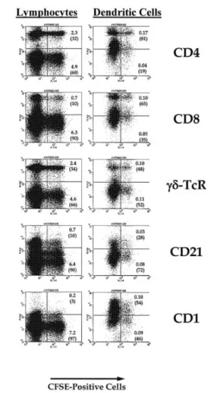
The large increase in labeling index using this in situ method indicated that tracking of low-frequency leukocyte populations was possible. Afferent lymph was collected either through direct cannulation or through the cannulation of pseudo-afferent lymph as previously described (14). The phenotype of labeled cells recovered in pseudo-afferent lymph 24±48 h after in vivo labeling is shown in Fig. 5. Cells were gated to include small lymphocytes according to previously estab-lished forward and side-scatter parameters (15). In order to enhance for DC inclusion, afferent lymph cells were stained with a panel of mAb, including CD1, mannose receptor, CD11c and CD21. It has previously been established that antibody 20-27 (CD1) speci®cally identi®es afferent lymph DC (16). Based on staining with CD1, cells were gated to include all afferent lymph DC. The majority of CD1+ cells (DC),
including the labeled population, were recovered in the large-cell (high forward scatter) gate, rather than the low forward scatter gate used to analyze lymphocytes. When this gate was used, labeled CD1+ cells accounted for ~0.1% of the total
population. More importantly, 54% of the larger, CFSE-labeled cells recovered in afferent lymph are CD1+.
In addition to the simple identi®cation of labeled DC in afferent lymph after in situ blood labeling, it was possible for the ®rst time to assess the kinetics and recovery of labeled afferent lymph DC. While the peak of the recovery of DC was slightly later than lymphocytes, the magnitude of recovery was similar for both populations (Fig. 6). These data were consist-ent independconsist-ent of whether CD1 or high expression of CD11c was used to identify DC in afferent lymph. When the number of labeled cells within each subset was calculated as a propor-tion of that subset, it became clear that similar numbers of labeled DC and labeled lymphocytes migrated into the afferent lymph after whole-blood labeling.
Discussion
This is the ®rst report of labeling the blood in situ, even though FITC and CFSE have been used to label cells in other tissues in situ (17±19), and PKH-26 has been able to selectively label neutrophils for 24 h (20). The present study is the ®rst to use DC markers to track this minor population from blood into normal afferent lymph.
CFSE is an ester combined with the ¯uorescein moiety. It is highly membrane permeable, and requires esterases found in cells to cleave the molecule and allow binding of the
Fig. 4. CFSE+ cells in spleen versus mesenteric lymph node 1 h
after in vivo labeling. Histograms showing cell count versus CFSE ¯uorescence intensity of cells from a biopsy performed on the spleen (A) and mesenteric lymph node (B) 1 h after in vivo labeling. Fig. 3. Reduction of labeled lymphocytes from the blood: in vitro
versus in vivo labeling. A direct comparison was made between in vitro and in vivo labeling by plotting the reduction of labeled lymphocytes from the blood compartment using both methods. Results show (A) the percent of CFSE+ lymphocytes in the blood
versus time, after the cells contained within a unit of blood were labeled in vitro with CFSE and returned to the sheep's venous circulation (n = 5); (B) the percent of CFSE+ lymphocytes in the
blood versus time, after in vivo labeling with CFSE (n = 3). Error bars indicate SEM.
¯uorochrome to intracellular proteins. We found, as was previously reported by others (21), that the intensity of CFSE-labeled cells declines over a 24-h period, but then stabilized, allowing long-term tracking of labeled cells. There may be further analytical procedures which could be devel-oped to distinguish cell division from other phenomena which affect loss of intensity, such as cell death or turnover of labeled intracellular elements. Labeling of platelets may allow future studies to make relative comparisons between lymphocyte or leukocyte subsets and these non-dividing, blood elements. Using our labeling conditions, only blood leukocytes were labeled. For example, insuf®cient CFSE reached lymph nodes to directly label cells in this compartment. This is supported by two observations made. (i) Lymph nodes collected 1 h after
blood labeling remained unlabeled. (ii) Kinetics of recovery of labeled lymphocytes in lymph were similar to the kinetics found with in vitro labeling studies. However, it is conceivable that by increasing the dye concentration, blood vascular cells and other organs/tissues might be directly labeled.
The present methodology enhances and validates a sub-stantial literature on the use of cell-tracking dyes to de®ne lymphocyte migration patterns (1,7,8). Leukocyte migration is a dynamic and complex process, which requires analysis of an ever-changing cellular mosaic in a physiological environ-ment. In vivo cell tracking has been the principal technique to investigate this phenomenon. While rodent studies have been particularly useful in establishing the molecular basis of leukocyte homing, particularly with respect to interactions with endothelial cells (22,23), investigations in sheep have de®ned the physiology of lymphocyte recirculation. This is a powerful experimental approach as large animals allow surgical techniques to sequentially sample blood and many different lymphatic compartments during the course of a single experiment. Chronic lymph collection in conscious sheep, combined with ¯uorescent cell-tracking compounds and ¯ow cytometry, has documented kinetics and quantitative recovery of i.v. injected labeled cells. Lymphocyte migration patterns for a variety of subsets and a variety of normal and pathological tissues have been well characterized (4±6,10). In fact, this physiological system has been crucial in validating cellular tracking labels. Only viable undamaged lymphocytes traf®c from blood to lymph (5).
Because DC have a fundamental role in antigen presenta-tion they have received considerable attenpresenta-tion over the last few years (24). It should be noted that the intimate association of veiled cells with lymphocytes was described in afferent lymph several decades ago (25). Typically the special status of lymphocytes has been recognized due to their recirculation capabilities, meaning that they continuously exit the blood compartment and return via the lymphatic system. However, a truncated migration pathway from blood to lymph nodes via afferent lymphatics exists for monocytes, DC and macro-phages. It is interesting that the cellular composition of afferent lymph in terms of the proportion of DC and macrophages to lymphocytes is very similar to blood (~1:10). We have shown that the capacity of DC to migrate from the blood to afferent lymph is similar to lymphocytes. In addition, the kinetics of the appearance of DC and lymphocytes in afferent lymph is comparable. Since monocytes, DC and macrophages are not found in normal efferent lymph, this indicates that selective cellular ®ltering solely resides in the lymph node.
Surprisingly there is a lack of detailed quantitative data on labeled leukocytes in the blood. In addition, the technical limitations associated with various labeling techniques used and sequential sampling have yielded a wide range of published half-times (10,26±29). We have been able to use the combination of this labeling technique with multiple sequential sampling to de®ne detailed blood curves for lymphocytes, monocytes, neutrophils and platelets (Hay and Ristevski, in preparation). We think the application of this method will permit a series of sound physiological experi-ments providing new information in the areas of normal hematology, leukemia, systemic infectious diseases and chronic in¯ammation.
Fig. 5. Phenotype of labeled peripheral blood cells recovered in afferent lymph. Cells collected 24±48 h after in vivo blood labeling were reacted with appropriate antibodies and analyzed by ¯ow cytometry. As DC are signi®cantly larger than lymphocytes, and to allow more detailed analysis of the lymphoid and non-lymphoid populations, cells were either gated as low side scatter/low forward scatter (lymphocytes) or as high forward scatter/high side scatter (DC). At least 100,000 cells were then analyzed for each antibody to determine either the population of gated cells which were positive for both CFSE and the antibody of interest (large number) or the proportion of labeled cells reactive for each antibody (bracketed number). Antibody controls not shown.
Abbreviations
CFSE carboxy¯uorescein diacetate succinimidyl ester DC dendritic cell
DMSO dimethyl sulfoxide
References
1 Seabrook, T., Au, B., Dickstein, J., Zhang, X., Ristevski, B. and Hay, J. 1999. The traf®c of resting lymphocytes through delayed hypersensitivity and chronic in¯ammatory lesions: a dynamic equilibrium. Semin. Immunol. 11:115.
2 Mackay, C., Kimpton, W., Brandon, M. and Cahill, R. 1988. Lymphocyte subsets show marked differences in their distribution between blood and the afferent and efferent lymph of peripheral lymph nodes. J. Exp. Med. 168:1744.
3 Chin, G. and Hay, J. 1980. A comparison of lymphocyte migration through intestinal lymph nodes, subcutaneous lymph nodes, and chronic in¯ammatory sites of sheep. Gastroenterology 79:1231.
4 Cahill, R., Poskitt, D., Frost, H. and Trnka, Z. 1977. Two distinct pools of recirculating T lymphocytes: migratory characteristics of nodal and intestinal T lymphocytes. J. Exp. Med. 145:420.
5 Andrade, W., Johnston, M. and Hay, J. 1998. The relationship of blood lymphocytes to the recirculating lymphocyte pool. Blood 91:1653.
6 Mackay, C., Marston, W. and Dudler, L. 1990. Naive and memory T cells show distinct pathways of lymphocyte recirculation. J. Exp. Med. 171:801.
7 Cahill, R. N. P., Kimpton, W. G., Washington, E. A., Holder, J. E. and Cunningham, C. P. 1999. The ontogeny of T cell recirculation during foetal life. Semin. Immunol. 111:105.
8 Young, A. 1999. The physiology of lymphocyte migration through the single lymph node in vivo. Semin. Immunol. 11:73.
9 Teare, G. F., Horan, P. K., Slezak, S. E., Smith, C. and Hay, J. B. 1991. Long-term tracking of lymphocytes in vivo: the migration of PKH-labeled lymphocytes. Cell. Immunol. 134:157.
10 Young, A. and Hay, J. 1995. Rapid turnover of the recirculating lymphocyte pool. Int. Immunol. 7:1607.
11 Seabrook, T. J. and Hay, J. B. 2001. Intracerebroventricular infusion of TNF-alpha preferentially recruit blood lymphocytes and induce a perivascular leukocyte in®ltrate. J. Neuroimmunol. 113:81. 12 Seabrook, T. J., Johnston, M. and Hay, J. B. 1998. Cerebral spinal
¯uid lymphocytes are part of the normal recirculating lymphocyte pool. J. Neuroimmunol. 91:100.
13 Boulton, M., Flessner, M., Armstrong, D., Hay, J. and Johnston, M. 1998. Determination of volumetric CSF absorption into extracranial lymphatics in sheep. Am. J. Physiol. 274:88.
14 Young, A., Hein, W. and Hay, J. 1997. Cannulation of lymphatic vessels and its use in the study of lymphocyte traf®c. In Lefkovits, I., ed., Handbook of Experimental Immunology, p. 2039. Academic Press, New York.
15 Young, A., Marston, W., Dessing, M., Dudler, L. and Hein, W. 1997. Distinct recirculating and non-recirculating B-lymphocyte pools in the peripheral blood are de®ned by coordinated expression of CD21 and L-selectin. Blood 90:4865.
16 Howard, J., Sopp, J., Brownlie, J., Kwong, S., Parsons, R. and Taylor G. 1997. Identi®cation of two distinct populations of dendritic cells in afferent lymph that vary in their ability to stimulate T cells. J. Immunol. 159:5372.
17 Cunningham, C. P., Kimpton, W. G., Holder, J. E. and Cahill, R. N. 2001. Thymic export in aged sheep: a continuous role for the thymus throughout pre- and postnatal life. Eur. J. Immunol. 31:802.
18 Graziano, M., St-Pierre, Y., Beauchemin, C., Desroslers, M. and Fig. 6. Appearance of labeled lymphocytes and DC in afferent lymph after in vivo labeling. Cells collected from pseudo-afferent lymph at various times after whole-blood labeling (t = 0) were reacted with appropriate antibodies to determine recirculation into the lymph. In order to quantitate the degree to which labeled cells recirculated into lymph, the number of labeled cells reactive with each antibody was expressed as a percentage of the total number of cells reactive with each antibody. (Clearly, CD1+ or CD11c+ DC migrate into afferent lymph in
Potworowski, E. F. 1998. The fate of thymocytes labeled in vivo with CFSE. Exp. Cell Res. 240:75.
19 Pabst, R. and Binns, R. M. 1981. In vivo labeling of spleen and mesenteric lymph nodes with ¯uorescein isothiocyanate for lymphocyte migration studies. Immunology 44:321.
20 Albertine, K. H. and Gee, M. H. 1996. In vivo labeling of neutrophils using a ¯uorescent cell linker. J. Leukoc. Biol. 59:631. 21 Parish, C. R. 1999. Fluorescent dyes for lymphocyte migration and
proliferation studies. Immunol. Cell Biol. 77:499.
22 Campbell, J. J. and Butcher, E. C. 2000. Chemokines in tissue-speci®c and microenvironmental-tissue-speci®c lymphocyte homing. Curr. Opin. Immunol. 12:336.
23 Springer, T. 1994. Traf®c signals for lymphocyte recirculation and leukocyte emigration: the multistep paradigm. Cell 76:301.
24 Hartgers, F. C., Figdor, C. G. and Adema, G. J. 2000. Towards a molecular understanding of dendritic cell immunobiology. Immunol. Today 21:542.
25 Heath, T., Lascelles, A. and Morris, B. 1962. The cells of sheep lymph. J. Anat. 96:397.
26 van Furth, R., Raeburn, J. and van Zwet, T. 1979. Characteristics of human mononuclear phagocytes. Blood 54:485.
27 Meuret, G. 1972. Monocytopoiesis and kinetics of blood monocytes in man. Blut 24:337.
28 Whitelaw, D. and Batho, H. 1972. The distribution of monocytes in the rat. Cell Tissue Kinet. 5:215.
29 Issekutz, T., Issekutz, A. and Movat, H. 1981. The in vivo quantitation and kinetics of monocyte migration into acute in¯ammatory tissue. Am. J. Pathol. 103:47.