S U R G I C A L T E C H N I Q U E
Arthroscopically Assisted Removal of Intraosseous Ganglion
Cysts of the Distal Tibia
Lorenz Bu¨chler MD, Harish Hosalkar MD, MBMS (Orth), FCPS (Orth), DNB (Orth), Martin Weber MD
Received: 23 September 2008 / Accepted: 17 February 2009 / Published online: 10 March 2009
Ó The Association of Bone and Joint Surgeons 2009
Abstract Intraosseous ganglia of the distal tibia are rare. We evaluated the feasibility of surgically treating these lesions with an arthroscopically assisted technique. Five patients with symptomatic distal tibial ganglia underwent surgical curettage and excision with this technique. All patients underwent de´bridement of the chondral lesion and hypertrophied synovial lining when present, probing of the portal to the ganglion, and subsequently thorough curettage with bone grafting performed through a cortical window made from a separate small incision. Biopsy confirmed the diagnosis in all patients. All patients had eventual relief of symptoms with good integration of bone graft at final followup. There were no recurrences at a minimum fol-lowup of 19 months (mean, 38.6 months; range, 19– 69 months). Mean time for return to full function was 15.4 weeks (range, 8–17 weeks). There were no intraop-erative or postoperative complications. The mean American Orthopaedic Foot and Ankle Society scores
increased from 73 points (range, 67–77 points) preopera-tively to 94 points (range, 90–100 points) postoperapreopera-tively. Arthroscopically assisted surgical treatment of ganglia of the distal tibia in the appropriate patient is a reasonably simple technique that relieves symptoms and helps the patient to regain normal gait and full function with no recurrence (in our small series).
Level of Evidence: Level IV, case series. See Guidelines for Authors for a complete description of levels of evidence.
Introduction
Although soft tissue ganglia are the most common benign tumors in the extremities and occasionally show evidence of bony erosions, intraosseous ganglia are rare [6,7, 12, 13]. Historically, cystic erosions located in the vicinity of a joint not associated with degenerative or inflammatory joint lesions have been reported under different names, mostly owing to the difference in interpretation of the etiology. Since the review article of Schajowicz et al. [15], the universal term for this lesion is ‘‘intraosseous gan-glion.’’ These lesions commonly are found in the lower extremity, the proximal tibia being the most common site [7,8,20]. There are some reports in the English literature regarding intraosseous ganglia around the ankle. Most of these lesions reportedly were in the tarsal bones and the medial malleolus and some in the lateral malleolus [4,8,12,14,16].
Intraosseous ganglia of the distal tibia are rare [7,12]. In the current scenario of advanced surgical techniques, arthroscopically assisted treatment of these juxtaarticular benign lesions is possible and can be meticulously accom-plished. This technique has been used successfully for
Each author certifies that he or she has no commercial associations (eg, consultancies, stock ownership, equity interest, patent/licensing arrangements, etc) that might pose a conflict of interest in connection with the submitted article.
Each author certifies that his or her institution has approved the human protocol for this investigation, that all investigations were conducted in conformity with ethical principles of research, and that informed consent for participation in the study was obtained. This work was performed at University of Bern, Inselspital. L. Bu¨chler, M. Weber (&)
Department of Orthopaedic Surgery, University of Bern, Inselspital, 3010 Bern, Switzerland
e-mail: [email protected] H. Hosalkar
Department of Orthopedic Surgery, University of Pennsylvania, School of Medicine, Philadelphia, PA, USA
treating intraosseous ganglia in other locations, such as the talus, the femoral condyle, and the lunate [2,11,16,18].
We explored the feasibility of achieving adequate mar-ginal excision of intraosseous ganglion cysts of the distal tibia via an arthroscopically assisted technique. Addition-ally, we report the presenting symptoms, patterns of distribution and dimensions of these lesions, associated changes of degree of arthritis in these ankles, intraoperative findings, postoperative American Orthopaedic Foot and Ankle Society (AOFAS) scores at latest followup, and recurrence rates at midterm followup.
Materials and Methods
We retrospectively reviewed all patients with histopatho-logically proven intraosseous ganglia of the distal tibial plafond treated with an arthroscopically assisted technique between January 2000 and December 2005. Five patients were found in our records (four males and one female), all of whom were included in the study. None of the five patients had any medical comorbidities. One patient described minor trauma to the medial malleolus with a metal bar approxi-mately 6 years ago, which we believe is merely coincidental. All other patients denied trauma in their his-tory. Patients’ charts, radiographs, advanced imaging studies, and arthroscopic images and videos, were reviewed. All patients presented with intermittent dull aching pain in the ankle. The mean duration of symptoms from onset to diagnosis was 5.5 years (range, 2–10 years). Except anal-gesic medication, there was no intervention before surgery. The mean age was 49.8 years (32–61 years). We reviewed plain radiographic and MRI features to confirm the benign and cystic natures of the lesion (therefore most likely pointing toward the diagnosis of a ganglion cyst) and noted the lesion dimensions and anatomic location; we graded radiographic arthritis based on the previously published classification of van Dijk et al. [19], where Grade 0 = normal joint or subchondral sclerosis, Grade 1 = os-teophytes without joint space narrowing, Grade 2 = joint space narrowing with or without osteophytes, and Grade
3 = (sub)total disappearance or deformation of the joint space. We recorded the intraoperative findings, biopsy confirmation, and postoperative course. AOFAS scores (a previously validated functional score for the ankle and hindfoot) [9] were obtained preoperatively and at the latest followup. Finally, we noted recurrences. The minimum followup was 19 months (mean, 38.6 months; range, 19– 69 months). Institutional review board approval was obtained.
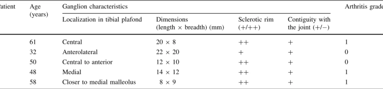
Preoperatively, standardized plain radiographs of the ankle with weightbearing and MRI were obtained for all patients. All MR images showed a juxtaarticular, localized, subchondral, cystic lesion with cancellous edema noted near the ganglion. All intraosseous lesions were connected to the joint, despite various locations in the distal tibia. Three patients had no degenerative joint disease (Grade 0) and two had mild degeneration (Grade 1) (Table 1).
Arthroscopy of the ankle was performed with the patient under epidural anesthesia. All patients were operated on by the senior surgeon (MW). A two-portal approach with anteromedial and anterolateral portals was used in all cases. In Patient 3, two posterior portals were used. After routine diagnostic arthroscopy to look at the entire joint and note changes of degeneration, the cartilage lesion corresponding to the ganglion cyst was identified. In all patients, we identified a portal of entrance to the lesion from within the joint. It appeared as a small area of soft cartilage with a central fissure, and on probing, the probe would sink through the subchondral plate into the ganglion cyst confirming the path (Fig.1). The portal between the joint and the ganglion was de´brided and the subchondral bone perforated to induce formation of a scar tissue and subsequent closure of the connection in all patients. In three of the five patients, we found moderate synovitis, which was de´brided. In one patient, a small osteophyte on the tibia at the anterolateral joint line was removed. In two patients, a chondral lesion in the joint (one centrally and one medially) was de´brided. The cyst then was approached through a separate incision where a cortical window was created based on the location of the cyst. All five lesions contained a viscous yellowish mucoid material in the cyst
Table 1. Preoperative evaluation and findings Patient Age
(years)
Ganglion characteristics Arthritis grade Localization in tibial plafond Dimensions
(length 9 breadth) (mm) Sclerotic rim (+/++) Contiguity with the joint (+/ ) 1 61 Central 20 9 8 ++ + 1 2 32 Anterolateral 22 9 20 + + 0 3 50 Central to anterior 12 9 10 ++ + 0 4 48 Medial 14 9 12 ++ + 1
cavity. The entire lining membrane was de´brided carefully and removed until the connection into the joint became visible (Fig.2). The cyst lining and de´brided material underwent histopathologic analysis. The osseous cavity
was packed with cancellous bone, which was obtained from the local distal tibial metaphysis.
Owing to the potential negative effect of fluid extrava-sation on bone graft healing, we kept postoperative joint irritation to a minimum by immobilizing the ankle in a nonweightbearing cast for 3 to 4 weeks. We hoped this would allow early healing of the graft and formation of a tight closure of the joint-side entrance to the ganglion by scar tissue. Afterward, the cast was removed and full weightbearing was resumed progressively, typically within 4 to 6 weeks.
Postoperative followup was at 2 weeks, 4 weeks, 8 weeks, and 6-month intervals thereafter. Biopsy revealed a simple benign ganglion cyst with mucoid degeneration in all cases. Postoperative weightbearing radiographs were obtained at 3 months, at 1 year, and at the latest followup. Repeat MRI was performed only if there was persistent or residual pain.
Results
All patients underwent complete excision of the cyst via the arthroscopically assisted technique that offered the added advantages of observing and inspecting the entire joint surface.
The postoperative course was uneventful for all the patients. The mean duration of hospitalization was 3.8 days (range, 3–5 days). Hospitalizing the patients for a few days is a standard practice in our health system. With different sociocultural circumstances and in other health systems, this relatively minor procedure could be performed with much shorter hospitalization or even on an outpatient basis. The mean time to return to full duty was 15.4 weeks (8– 17 weeks). The mean AOFAS scores increased from 73 points (range, 67–77 points) preoperatively to 94 points (range, 90–100 points) postoperatively (Table2). The AOFAS subscores for alignment (10 points), walking dis-tance (5 points), ankle motion (6 points), and subtalar motion (8 points) remained unchanged preoperatively and postoperatively, with the maximum of points allocated for
Fig. 1 An arthroscopic view shows the anterior part of ankle. The tip of the probe marks the chondral lesion that leads into the ganglion cyst (arrow). A = tibial plafond; B = talus.
Fig. 2 An endoscopic view from within the cavity of the ganglion (A) is shown. The arrow marks the cartilage of the talus, seen through the portal between the ganglion and the joint. In all patients, we noted a communication between ganglion and joint.
Table 2. Results after arthroscopically assisted surgical treatment Patient Followup (months) AOFAS score (preoperative/ postoperative) Arthritis grade at last followup 1 24 72/90 1 2 26 77/100 0 3 19 74/100 0 4 55 76/90 1 5 69 67/90 1
all patients in all of these subscores. The subscores for function (maximum 10 points) improved from an average of 8.2 points to 10 points, for walking on uneven surfaces (maximum 5 points) from 3.6 points to 5 points, and for ankle stability (maximum 8 points) from 6.4 points to 8 points. The score for pain was the only subscore that did not improve to the maximum (40 points), with the average score increasing from 18 points to 34 points.
The plain radiographs taken at latest followup showed no signs of progression of osteoarthritis in any of the patients. None of the patients had any recurrence of the lesion. In four patients, the bone grafts were well integrated by the 6-month followup and they remained asymptomatic (Fig.3). One patient (Patient 5) who initially did well postoperatively had intermittent ankle pain at the 2-year 9-month followup. MRI was repeated at that point and showed evidence of scarring with some intraosseous edema at the location of the bone graft (Fig.4). Inflammatory and infection markers were negative and there was no recur-rence of the ganglion cyst. At the latest followup,
69 months after surgery, the patient was asymptomatic with good function. Plain radiographs revealed no signs of recurrence.
Discussion
Intraosseous ganglia are rare, benign lesions of the bone. Various terms such as subchondral bone cyst, synovial bone cyst, intraosseous mucoid cyst, or juxtaarticular bone cyst have been proposed. Radiographs typically show a well-demarcated circular to oval radiolucent defect without internal calcification, outlined by a rim of sclerotic bone without altering the cortex. In the current scenario of advanced surgical techniques, arthroscopically assisted treatment of intraosseous ganglion cysts is possible. We explored the feasibility of achieving adequate marginal excision of intraosseous ganglion cysts of the distal tibia via an arthroscopically assisted technique. We also reported the presenting symptoms, patterns of distribution
Fig. 3A–F Preoperative (A) anteroposterior and (B) lateral plain radiographs show the cystic lesion with a surrounding mild sclerotic rim. (C) T1-weighted and (D) T2-weighted coronal MR images of the ankle show the juxtaarticular cystic lesion with surrounding mild changes of edema and chondral changes. The arrow marks a defect in
the cartilage and subchondral bone. Arthroscopically, a connection between the joint and the ganglion was seen. (E) Anteroposterior and (F) lateral plain radiographs taken 3 months after arthroscopically assisted excision and bone grafting show graft incorporation with no progression in arthritis and no evidence of recurrence.
and dimensions of these lesions, associated changes of degree of arthritis in these ankles, intraoperative findings, postoperative AOFAS scores at latest followup, and recurrence rates at midterm followup.
There are limitations to this study. This observational analysis was performed on a relatively small sample size of five patients, which, considering the rarity of the diagnosis, may be a relatively large number. The length of followup is short (mean, 19 months) and certainly a limitation; how-ever, all patients were followed carefully until they became symptom-free and had functional improvement. All patients and surgical outcomes including AOFAS scores were evaluated by the senior author and not by an inde-pendent observer, potentially introducing systematic bias. The study reflects the perspective of one institution regarding surgical approach and treatment of this rare disorder of the distal tibia (although one of the largest single series of this condition with surgical treatment and followup). A multi-institutional involvement or study design would have allowed not only a greater number of patients to be analyzed but also variations in treatment
regimens. Unfortunately, because of the rarity of the diagnosis, such a prospective study would require a long time and recruitment of patients from multiple institutions. One of the major problems in our study design is the lack of a control group (with probably an isolated arthroscopy group and another isolated open-surgical group). Given the fact that we were convinced of the advantages of using the dual arthroscopically assisted technique and that it allowed thorough excision of the lesion with inspection and treat-ment of associated joint structures, we believed it was unethical to withhold parts of this treatment to other patients in the interest of creating a control group. Some surgeons in our area and regional hospitals who use only the open technique may be a reasonable source of control patients for future studies. Given that all patients initially were treated at other centers and referred for surgical consideration, it is possible we examined a group of patients for whom nonoperative treatments already had failed and had more of a joint involvement attributable to delay in treatment, resulting in selection bias. The rarity of the condition and its vague initial presentation, with likely failure of nonoperative measures and subsequent referral to a tertiary institution, are probably the scenario most sub-specialists in different parts of the world will encounter.
We did not offer nonoperative treatment (eg, a non-weightbearing cast for a period of time) to these patients as we did not believe the chances of success were likely after the many years of symptoms. The average duration of symptoms was 5.5 years. The reason for the long delay until diagnosis and referral is that the patients typically present to their family physician with intermittent mild to moderate pain of several months’ to years’ duration. With negative physical examination and normal plain radio-graphs, the patients receive no treatment or only analgesics. Only on further recurrence or increasing pain would MRI be performed, leading to diagnosis and referral. The bone marrow edema surrounding the ganglion would seem more likely the cause of the dull, deep-seated pain than the associated synovitis in the recesses, which was only mild to moderate. Therefore, we believe a synovectomy alone would not have been a treatment option with sufficient chance of lasting relief of symptoms. In all our patients, review of the MRI by an experienced musculoskeletal radiologist and the treating team of orthopaedic surgeons led to the unanimous diagnosis of a ganglion. In case of doubt, it is imperative a biopsy of the lesion be done extraarticularly first and to avoid a caveat arthroscopy [10,17].
Arthroscopy never should be used as a substitute for proper clinical and radiographic examinations before sur-gery. Surgeons should only proceed with surgery if they are absolutely certain about the benign nature of the lesion. If there is any doubt regarding the nature of the tumor, the
Fig. 4 A MR image of the patient with prolonged symptoms 33 months postoperatively shows some scarring and intraosseous edema at the location of the bone graft (arrow).
patient should be referred to a center well-conversant with the treatment of bone tumors before surgery or biopsy of the lesion. Differential diagnosis includes epiphyseal lesions in infection or osteomyelitis, but there one would expect clinical findings. Chondroblastomas usually are seen in young patients with open physes. They can have a similar appearance with a central region of bone destruc-tion surrounded by a rim of sclerotic bone. The presence of surrounding marrow edema seen on MRI favors the diag-nosis of a ganglion. Giant cell tumors classically present with nonsclerotic well-defined margins. Absence of arthritic changes and younger age favor an intraosseous ganglion over a subchondral cyst of osteoarthritis.
The constant finding of a small area of soft and fissured cartilage with an underlying defect of the subchondral bone giving direct portal of access (valve) to the ganglion seems to support the theory of an articular origin of the ganglion [5, 14], rather than a degenerative process in epiphyseal cancellous bone penetrating the joint [7, 8, 15, 20]. Hypothetically, the portal of access then would allow joint fluid penetration into the subchondral cancellous bone with pressure erosion of the trabeculae and subsequent forma-tion of a cyst. De´bridement of the cartilage lesion, in conjunction with the cancellous packing of the cyst, we believe, induced scarring and closure of this portal, thus effectively sealing it and preventing recurrence. Lacking a control group of patients with a similar disorder treated with bone grafting alone, this explanation cannot be confirmed.
Crabbe [5] initially reported 10 cases of a cystic lesion adjacent to a joint, surrounded by a zone of sclerosis, and introduced the term ‘‘intraosseous ganglion.’’ By 1973, fewer than 40 cases of intraosseous ganglia had been reported in the English literature, according to Feldman and Johnston [6]. Schajowicz et al. [15] reported 88 cases over 17 years with followups from 1 to 12 years. Sixteen of the 88 cases were about the ankle. All were treated with open excision of the ganglion and bone grafting, with two recurrences. In general, recurrences seem rare, as the reported recurrence rates are only 2.2% to 7% [12,15].
Arthroscopic techniques for treating benign lesions of the distal tibial plafond have been described [1, 3], but apparently not for intraosseous ganglia of the distal tibia. Our hypothesis of the valve mechanism in the etiology of the ganglion requires removal of this circumscript osteo-chondral lesion. This can be accomplished from the joint side or from the ganglion side. We fear de´briding the lesion from the ganglion side either will be inadequate or possibly lead to excessive joint damage. From the joint side, this can be obtained much more precisely and reliably. We believe the arthroscopic approach is more comfortable for the patient. We were able to observe the joint well in all cases, locate the portal or valve into the ganglion cyst, and
perform thorough de´bridement of chondral debris and hypertrophic synovial lining through the portal. Addition-ally, the small incision to make a cortical window helped in thorough curettage and complete excision of the lining of the cyst wall. We believe the combined technique was responsible for complete relief of symptoms noted in all our patients (as the intraarticular de´bridement helped to relieve symptoms emanating from the joint). Also, the bony window, with curettage and grafting and eventual consolidation of the graft in the cavity, was helpful in preventing recurrence. Although we had few patients in this series, we believe the recurrence rate is low with this technique.
Based on our small series, it seems feasible to treat distal tibial intraosseous ganglion cysts with arthroscopically assisted surgical intervention, de´bridement and curettage with bone grafting, with good results and improvement of function. All patients became symptom-free and we observed no recurrences.
References
1. Andrews JR, Tedder JL, Godbout BP. Simple bone cyst of the distal tibia: a case for ankle arthroscopy. Arthroscopy. 1991;7: 381–384.
2. Ashwood N, Bain GI. Arthroscopically assisted treatment of intraosseous ganglions of the lunate: a new technique. J Hand Surg Am. 2003;28:62–68.
3. Banerjee D, Eriksson K, Morris H. Arthroscopically treated intraarticular osteoid osteoma in the ankle: a report of 3 cases. Acta Orthop. 2005;76:721–724.
4. Coulier B, Devyver B, Hamels J. Imaging demonstration of fis-tulous gas communication between joint and ganglion of medial malleolus. Skeletal Radiol. 2002;31:57–60.
5. Crabbe WA. Intra-osseous ganglia of bone. Br J Surg. 1966;53: 15–17.
6. Feldman F, Johnston A. Intraosseous ganglion. Am J Roentgenol Radium Ther Nucl Med. 1973;118:328–343.
7. Ferkel RD, Field J, Scherer WP, Bernstein ML, Kasimian D. Intraosseous ganglion cysts of the ankle: a report of three cases with long-term follow-up. Foot Ankle Int. 1999;20:384–388. 8. Helwig U, Lang S, Baczynski M, Windhager R. The intraosseous
ganglion: a clinical-pathological report on 42 cases. Arch Orthop Trauma Surg. 1994;114:14–17.
9. Ibrahim T, Beiri A, Azzabi M, Best AJ, Taylor GJ, Menon DK. Reliability and validity of the subjective component of the American Orthopaedic Foot and Ankle Society clinical rating scales. J Foot Ankle Surg. 2007;46:65–74.
10. Joyce M, Mankin H. Caveat arthroscopos: extra-articular lesions of bone simulating intra-articular pathology of the knee. J Bone Joint Surg Am. 1983;65:289–292.
11. Kubota C, Kobayashi S, Miyazaki T, Kokubo Y, Yayama T, Uchida K, Sato R, Bangirana A, Baba H. Exceedingly large femoral condyle intraosseous ganglion cyst following high tibial osteotomy. J Orthop Sci. 2007;12:592–596.
12. Murff R, Ashry HR. Intraosseous ganglia of the foot. J Foot Ankle Surg. 1994;33:396–401.
13. Pope TL Jr, Fechner RE, Keats TE. Intra-osseous ganglion: report of four cases and review of the literature. Skeletal Radiol. 1989;18:185–187.
14. Rozbruch SR, Chang V, Bohne WH, Deland JT. Ganglion cysts of the lower extremity: an analysis of 54 cases and review of the literature. Orthopedics. 1998;21:141–148.
15. Schajowicz F, Clavel Sainz M, Slullitel JA. Juxta-articular bone cysts (intra-osseous ganglia): a clinicopathological study of eighty-eight cases. J Bone Joint Surg Br. 1979;61:107– 116.
16. Scholten PE, Altena MC, Krips R, van Dijk CN. Treatment of a large intraosseous talar ganglion by means of hindfoot endos-copy. Arthrosendos-copy. 2003;19:96–100.
17. Schwartz H, Limbird T. Caveat arthroscopy: definition and guidelines for prevention. J South Orthop Assoc. 1996;5:96–100. 18. Uysal M, Akpinar S, Ozalay M, Ozkoc G, Cesur N, Hersekli MA, Tandogan RN. Arthroscopic debride-ment and grafting of an intraosseous talar ganglion. Arthroscopy. 2005;21:1269. 19. van Dijk CN, Verhagen RA, Tol JL. Arthroscopy for problems
after ankle fracture. J Bone Joint Surg Br. 1997;79:280–284. 20. Williams HJ, Davies AM, Allen G, Evans N, Mangham DC.
Imaging features of intraosseous ganglia: a report of 45 cases. Eur Radiol. 2004;14:1761–1769.