www.elsevier.com / locate / cardiores www.elsevier.nl / locate / cardiores
Connexin expression in cultured neonatal rat myocytes reflects the pattern
of the intact ventricle
a ,
*
a b b´
Brenda R. Kwak
, Marjan J.A. van Kempen , Magali Theveniau-Ruissy , Daniel B. Gros ,
a
Habo J. Jongsma
aDepartment of Medical Physiology and Sports Medicine, Utrecht University, Utrecht, The Netherlands
b
´ ´ ´ ´ ´ ´ ´
Laboratoire de Genetique et Physiologie du Developpement, Institut de Biologie du Developpement de Marseille, Universite de la Mediterranee, Marseille, France
Received 26 February 1999; accepted 25 May 1999
Abstract
Objective: Primary cultures of neonatal rat ventricular myocytes have become a widely used model to examine a variety of functional,
physiological and biochemical cardiac properties. In the adult rat, connexin43 (Cx43) is the major gap junction protein present in the working myocardium. In situ hybridization studies on developing rats, however, showed that Cx40 mRNA displays a dynamic and heterogeneous pattern of expression in the ventricular myocardium around birth. The present studies were performed to examine the expression pattern of the Cx40 protein in neonatal rat heart, and to examine the connexins present in cultures of ventricular myocytes obtained from those hearts. Methods: Cryosections were made of hearts of 1-day-old Wistar rats. Cultures of ventricular myocytes obtained from these hearts by enzymatic dissociation were seeded at various densities (to obtain .75, |50%, and ,25% confluency) and cultured for 24, 48 or 96 h. Cx40 and Cx43 were detected by immunofluorescence and immunoblotting. Results: Immunohistochemical stainings confirmed that gap junctions in the atrium and His–Purkinje system were composed of at least Cx43 and Cx40. From the subendocardium towards the subepicardium Cx40 expression gradually decreased, resulting in the sole expression of Cx43 in the subepicardial part of the ventricular wall. In ventricular myocytes cultured at high density (.75% confluency) Cx43 and Cx40 immunoreactivity could be detected. In contrast to Cx43 immunolabeling which showed a homogeneous distribution pattern, Cx40 staining was heterogeneous, i.e. in some clusters of cells abundant labeling was present whereas in others no Cx40 staining could be detected. The pattern of Cx43 immunoreactivity was not altered by the culture density. In contrast, in isolated ventricular myocytes cultured at low density (,25% confluency) the relative number of cell–cell interfaces that were Cx40-immunopositive decreased as compared to high density cultures (35 vs. 70%). Western blots did not reveal significant differences in the level of Cx40 and Cx43 expression at different culture densities. Conclusions: These results show that cultured ventricular myocytes retained typical features of the native neonatal rat ventricular myocardium with regard to their composition of gap junctions. This implicates that these cultures may serve as a good model for studying short-term and long-term regulation of cardiac gap junction channel expression and function.
1999 Elsevier Science B.V. All rights reserved.
Keywords: Cell culture / isolation; Developmental biology; Gap junctions; Histo(patho)logy; Myocytes
1. Introduction proteins, which are coded for by a multigene family
consisting of at least 14 members in mammals (for Cardiac gap junction channels are essential for the reviews, see Bruzzone et al. [2], Gros and Jongsma [3] and successful propagation of electrical activity throughout the Willecke and Haubrich [4]). Six connexin protein subunits heart [1]. Gap junction channels are formed by connexin form a hemichannel (or connexon) in the plasma mem-brane of one cell that can dock to its counterpart in the membrane of contacting cells (for reviews, see Kumar and
*Corresponding author. Present address: Fondation pour la Recherches
Gilula [5] and Sosinsky [6]). Expression of several
con-´
Medicales, Division of Cardiology, Geneva Medical School, 64 Avenue de la Roseraie, CH-1211 Geneva, Switzerland. Tel.: 141-22-382-7239;
fax: 141-22-347-5979. Time for primary review 27 days.
0008-6363 / 99 / $ – see front matter 1999 Elsevier Science B.V. All rights reserved. P I I : S 0 0 0 8 - 6 3 6 3 ( 9 9 ) 0 0 1 9 6 - 0
nexins by stable transfection of communication-incompe- erties. However, several laboratories use different isolation tent cell lines have revealed that different connexins form procedures and culture conditions, which might affect the channels with distinct conductance, permeability and reg- basic and regulatory properties of the ventricular cells. In
ulatory properties [7–9]. fact, it has been shown that after collagenase isolation
Four different gap junction proteins, connexin43 (Cx43), neonatal rat cardiomyocytes restore their typical heart cell Cx40, Cx45, and Cx37, have been identified to be ex- morphology after 2 days whereas after trypsine isolation pressed in the adult mammalian myocardium [10–12]. In usually a week is needed to reach a similar appearance addition to these four connexins, Cx46 mRNA has been [35]. It has also been reported that the expression of some detected in homogenates of mammalian hearts [13]. Al- cardiac genes, like SERCA2 and Cx43, might vary with though occasionally detected between human atrial and the density of the plated cells and the amount of time in ventricular myocytes [14], Cx46 is considered to be culture [36–38]. As a result a similar cultures prepared in attributable to valvular tissues in the rat heart [15]. Cx43 is different laboratories may display different characteristics generally regarded to be the most abundant cardiac con- due to differential expression of certain cardiac genes nexin. It can be readily observed in gap junctions in the which might not always reflect the actual situation in the atrial and ventricular working myocardium and in the heart.
distal His–Purkinje system [16–20]. The distribution of The objectives of the present study were two-fold. First, Cx40 in the adult heart is more restricted. Cx40 is most we wanted to determine the distribution of Cx40 in the abundantly present in the atrium and in the conduction neonatal rat ventricular myocardium at the protein level. In system [17,18,21–24]. Cx45 has been reported to be addition, we have examined the distribution pattern of expressed in myocytes of the conduction system as well as Cx43 and Cx40 in cultures of isolated neonatal rat in the atrium and ventricle of dog, rat and human hearts ventricular myocytes seeded at different densities and [20,25,26]. Recently it has been shown, however, that cultured for different length of time. The results presented Cx45 expression is confined to the ventricular conduction in this study show that in sections of ventricle as well as in system only [27]. Finally, Cx37 is expressed in the isolated ventricular myocytes both Cx43 and Cx40 im-endocardial layer of atria and ventricles [12,28]. munoreactivity could be detected although in different The role of the different connexins expressed in heart is distribution profiles. Furthermore, they show that our best illustrated in studies on cardiac conduction in connex- cultured neonatal rat ventricular myocytes retained typical
2 2
in knock-out mice. Homozygous Cx43 / mice die features of the native neonatal rat ventricular myocardium shortly after birth because of an obstruction of the right with regard to their composition of gap junctions. ventricular outflow tract [29,30]. However, studies on
1 2
heterozygous Cx43 / mice, expressing about half the
normal level of Cx43 protein, indicated that Cx43 is the 2. Methods
main determinant of conduction in the ventricular, but not
in the atrial myocardium [31]. Studies of homozygous The investigation conforms with the Guide for the Care
2 2
Cx40 / mice, on the other hand, demonstrated the and Use of Laboratory Animals published by the US importance of this Cx for atrial and His–Purkinje conduc- National Institutes of Health (NIH Publication No. 85-23,
tion [32,33]. revised 1996).
In the rat heart, the expression of Cx40 mRNA is
developmentally regulated [34]. A strong expression of 2.1. Isolation and culture of neonatal rat ventricular Cx40 mRNA in both atrial and ventricular myocytes of cardiomyocytes
young embryos was shown using the in situ hybridisation
technique. As development proceeds, the expression of Primary cultures of neonatal rat ventricular car-Cx40 mRNA in the atria gradually extinguishes and is no diomyocytes were obtained from 1-day-old Wistar rats as longer detectable in the adult rat. The developing ventri- described previously [39]. Briefly, the hearts were excised cles, on the other hand, show a more dynamic and aseptically and atrial tissue and large vessels were trimmed 3 heterogenous expression of Cx40 mRNA. The mRNA off. Ventricles were cut into small pieces of about 1 mm , disappears progressively from the ventricular working and incubated in 0.75 ml dissociation medium per heart. myocardium, being just after birth still expressed in a The dissociation medium was a HEPES-buffered physio-subendocardial layer in the ventricles. In the adult rat, logical saline containing 450 U / ml collagenase (Worth-Cx40 mRNA is confined to the conduction system. With ington, CLS type 1), 0.001% DNAse (Worthington), 0.01
21 21
the exception of the atria, where Cx40 persists in adult life, mM Ca and 0.6 mM Mg . Dissociation was obtained for the developing mouse heart a comparable pattern of by gently stirring at 378C. After 10 min the dissociation expression has been described [23]. medium was discarded and replaced by a fresh aliquot. A Primary cultures of neonatal rat cardiac myocytes have further 40 min of stirring resulted in complete dissociation become a widely used model to examine a variety of of the remaining fragments. The cell suspension was then functional, physiological and biochemical cardiac prop- cooled to 08C for 5 min to inhibit enzyme activity. After
centrifugation, the cell pellet was washed and suspended in secondary antibody (Texas red-conjugated donkey-anti-Ham’s F10 culture medium (GIBCO) supplemented with rabbit IgG at 1:100 or FITC-conjugated donkey-anti-mouse 5% fetal calf serum and 5% horse serum (GIBCO). The IgG at 1:100; Jackson) for 4 h followed by rinsing steps as fibroblast content of the cell suspension was reduced using described above. All steps were performed at room tem-a differentitem-al tem-atttem-achment method [40]. The cell suspension perature. Coverslips were mounted on slides in Vectashield was transferred to 60-mm plastic culture dishes (Falcon (Vector Laboratories) to reduce photobleaching. Cells were 3004) and was kept for 90 min in an incubator at 378C. examined with a Nikon epifluorescence microscope After this preincubation, the myocytes remaining in sus- equipped with appropriate filters.
pension were counted with a hemocytometer and diluted to 5
achieve 3310 cells / ml. Aliquots of the cell suspension
were plated at three different densities on glass coverslips 2.4. Immunofluorescent labeling of heart cryosections or into plastic culture dishes (Falcon 3001). For high
density cultures (.75% confluency), 1 ml of cell suspen- Immunofluorescent studies on cardiac tissue were per-sion was used per 35-mm culture dish. Consequently, 0.6 formed as previously described [22]. Briefly, whole hearts and 0.3 ml of the cell suspension was used to obtain were infiltrated with Tissue Tek OCT compound (Miles medium density (|50% confluency) and low density Laboratories) and rapidly frozen in liquid nitrogen. Serial (,25% confluency) cultures. These cultures, consisting sections (10 mm) were cut on a cryostat, collected on predominantly of non-dividing ventricular myocytes, were 3-aminopropyltriethoxysilane-coated slides and stored at incubated for 24, 48 or 96 h in a 5% CO / 95% air- and2 2808C until use. After equilibration to room temperature, humidity-controlled incubator at 378C. Culture medium sections were rehydrated in PBS and subjected to the same
was refreshed each 24 h. procedure as described above for immunofluorescent
label-ing of cultured ventricular myocytes. 2.2. Antibodies
2.5. Western blotting of cultured ventricular myocytes Polyclonal antibodies were raised in rabbit against
oligopeptides of the carboxy-terminus of the gap junction Western blotting was performed as previously described proteins Cx40 (amino acids 335–356 [22]), and Cx43 [23]. Briefly, cells cultured at three different densities (amino acids 314–322 [41]). A mouse monoclonal anti- (,25, |50, and .75% confluency) for 48 h were rinsed body raised against another part of the gap junction protein with cold PBS and subsequently scraped in ice-cold RIPA Cx43 (amino acids 252–270) was purchased from Zymed solubilization buffer consisting of 50 mM Tris–HCl (pH Laboratories Inc. To delineate the myocardium, a mouse 7.4), 150 mM NaCl, 1% sodium deoxycholate, 1% monoclonal antibody against human desmin was used Nodinet-P40, 0.1% sodium dodecylsulfate, 2.5 mM pre-(Monosan). A mouse monoclonal antibody raised against treated sodium orthovanadate, 125 mM phenylarsine oxide Von Willebrandt factor (vWf) was used as a marker for and 2 mM phenylmethyl sulphonyl fluoride. Samples were
endothelial cells (Dako). maintained at 2808C, thereafter thawed and centrifuged
for 30 min at 13 0003g at 48C. Supernatants containing solubilized material were recovered and the total amounts 2.3. Immunofluorescent labeling of cultured ventricular of protein were quantified using a bicinchoninic acid myocytes quantification assay (Sigma). Fifty mg of protein was loaded on 12.5% SDS-polyacrylamide gel, electrophoresed Cells cultured on glass coverslips at three different and electrotransferred onto Nitroscreen membrane (Amer-densities (,25, |50, and .75% confluency) for 24, 48 or sham). Membranes were then soaked for 1 h at room 96 h were fixed in methanol at 2208C for 5 min. After temperature in a 4% defatted milk saturation buffer fixation, cells were rinsed and incubated in 0.2% Triton consisting of 40 mM Tris–HCl (pH 7.5) and 0.1% Tween X-100 in PBS for 1 h and subsequently incubated in 0.5 M 20. Blotted proteins were probed for the presence of Cx40 NH Cl in PBS for 15 min. Cells were preincubated with4 or Cx43 by an overnight incubation at 48C with rabbit 2% bovine serum albumin (BSA, Amersham) in PBS for polyclonal Cx40 (1 mg / ml; [22]) or Cx43 (1 mg / ml; [41]) 30 min and incubated overnight with primary antibody at antibodies. Complexed antigen-antibodies were detected appropriate dilutions (for mouse monoclonal anti-Cx43 with a BM chemiluminescence detection Western blotting 1:1000, for affinity-purified rabbit polyclonal anti-Cx43 reagent kit according to the manufacturer’s instructions 1–3 mg / ml, for anti-Cx40 3–5 mg / ml, for anti-desmin (Boehringer Mannheim). The chemiluminescence reaction 1:50, and for anti-vWf 1:200) and 10% normal donkey was visualized on Hyperfilm MP (Amersham). Specificity serum (Jackson) in PBS. In between incubation steps cells of the labeling was checked by replacement of the anti-were rinsed in PBS. After this period, the coverslips anti-were body by an identical antibody preincubated overnight a rinsed three times for 5 min with PBS and incubated with 48C with its immunogenic peptide (10 mg / ml).
3. Results whereas the left side of the picture shows the myocardium of the ventricular free wall adjacent to the subendocar-The distribution patterns of Cx43 and Cx40 in the heart dium. Cx43 (Fig. 1F) can be detected in the Purkinje fibers of a 1-day-old rat were investigated in serial sections by and throughout the whole ventricular wall. However, Cx40 immunofluorescence labeling. Fig. 1A shows the expres- is abundantly labeled in both the Purkinje fibers and the sion of the myocardial marker desmin in the left atrium subendocardial myocardium but staining intensity gradual-(upper right corner) and the left ventricle (lower left ly decreases towards the outer layer of the ventricular free corner). As expected Cx43 (Fig. 1E) is abundantly present wall (Fig. 1D; left side of picture). Double immunostaining in both atrial and ventricular myocytes, whereas the with mouse anti-Cx43 and rabbit anti-Cx40 antibodies expression of Cx40 is restricted to the atrium (Fig. 1C). examined under high magnification revealed colocalization Fig. 1B shows the expression of desmin in a part of the of Cx40 with Cx43. However, whereas in the ‘transition ventricular myocardium, where some subendocardial Pur- region’ all cardiomyocytes showed a homogeneous Cx43 kinje fibers are located on the right side of the picture expression pattern, clusters of cardiomyocytes virtually
Fig. 1. Distribution of Cx40 and Cx43 in the neonatal rat heart. Immunofluorescence images of frozen sections (10 mm) of a 1-day-old rat are shown. Panels A, C and E (left side) and panels B, D and F (right side) display two groups of three adjacent sections incubated with antibodies against desmin, Cx40 and Cx43, respectively. Sections at the left side show the left atrium and the upper part of the left ventricular wall. Sections at the right side show part of the ventricular myocardium with some subendocardial Purkinje fibers. Note the gradual decline of Cx40 in D going from the endocardium (right) to epicardium (left). Bar represent 25 mm for panels A, C and E, and 50 mM for panels B, D and F.
devoid of Cx40 immunoreactivity were located next to produced punctate labeling at appositional membranes clusters showing abundant Cx40 immunoreactivity (data between all cells, whereas the anti-Cx40 antibody labeled
not shown). only between a few cells within a cluster. The
colocaliza-Immunofluorescent staining of ventricular myocytes was tion of Cx40 with Cx43 at most of its immunoreactive sites performed on cultures obtained by enzymatic dissociation is illustrated in Fig. 3, where the anti-Cx43 antibody is of 1-day-old rat hearts, seeded at different densities and stained with FITC (green labeling) and the Cx40 antibody cultured for 24, 48 or 96 h. All anti-Cx antibodies with Texas Red (red labeling), resulting in yellow labeling produced identical staining patterns whatever the time of at gap junctional plaques containing both types of Cxs. culture and therefore only the 48 h cultures will be Only sparse red labeling, indicating Cx40 immunoreactivi-discussed. Fig. 2A,B show a representative field from ty solely at a particular site, was observed throughout the ventricular myocytes seeded at .75% confluency and preparation. Almost no intracellular labeling was observed double-labeled with mouse anti-Cx43 (panel A) and rabbit with both antibodies. Control experiments using secondary anti-Cx40 (panel B) antibodies. The anti-Cx43 antibody antibodies alone yielded no significant labeling.
Fig. 2. Distribution of Cx40 in cultured neonatal rat ventricular myocytes varies with density. Panels A and B are a set of fluorescence images of neonatal rat ventricular myocytes cultured for 48 h at .75% confluency double-labeled with mouse anti-Cx43 (A) and rabbit anti-Cx40 (B). Whereas anti-Cx43 antibodies label virtually all cell–cell interfaces, the anti-Cx40 antibody labels only a few interfaces in a cluster of myocytes. Panels C and D and panels E and F are sets of fluorescence images of neonatal rat ventricular myocytes cultured for 48 h at .75% confluency and at |50% confluency, respectively, double-labeled with mouse anti-desmin (C and E) and rabbit anti-Cx40 (D and F). Note the decrease in number of anti-Cx40 stained cell–cell interfaces going from .75 to |50% confluency. Bar represents 25 mm.
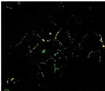
Fig. 3. Distribution of Cx43 and Cx40 in cultured neonatal rat ventricular myocytes. Fluorescent image of neonatal rat ventricular myocytes cultured at
.75% confluency for 48 h double-labeled with mouse anti-Cx43 (green) and rabbit anti-Cx40 (red). As can be concluded from the frequent yellow and
only sparse red labeling, most Cx40 is colocalized with Cx43. Bar represents 25 mm.
Most of the cells, including anti-Cx40 labeled cells, anti-Cx40 staining. A further reduction to ,25% con-were positive for the myocardial marker desmin and their fluency allowed us to observe many isolated cell pairs (see contractile phenotype was sometimes established by a clear Fig. 5, left upper panel). We never observed Cx40 staining cross-striation indicative for cardiac muscle cells. How- between the more than 50 desmin-positive cell pairs we ever, by isolating cells from whole ventricle some con- examined (see Fig. 4A,B), whereas Cx43 is clearly de-tamination of non-muscle cells like endothelial cells or tected between such cell pairs (see Fig. 4C,D). In double-fibroblasts may be expected, although the bulk of fi- labeling experiments performed with mouse anti-Cx43 and broblasts was removed from the cultures by including a rabbit anti-Cx40 antibodies this point is even further differential plating step of 1 h in the isolation procedure stressed (Fig. 4E,F).
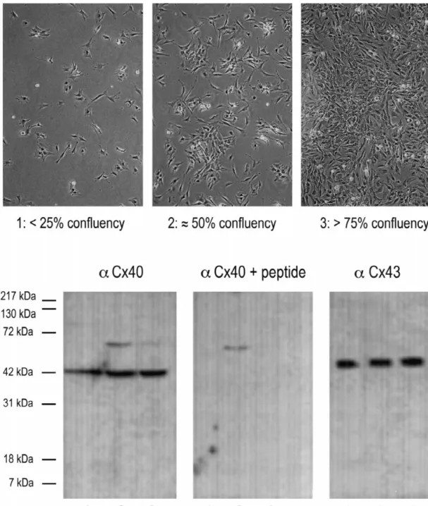
[40]. In order to exclude the possibility that Cx40 labeling In an attempt to quantify these apparent differences in is confined to vascular endothelial cells, double labeling Cx40 expression in high and low density cultures we experiments were performed where mouse anti-desmin or counted the relative number of Cx40 positive interfaces anti-vWf antibodies were combined with rabbit anti-Cx40 under both conditions and compared this with the relative antibody. Cultures of neonatal rat ventricle incubated with number of Cx43 positive interfaces in parallel cultures anti-vWf antibody revealed no positive staining (not (Table 1). Whereas the relative number of Cx43 positive shown), whereas immunolabeling could be observed in interfaces was identical at both densities (95%), the cultures of human umbilical cord endothelial cells using a relative number of Cx40 positive interfaces was con-similar procedure [42]. This may indicate that endothelial siderably less in low density cultures (35%) compared to cells were removed during the differential plating step or high density cultures (70%).
died during the enzymatic isolation of cells. Double-label- Western blotting was performed in order to compare the ing of mouse anti-desmin with rabbit anti-Cx40 antibodies levels of Cx40 expression at different culture densities. revealed anti-Cx40 immunoreactivity in anti-desmin posi- Proteins were extracted from cultures of neonatal rat
tive cells (e.g. Fig. 2C–F). ventricular myocytes grown to ,25, |50 and .75%
All immunostainings that were performed on myocyte confluency (see Fig. 5 top panels). From cultures of each cultures at .75% confluency showed a homogenous density equal amounts of total protein were blotted and pattern of anti-Cx43 labeling and a heterogeneous pattern probed with anti-Cx40 or anti-Cx43 antibody. Subsequent of anti-Cx40 labeling. The reduction of culture density chemiluminescence detection revealed for Cx40 at all three from .75% confluency to |50% confluency, leads to a densities a single band with a M of |42 kDa (Fig. 5,r reduced percentage of cell–cell contacts which were anti- aCx40, lanes 1–3). This protein could not be detected in Cx40 positive in the latter cultures (compare Fig. 2C,D control blots incubated with the primary antibody preincu-with E,F). Cx43 immunostaining showed no gross altera- bated with the immunogenic peptide (Fig. 5, aCx401 tions between both culture densities (not shown). Note that peptide, lanes 1–3). Although the band detected in lane 1 the muscle cell pair in the middle of Fig. 2E,F shows no (representing cultures of ,25% confluency) may seem
Fig. 4. Distribution of Cx43 and Cx40 in cultured neonatal rat ventricular myocyte pairs. Fluorescence images of neonatal rat ventricular myocytes cultured at ,25% confluency for 48 h showing cell pairs double-labeled with mouse anti-desmin (A) and rabbit anti-Cx40 (B), or mouse anti-desmin (C) and rabbit anti-Cx43 (D), or mouse anti-Cx43 (E) and rabbit anti-Cx40 (F). Bar represents 25 mm.
slightly less intense, no clear differences in the intensity of preincubation in the samples from cultures at |50% the 42-kDa band in any of the three lanes were observed in confluency. For Cx43 a single band with a M of |45 kDar repetitive experiments. An unspecific band of unknown and of equal intensity could be detected at all three identity at |65 kDa was observed with and without peptide densities (Fig. 5, aCx43, lanes 1–3).
Table 1
a
Gap junctional expression of Cx43 and Cx40 in neonatal rat ventricular myocytes cultured at various densities for 48 h
Cx Culture Number of Total number of Relative number of
density positive interfaces counted interfaces positive interfaces (%)
Cx43 High 398 420 95
Cx43 Low 96 101 95
Cx40 High 380 546 70
Cx40 Low 45 129 35
a
Cx40 or Cx43 labeling at cell–cell interfaces in cultures of low (,25% confluency) and high (.75% confluency) density was counted and divided by the total number of cell–cell interfaces present in this particular area of the coverslip.
Fig. 5. Expression of Cx40 and Cx43 in neonatal rat ventricular myocytes cultured at various densities for 48 h. Top panels are representive phase-contrast photographs illustrating the different culture densities used. Lower panels show Western blots with Cx40 antibody (left), Cx40 antibody after preabsorption with its immunogenic peptide (middle) and Cx43 antibody (right). Lanes 1: ,25% confluency, lanes 2: |50% confluency and lanes 3 .75% confluency. Equal amounts of total protein (50 mg) were loaded per lane on 12.5% SDS-PAGE and the connexins were detected by immunoblotting. For Cx40, at all three densities a single band at |42 kDa was detected. This band, which represents Cx40, was not detected in blots incubated with the Cx40 antibody preincubated with the immunogenic peptide. An unspecific band of unknown identity at |65 kDa was observed with and without peptide preincubation in the samples from cultures at |50% confluency. For Cx43, at all three densities a single band at |45 kDa was detected. Migration of molecular mass markers is indicated on the left of the figure.
4. Discussion ever, recent studies on the expression of Cx40 in
em-bryonic and fetal hearts revealed a dynamic and heteroge-The homogeneous expression of gap junction protein neous distribution pattern during development that has not Cx43 in the ventricular working myocardium of neonatal yet fully matured in the neonatal heart just after birth rats is unambiguously shown, both in sections of whole [23,34]. Cx40 expression is most extensive in the fetal hearts and in isolated cardiomyocytes [15,16,26]. How- period. Towards birth Cx40 mRNA gradually disappears
from the rat ventricular myocardium; from the ventricular contact with their neighboring cells. In an attempt to verify free wall towards the trabeculations. Just after birth Cx40 such density-dependent expression of Cx40, Western blots mRNA is still expressed in a subendocardial layer in the detecting Cx43 or Cx40 from equal amounts of total ventricles and in the atria. In the adult rat, Cx40 is only protein obtained from cultures grown at three different detectable in the conduction system and has disappeared densities were carried out. Although the 42-kDa band in from both the atrial and ventricular working myocardium lane 1 of Fig. 5 (representing cultures of ,25% con-[24]. The distribution pattern of Cx40 protein we report in fluency) may seem slightly less intense, no clear differ-this study confirms the data on the Cx40 mRNA dis- ences could be observed in the intensity of the 42-kDa tribution pattern. Moreover, it emphasizes the dynamic and bands in the three lanes in repetitive experiments. Indeed, heterogeneous nature of the ventricular myocyte popula- cultures of ,25% confluency contain many cell pairs, tion with respect to Cx40 expression. The age of the however, many small clusters of three to six cells are also neonatal rats used to obtain ventricular cell cultures may present. These clusters express Cx40 (a part of such a thus determine the proportion of cells able to express Cx40 cluster can be seen at the left top corner of Fig. 4A,B) and in those cultures. We have attempted to standardize for this thus may account for the Cx40 protein detected in Western variable in our experiments by using rats between 24–48 h blots of these cultures. In addition, the possibility that
after birth. some of the Cx40 detected in Western blots is from
Oyamada et al. [38] studied dye coupling and Cx43 intracellular (non-plasmalemmal) origin cannot be ex-expression in confluent cultures of neonatal rat ventricular cluded. An alternative explanation for the density-depen-myocytes over a period of 7 days [38]. They observed an dent expression of Cx40 may be that only a relative small increase both in cell-to-cell coupling and in Cx43 expres- proportion of subendocardial cells from the total ventricu-sion with time, which occurred mainly between day 3 and lar myocyte population still have the ability to express both day 7. We have performed immunocytochemical stainings Cxs. Actual formation of Cx40 gap junction channels will on ventricular myocyte cultures of three different densities then only occur in the case that two neighboring cells form ranging from very low density (,25% confluency) to Cx40 hemichannels. The probability that this occurs is subconfluent (.75% confluency). In all cultures Cx43 and much more frequent in clusters, where cells have several Cx40 could be detected. Moreover, for a given density neighbors (sometimes up to eight), than in a cell pair or identical Cx43 or Cx40 staining patterns were observed at triplet. Observing anti-Cx40 immunolabeling in one 24, 48 and 96 h of culturing. Immunolabeling experiments myocyte exclusively at the cell–cell contact with some of were initially performed at .75% and |50% confluency its neighbors and not with all of its neighbors will favor and showed a homogeneous pattern of anti-Cx43 labeling this hypothesis. Indeed, as can be observed in Fig. 3 and a heterogeneous pattern of anti-Cx40 labeling. How- (asterisk) this is sometimes the case. The indicated cell ever, in electrophysiological recordings (dual whole-cell or shows only Cx40 immunolabeling at the gap junction it perforated patch voltage-clamp) performed on cell pairs in formed with two of its neighbors (at the right and bottom cultures of ,25% confluency we did not observe any side) and not at its gap junctions with three to four other indication of the presence of Cx40 gap junction channels neighboring cells, where, however, Cx43 immunolabeling between ventricular cell pairs (e.g. [43,44]). The observed is apparent.
single channel conductances were |20, 40–45, and 70 pS In summary, it may be concluded that our cultured which are indicative for Cx43 gap junction channels neonatal rat ventricular myocytes compare very well with [8,45,46]. The expected size for Cx40 gap junction chan- cells from the intact ventricular myocardium with regard to nels, on the other hand, would be 120–160 pS [47–49]. their composition of gap junctions. They will thus be a Events of this size were not observed in our recordings. In good model for studying short-term and long-term regula-an attempt to explain this apparent controversy, we per- tion of cardiac gap junction channel expression and formed immunostaining experiments on ventricular function. Care should be taken, however, in comparing myocyte cultures of low density (,25% confluency) as experiments performed on high and low density cultures. also used for dual patch clamp studies. The pattern of Furthermore, the heterogeneity of Cx40 expression may Cx43 immunoreactivity was not affected by the culture account for large scatter in results of experiments per-density. In contrast, in ventricular myocyte cultures of low formed on (sub)confluent cultures.
density the relative number of cell–cell interfaces that were Cx40-immunopositive decreased as compared to high
density cultures. Moreover, Cx40 staining was detected in Acknowledgements
these cultures only between clusters of three or more cells
and not between cell pairs. A possible explanation for this The authors wish to thank Anton van der Wardt for phenomenon may be that isolated neonatal rat ventricular excellent technical assistance and Dr Paolo Meda for cells are all able to express Cx40, however, this expression critical reading of the manuscript. The investigators were is upregulated with increasing culture density. In other supported by grants from the Netherlands Organization for words, cells may only express Cx40 if they are in close Scientific Research (grant 902-16-093), the Netherlands
[20] Davis LM, Kanter HL, Beyer EC, Saffitz JE. Distinct gap junction
Heart Foundation (grant 95.077), the Association Franc¸aise
protein phenotypes in cardiac tissues with disparate conduction
contre les Myopathies and the European Economic
Com-system. J Am Coll Cardiol 1994;24:1124–1132.
munity (grant BMH4-CT96-1412). [21] Bastide B, Neyses L, Ganten D, Paul M, Willecke K, Traub O. Gap
junction protein connexin40 is preferentially expressed in vascular endothelium and conductive bundles of rat myocardium and is increased under hypertensive conditions. Circ Res 1993;73:1138–
References 1149.
[22] Gros D, Jarry-Guichard T, Ten Velde I et al. Restricted distribution [1] Barr L, Dewey MM, Berger W. Propagation of action potentials and of connexin40, a gap junction protein, in mammalian heart. Circ Res
the structure of the nexus in cardiac muscle. J Gen Phys 1994;74:839–851.
1965;48:797–823. [23] Delorme B, Dahl E, Jarry-Guichard T et al. Developmental regula-[2] Bruzzone R, White TW, Paul DL. Connections with connexins: the tion of connexin 40 gene expression in mouse heart correlates with molecular basis of intercellular signaling. Eur J Biochem the differentiation of the conduction system. Dev Dyn
1996;238:1–27. 1995;204:358–371.
[3] Gros DB, Jongsma HJ. Connexins in mammalian heart function. [24] Van Kempen MJA, Ten Velde I, Wessels A et al. Differential BioEssays 1996;18:719–730. connexin distribution accommodates cardiac function in different
species. Microsc Res Tech 1995;31:420–436. [4] Willecke K, Haubrich S. Connexin expression systems: to what
extent do they reflect the situation in the animal? J Bioenerg [25] Chen SC, Davis LM, Westphale EM, Beyer EC, Saffitz JE. Biomembr 1996;28:319–326. Expression of multiple gap junction proteins in human fetal and
infant hearts. Pediatr Res 1994;36:561–566. [5] Kumar NM, Gilula NB. The gap junction communication channel.
Cell 1996;84:381–388. [26] Darrow BJ, Laing JG, Lampe PD, Saffitz JE, Beyer EC. Expression of multiple connexins in cultured neonatal rat ventricular myocytes. [6] Sosinsky GE. Molecular organization of gap junction membrane
Circ Res 1995;76:381–387. channels. J Bioenerg Biomembr 1996;28:297–309.
´ [27] Coppen SR, Dupont E, Rothery S, Severs NJ. Connexin45 expres-[7] Elfgang C, Eckert R, Lichtenberg-Frate H et al. Specific
permeabili-sion is preferentially associated with the ventricular conduction ty and selective formation of gap junction channels in
connexin-system in mouse and rat heart. Circ Res 1998;82:232–243. transfected HeLa cells. J Cell Biol 1995;129:805–817.
[8] Kwak BR, Hermans MMP, DeJonge HR, Lohmann SM, Jongsma [28] Delorme B, Dahl E, Jarry-Guichard T et al. Expression pattern of HJ, Chanson M. Differential regulation of distinct types of gap connexin gene products at early developmental stages of the mouse junction channels by similar phosphorylating conditions. Mol Biol cardiovascular system. Circ Res 1997;81:423–437.
Cell 1995;6:1707–1719. [29] Reaume AG, De Sousa PA, Kulkarni S et al. Cardiac malformation [9] Veenstra RD. Size and selectivity of gap junction channels formed in neonatal mice lacking connexin43. Science 1995;267:1831–1834. from different connexins. J Bioenerg Biomembr 1996;28:327–337. [30] Ya J, Erdtsieck-Ernste EB, de Boer PA et al. Heart defects in [10] Beyer EC, Paul DL, Goodenough DA. Connexin43: a protein from connexin43-deficient mice. Circ Res 1998;82:360–366.
rat heart homologous to a gap junction protein from liver. J Cell [31] Thomas SA, Schuessler RB, Berul CI et al. Disparate effects of Biol 1987;105:2621–2629. deficient expression of connexin43 on atrial and ventricular conduc-[11] Kanter HL, Saffitz JE, Beyer EC. Cardiac myocytes express multiple tion. Circulation 1988;97:686–691.
gap junction proteins. Circ Res 1992;70:438–444. [32] Kirchhoff S, Nelles E, Hagendorff A, Kruger O, Traub O, Willecke [12] Verheule S, Van Kempen MJA, Te Welscher PHJA, Kwak BR, K. Reduced cardiac conduction velocity and predisposition to Jongsma HJ. Characterization of gap junction channels in adult arrhythmias in connexin40-deficient mice. Curr Biol 1998;8:299– rabbit atrial and ventricular myocardium. Circ Res 1997;80:673– 302.
681. [33] Simon AM, Goodenough DA, Paul DL. Mice lacking connexin40
[13] Paul DL, Ebihara L, Takemoto LJ, Swenson KI, Goodenough DA. have cardiac conduction abnormalities characteristic of atrioven-Connexin 46: a novel lens gap junction protein, induces voltage- tricular block and bundle branch block. Curr Biol 1998;8:295–298. gated currents in non-junctional plasma membrane of Xenopus [34] Van Kempen MJA, Vermeulen JLM, Moorman AFM, Gros D, Paul oocytes. J Cell Biol 1991;115:1077–1089. DL, Lamers WH. Developmental changes of connexin40 and [14] Davis LM, Rodefeld ME, Green K, Beyer EC, Saffitz JE. Gap connexin43 mRNA distribution patterns in the heart. Cardiovasc Res
junction protein phenotypes of the human heart and conduction 1996;32:886–900. ´
system. J Cardiovasc Electrophysiol 1995;6:813–822. [35] Masson-Pevet M, Jongsma HJ, De Bruijne J. Collagenase- and [15] Gourdie RG, Green CR, Severs NJ, Thompson RP. Immunolabeling trypsin-dissociated heart cells: a comparative ultrastructural study. J
patterns of gap junction connexins in the developing and mature Mol Cell Cardiol 1976;8:747–757.
heart. Anat Embryol 1992;185:363–378. [36] Bassani JW, Qi M, Samarel AM, Bers DM. Contractile arrest [16] Van Kempen MJA, Fromaget C, Gros D, Moorman AFM, Lamers increases sarcoplasmic reticulum calcium uptake and SERCA2 gene WH. Spatial distribution of connexin-43, the major cardiac gap- expression in cultured neonatal rat heart cells. Circ Res junction protein, in the developing and adult rat heart. Circ Res 1994;74:991–997.
1991;68:1638–1651. [37] De Leon JR, Buttrick PM, Fishman GI. Functional analysis of the [17] Gourdie RG, Severs NJ, Green CR, Rothery S, Germroth P, connexin43 gene promoter in vivo and in vitro. J Mol Cell Cardiol
Thompson RP. The spatial distribution and relative abundance of 1994;26:379–389.
gap-junctional connexin40 and connexin43 correlate to functional [38] Oyamada N, Kimura H, Oyamada Y, Miyamoto A, Ohshika H, Mori properties of components of the cardiac atrioventricular conduction M. The expression, phosphorylation, and localization of connexin 43 system. J Cell Sci 1993;105:985–991. and gap-junctional intercellular communication during the establish-[18] Kanter HL, Laing JG, Beau SL, Beyer EC, Saffitz JE. Distinct ment of a synchronized contraction of cultured neonatal rat cardiac
patterns of connexin expression in canine Purkinje fibers and myocytes. Exp Cell Res 1994;212:351–358.
ventricular muscle. Circ Res 1993;72:1124–1131. [39] De Bruijne J, Jongsma HJ. Membrane properties of aggregates of [19] Oosthoek PW, Viragh S, Lamers WH, Moorman AFM. Immuno- collagenase-dissociated rat heart cells. In: Tajuddin M, Das PK, histochemical delineation of the conduction system. II. The atrioven- Tariq M, Dhalla NS, editors, Advances in myocardiology, Balti-tricular node and Purkinje fibers. Circ Res 1993;73:482–491. more: University Park Press, 1980, pp. 231–242.
[40] Blondel B, Roijen I, Cheneval JP. Heart cells in culture: a simple connexins confer distinct regulatory and conductance properties of method for increasing the proportion of myoblasts. Experientia gap junctions in developing heart. Circ Res 1992;71:1277–1283. 1971;27:356–358. [46] Moreno AP, Saez JC, Fishman GI, Spray DC. Human connexin43 [41] El Aoumari A, Fromaget C, Dupont E et al. Conservation of a gap junction channels. Regulation of unitary conductances by
cytoplasmic carboxy-terminal domain of connexin 43, a gap junc- phosphorylation. Circ Res 1994;74:1050–1057. ´
tion protein, in mammalian heart and brain. J Membr Biol [47] Traub O, Eckert R, Lichtenberg-Frate H et al. Immunochemical and 1990;115:229–240. electrophysiological characterization of murine connexin40 and -43 [42] Van Rijen HVM, Van Kempen MJA, Analbers LJS et al. Gap in mouse tissues and transfected human cells. Eur J Cell Biol
junctions in human umbilical cord endothelial cells contain multiple 1994;64:101–112.
connexins. Am J Phys 1997;272:C117–C130. [48] Beblo DA, Wang HZ, Beyer EC, Westphale EM, Veenstra RD. [43] Takens-Kwak BR, Jongsma HJ, Rook MB, Van Ginneken ACG. Unique conductance, gating, and selective permeability properties of Mechanism of heptanol-induced uncoupling of cardiac gap junc- gap junction channels formed by connexin40. Circ Res tions: a perforated patch-clamp study. Am J Phys 1992;262:C1531– 1995;77:813–822.
C1538. [49] Bukauskas FF, Elfgang C, Willecke K, Weingart R. Biophysical
[44] Kwak BR, Jongsma HJ. Regulation of cardiac gap junction channel properties of gap junction channels formed by mouse connexin40 in permeability and conductance by several phosphorylating condi- induced pairs of transfected human HeLa cells. Biophys J tions. Mol Cell Biochem 1996;157:93–99. 1995;68:2289–2298.