Original article
Long-term evolution of renal function in patients with ovarian cancer after
whole abdominal irradiation with or without preceding cisplatin*
D. P. Schneider,
1H.-P. Marti,
2C.Von Briel,
1F. J. Frey
2& R. H. Greiner
11
Department of Radiation Oncology. 2Division of Nephrology, University of Bern. Inselspital, Bern. Switzerland
Summary
Background: The upper limit of the natural decline in
creati-nine clearance is 1 ml/min/year. To define the loss of renal
function, we started a long-term assessment of patients with
ovarian cancer treated by whole abdominal irradiation (WAI)
with preceding cisplatin chemotherapy (CDDP) and
second-look laparotomy (SLL).
Patients and methods- We analyzed the creatinine clearance
over time of 56 patients treated from 1982 to 1988 for ovarian
cancer. Thirty-one of 56 patients had received WAI after their
initial surgery, and 25 of 56 patients had undergone CDDP
therapy followed by SLL, and then WAI after their initial
surgery. Median follow-up was 99 months (7-156). Twenty of
56 patients accepted our invitation for additional assessment
of tubular function, nine of the 31 patients without CDDP
therapy and SLL, and 11 of the 25 patients with CDDP
followed by SLL and WAI.
Ten of twenty patients had received four to six cycles
CDDP, 80 mg/m
2/cycle, and one patient nine cycles. The
median total dose for each kidney was 1450 cGy (480-1690).
Results: The mean creatinine clearance decreased from 84
ml/min to 66 ml/min. Seventy-six percent of the 25 patients
who had undergone CDDP therapy, SLL and WAI had
de-clines of more than 1 ml/min/year, 64% of these patients of
more than 2 ml/min/year. For the 31 patients who had
re-ceived WAI after their initial surgery, the corresponding
num-bers were 71% and 55%, respectively. The tubular function of
the 20 patients who had undergone the additional
investiga-tions was not impaired.
Conclusion: The decline in renal function after WAI is more
pronounced than in healthy subjects. The treatment with
cis-platin and SLL prior to WAI does not seem to contribute to
this loss of kidney function.
Key words: cisplatin (CDDP), nephrotoxicity, ovarian cancer,
whole abdominal irradiation (WAI)
Introduction
Adverse effects of irradiation on kidneys were first
dis-cussed by Baerman et al. in 1904 [1]. In 1952 and 1964,
Kunkler and Luxton described clinical syndromes of
radiation nephropathy by the analysis of patients treated
for seminoma [2, 3]. Many patient reports of renal
disorders after irradiation subsequently followed in
rapid sequence [4-18]. Hallmarks of chronic radiation
nephropathy are arterial hypertension, glomerular
scle-rosis/hyalinization, fibrinoid changes in arterioles and
interlobular arteries, tubular atrophy and interstitial
fibrosis [19].
A task force under the direction of the National
Cancer Institute defined tolerance doses of various organs
and tissues to therapeutic irradiation and published
their recommendations in 1991 [20]. To support these
recommendations, further systematic clinical studies are
warranted. Cassady stressed in a recent article the need
for well designed studies with long follow-up periods to
accurately assess late radiation toxicity [21].
In this context, we examined the radiation
nephrop-athy of our patients with advanced and non-advanced
ovarian cancer who were treated in controlled protocols
by surgery followed by either CDDP, second-look
laparotomy and WAI or WAI exclusively following
sur-gery. The long-term assessment examined especially the
impact and mutual influence of the two potentially
nephrotoxic agents, CDDP and irradiation, sequentially
given, on renal function.
Patients and methods
From 1982 to 1988, a total of 136 patients, aged 14 to 82 years, all with epithelial ovarian cancer, were treated by surgery followed by either cisplatin (CDDP), second-look laparotomy and WAI. or by WAI alone. Of these 136, nine foreign patients were lost to follow-up, and 71 died sometime after therapy. 67 of causes related to the ovarian cancer; none, however, suffered from kidney failure.
Study group
The remaining 56 patients comprised our study group. The median follow-up period was 99 months (7-156). Twenty of 56 patients agreed
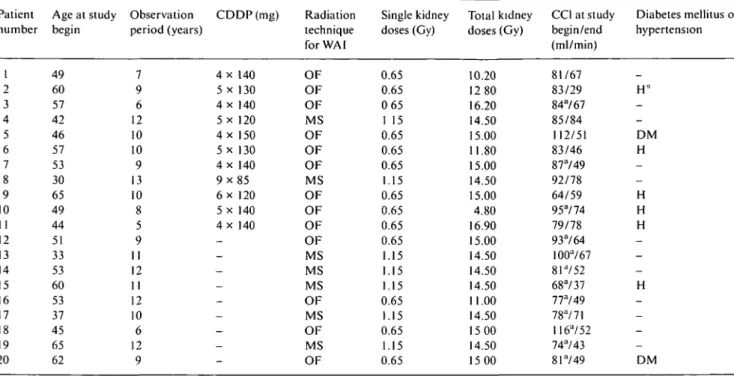
Table 1 Characteristics of the 20 investigated patients. Patient number 1 2 3 4 5 6 7 8 9 10 11 12 13 14 15 16 17 18 19 20 Age at study begin 49 60 57 42 46 57 53 30 65 49 44 51 33 53 60 53 37 45 65 62 Observation period (years) 7 9 6 12 10 10 9 13 10 8 5 9 11 12 11 12 10 6 12 9 CDDP(mg) 4 x 140 5 x 130 4 x 140 5 x 120 4 x 150 5 x 130 4 x 140 9 x 8 5 6 x 120 5 x 140 4 x 140 _ — _ -_ — — -Radiation technique for WAI OF OF OF MS OF OF OF MS OF OF OF OF MS MS MS OF MS OF MS OF Single kidney doses (Gy) 0.65 0.65 0 65 1 15 0.65 0.65 0.65 1.15 0.65 0.65 0.65 0.65 1.15 1.15 1.15 0.65 1.15 0.65 1.15 0.65 Total kidney doses (Gy) 10.20 12 80 16.20 14.50 15.00 11.80 15.00 14.50 15.00 4.80 16.90 15.00 14.50 14.50 14.50 11.00 14.50 15 00 14.50 15 00 C O at study begin/end (ml/min) 81/67 83/29 84"/67 85/84 112/51 83/46 87"/49 92/78 64/59 95"/74 79/78 93"/64 IOOa/67 81-/52 68"/37 77"/49 78a/71 H6a/52 74a/43 81"/49 Diabetes mellitus or hypertension _ H° -DM H -H H H -H -DM
Abbreviations: CDDP - cis-diamminedichloride platinum; WAI - whole abdominal irradiation; CC1 - creatinine clearence. values followed by " were calculated according to Cockcroft et al. [22] or Jeliffe et al. [23]; OF open field; MS moving strip: DM diabetes mellitus, H -hypertension (H° = in addition to -hypertension, history of excessive consumption of phenacetin).
to undergo investigaton of renal-tubular function in addition to assess-ment of glomerular filtration rate. These 20 patients constituted the investigated group, while the remaining 36 patients formed the non-investigated group.
In the investigated group, 11 of 20 patients (FIGO stage: 2 x IC. 1 x IIB, 2 x IIC, 6 x HI) had received C D D P chemotherapy, 10 patients four to six cycles with 80/m2/cycle and 1 patient nine cycles. In addition to CDDP as the only nephrotoxic agent, combination chemo-therapy contained either melphalan, melphalan and hexamethylmela-min, or cyclophosphamide. The FIGO stage of the nine investigated patients without CDDP chemotherapy were 5 x IB/C, 2 x MB, and 2 x III. In the non-investigated group 14 of 36 patients received CDDP chemotherapy (FIGO stage: 3 x IC. 5 x 2B/C. and 6 x III). The FIGO stage of the 22 patients in the non-investigated group who did not receive C D D P chemotherapy were 21 x IB/C, and 1 x III.
For the investigation of a possible adverse effect of cisplatin on renal function, we pooled patient data from the investigated (n = 20) and non-investigated (;; = 36) patients and classified these individuals into two new groups, WAI patients treated with cisplatin (/; = 25) and those receiving no cisplatin chemotherapy (;J = 31). Median age and follow-up periods for the former group were 52 years (28-72) and 83 months (26-156). respectively, and for the latter group 53 years (33-82) and 102 months (7-148). respectively.
Kidney irradiation dose
To deliver whole abdominal irradiation (WAI), the moving strip and. later, the open field techniques were used. Patients were treated in prone position using anterior-posterior fields for a target volume extending from the domes of the diaphragms to the lower border of the obturator foramina: 5 H.V.L. lead shielding were inserted to shield the kidneys from the posterior field. Kidney shielding was localised by an intravenous pyelogram at the time of treatment simulation. Cobalt-60 beam was employed for the moving strip technique. The single dose was 230 cGy adapted to an isodose. which encompassed the whole abdomen rather homogeneously. For the open field technique the energies of 8 or 16 MeV from the linear accelerator were used. The
single dose of 130 cGy was calculated for mid-plane. The median total kidney dose for both techniques was 14.5 Gy (4.8-16.9 Gy) with single kidney doses in the range of 1 15 Gy (moving strip) and of 0.65 Gy (open field). Terminations of treatment, principally because of hemato-toxicity, are the reason for the broad range of the kidney doses.
Study group: Investigated patients (n = 20)
The median age of the investigated group at diagnosis was 52 years (30-65). All 20 patients were tumor-free at the time of the study. The median follow-up period of these patients was 117 months (61-156). Additional clinical data and details of radiation and chemotherapy are given in Table 1. Analyses of all 20 patients included clinical history, physical examination, blood and urine tests and kidney ultrasound. At the end of the observation period, 24-hour urine specimens were collected from all patients in order to determine creatinine clearance,
proteinuria. and tubular function by excretion of uric acid, phosphate
and glucose. Urine analysis including measurement of pH for assess-ment of tubular acidification was performed in random morning urine samples. Prior to any therapy such as WAI or chemotherapy, creatinine clearance was measured from 24-hour urine specimens in eight pa-tients. For the remaining 12 patients, the creatinine clearance was calculated using the formulas reported by Cockcroft et al. [22] and Jelliffe et al. [23]. To predict creatinine clearance, the former relies on serum creatinine. body weight and age. and the latter on serum creatinine and age only. In our analyses, we used the first formula, except in a few patients with significant obesity (body mass index > 3 0 kg/m2), in whom we applied the second formula because of overestimation of creatinine clearance by inclusion of excessive body weight into the calculations.
Study group: Non-investigated patients (n = 36)
The median age of the non-investigated group at diagnosis was 57 years (28-72). Twenty-three of the 36 patients were tumor-free at the time of the renal function investigations. The median follow-up period
140 • • • I • • • • • • • • • I
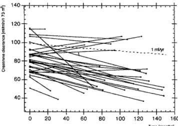
Figure 1 Creatinine clearance as a function of time in 25 patients
treated with CDDP and WAI. The dashed line indicates the natural age-related decline of the creatinine clearance (1 ml/year) according to Bjornsson [24]. A total of 76% of the patients had a decline greater than 1 ml/year, and 64% of more than 2 ml/year.
140
1 2 0
-
100S4 0
-Figure 2. Creatinine clearance as a function of time in 31 patients
treated with WAI only. The dashed line represents the natural decrease of the creatinine clearance with age (1 ml/year), according to Bjorns-son [24]. A total of 71% of the patients demonstrated a decline greater than 1 ml/year, and 55% of more than 2 ml/year.
of these patients was 81 months (7-158) Creatinine clearance values were calculated in all of these patients, as stated above.
Kidney function
Kidney function in all 56 patients of both the investigated and the
non-investigated groups was analyzed as a function of time. In addition,
the evolution of the glomerular filtration rate in these patients was compared to the age-related, natural decline in creatinine clearance [22-27] Importantly, according to Bjornsson et al [24], creatinine clearance in adults, such as our study patients, decreases by approx-imately 1 ml/min/year independent of gender. This value was used as reference for the analyses of our patients.
Results
General evolution of glomerular filtration rate
The main purpose of this study was to investigate the
glomerular filtration rate as a function of time after
WAI. In our 56 patients the decline in creatinine
clear-ance was more pronounced than in healthy subjects
during an identical observation period. The mean
crea-tinine clearance in our patients decreased by 18 ml/min
(from 84 ± 16 to 66 ± 21 ml/min; P < 0.05) during the
median and mean observation period of eight years.
Effect of CDDP and WAI versus WAI alone on glomerular
filtration rate
During the study period, the decline in mean creatinine
clearance of patients exposed to CDDP and irradiation
(n = 25) was 19 ml/min, very close to the 16 ml/min in
patients treated by radiotherapy alone (n = 31), when a
comparable observation time was considered.
Addition-ally, the results of the creatinine clearances of these two
patient groups were plotted against time (Figures 1 and
2). As shown in Figure 1, 19 of 25 (76%) of the patients
in the group treated with chemotherapy and irradiation
had declines in creatinine clearance greater than 1
ml/year (with 1 ml/year as the natural decline in renal
function of a normal adult population of comparable
age; 24). Moreover, 16 of 25 (64%) of these patients had
losses of more than 2 ml/year. In the group with
irradi-ation alone, the numbers were 71% and 55%,
respec-tively (Figure 2).
Effect of CDDP and WAI on kidney function of the
investigated patient group
Kidney function in the 20 patients in the investigated
group was analyzed in greater detail with respect to
glomerular filtration rate, kidney size, and renal tubular
function. Relevant data of patients, treatment methods
and laboratory results are summarized in Tables 1 and 2.
Glomerular filtration rate. Altogether, during the
follow-up period of 117 months (61-156), the mean creatinine
clearance significantly decreased from 86 ± 13 ml/min
to 58 ± 15 ml/min (P < 0.05), with a mean value for the
patients with CDDP and WAI of 62± 16 ml/min and for
the patients with WAI alone 54 ±11 ml/min. At the end
of the observation period, 11 of the 20 patients
demon-strated creatinine clearances below 60 ml/min; in 9 of
these 11 patients, creatinine clearance was 40-59 ml/min,
and in the remaining two, 39 and 29 ml/min.
In these 20 patients, we were not able to correlate
renal radiation dose and decline in kidney function on
the basis of the data reported in Table 1.
Tubular function. Renal tubular function of the 20
investigated-group patients was analyzed in detail. The
results are summarized in Table 2.
Tubular reabsorption of phosphate (TRP) measured in
individuals with normal renal function (creatinine
clear-ance) was previously reported by Popovtzer et al. to be
Table 2. Renal tubular function of the 20 investigated patients.
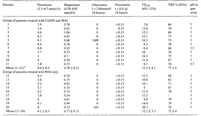
Patients Potassium Magnesium Glucosuria Proteinuria FEU R (3.5-4.7 mmol/1) (0.70-0.95 (<2.80mmol/ (<0.15g/ (6%-12%)
mmol/l) 24 hours) 24 hours)
TRP%(85%) pH in spot urine
Group of patients treated with C D D P a n d WAI 1 2 3 4 5 6 7 8 9 10 11 Mean (1-1 l )a oup of patients tn 12 13 14 15 16 17 18 19 20 Mean (12-20) 3.9 3.6 4.6 4.3 4.3 4.4 4.8 3.7 3 4 3.8 4.0 ±0.5
eated with WAI only 4 4 3.6 4 2 5.3 4.2 3.8 4 4.5 4 4.2 ± 0 . 5 0.78 0.62 1.06 0.82 0.68 0.78 0.92 0.75 0.7 0.74 0.75 0.78 ±0.12 0.78 0.75 0.83 0.76 0.74 0.54 0.81 0.94 0.75 0.77 ±0.11 0 0 0 0 1049 0 0 0 0 0 0 0 0 0 0 0 0 0 0 185 <0.15 0.25 <0.15 <0.15 <0.15 <0.15 <0.15 <0.15 <0.15 < 0 15 < 0 15 <0.15 <0.15 <0.15 <0.15 <0.15 <0.15 <0.15 <0.15 <0.15 7.6 136 13.5 13.1 18.3 9.2 6.4 19 16.3 11.4 8.7 12.5 ±4.1 11.5 10.6 10 1 9 11 4 13.3 8.8 14.4 20.7 12.2 ± 3 . 5 86 70 84 77 73 78 66 76 73 87 76 77 ±6 68 63 71 63 78 -79 79 76 72 ±6 3 5 5 5 5 5 5.5 5 5 5 6.5 5 5 5 5 5 5 5 5 5
All biochemical results were obtained at the end of the observation period. a Patients 1-11 received WAI and additional chemotherapy as depicted in Table 1
FEu r (%): Fractional excretion of uric acid = "™^d£ZL x 100.
TRP: Tubular reabsorption of phosphate = I — fractional excretion of phosphate.
in the order of 85% [28]. In our 20 patients, TRP was
77% ± 6% in the 11 cases treated with CDDP and WAI
and 72% ± 6% in the 9 individuals treated with
radio-therapy alone. Considering reduced mean creatinine
clearances of 62 ± 16 ml/min in the former and of 54 ±
11 ml/min in the latter group, the small reduction in
TRP due to the increase in fractional excretion of
phos-phate is an expected finding according to Popovtzer et
al. [28]. Thus, the small reduction in TRP due to
in-creased fractional phosphate excretion merely reflects
the reduction in renal function and may in addition be
interpreted as a consequence of mild secondary
hyper-parathyroidism.
The fractional excretion ofuric acid (FE
ur) was 12.5%
± 4.1% in patients treated with CDDPand 12.2% ± 3.5%
in patients not exposed to chemotherapy. These results
are not significantly different from the normal excretion
of uric acid, which is generally on the order of 6% to
12% of the filtered load [29] and 12.0% ± 2.9% in
non-pregnant women [30]. However, as renal function
dete-riorates, there is a progressive increase in excretion and
a relative clearance of uric acid because of increased
tubular secretion of urate and incomplete reabsorption
of filtered urate [31]. According to a plot of glomerular
filtration rate versus FE
urby Calabrese et al. [32], both
mean values of FE
urobtained in our patient subgroups
are only slightly above the expected value of about 11%:
however, in that publication 10% was considered to be
the upper limit of a normal FE
urin unimpaired kidney
function.
Except in the two patients with known diabetes
melli-tus, no glucosuria due to tubular damage was found.
Only one patient showed a slight proteinuria of 0.25 g/day,
which may well be explained by hypertension and past
abuse of phenacetin. In 18 of the 20 patients, a pH of 5.0
was measured in random urine samples. Although this
finding does not entirely exclude an acidification defect
[33], a significant distal renal tubular disorder with
respect to acid excretion seems unlikely.
Finally, potassium and magnesium measured in serum
were in the normal range except in one patient with a
potassium of only 3.0 mmol/1 (3.5-4.7 mmol/1) and in
three patients with borderline to mild hypomagnesemia
of 0.54-0.68 mmol/1 (0.70-0.95 mmol/I). These findings
satisfactorily excluded tubulopathy with loss of
electro-lytes, typically found in cisplatin- induced nephropathy.
Kidney size. The kidney length found in normal adult
females is slightly less than 11 cm [34]. However, renal
length decreases by 2 cm between the ages of 50 and 80
years [35]. Therefore, as measured by ultrasound in the
investigated group, renal size was practically not affected,
with a mean of 10.0 ± 1.1 cm in the CDDP-treated group
and of 10.3 ± 1.1 cm in the group with radiotherapy
alone. Most likely, the only minimal reduction of kidney
size reflected the glomerular function slightly reduced
Table 3. Patient reports of radiation nephropathy.
Author, year [Ref.]
Luxton. 1964 [3] Thompson. 1971 [17] Arneil, 1974 [4] Keane, 1976 [12] Churchill, 1978 [6] Le Bourgeois, 1979 [15] Birkhead, 1979(5] Kim, 1984 [13] Willet. 1986 [18] Markoe. 1989 [44] Dewitt, 1990 [7] Flentje, 1993 [9] Irwin, 1996 [36]
Schneider [present study]
Patient number 2.54 2.67 2.2 2.2 2.1 1.74 2.17 1 18 2.86 2.12 2.26 2.142 1.60 56 Underlying disease Mostly seminoma Gastric hypersecretion Nephro-blastoma Ovarian carcinoma Testicular carcinoma Lymphoma Hodgkin lymphoma NHL Abdominal carcinoma, lymphoma and sarcoma Ovarian cancer, lymphoma, lesticular tumor NHL, Hodgkin lymphoma, ovarian cancer, seminoma Seminoma Ovarian cancer. NHL, carcinoid Ovarian cancer
Total kidney dose (Gy) 25-30 15-20 to left kidney 15/20 to not nephrectomized kidney 25 and 27 to both kidneys 38 to both kineys 15-45 to left kidney 40 to left kidney 22-45 to one kidney 26-61 to one kidney 17-25 to both kidneys 17-40 (uni-and bilateral) 19-28 to both kidneys 19 (7-23) to both kidneys 14.5 (5-17) to both kidneys by WAI Chemotherapy Not reported Not given Actinomycin D and vincristine Not given Bleomycin and vinblastine Not reported
Only for 1 patient reported Not reported Not reported Not reported Not reported Not reported Not given Cisplatin to 25/56 pat. prior WAI
Follow-up (years) =£14 8-19 2 10 and 11 months 9 months 3-5 3-6 2-8 3(1-9) 0.5-14 3-5 8(2-21) 9 (5-20) 8(0.5-13) Kidney function (GFR) ARN:20. and CRN: 22 patients RN: 31 patients (9 deaths) ARN: 2 patients ESRD and CClof30ml/min ARN (CCI: 67 ml/min) Stable renal function Stable renal function RN: 9 patients (CCI 5216 ml) Mean decrease of CC1= 17/min CRN: 1 patient (CCI- 48 ml/min) 5 patients with 25% decline of CCI 'Clinical manifest' CRN: 7 patients Stable renal function Decrease in mean CCL from 84 to 66 ml/min
Abbreviations: ARN - acute radiation nephritis. CRN - chronic radiation nephritis: NHL - non-Hodgkin's lymphoma: CCL - creatinine clearance; ESRD - end-stage renal disease.
over many years and present at the observation time.
Even in the two patients with the lowest creatinine
clearances of 29 ml/min and 37 ml/min, kidney length
was still close to 9 cm. These findings confute pre-existing
chronic renal failure. In addition, renal ultrasound
ex-amination revealed no further pathological findings
such as increased echogenicity, cortical atrophy or
pa-renchymal cysts in any of the 20 patients.
Comorbidity. Co-morbid conditions were found in eight
of the patients in this group which could potentially have
an adverse effect on kidney function, as shown in Table 2.
Arterial hypertension was treated in six patients,
includ-ing the one with excessive consumption of phenacetin,
for durations of five months to six years. However, the
mean creatinine clearances of hypertensive and
normo-tensive patients were almost identical: 54 ml/min and
60 ml/min, respectively. Two patients suffered from
diabetes mellitus. Only one patient with hypertension,
who also had the worst renal function of the 20 patients
analyzed, showed borderline proteinuria of 0.25 g/24 h
(patient 2, Table 1). In addition, this patient was the
only one who demonstrated significant abnormality in
urinalysis, namely microhematuria.
Discussion
Radiation nephritis is a well-known entity which is,
however, difficult to analyze quantitatively because of
many confounding variables. The key findings of 13
previously published studies or case reports are
sum-marized in Table 3. The observed effects of irradiation on
renal function are heterogeneous, depending on total
renal dose, irradiated renal volume, on uni- or bilateral
kidney irradiation, and on length of patient follow-up
periods. The duration of the present study is extensive
and the patient numbers high, comparable to only four
other studies (Table 3) [3, 9, 17, 36].
Chemotherapy is a common variable in the
assess-ment of radiation nephritis, and chronic renal failure is
a rare but well-described complication of cisplatin
[37-39]. Remarkably, there is no published report separately
analyzing the effects of chemotherapy on the course of
radiation nephropathy, so our study is thus unique with
respect to its direct comparison of irradiated patients
treated with and without cisplatin. However, we found
no differences with respect to cisplatin. Experimental
studies analyzing mice or rats clearly demonstrated an
enhancing effect of cisplatin on kidney damage caused by
irradiation [40-43]. In addition, second-look laparotomy
had no negative effect on renal function in the patients
who received chemotherapy.
Other important confounding factors are hypertension
and diabetes mellitus. As in most other studies, the
precise adverse effects of these two underlying diseases
on kidney function in our subjects is difficult to judge,
since the durations and treatments of hypertension and
diabetes mellitus are not known. Tn addition, high blood
pressure in our patients may be a result of radiation
nephropathy but could also represent essential
hyper-tension, especially in the presence of well-preserved
kidney function, such as in the study patients 10 and 11
(Table 1). However, there were no cases of malignant
hypertension and in our investigated-patient group kidney
function did not differ in hypertensive and normotensive
individuals.
The decline of renal function in our population was
pronounced despite the low single doses of 0.65 and
1.15 Gy. The decrease of the mean glomerular filtration
rate in our 56 patients was more than twice the expected
natural age-related decline of 1 ml/min/year [24].
In addition to creatinine clearance, we analyzed several
factors reflecting renal tubular functions. Despite the
reduction of glomerular filtration rate, renal tubular
function was well preserved. Therefore, most of the
late-effect damage of low-dose irradiation to the kidney may
have occurred to endothelial cells present in arterioles
and in glomerular capillaries rather than to tubular cells
due to interstitial inflammation [13].
In summary, we found no patients with chronic
radi-ation nephropathy in our series of 56 patients who
received WAI or CDDP and SLL followed by WAI after
epithelial ovarian cancer surgery. Nevertheless, analyses
of kidney function over time showed a more pronounced
decline of creatinine clearance in our patients than in
the normal population. Interestingly, the addition of
cisplatin and SLL did not adversely influence the
de-crease in renal function.
Acknowledgements
We particularly thank M. Giudici, MD, Institute of
Diagnostic Radiology, Inselspital, for the ultrasound
examinations in our patients.
References
1. Baerman G, Linser P. Ueber die lokale und allgemeine Wirkung der Rontgenstrahlen. Munchen Med Wochenschr 1904; 7: 996. 2. Kunkler PB, Farr RF. Luxton RW. The limit of renal tolerance to
X-rays. Br J Radiol 1952; 25: 190-201.
3. Luxton RW. Kunkler PB. Radiation nephritis. Acta Radiolog Ther Phys Biol 1964, 2: 169-78.
4. Arneil GC, Emmanuel IC. Flatman GE et al Nephritis in two children after irradiation and chemotherapy for nephroblastoma. Lancet 1974; 18- 960-3
5. Birkhead BM, Dobbs CE, Beard MF et al. Assessment of renal function following irradiation of the intact spleen for Hodgkin's disease. Radiology 1979: 130: 473-5.
6. Churchill DN, Hong K. Gault MH. Radiation nephritis following combined abdominal radiation and chemotherapy (bleomycin-vinblastine). Cancer 1978; 41: 2162-4
7. Dewitt L. Anninga JK, Hoefnagel CA et al. Radiation injury in the human kidney: A prospective analysis using specific scinti-graphic and biochemical endpoints. Int J Radiat Oncol Biol Phys 1990; 19: 977-83.
8. Fajardo LF, Brown JM, Glatstein E. Glomerular and juxta-glomerular lesions in radiation nephropathy. Radiat Res 1976; 68: 177-83.
9. Flentje M, Hensley F, Gademann G et al. Renal tolerance to non homogeneous irradiation: Comparison of observed effects to pre-dictions of normal tissue complication probability from different biophysical models. Int J Radiat Oncol Biol Phys 1993; 27: 25-30. 10. Greenberger JS, Weichselbaum RR. Cassady JR. Radiation nephropathy In Rieselbach RE, Garnick MB (eds): Cancer and Kidney. Philadelphia. PA: Lea & Febiger 1982. 814-23.
11. Jordan SW. Anderson RE, Lane RG et al. Fraction size, dose and time dependence of X-ray induced late renal injury Int J Radiat Oncol Biol Phys 1985; 11: 1096-101.
12. KeaneWF, Crosson JT. Staley NA et al. Radiation-induced renal disease. Am J Med 1976; 60: 127-37.
13. Kim TH, Sommerville PJ, Freeman CR. Unilateral radiation nephropathy. The long-term significance. Int J Radiat Oncol, Biol Phys 1984: 10: 2053 9.
14. Lebesque JV, Stewart FA. Hart AAM. Analysis of the rate of expression of radiation-induced renal damage and the effects of hyperfractionation. Radiother Oncol 1986: 5: 147-57.
15. Le Bourgeois JP. Meignan M. Parmentier C et al. Renal conse-quences of irradiation of the spleen in lymphoma patients. Br J Radiol 1979: 52: 56-60.
16. Madrazo AA. Churg J. Radiation nephritis chronic changes following moderate doses of radiation. Laborat Invest 1976; 34: 283-90. ~
17. Thompson PL. Mackay IR. Robson GSW et al. Late radiation nephritis after gastric X-irradiation for peptic ulcer. Q J Med
1971: 157 (new series. XL): 145 57.
18. Willet CG. Tepper JE. Orlow EL et al. Renal complications secondary to radiation treatment of upper abdominal malignan-cies. Int J Radiat Oncol Biol Phys 1986: 12: 1601 ^4.
19. Heptinstall RH. Irradiation inury and effects of heavy metals. In Heptinstall RH (ed): Pathology of the Kidney (4th edition). Bos-ton/Toronto/London: Little. Brown and Company 1992: 2085-95. 20. Emami B. Lyman J. Brown A et al. Tolerance of normal tissue to therapeutic irradiation Int J Radiat Oncol Biol Phys 1991: 21: 109-22.
21. Cassady JR. Clinical radiation nephropathy. Int J Radiat Oncol Biol Phys 1995: 31: 1249-56.
22. Cockcroft DW. Gault MH. Prediction of creatinine clearance from serum creatinine. Nephron 1976: 16: 31-41.
23. Jelliffe RW. Creatinine clearance. Bedside estimate. Ann Intern Med 1973: 79: 604-5.
24. Bjornsson TD. Use of serum creatinine concentrations to deter-mine renal function. Clin Pharmacokinet 1979: 4: 200-2. 25. Kampman J. Siersbaek-Nielsen K, Kristensen M et al. Rapid
evaluation of creatinine clearance. Acta Med Scand 1974: 196: 517-20.
26. Rowe JW. Andres R, Tobin JD et al. Age-adjusted standards for creatinine clearance. Ann Intern Med 1976; 84: 567-9.
27. Rowe JW, Andres R, Tobin JD et al. The effect of age on creatinine clearance in man. A cross-sectional and longitudinal study. J Gerontol 1976; 31: 155-63.
28. Popovtzer MM. Schainuck LI, Massry SG et al. Divalent ion excretion in chronic kidney disease: Relation to degree of renal insufficiency. Clin Sci 1970; 38: 297-307.
29. Rose BD. Clinical Physiology of Acid-Base and Electrolyte Dis-orders. 4th edition. New York: McGraw-Hill 1994; 91.
30. Dunlop W, Davidson JM. The effect of normal pregnancy upon the renal handling of uric acid. Br J Obs Gynecol 1977; 84: 13-21. 31. Danovitch GM, Weinberger J, Berlyne GM. Uric acid in
ad-vanced renal failure. Clin Sci 1972, 43: 331-41
32. Calabrese G, Simmonds HA, Cameron JS et al. Precocious familial gout with reduced fractional urate clearance and normal purine enzymes. Q J Med 1990; 75' 441-50.
33. Halperin ML, Richardson RMA. Bear RA et al. Urine ammo-nium: The key to the diagnosis of distal renal tubular acidosis. Nephron 1988; 50: 1-4.
34. Venkatachalam MA, Kriz W. Anatomy. In Heptinstall RH (ed): Pathology of the Kidney, 4th edition. Boston/Toronto/London: Little, Brown and Company 1992; 1-93.
35. McLachlan M. Anatomic structural and vascular changes in the aging kidney. In Macias-Nunez JF. Cameron JS (eds): Renal Function and Disease in the Elderly. London: Buttersworths 1987: 67-93.
36. lrwin C. Fyles A. Wong CS et al. Late renal function following whole abdominal irradiation. Radiother Oncol 1996; 38: 257-61. 37. Brillet G. Deray G. Lucsko M et al. Definitive end-stage chronic
kidney failure after cisplatin treatment. Nephrol 1993: 14 (5). 227-9.
38. Dentino M. Luft FC. Yum MN et al. Long term effect of cis-diamminedichlonde platinum (CDDP) on renal function and structure in man. Cancer 1978.41 (4): 1274-81
39. Jakob S. Arnold W. Marti HP. Progressive renal failure after cisplatin therapy. Nephrol Dial Transplant 1996: 11: 370-3. 40. Moulder JE, Fish BL. Effect of sequencing on combined toxicity
of renal irradiation and cisplatin. Natl Cancer Inst Monogr 1988. 6. 35-9.
41. Stewart FA. Bohlken S. Begg AC et al. Renal damage in mice after treatment with cisplatin alone or in combination with X-irradiation. Int J Radiat Oncol Biol Phys 1986; 12 (6): 927-33. 42 Stewart FA, Luts A, Begg AC. Tolerance of previously irradiated
mouse kidneys to cis-diaminedichloroplatinum. Cancer Res 1987; 47. 1016-21.
43. Stewart FA, Williams MV. The urinary tract. In Scherer E, Streffer C, Trott K (eds): Radiopathology of Organs and Tissues Berlin/Heidelberg/New-York: Spnnger-Verlag 1991; 405-52. 44. Markoe AM, Brady LW, Swartz C et al. Radiation effects on renal
function. In Vaelh M, Meyer JL (eds): Radiation Tolerance of Normal Tissues. Basel: Karger, Front Radiat Ther Oncol 1989; 23: 310-22.
Received 25 November 1998: accepted 9 March 1999.
Correspondence to:
R. H. Greiner. MD
Department of Radiation Oncology Inselspital Bern
CH-3010 Bern Switzerland