79. Komohara Y, Horlad H, Ohnishi K et al. M2 macrophage/microglial cells induce activation of Stat3 in primary central nervous system lymphoma. J Clin Exp Hematop 2011; 51: 93–99.
80. Yang SH, Lee KS, Kim IS et al. Long-term survival in primary CNS lymphoma treated by high-dose methotrexate monochemotherapy: role of STAT6 activation as prognostic determinant. J Neurooncol 2009; 92: 65–71.
81. Ngo VN, Young RM, Schmitz R et al. Oncogenically active MYD88 mutations in human lymphoma. Nature 2011; 470: 115–119.
82. Sung KH, Lee EH, Kim YZ. Factors influencing the response to high dose methotrexate-based vincristine and procarbazine combination chemotherapy for primary central nervous system lymphoma. J Korean Med Sci 2011; 26: 551–560.
83. He M, Zuo C, Wang J et al. Prognostic significance of the aggregative perivascular growth pattern of tumor cells in primary central nervous system diffuse large B-cell lymphoma. Neuro Oncol. 2013; 15: 727–734.
84. Walter P, Ron D. The unfolded protein response: from stress pathway to homeostatic regulation. Science 2011; 334: 1081–1086.
85. Wiseman RL, Balch WE. A new pharmacology—drugging stressed folding pathways. Trends Mol Med 2005; 11: 347–350.
86. Muta D, Makino K, Nakamura H et al. Inhibition of eIF4E phosphorylation reduces cell growth and proliferation in primary central nervous system lymphoma cells. J Neurooncol 2011; 101: 33–39.
87. Fischer L, Korfel A, Pfeiffer S et al. CXCL13 and CXCL12 in central nervous system lymphoma patients. Clin Cancer Res 2009; 15: 5968–5973.
88. Smith JR, Braziel RM, Paoletti S et al. Expression of B-cell-attracting chemokine 1 (CXCL13) by malignant lymphocytes and vascular endothelium in primary central nervous system lymphoma. Blood 2003; 101: 815–821.
89. Smith JR, Falkenhagen KM, Coupland SE et al. Malignant B cells from patients with primary central nervous system lymphoma express stromal cell-derived factor-1. Am J Clin Pathol 2007; 127: 633–641.
90. Chunsong H, Yuling H, Li W et al. CXC chemokine ligand 13 and CC chemokine ligand 19 cooperatively render resistance to apoptosis in B cell lineage acute and chronic lymphocytic leukemia CD23+CD5+ B cells. J Immunol 2006; 177: 6713–6722.
91. Saez de Guinoa J, Barrio L, Mellado M et al. CXCL13/CXCR5 signaling enhances BCR-triggered B-cell activation by shaping cell dynamics. Blood 2011; 118: 1560–1569.
92. Hoellenriegel J, Meadows SA, Sivina M et al. The phosphoinositide 30-kinase delta inhibitor, CAL-101, inhibits B-cell receptor signaling and chemokine networks in chronic lymphocytic leukemia. Blood 2011; 118: 3603–3612.
93. Kawaguchi A, Orba Y, Kimura T et al. Inhibition of the SDF-1alpha-CXCR4 axis by the CXCR4 antagonist AMD3100 suppresses the migration of cultured cells from ATL patients and murine lymphoblastoid cells from HTLV-I Tax transgenic mice. Blood 2009; 114: 2961–2968.
94. Ponzoni M, Berger F, Chassagne-Clement C et al. Reactive perivascular T-cell infiltrate predicts survival in primary central nervous system B-cell lymphomas. Br J Haematol 2007; 138: 316–323.
95. Bashir R, Chamberlain M, Ruby E et al. T-cell infiltration of primary CNS lymphoma. Neurology 1996; 46: 440–444.
96. Kadoch C, Dinca EB, Voicu R et al. Pathologic correlates of primary central nervous system lymphoma defined in an orthotopic xenograft model. Clin Cancer Res 2009; 15: 1989–1997.
97. Isobe Y, Sugimoto K, Masuda A et al. Central nervous system is a sanctuary site for chronic myelogenous leukaemia treated with imatinib mesylate. Intern Med J 2009; 39: 408–411.
98. Porkka K, Koskenvesa P, Lundan T et al. Dasatinib crosses the blood–brain barrier and is an efficient therapy for central nervous system Philadelphia chromosome-positive leukemia. Blood 2008; 112: 1005–1012.
99. Muldoon LL, Soussain C, Jahnke K et al. Chemotherapy delivery issues in central nervous system malignancy: a reality check. J Clin Oncol 2007; 25: 2295–2305.
Annals of Oncology 25: 322–331, 2014 doi:10.1093/annonc/mdt405 Published online 26 November 2013
Antibody-based immunotherapy for ovarian cancer:
where are we at?
B. W. C. Tse
1, A. Collins
2, M. K. Oehler
3, A. Zippelius
4& V. A. Heinzelmann-Schwarz
1,5*
1
Ovarian Cancer Group, Lowy Cancer Research Centre, Prince of Wales Clinical School;2
School of Biotechnology and Biomolecular Sciences, University of New South Wales, Sydney;3
Department of Gynaecological Oncology, Royal Adelaide Hospital, Adelaide, Australia;4
Department of Medical Oncology, University Hospital Basel; 5
Women’s University Hospital and Department of Biomedicine, University of Basel, Basel, Switzerland
Received 3 December 2012; revised 24 June 2013 & 6 August 2013; accepted 27 August 2013
Cytoreductive surgery and chemotherapy continue to be the mainstay of ovarian cancer treatment. However, as mortality from advanced ovarian cancer remains very high, novel therapies are required to be integrated into existing treatment regi-mens. Immunotherapy represents an alternative and rational therapeutic approach for ovarian cancer based on a body of evidence supporting a protective role of the immune system against these cancers, and on the clinical success of im-munotherapy in other malignancies. Whether or not imim-munotherapy will have a role in the future management of ovarian cancer is too early to tell, but research in thisfield is active. This review will discuss recent clinical developments of selected immunotherapies for ovarian cancer which fulfil the following criteria: (i) they are antibody-based, (ii) target a dis-tinct immunological pathway, and (iii) have reached the clinical trial stage. Specifically, the focus is on Catumaxomab
*Correspondence to: Prof V. A. Heinzelmann-Schwarz, Department of Biomedicine, University Hospital Basel, Hebelstrasse 20, Basel CH-4031, Switzerland. Tel: +41-79-793-07-13; Fax: +41-61-265-93-99; E-mail: [email protected]
© The Author 2013. Published by Oxford University Press on behalf of the European Society for Medical Oncology. All rights reserved. For permissions, please email: [email protected].
(anti-EpCAM × anti-CD3), Abagovomab, Oregovomab (anti-CA125), Daclizumab (anti-CD25), Ipilimumab (anti-CTLA-4), and MXD-1105 (anti-PD-L1). Catumaxomab has reached phase III clinical trials and exhibits promise with reports, showing that it can cause a significant and sustained reduction in ascites. Phase I–III clinical trials continue to be con-ducted on the other antibodies, some of which have had encouraging reports. We will also provide our perspective on the future of immunotherapy for ovarian cancer, and how it may be best employed in treatment regimens.
Key words:antibody, clinical trials, diagnosis, gynaecological cancers, immunology, treatment regimens
introduction
Epithelial ovarian cancer is thefifth most common malignancy in women and the second leading cause of gynaecological cancer death worldwide [1]. The majority of patients are diagnosed at an advanced FIGO stage due to limited screening tools and the non-specific nature of symptoms. The 5-year survival rate for women with early pelvic disease is over 70% but less than 30% for those with advanced metastatic disease [2]. Ovarian carcinomas are histologically categorized into serous (75%), mucinous (10%), endometrioid (10%), clear cell (1%), and undifferentiated (1%) subtypes. Maximal cytoreductive therapy and chemotherapy (car-boplatin (Hospira Australia Pty Ltd., Mulgrave, Australia) and paclitaxel (Bristol-Myers-Squibb, Mulgrave, Australia)) are the two mainstays of adjuvant therapy, but∼70% of patients with advanced disease will relapse despite response to initial treatments [3].
cancer immunotherapy
Immunotherapy represents an alternative and rational approach for the treatment of cancer, including ovarian cancer. A major function of the immune system is to continually seek out and eliminate cancer cells as they arise in a process described as cancer immunosurveillance [4]. This involves both innate and adaptive immune mechanisms that function complimentarily to promote tumour immunity (supplementary 1, available at Annals of Oncology online). Most importantly is that anti-tumour immune responses can be induced by immunological agents. Various forms of immunotherapies ( passive and active) are central components of treatment regimens for a number of malig-nancies [5]. Several lines of clinical evidence collectively suggest that the immune system is protective against ovarian cancer, and thus forms the basis of immunotherapy for this disease (supple-mentary 2, available at Annals of Oncology online).
antibodies as therapeutic agents
for cancer
Antibodies are glycoproteins composed of two heavy chains and two light chains joined by disulphide bonds, with the antigen-binding site located at the C-terminus and the constant region at the N-terminus. Antibodies are excellent anti-cancer agents by virtue of their high specificity for antigen, stability, and sim-plicity to be mass-produced by bioengineering technology. As therapeutic molecules, they can exist in various formats: fully mouse, chimeric [murine variable regions fused to the constant region (Fc) of the human antibody], humanized (murine com-plementarity determining regions fused to the human antibody backbone), or fully human [6]. Antibodies can potentially
induce tumour cell apoptosis via a number of mechanisms: (i) complement-dependent cytotoxicity (CDC) is initiated when the Fc portion of immunoglobulins (Igs) activates the comple-ment system, leading to the assembly of the membrane attack complex, which disrupts the plasma membrane causing cell lysis [7]. (ii) Antibody-dependent cellular cytotoxicity (ADCC) involves the cross-linking of the Fc domain of antigen-bound Ig (usually IgG1 or IgG3) with Fcγ receptors (FcγRs), such as FcγRIIIA (CD16A) on natural killer (NK) cells, promoting the release of perforin and granzymes that mediate lysis of tumour cells [8]. (iii) Antibodies can also indirectly induce tumour cell death through modulation of anti-tumour immunity via block-ade of immune-checkpoint inhibitors. By targeting regulatory molecules on immune cells that would normally dampen their activity [e.g. programmed cell death 1 protein (PD-1)], more robust and durable anti-tumour immune responses could result. (iv) Antibodies can also limit tumour growth by binding to growth receptors, preventing interactions with endogenous ligands, and hence inhibiting downstream signalling events. This review will review recent developments in various anti-body-based immunotherapies undergoing clinical trials for ovarian cancer. The discussed anti-cancer agents fulfil the fol-lowing criteria: (i) they are antibody-based, (ii) target various immunological pathways (in order to highlight the broad spec-trum of strategies used in thefield), and (iii) have reached the clinical trial level (Table1).
catumaxomab
The human epithelial cell adhesion molecule (EpCAM) is a Type I transmembrane glycoprotein frequently expressed on a variety of cancer types, including ovarian, gastric, prostate, and breast cancers [9–11]. It is either expressed at very low levels or not at all on normal ovarian surface epithelium [12], but is frequently over-expressed in ovarian cancers of serous (68%), endometrioid (82%), clear cell (92%), and mucinous (49%) histological subtypes, and over-expression correlates with lower overall survival (OS) [10]. Over 90% of ovarian cancer patients have EpCAM over-expressed on tumour cells present in ascites [13]. Pathophysiologically, EpCAM has been reported to impact on tumour cell proliferation by upregulating the oncogene c-myc [14] and to dampen anti-tumour immunity by blocking antigen presentation on dendritic cells [15]. Therefore, EpCAM has attracted much interest as a target in cancer immunotherapies.
Catumaxomab (Removab) is a monoclonal bispecific anti-body approved in 2009 in the European Union for the intraperi-toneal treatment of patients with malignant ascites. The antibody has two different antigen-binding sites: one for human EpCAM (via a heavy and light chain of a rat IgG2b antibody) and another for human CD3 (via a heavy and light chain of a
Volume 25 | No. 2 | February 2014 doi:10.1093/annonc/mdt405 |
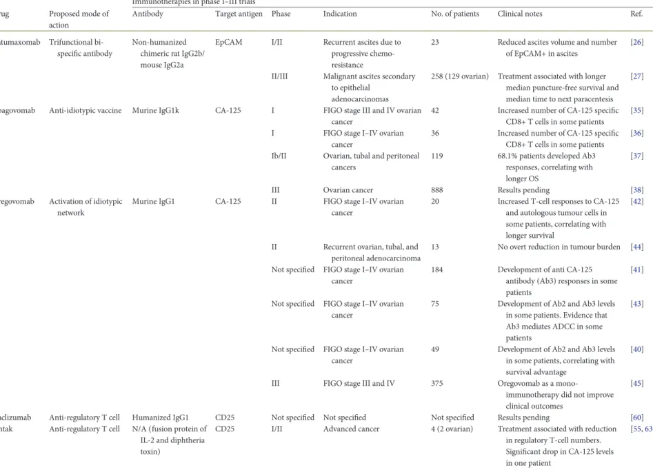
Table 1. Clinical trials of selected antibody-based immunotherapies for ovarian cancer Immunotherapies in phase I–III trials Drug Proposed mode of
action
Antibody Target antigen Phase Indication No. of patients Clinical notes Ref.
Catumaxomab Trifunctional bi-specific antibody
Non-humanized chimeric rat IgG2b/ mouse IgG2a
EpCAM I/II Recurrent ascites due to progressive chemo-resistance
23 Reduced ascites volume and number of EpCAM+ in ascites
[26]
II/III Malignant ascites secondary to epithelial
adenocarcinomas
258 (129 ovarian) Treatment associated with longer median puncture-free survival and median time to next paracentesis
[27]
Abagovomab Anti-idiotypic vaccine Murine IgG1k CA-125 I FIGO stage III and IV ovarian cancer
42 Increased number of CA-125 specific CD8+ T cells in some patients
[35] I FIGO stage I–IV ovarian
cancer
36 Increased number of CA-125 specific CD8+ T cells in some patients
[36] Ib/II Ovarian, tubal and peritoneal
cancers
119 68.1% patients developed Ab3 responses, correlating with longer OS
[37]
III Ovarian cancer 888 Results pending [38]
Oregovomab Activation of idiotypic network
Murine IgG1 CA-125 II FIGO stage I–IV ovarian cancer
20 Increased T-cell responses to CA-125 and autologous tumour cells in some patients, correlating with longer survival
[42]
II Recurrent ovarian, tubal, and peritoneal adenocarcinoma
13 No overt reduction in tumour burden [44] Not specified FIGO stage I–IV ovarian
cancer
184 Development of anti CA-125 antibody (Ab3) responses in some patients
[41]
Not specified FIGO stage I–IV ovarian cancer
75 Development of Ab2 and Ab3 levels in some patients. Evidence that Ab3 mediates ADCC in some patients
[43]
Not specified FIGO stage I–IV ovarian cancer
49 Development of Ab2 and Ab3 levels in some patients, correlating with survival advantage
[40]
III FIGO stage III and IV 375 Oregovomab as a
mono-immunotherapy did not improve clinical outcomes
[45]
Daclizumab Anti-regulatory T cell Humanized IgG1 CD25 Not specified Not specified Not specified Results pending [60] Ontak Anti-regulatory T cell N/A (fusion protein of
IL-2 and diphtheria toxin)
CD25 I/II Advanced cancer 4 (2 ovarian) Treatment associated with reduction in regulatory T-cell numbers. Significant drop in CA-125 levels in one patient [55,63] | Tse et al. V olume 25 | No. 2 | F ebruary 2014
ws
Annals of Oncologymouse IgG2a). It also contains a functional Fc region that binds to activating receptors on accessory cells (macrophages, dendrit-ic cells, and NK cells) for immunologdendrit-ical effector function. The rationale of using Catumaxomab is to activate and recruit T cells to EpCAM-expressing tumours while simultaneously stimulating accessory cells via their FcγRs. The result is tumour cell destruc-tion by ADCC and/or T-cell-mediated cytotoxicity via perforin and granzyme B [16].
In a phase I/II clinical trial involving 23 ovarian cancer patients with recurrent ascites due to progressive chemo-resist-ant disease, treatment with Catumaxomab resulted in a signi fi-cant and sustained reduction in ascites [17]. Catumaxomab substantially reduced the number of EpCAM-positive tumour cells within ascites by up to 5 log, with six patients even showing negative cytology. A recently completed phase II/III clinical trial compared the efficacy of Catumaxomab plus paracentesis (C + P) to paracentesis alone (P) in 258 patients with recurrent chemo-resistant adenocarcinoma (ovarian, gastric, breast, pan-creas, colon, and endometrial) presenting with ascites [18]. In the ovarian cancer arm, the median puncture-free survival, defined as the time after treatment (Day 0) to the first need for paracentesis, was 52 days for C + P versus 11 days for P alone (P < 0.0001). This suggests that Catumaxomab functions rela-tively rapidly to alleviate ascites accumulation. Similarly, the median time to next paracentesis was significantly longer in the C + P group (71 versus 11 days) (P < 0.0001). This represented a six- to seven-fold prolongation and would be highly beneficial to patients since ascites has a high morbidity and repeated para-centesis increases the risk of infection, bowel perforation, and adhesions. Catumaxomab was also shown to increase the ratio of CD45+ cells (immune cells) to EpCAM-positive tumour cells in ascites throughout the course of treatment, with some patients having EpCAM-positive cells no longer detectable. Since Catumaxomab is composed of antibody chains of mouse and rat origin, it is expected that human anti-mouse antibodies (HAMAs) will be induced when administered to patients. Indeed, Catumaxomab induced greater clinical benefits for patients who developed HAMAs [19]. In ovarian cancer patients, the median puncture-free survival was 64 versus 30 days for HAMA-positive/-negative responders, respectively. The median time to next puncture was 104 versus 32 days, and OS was 163 compared with 82 days. From these clinical trial outcomes, Catumaxomab appears to be a promising immunotherapy for ovarian cancer and may be particularly useful for patients pre-senting with chemotherapy-resistant disease with recurrent ascites. Although some immunotherapy can be compromised by the HAMA response, it can sometimes augment the therapeutic response. In these studies, Catumaxomab was generally well tolerated with most common adverse events being reversible mild-to-moderate nausea and abdominal pain.
abagovomab and oregovomab
Antibodies can function as antigens in the form of ‘anti-idioty-pic antibodies’. Immunization with a given antigen (x) results in the generation of anti-x antibodies, which can be referred to as Ab1 [20]. Neils Jerne proposed that these antibodies may them-selves be immunogenic and defined the immunogenic determi-nants of the antibody as idiotopes. The idiotopes of Ab1 can
Ipili mumab Activa tion of ef fector T cells Huma n IgG1 CT LA-4 I P re-t re ated advanced melano ma or ovarian cancer 9 (2 ovarian) C A-125 le ve ls tabili za tion (o ne pa tient ) and reduct ion (one pa tient ) [ 72 ] I FIGO stag e IV ovarian cancer pr eviou sly recei ve d G V A X 9 Ant i-tumo ur activit y in one pa tient [ 73 ] MDX-1 105 Immun e-che ckpoint inhibi tor Huma n IgG4 PD -L1 I Not speci fied 17 P artial respo nse in one pa tie nt. Disease stabili za tion in thr ee pa tient s [ 77 ]
Volume 25 | No. 2 | February 2014 doi:10.1093/annonc/mdt405 |
function as antigen that evokes the generation of antibodies (anti-idiotypic). Ab2 are antibodies that result from exposure to Ab1, some of which are anti-idiotypic. Since idiotopes are con-centrated in the highly variable region of the antigen-binding site, some Ab2 will have idiotopes that effectively mimic the three-dimensional structure of antigen x. Experimental evidence suggests that, in some circumstances, exposure to Ab2 may pro-voke a more effective response than exposure to antigen. These antibodies, in turn, may stimulate the production of Ab3, some of which target the idiotopes of Ab2, and bind to antigen x.
CA125 is a high-molecular-weight mucin-like glycoprotein over-expressed on the surface of ovarian cancer cells. It is also shed into the bloodstream and is currently the most widely used tumour marker for ovarian cancer. A high level of serum CA125 at ovarian cancer diagnosis frequently indicates widespread peritoneal dissemination, and a continual increase in its value generally indicates disease progression and/or relapse [21,22]. Although CA125 is in clinical use as a tumour marker, its bio-logical function is still poorly understood; some studies suggest that it is involved in cell adhesion, migration, and invasion, and may have immunosuppressive properties [23,24]. CA125 binds with high affinity to mesothelin, a protein highly expressed on the surface of the peritoneal lining [23]. The human serous ovarian cancer cell line OVCAR-3 (CA125-proficient) adheres strongly to mesothelin-expressing cells but not when CA125 was silenced through siRNA, suggesting that CA125 may con-tribute to peritoneal metastasis at some levels. CA125 knock-down was shown in ovarian cancer cell lines to reduce their motility and invasiveness (associated with matrix metalloprotei-nase-2 down-regulation) and to suppress their growth by induc-tion of caspase-dependent apoptosis [25]. In another study, incubation of NK cells with CA125 decreased their cytolytic ac-tivity against target cells by 50%–70%, suggesting that CA125 exhibits immune suppressive properties as well [24].
Abagovomab (ACA-126) is a murine IgG1k monoclonal anti-body (Ab2) with its idiotope imitating CA125 and is currently being investigated as an anti-idiotypic vaccine for ovarian cancer. In a phase I trial involving 42 patients with chemother-apy-resistant ovarian cancers (93% with FIGO stages III–IV; 67% serous histotype), Abagovomab induced Ab3 and HAMA responses in all patients [26]. Moreover, 5 of 5 patients also had detectable CA125-specific interferon (IFN)-γ-producing T cells post vaccination, whereas none were detectable in any subjects before treatment. Abagovomab also strongly increased serum levels of IFN-γ, which indicates the induction of Th1 immune responses, in a subset of 25 patients. In another phase I clinical trial involving 36 patients with similar clinicopathological characteristics, a two-fold increase in the number of CA125-specific CD8+ T cells was found in those that received nine (75%) injections of Abagovomab compared with those with six (17.6%) injections, suggesting that the strength of the immune re-sponse is dose-dependent [27]. A phase Ib/II clinical trial with 119 patients with CA125-positive ovarian, tubal, or peritoneal cancers showed that 68.1% of patients that received Abagovomab developed Ab3 responses [28]. Strikingly, Ab3 responders had a significantly longer OS (23.4 months) compared with Ab3 non-responders (4 months) (P < 0.0001). When the patient cohort was stratified for FIGO stage, type of first-line chemotherapy, and the number of previous therapies, the trend of longer survival in
Ab3 responders was maintained. ADCC was also observed in some patients (26.9%) and they had significantly longer median survival than those that did not (25 versus 10 months; P = 0.0126), suggesting that ADCC may be a mechanism that mediates the anti-tumour effect of Abagovomab. In these clinical trials, the drug was generally well tolerated with common side-effects including local reactions at the injection site and fatigue. Such promising data have resulted in the initiation of a rando-mized, double-blind, placebo-controlled phase III trial involving 888 patients with FIGO stage III/IV ovarian cancer known as ‘Monoclonal antibody Immunotherapy for Malignancies of Ovary by Subcutaneous Abagovomab’ (MIMOSA) trial [29]. While Abagovomab was shown to be safe and induced measur-able immune responses, the study concluded that when the anti-body was employed as a maintenance therapy for patients with first remission, it did not prolong relapse-free and OS [30].
Oregovomab (B43.13, OvRex) is a murine monoclonal antibody of IgG1 subclass with high affinity for CA125 (KD= 1.2 × 10¹⁰
M−1). It was initially developed as a Technetium 99c-labelled anti-body for the immunoscintigraphic detection of recurrent ovarian cancer by virtue of their expression of CA125 [31]. However, some patients that received this tumour-imaging agent had an un-expected survival advantage, prompting investigations into the antibody’s potential as a therapy for ovarian cancer. Oregovomab does not directly inhibit tumour growth nor does it induce CDC or ADCC by itself. However, it elicits tumour-specific, T-cell responses. Noteworthy, anti-CA125 antibodies evoked from Oregovomab treatment are not necessarily Ab3. Other mechan-isms such as the formation of immunostimulating complexes may also lead to the generation of anti-CA125 antibodies.
In a trial involving 184 patients with ovarian cancer (FIGO stages I–IV), a single treatment of Oregovomab caused a rapid reduction in serum levels of CA125 due to the formation of immune complexes [32]. After multiple treatments (1–10 infu-sions), 26 of 60 (43%) patients had a greater than three-fold in-crease in anti-CA125 antibody levels, and the level of inin-crease correlated with the amount of circulating CA125 at the time of injection. Anti-CA125 antibody responders also had longer sur-vival times compared with non-responders (22.9 versus 13.5 months; P = 0.0089). In addition, 9 of 17 (53%) patients showed an increase in T-cell proliferation in response to CA125 and had significantly longer survival time than non-responders (>84 versus 13.2 months; P = 0.0202). These findings suggest that generation of humoural and cellular anti-CA125 responses con-tribute to the clinical benefits of Oregovomab treatment. Similarly, in a phase II trial which recruited 20 patients with platinum-resistant recurrent ovarian cancer (FIGO stages I–IV), Oregovomab increased T-cell responses to CA125 in 7 of 18 (39%) patients and to autologous tumour cells in 5 of 8 (63%) [33]. Patients who developed a T-cell response to CA125 and/or autologous tumours also had longer survival compared with non-responders (median not reached versus 51.9 weeks). In another trial involving 75 patients with ovarian cancer (FIGO stages I–IV) treated with Oregovomab (1–10 infusions), 48 (64%) developed Ab2 antibodies, and 18 (24%) developed anti-CA125 antibodies [34]. Incubation of OVCAR3-NU3 cells with anti-CA125 antibodies purified from the serum of these patients in the presence of peripheral blood mononuclear cells led to ef-fective lysis of tumour cells, indicating that ADCC was
operative. However, these antibodies did not mediate killing of tumour cells via CDC in the study. In a retrospective analysis of a previous trial including 44 patients with recurrent ovarian cancer (majority being FIGO stages III–IV), treatment with Oregovomab resulted in the development of HAMA in 27 of 40 (67.5%) patients, anti-idiotypic antibodies (Ab2) in 76.7% of patients, and an increase by more than three-fold in the level of anti-CA125 antibodies in 28% of patients [31]. The ability of patients to generate these idiotypic network-related antibodies also correlated with a survival advantage; median survival for HAMA responders was 22.6 versus 7.2 months for non-responders (P = 0.0016). Similarly, for Ab2 responders and non-responders, the median survival was 18.3 versus 9.3 months (P = 0.075), and for anti-CA125 responders versus non-responders, 18.2 versus 13.1 months (P = 0.0896). However, such clinical benefits of Oregovomab were not demonstrable in one pilot phase II study. No overt reduction in tumour burden was observed in any of the 13 patients treated with Oregovomab, despite most having measurable T- and B-cell responses [35]. Similarly, a phase III trial which recruited 375 ovarian cancer patients with FIGO stage III and IV showed that while bioactivity was demonstrable and the drug was well tolerated, patients receiving Oregovomab as a mono-immunotherapy had similar clinical outcomes as those given placebo [36]. Recently, the therapeutic potential of combining Oregovomab with conventional chemotherapies, carboplatin and paclitaxel, were investigated in a phase II clinic-al triclinic-al [37]. In that study, 40 patients with FIGO stages III/IV were split into two groups and received Oregovomab either sim-ultaneously or 1 week after carboplatin–paclitaxel treatment. Although the main objectives of that trial were to compare the magnitude of antibody and cellular responses to CA125 evoked by the two treatment schedules, the authors concluded that the immune responses triggered by this chemo-immunotherapy were stronger than those measured in previously published mono-immunotherapy protocols.
daclizumab and ontak
Regulatory T cells (Tregs) are a subset of T cells that mediate immune suppression and are involved in the prevention of auto-immunity [38]. They inhibit the proliferation, cytokine produc-tion, and cytotoxicity of Th1 T cells, CD8+ T cells, and NK cells via cell–cell contact and the secretion of immunosuppressive factors [39–42]. Regulatory T cells also suppress immune activa-tion by down-regulating co-stimulatory molecules that are ne-cessary for T-cell activation on dendritic cells [43].
Early studies reported that regulatory T cells were CD4+ and CD25+; CD25 is the interleukin (IL)-2 receptorα chain [44,
45]. However, the best current marker of regulatory T cells is the transcription factor ‘foxhead box P3 (foxp3)’. Importantly, CD25 is not a specific marker of regulatory T cells as it is also expressed on activated effector T cells. One study reported that malignant ascites from previously untreated ovarian cancer patients contained significant numbers of CD4+ CD25+ CD3+ T cells, whereas these cells were rarely seen in non-malignant ascites [46]. FIGO stages III–IV ovarian cancers were also
shown to be more abundant with CD4+ CD25+ cells than stages I–II tumours [46]. Moreover, higher tumour infiltration of regu-latory T cells correlated with shorter patient survival [47, 48],
suggesting that regulatory T cells may facilitate ovarian cancer progression. Therefore, strategies that block or transiently deplete these cells may prove useful in treating cancer patients. This is supported by animal studies, whereby systemic removal of CD25+ cells with a cell-depleting monoclonal antibody can elicit potent and durable anti-tumour responses [49,50].
Daclizumab (Zenapax) is a cell-depleting humanized IgG1 monoclonal antibody specific for CD25. It is currently being evaluated in clinical trials as an immunotherapy for a variety of cancers, including ovarian cancer [51]. Although results from clinical trials of Daclizumab in ovarian cancer have yet to be released, data from clinical trials of Ontak (Denileukin Diftitox), another form of CD25-targeted therapy, are encouraging. Ontak is an engineered protein combining IL-2 with diphtheria toxin causing apoptosis of CD25+ cells. It is FDA-approved for the treatment of cutaneous T-cell leukaemia and currently being investigated as a therapy for other cancer types, including meta-static ovarian, renal [52], and breast cancers [53]. In a phase I/II clinical trial involving seven patients with advanced adenocar-cinomas, including ovarian cancers, treatment with Ontak was associated with a reduction in peripheral blood CD3+ CD4+ CD25+ cells and an increase in the number of circulating IFN-γ-producing T cells [46]. In one patient with FIGO stage IV ovarian cancer, blood CA125 dropped from 121–17 units/ml 39 days after herfirst of seven infusions [54]. A positron emission tomography scan revealed that all lymph node, visceral, and bone metastases had resolved with the exception of one lesion in the left groin which had progressed. In all patients, Ontak was generally well tolerated and further trials are ongoing [54].
ipilimumab
Effective T-cell activation requires two signals, namely (i) inter-action of the T-cell receptor with antigen in association with (MHC) molecules, and (ii) co-stimulation of CD28 on T cells with CD86 and CD80 on antigen-presenting cells (APCs). A lack of co-stimulatory signals can result in T-cell anergy. Cytotoxic T-lymphocyte-associated antigen 4 (CTLA-4) is a homologue of CD28 expressed on the surface of T cells upon ac-tivation and is a key molecule in the down-regulation of T-cell activity as a means to limit self-damage. It inhibits the activation of T cells by effectively outcompeting CD28 for its ligands on APCs (CTLA-4 has a higher affinity for CD86 and CD80) and/ or delivering inhibitory signals to T cells [55]. In this sense, CTLA-4 functions as an immune-checkpoint receptor. CTLA-4 is also constitutively expressed on regulatory T cells and is a mechanism by which they mediate immune suppression [56]. The use of therapeutic agents to block CTLA-4 function pre-vents immune inactivation and is a rational approach to evoke effective anti-tumour immune responses.
Ipilimumab (MDX-CTLA-4, Yervoy) is a fully human IgG1 monoclonal antibody, which binds to and blocks the activity of CTLA-4. It was recently approved by the FDA for the treatment of advanced melanoma [57]. A phase III clinical trial comprised of patients with unresectable pre-treated stages III–IV melan-oma, demonstrated that treatment with Ipilimumab prolonged the median survival by∼4 months—the first compound shown to have such beneficial effects in these patients [58]. Results from several studies suggest that the anti-tumour effect of
Volume 25 | No. 2 | February 2014 doi:10.1093/annonc/mdt405 |
Ipilimumab is mediated by immune modulation. Treatment with Ipilimumab has been reported to increase (i) absolute lymphocyte count [59], (ii) expression of inducible co-stimula-tor (ICOS) on CD4+ T cells [60], and (iii) antibody and T-cell responses to the cancer-testis antigen NY-ESO-1 [60, 61]. In most cases, such responses correlated with clinical benefit and OS. Moreover, in some trials, response rates have been substan-tially higher in patients who developed immune-related adverse events such as enterocolitis [62] or hypophysitis [63] than those who did not. Although the majority of Ipilimumab trials, to date, have been on melanoma, a few ovarian cancer trials have been conducted. In a phase I/II trial on 11 patients with FIGO stage IV ovarian cancers which had previously either received chemotherapy or GVAX [a vaccine product comprised on au-tologous, irradiated tumour cells engineered to secrete the immune stimulatory cytokine, granulocyte macrophage colony-stimulating factor), Ipilimumab was generally well tolerated with the exception of some grade 3 inflammatory toxicities [64,
65]. Significant anti-tumour effects were noted in one particular ovarian cancer patient who showed a dramatic fall of serum CA125 levels during treatment with a substantial regression of a large hepatic metastasis, mesenteric lymph nodes, and an omental cake [65]. Moreover, generation of antibody responses to NY-ESO-1 was detectable and this correlated with the observed therapeutic effects. Another ovarian cancer patient had a reduction in pain and ascites, which correlated with stabiliza-tion of CA125 levels. Four other patients had stable disease as assessed by blood CA125 levels and imaging.
current stage of immunotherapy
in ovarian cancer
It is still unclear if immunotherapy has the potential to be incor-porated into treatment regimens against ovarian cancer. Over the past decade, there has been an increase in the number of anti-body-based immunotherapeutics that have reached the clinical trial stage. While Catumaxomab, Abagovomab, and Oregovomab have already reached phase III, many other new antibody-based treatments have just entered phase I and II clinical trials. Catumaxomab particularly shows promise and was recently approved by the European Community for the treatment of ma-lignant ascites. Results from larger trials of this antibody are greatly anticipated. Abagovomab and Oregovomab, which showed promise based on early clinical trials, have largely been disappointing in recent phase III trials with no overt anti-tumour efficacy reported. In recent years, immune-checkpoint inhibitors, such as Ipilimumab, MDX-1105, and MDX-1106, the latter two targeting the PD-1/PD-L1 axis (supplementary 3, available at Annals of Oncology online), have emerged as candidates for cancer immunotherapy [66], although few clinical trials for ovarian cancer have been conducted so far. For this researchfield to move forward, it is important to consider the limiting factors of clinical trials in order to improve the design of future studies.
A limitation common to almost all cancer clinical trials is that the patients already have advanced disease. This is a major issue especially for immunotherapy because the ability to initiate an immune response, or its magnitude, is limited by the extent of disease burden, the suppressive effect of the tumour
microenvironment, and the multiple layers of immunological tolerance mechanisms (e.g. regulatory T cells), which keep the immune response in check. As a consequence, immune-based therapies are more likely to be effective in patients with low volume disease such as earlier stage cancers or after cytoreduc-tive treatment with minimal residual disease.
Realistically, if immunotherapy is to be applied to ovarian cancer treatment regimens in the short-to-medium term, it would be as an adjunct therapy rather than as a front-line monotherapy. Future clinical trials should investigate the syner-gistic potential of immune-modulating agents with established chemotherapies. Indeed, Oregovomab was recently investigated as a combination therapy with standard carboplatin–paclitaxel in a phase II clinical trial, and stronger immune responses were measured than those reported in a previous mono-immunother-apy protocol [37]. Chemo-immunotherapy is appealing not only because chemotherapies directly induce apoptosis of tumour cells and result in the release of antigen to drive immune responses, but they often also disrupt essential immune regulatory mechanisms that limit the development of immunity, an increasingly appreciated attribute [67]. Low-dose cyclophos-phamide andfludarabine have been shown to selectively deplete and suppress regulatory T-cell activity, rendering tumours cells more susceptible to immune-mediated cytotoxicity [68,69]. An ideal combination of chemo-immunotherapy would be one where both agents have minimal overlapping toxicities, work via independent mechanisms but have additive or synergistic anti-tumour effects. A successful example of this approach is the combination offludarabine, cyclophosphamide, and rituximab (FCR) for chronic lymphocytic leukaemia (CLL). The addition of rituximab to the chemotherapies increases the rate of com-plete remission and OS [70]. However, it is critical to optimize the dosage, sequence, and timing of administering the each drug as all these parameters could impact on the outcome.
Another point to consider in the design of future clinical trials is to acknowledge that ovarian cancer is a heterogeneous disease with four major histological subtypes, each of which have distinct genetic profiles which may affect the ability and/or magnitude of immune responses evoked by an immunotherapy. The heterogeneity of ovarian cancer can be further highlighted by differences in the anti-glycan antibody profiles of patients with various histotypes (supplementary 4, available at Annals of Oncology online). As a consequence, it is necessary to conduct clinical trials on more homogenous populations, for example, serous cancer patients only, to have greater power to detect po-tential clinical efficacy. Pooling patients into a single cohort may reduce the sensitivity to detect clinical benefits to certain types. The importance of stratifying treatment based on histo-type is supported by recent evidence of contrasting clinical outcomes in mucinous and serous ovarian cancers when treated with platinum-based chemotherapy [71]. These two histotypes may be distinct entities altogether [71], and investigations are underway to determine how each could be best managed.
An important lesson learnt in recent years is not become overly excited with novel drugs that show particular promise in phase I and/or II clinical trials because quite often they fail to meet the high expectations in phase III studies. This can be highlighted through Abagovomab, Oregovomab, and more re-cently Farletuzumab, a humanized IgG1κ antibody that binds to
folate receptorα (FRα), which is highly expressed on 90% epi-thelial ovarian cancers but not on normal ovarian tissues [72,
73]. Farletuzumab has been shown to inhibit tumour growth through CDC, ADCC, and direct anti-proliferative actions, and a phase I trial reported it to be well tolerated [74]. A phase II clinical trial involving platinum-sensitive patients experiencing first relapse showed that Farletuzumab in combination with car-boplatin/taxane improved objective response rates (ORRs) com-pared with historical controls, and increased duration of second remission compared withfirst remission in some patients [75]. However, a recent phase III trial using this combination did not meet the primary end point of progression-free survival (PFS), and another phase III trial was terminated early due to the lack of efficacy in interim analyses [76,77]. Although these results are rather disappointing, it is important for our researchfield to not give up on exploring immunotherapy in combination with chemotherapy because there are glimpses of efficacy in some patients from initial studies. We need to think critically the reasons for this, and design future clinical trials accordingly.
conclusion
A wide range of immune-modulating approaches are currently being evaluated for the treatment of ovarian cancer. As the results of clinical trials of the discussed antibody-based im-munotherapies are inconsistent, it is still too early to conclude if these drugs will be incorporated into treatment regimens against ovarian cancer. Furthermore, their true potential may lie within being strategically employed in chemo-immunotherapy regi-mens.
acknowledgements
We would like to acknowledge André Fedier for critically reading the manuscript and Mei-Chun Yeh for designing the figure.
funding
This work was supported by the Cancer Institute of NSW (09/ CRF/2-02), William Maxwell Trust, Mary Elisabeth Courier Scholarship (RANZCOG), RHW Foundation, and GO Fund.
disclosure
The authors have declared no conflicts of interest.
references
1. Jemal A, Bray F, Center MM et al. Global cancer statistics. CA Cancer J Clin 2011; 61: 69–90.
2. Siegel R, Naishadham D, Jemal A. Cancer statistics, 2012. CA Cancer J Clin 2012; 62: 10–29.
3. Ushijima K. Treatment for recurrent ovarian cancer-atfirst relapse. J Oncol 2010; 2010: 497429.
4. Smyth MJ, Dunn GP, Schreiber RD. Cancer immunosurveillance and immunoediting: the roles of immunity in suppressing tumor development and shaping tumor immunogenicity. Adv Immunol 2006; 90: 1–50.
5. Marcus R, Hagenbeek A. The therapeutic use of rituximab in non-Hodgkin’s lymphoma. Eur J Haematol Suppl 2007; January 5–14.
6. Harris M. Monoclonal antibodies as therapeutic agents for cancer. Lancet Oncol 2004; 5: 292–302.
7. Nicodemus CF, Smith LM, Schultes BC. Role of monoclonal antibodies in tumor-specific immunity. Expert Opin Biol Ther 2007; 7: 331–343.
8. Weiner LM, Surana R, Wang S. Monoclonal antibodies: versatile platforms for cancer immunotherapy. Nat Rev Immunol 2010; 10: 317–327.
9. Spizzo G, Went P, Dirnhofer S et al. High Ep-CAM expression is associated with poor prognosis in node-positive breast cancer. Breast Cancer Res Treat 2004; 86: 207–213.
10. Spizzo G, Went P, Dirnhofer S et al. Overexpression of epithelial cell adhesion molecule (Ep-CAM) is an independent prognostic marker for reduced survival of patients with epithelial ovarian cancer. Gynecol Oncol 2006; 103: 483–488. 11. Went P, Vasei M, Bubendorf L et al. Frequent high-level expression of the
immunotherapeutic target Ep-CAM in colon, stomach, prostate and lung cancers. Br J Cancer 2006; 94: 128–135.
12. Heinzelmann-Schwarz VA, Gardiner-Garden M, Henshall SM et al. Overexpression of the cell adhesion molecules DDR1, claudin 3, and Ep-CAM in metaplastic ovarian epithelium and ovarian cancer. Clin Cancer Res 2004; 10: 4427–4436.
13. Diaz-Arias AA, Loy TS, Bickel JT et al. Utility of BER-EP4 in the diagnosis of adenocarcinoma in effusions: an immunocytochemical study of 232 cases. Diagn Cytopathol 1993; 9: 516–521.
14. Munz M, Kieu C, Mack B et al. The carcinoma-associated antigen EpCAM upregulates c-myc and induces cell proliferation. Oncogene 2004; 23: 5748–5758.
15. Gutzmer R, Li W, Sutterwala S et al. A tumor-associated glycoprotein that blocks MHC class II-dependent antigen presentation by dendritic cells. J Immunol 2004; 173: 1023–1032.
16. Seimetz D, Lindhofer H, Bokemeyer C. Development and approval of the trifunctional antibody catumaxomab (anti-EpCAM x anti-CD3) as a targeted cancer immunotherapy. Cancer Treat Rev 2010; 36: 458–467.
17. Burges A, Wimberger P, Kumper C et al. Effective relief of malignant ascites in patients with advanced ovarian cancer by a trifunctional anti-EpCAM x anti-CD3 antibody: a phase I/II study. Clin Cancer Res 2007; 13: 3899–3905.
18. Heiss MM, Murawa P, Koralewski P et al. The trifunctional antibody catumaxomab for the treatment of malignant ascites due to epithelial cancer: results of a prospective randomized phase II/III trial. Int J Cancer 2010; 127: 2209–2221. 19. Ott MG, Marme F, Moldenhauer G et al. Humoral response to catumaxomab
correlates with clinical outcome: results of the pivotal phase II/III study in patients with malignant ascites. Int J Cancer 2012; 130: 2195–2203.
20. Jerne NK. Clonal selection in a lymphocyte network. Soc Gen Physiol Ser 1974; 29: 39–48.
21. Rustin GJ, Marples M, Nelstrop AE et al. Use of CA-125 to define progression of ovarian cancer in patients with persistently elevated levels. J Clin Oncol 2001; 19: 4054–4057.
22. Bast RC, Jr. CA 125 and the detection of recurrent ovarian cancer: a reasonably accurate biomarker for a difficult disease. Cancer 2010; 116: 2850–2853. 23. Gubbels JA, Belisle J, Onda M et al. Mesothelin-MUC16 binding is a high affinity,
N-glycan dependent interaction that facilitates peritoneal metastasis of ovarian tumors. Mol Cancer 2006; 5: 50.
24. Patankar MS, Jing Y, Morrison JC et al. Potent suppression of natural killer cell response mediated by the ovarian tumor marker CA125. Gynecol Oncol 2005; 99: 704–713.
25. Reinartz S, Failer S, Schuell T et al. CA125 (MUC16) gene silencing suppresses growth properties of ovarian and breast cancer cells. Eur J Cancer 2012; 48: 1558–1569.
26. Sabbatini P, Dupont J, Aghajanian C et al. Phase I study of abagovomab in patients with epithelial ovarian, fallopian tube, or primary peritoneal cancer. Clin Cancer Res 2006; 12: 5503–5510.
27. Pfisterer J, du Bois A, Sehouli J et al. The anti-idiotypic antibody abagovomab in patients with recurrent ovarian cancer. A phase I trial of the AGO-OVAR. Ann Oncol 2006; 17: 1568–1577.
28. Reinartz S, Kohler S, Schlebusch H et al. Vaccination of patients with advanced ovarian carcinoma with the anti-idiotype ACA125: immunological response and survival ( phase Ib/II). Clin Cancer Res 2004; 10: 1580–1587.
Volume 25 | No. 2 | February 2014 doi:10.1093/annonc/mdt405 |
29. Sabbatini P, Berek JS, Casada A et al. Abagovomab maintenance therapy in patients with epithelial ovarian cancer after complete response (CR) post-first-line chemotherapy (FLCT): preliminary results of the randomized, double-blind, placebo-controlled, multicenter MIMOSA trial. ASCO Meeting Abstracts 2010; 14:5036.
30. Sabbatini P, Harter P, Scambia G et al. Abagovomab as maintenance therapy in patients with epithelial ovarian cancer: a Phase III trial of the AGO OVAR, COGI, GINECO, and GEICO—The MIMOSA Study. J Clin Oncol 2013; 31: 1554–1561.
31. Mobus VJ, Baum RP, Bolle M et al. Immune responses to murine monoclonal antibody-B43.13 correlate with prolonged survival of women with recurrent ovarian cancer. Am J Obstet Gynecol 2003; 189: 28–36.
32. Noujaim AA, Schultes BC, Baum RP et al. Induction of CA125-specific B and T cell responses in patients injected with MAb-B43.13—evidence for antibody-mediated antigen-processing and presentation of CA125 in vivo. Cancer Biother Radiopharm 2001; 16: 187–203.
33. Gordon AN, Schultes BC, Gallion H et al. CA125- and tumor-specific T-cell responses correlate with prolonged survival in oregovomab-treated recurrent ovarian cancer patients. Gynecol Oncol 2004; 94: 340–351.
34. Schultes BC, Baum RP, Niesen A et al. Anti-idiotype induction therapy: anti-CA125 antibodies (Ab3) mediated tumor killing in patients treated with Ovarex mAb B43.13 (Ab1). Cancer Immunol Immunother 1998; 46: 201–212. 35. Ehlen TG, Hoskins PJ, Miller D et al. A pilot phase 2 study of oregovomab murine
monoclonal antibody to CA125 as an immunotherapeutic agent for recurrent ovarian cancer. Int J Gynecol Cancer 2005; 15: 1023–1034.
36. Berek J, Taylor P, McGuire W et al. Oregovomab maintenance monoimmunotherapy does not improve outcomes in advanced ovarian cancer. J Clin Oncol 2009; 27: 418–425.
37. Braly P, Nicodemus CF, Chu C et al. The immune adjuvant properties of front-line carboplatin–paclitaxel: a randomized phase 2 study of alternative schedules of intravenous oregovomab chemoimmunotherapy in advanced ovarian cancer. J Immunother 2009; 32: 54–65.
38. Setoguchi R, Hori S, Takahashi T et al. Homeostatic maintenance of natural Foxp3(+) CD25(+) CD4(+) regulatory T cells by interleukin (IL)-2 and induction of autoimmune disease by IL-2 neutralization. J Exp Med 2005; 201: 723–735.
39. Takahashi T, Kuniyasu Y, Toda M et al. Immunologic self-tolerance maintained by CD25+CD4+ naturally anergic and suppressive T cells: induction of autoimmune disease by breaking their anergic/suppressive state. Int Immunol 1998; 10: 1969–1980.
40. Thornton AM, Shevach EM. CD4+CD25+ immunoregulatory T cells suppress polyclonal T cell activation in vitro by inhibiting interleukin 2 production. J Exp Med 1998; 188: 287–296.
41. Piccirillo CA, Shevach EM. Cutting edge: control of CD8+ T cell activation by CD4 +CD25+ immunoregulatory cells. J Immunol 2001; 167: 1137–1140. 42. Smyth MJ, Teng MW, Swann J et al. CD4+CD25+ T regulatory cells suppress NK
cell-mediated immunotherapy of cancer. J Immunol 2006; 176: 1582–1587. 43. Onishi Y, Fehervari Z, Yamaguchi T et al. Foxp3+ natural regulatory T cells
preferentially form aggregates on dendritic cells in vitro and actively inhibit their maturation. Proc Natl Acad Sci USA 2008; 105: 10113–10118.
44. Woo EY, Chu CS, Goletz TJ et al. Regulatory CD4(+)CD25(+) T cells in tumors from patients with early-stage non-small cell lung cancer and late-stage ovarian cancer. Cancer Res 2001; 61: 4766–4772.
45. Sakaguchi S. Naturally arising CD4+ regulatory t cells for immunologic self-tolerance and negative control of immune responses. Annu Rev Immunol 2004; 22: 531–562.
46. Barnett B, Kryczek I, Cheng P et al. Regulatory T cells in ovarian cancer: biology and therapeutic potential. Am J Reprod Immunol 2005; 54: 369–377.
47. Barnett JC, Bean SM, Whitaker RS et al. Ovarian cancer tumor infiltrating T-regulatory (T(reg)) cells are associated with a metastatic phenotype. Gynecol Oncol 2010; 116: 556–562.
48. Sato E, Olson SH, Ahn J et al. Intraepithelial CD8+ tumor-infiltrating lymphocytes and a high CD8+/regulatory T cell ratio are associated with favorable prognosis in ovarian cancer. Proc Natl Acad Sci USA 2005; 102: 18538–18543.
49. Shimizu J, Yamazaki S, Sakaguchi S. Induction of tumor immunity by removing CD25+CD4+ T cells: a common basis between tumor immunity and autoimmunity. J Immunol 1999; 163: 5211–5218.
50. Tien AH, Xu L, Helgason CD. Altered immunity accompanies disease progression in a mouse model of prostate dysplasia. Cancer Res 2005; 65: 2947–2955. 51. Vasievich EA, Huang L. The suppressive tumor microenvironment: a challenge in
cancer immunotherapy. Mol Pharm 2011; 8: 635–641.
52. Dannull J, Su Z, Rizzieri D et al. Enhancement of vaccine-mediated antitumor immunity in cancer patients after depletion of regulatory T cells. J Clin Invest 2005; 115: 3623–3633.
53. Mahnke K, Schonfeld K, Fondel S et al. Depletion of CD4+CD25+ human regulatory T cells in vivo: kinetics of Treg depletion and alterations in immune functions in vivo and in vitro. Int J Cancer 2007; 120: 2723–2733.
54. Curiel TJ, Barnett B, Kryczek I et al. Regulatory T cells in ovarian cancer: biology and therapeutic potential. Cancer Immun 2006; 6: 20.
55. Hoos A, Ibrahim R, Korman A et al. Development of ipilimumab: contribution to a new paradigm for cancer immunotherapy. Semin Oncol 2010; 37: 533–546. 56. Wing K, Onishi Y, Prieto-Martin P et al. CTLA-4 control over Foxp3+ regulatory T
cell function. Science 2008; 322: 271–275.
57. Sondak VK, Smalley KS, Kudchadkar R et al. Ipilimumab. Nat Rev Drug Discov 2011; 10: 411–412.
58. Hodi FS, O’Day SJ, McDermott DF et al. Improved survival with ipilimumab in patients with metastatic melanoma. N Engl J Med 2010; 363: 711–723. 59. Ku GY, Yuan J, Page DB et al. Single-institution experience with ipilimumab in
advanced melanoma patients in the compassionate use setting: lymphocyte count after 2 doses correlates with survival. Cancer 2010; 116: 1767–1775. 60. Liakou CI, Kamat A, Tang DN et al. CTLA-4 blockade increases
IFNgamma-producing CD4+ICOShi cells to shift the ratio of effector to regulatory T cells in cancer patients. Proc Natl Acad Sci USA 2008; 105: 14987–14992.
61. Yuan J, Gnjatic S, Li H et al. CTLA-4 blockade enhances polyfunctional NY-ESO-1 specific T cell responses in metastatic melanoma patients with clinical benefit. Proc Natl Acad Sci USA 2008; 105: 20410–20415.
62. Beck KE, Blansfield JA, Tran KQ et al. Enterocolitis in patients with cancer after antibody blockade of cytotoxic T-lymphocyte-associated antigen 4. J Clin Oncol 2006; 24: 2283–2289.
63. Blansfield JA, Beck KE, Tran K et al. Cytotoxic T-lymphocyte-associated antigen-4 blockage can induce autoimmune hypophysitis in patients with metastatic melanoma and renal cancer. J Immunother 2005; 28: 593–598.
64. Hodi FS, Mihm MC, Soiffer RJ et al. Biologic activity of cytotoxic T lymphocyte-associated antigen 4 antibody blockade in previously vaccinated metastatic melanoma and ovarian carcinoma patients. Proc Natl Acad Sci USA 2003; 100: 4712–4717. 65. Hodi FS, Butler M, Oble DA et al. Immunologic and clinical effects of antibody
blockade of cytotoxic T lymphocyte-associated antigen 4 in previously vaccinated cancer patients. Proc Natl Acad Sci USA 2008; 105: 3005–3010.
66. Pardoll DM. The blockade of immune checkpoints in cancer immunotherapy. Nat Rev Cancer 2012; 12: 252–264.
67. Emens LA. Chemotherapy and tumor immunity: an unexpected collaboration. Front Biosci 2008; 13: 249–257.
68. Ghiringhelli F, Menard C, Puig PE et al. Metronomic cyclophosphamide regimen selectively depletes CD4+CD25+ regulatory T cells and restores T and NK effector functions in end stage cancer patients. Cancer Immunol Immunother 2007; 56: 641–648.
69. Beyer M, Kochanek M, Darabi K et al. Reduced frequencies and suppressive function of CD4+CD25hi regulatory T cells in patients with chronic lymphocytic leukemia after therapy withfludarabine. Blood 2005; 106: 2018–2025. 70. Tam CS, Keating MJ. Chemoimmunotherapy of chronic lymphocytic leukemia. Nat
Rev Clin Oncol 2010; 7: 521–532.
71. Naik JD, Seligmann J, Perren TJ. Mucinous tumours of the ovary. J Clin Pathol 2012; 65: 580–584.
72. Bagnoli M, Canevari S, Figini M et al. A step further in understanding the biology of the folate receptor in ovarian carcinoma. Gynecol Oncol 2003; 88: S140–S144. 73. Miotti S, Bagnoli M, Ottone F et al. Simultaneous activity of two different
mechanisms of folate transport in ovarian carcinoma cell lines. J Cell Biochem 1997; 65: 479–491.
74. Konner JA, Bell-McGuinn KM, Sabbatini P et al. Farletuzumab, a humanized monoclonal antibody against folate receptor alpha, in epithelial ovarian cancer: a phase I study. Clin Cancer Res 2010; 16: 5288–5295.
75. Armstrong DK, White AJ, Weil SC et al. Farletuzumab (a monoclonal antibody against folate receptor alpha) in relapsed platinum-sensitive ovarian cancer. Gynecol Oncol 2013; 129: 452–458.
76. Clinicaltrials.gov. An Efficacy and Safety of Study of MORAb-003 in Subjects with Platinum-Resistant or Refractory Relapsed Ovarian Cancer (FAR-122). Morphotek Inc., Exton PA, USA.
77. Clinicaltrials.gov. Efficacy and Safety of Study of MORAb-003 in Subjects with Platinum-Sensitive Ovarian Cancer in First Relapse. Morphotek Inc., Exton PA, USA.
Annals of Oncology 25: 331–338, 2014 doi:10.1093/annonc/mdt425 Published online 24 November 2013
A meta-analysis of patient outcomes with
subcentimeter disease after chemotherapy for
metastatic non-seminomatous germ cell tumor
P. Ravi
1, K. P. Gray
2,4, E. K. O
’Donnell
3& C. J. Sweeney
3,5*
1
Christ’s College, University of Cambridge, Cambridge, UK; Departments of2
Biostatistics and Computational Biology Boston;3
Medicine, Dana Farber Cancer Institute, Boston, MA;4
Harvard School of Public Health, Boston, MA;5
Harvard Medical School, Boston, MA, USA
Received 7 May 2013; revised 2 July 2013 & 18 August 2013; accepted 4 September 2013
Background:Approximately a quarter of men with metastatic non-seminomatous germ cell tumor (NSGCT) have a
re-sidual mass, typically in the retroperitoneum, after chemotherapy. The management of small rere-sidual masses (≤1 cm) is controversial, with good outcomes seen with either post-chemotherapy retroperitoneal lymph node dissection (PC-RPLND) or surveillance. We sought to review our experience of surveillance and synthesize the cumulativefindings with the current literature in the form of a meta-analysis.
Patients and methods:We searched PubMed, EMBASE and abstracts from ASCO and AUA to identify relevant,
English-language studies for the meta-analysis. The DFCI (Dana Farber Cancer Institute) database was constructed from a database of men undergoing cisplatin-based chemotherapy for metastatic NSGCT. The outcomes of interest were the proportion with necrosis, teratoma or active cancer on histology at PC-RPLND (literature) and the total number of relapses, RP-only relapses and overall survival in men undergoing surveillance (literature and DFCI cohort).
Results:Three of 47 men undergoing post-chemotherapy surveillance at our institution relapsed over a median
follow-up of 5.4 years. All three were alive at a median of 4.2 years after relapse. On meta-analysis, the pooled estimates of ne-crosis, teratoma and active cancer in the 588 men who underwent PC-RPLND were 71, 24 and 4%, respectively. Of the combined 455 men who underwent surveillance, the pooled estimate of the relapse rate was 5%, with an RP-only relapse rate of 3%. Of the 15 men who suffered an RP-only relapse on surveillance, two died of disease.
Conclusion:Surveillance is a reasonable strategy for men with minimal residual RP disease after chemotherapy and
avoids an RPLND in∼97% of men who are cured with chemotherapy alone.
Key words:complete remission, retroperitoneal lymph node dissection, testicular cancer
introduction
Testicular cancer is the commonest solid tumor in men aged between 15 and 34 and notable for the fact that the vast majority of men with metastatic disease are cured with cisplatin-based chemo-therapy [1]. However, around 20–25% of patients with
non-semino-matous germ cell tumor (NSGCT) will have a residual tumor mass,
most often in the retroperitoneum (RP) [2]. Surgery, in the form of post-chemotherapy RP lymph node dissection (PC-RPLND), is routinely carried out for patients with residual masses greater than 1 cm as∼45% will harbor teratoma and 10% viable cancer [3–6].
The management of NSGCT with small residual masses (≤1 cm in short axial dimension in accordance with RECIST 1.1) post-chemotherapy is controversial. Men achieving serologic and radiologic complete remission are treated with PC-RPLND at certain institutions as routine policy, with its advantage of being able to resect potential low volume chemoresistant tera-toma or viable residual cancer [7]. However, recent reports have suggested that surveillance may be an option in this situation [8,9]
*Correspondence to: Dr C. J. Sweeney, Department of Medicine, Lank Center for Genitourinary Oncology, Dana-Farber Cancer Institute, 450 Brookline Ave., Suite D1230 Boston, MA 02215, USA. Tel: +1-617-632-4524; Fax: + 1-617-632-2165; E-mail: [email protected]
© The Author 2013. Published by Oxford University Press on behalf of the European Society for Medical Oncology. All rights reserved. For permissions, please email: [email protected].