Alexander Semmler , Susanna Moskau-Hartmann , Birgit Stoffel-Wagner , Christian Elger
and Michael Linnebank*
Homocysteine plasma levels in patients treated
with antiepileptic drugs depend on folate and
vitamin B12 serum levels, but not on genetic
variants of homocysteine metabolism
Abstract
Background : Antiepileptic drugs (AEDs) are commonly
used in the treatment of epilepsy, psychiatric diseases
and pain disorders. Several of these drugs influence blood
levels of folate and vitamin B12 and, consequently,
homo-cysteine. This may be relevant for AED effects and side
effects. However, not only folate and vitamin B12, but also
genetic variants modify homocysteine metabolism. Here,
we aimed to determine whether there is a
pharmacoge-netic interaction between folate, vitamin B12 and gepharmacoge-netic
variants and homocysteine plasma level in AED-treated
patients.
Methods : In this mono-center study, we measured
homo-cysteine, folate and vitamin B12 plasma levels in a
popu-lation of 498 AED-treated adult patients with epilepsy. In
addition, we analyzed the genotypes of seven common
genetic variants of homocysteine metabolism:
methyl-enetetrahydrofolate reductase (
MTHFR ) c.677C > T and
c.1298A
> C, methionine synthase ( MTR ) c.2756A > G,
dihy-drofolate reductase (
DHFR ) c.594 + 59del19bp,
cystathio-nine
β -synthase ( CBS ) c.844_855ins68, transcobalamin
2 (
TC2 ) c.776C > G and methionine synthase reductase
(
MTRR ) c.66G > A.
Results : On multivariate logistic regression, folate and
vitamin B12 levels, but none of the genetic variants, were
predictive for homocysteine levels.
Conclusions
: These data suggest that, in AED-treated
patients, folate and vitamin B12 play important roles in
the development of hyperhomocysteinemia, whereas
genetic variants of homocysteine metabolism do not and
thus do not contribute to the risk of developing
hyper-homocysteinemia during AED treatment.
Keywords: antiepileptic drugs; folate; single nucleotide
polymorphism; vitamin B12.
*Corresponding author: PD Dr. Michael Linnebank , Department of Neurology, University Hospital Zurich, Frauenklinikstrasse 26, 8091 Zurich, Switzerland, Phone: +41 44 2555544, Fax: +41 44 2554507, E-mail: michael.linnebank@usz.ch
Alexander Semmler: Department of Neurology, University of Zurich, Switzerland
Susanna Moskau-Hartmann and Christian Elger: Department of Epileptology, University of Bonn, Germany
Birgit Stoffel-Wagner: Institute of Clinical Chemistry and Clinical Pharmacology, University of Bonn, University Hospital of Bonn, Germany
Introduction
Antiepileptic drugs (AEDs) are widely used to treat epileptic
seizures, psychiatric diseases and pain syndromes. Several
side effects and risks limit the usage of AEDs. A
character-istic of several AEDs is a reduction of folate and vitamin B12
serum levels accompanied by an increase of homocysteine
plasma levels [ 1 – 4 ]. A mild to moderate increase in
homo-cysteine plasma levels has been firmly established as an
independent risk factor for cardiovascular [ 5 , 6 ] and
neu-rodegenerative [ 7 – 9 ] diseases in the general population.
Epidemiologic studies show that patients with epilepsy
have an increased risk for ischemic heart disease, fatal
cardiovascular outcome and neurodegenerative diseases
such as dementia and Parkinson ’ s disease, which may be
attributed to hyperhomocysteinemia [ 10 – 13 ]. In addition,
increased homocysteine plasma levels potentially lead to
the aggravation of seizures, as indicated by experimental
data [ 14 ] and in patients with alcohol withdrawal seizures
[ 15 ]. Therefore, the effect of AEDs on vitamin and
homo-cysteine metabolism and ultimately on cardiovascular
and neurological disease is a relevant research area in the
patient population receiving chronic AED treatment.
Not only vitamin status, but also genetic variants may
modify folate, vitamin B12 and homocysteine metabolism
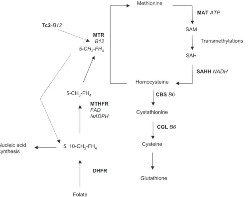
( Figure 1 ) [ 16 ]. Several studies have reported that
eleva-tion of homocysteine plasma levels during AED treatment
is enhanced by the presence of genetic risk factors such as
the presence of the T allele of the common
methylenetet-rahydrofolate reductase (
MTHFR ) c.677C > T polymorphism
[ 18 – 22 ]. However, these studies are limited due to their
small study populations and the small number of genetic
variants of homocysteine metabolism tested. In this study,
we investigated whether there is a relevant
pharmacoge-netic relationship between folate, vitamin B12 and seven
genetic variants of homocysteine metabolism and
homo-cysteine plasma level in 498 AED-treated patients.
Materials and methods
Patients
Inclusion criteria: This mono-center study included adult se-rial in- and out-patients with epilepsy seen in the Department for
Epileptology of the University Hospital Bonn, Germany. The patients were treated with various commonly used AEDs in mono- or com-bined therapy [ 4 ].
Exclusion criteria: Patients with conditions that could potential-ly infl uence folate, vitamin B12 or homocysteine plasma levels, such as renal insuffi ciency, atrophic gastritis and alcohol or drug abuse, were excluded from the study. Patients who were taking vitamin sup-plements were also excluded.
This study was approved by the Local Ethics Committee. All pa-tients gave their informed written consent.
Laboratory investigations
Serum concentrations of vitamin B12 and folate were measured by means of a competitive chemiluminescent immunoassay with an Ac-cess ™ Immunoassay System (Beckman Coulter, Krefeld, Germany). The intra-assay coeffi cient of variation of the folate assay was 3.1% (mean: 14.1 nmol/L; n = 20); the inter-assay coeffi cient of variation was 3.6% (mean: 14.3 nmol/L; n = 20). The intra-assay coeffi cient of variation of the vitamin B12 assay was 3.8% (mean: 487 pmol/mL; n = 20); the inter-assay coeffi cient was 4.2% (mean: 492 pmol/L; n = 20). Homocysteine was determined by fully automated particle-enhanced immunonephelometry with a BN II System (Siemens Methionine SAM SAH Homocysteine Cystathionine Cysteine Folate 5, 10-CH2-FH4 5-CH3-FH4 5-CH3-FH4 Nucleic acid synthesis Glutathione Transmethylations MAT ATP SAHH NADH MTR B12 Tc2-B12 CBS B6 MTHFR FAD NADPH CGL B6 DHFR
Figure 1 Homocysteine metabolism.
The sulfur-containing amino acid methionine is activated to S-adenosylmethionine (SAM), which is a ubiquitous methyl group donor. The degradation product of SAM is S-adenosylhomocysteine (SAH), which is hydrolyzed to homocysteine. Homocysteine can be remethylated to methionine and SAM via methionine synthase (MTR), which depends on derivatives of folate and vitamin B12 as cofactors. Lack of these vitamins is a common cause of hyperhomocysteinemia [ 17 ]. The folate derivative is synthesized by methylenetetrahydrofolate reductase (MTHFR) and dihydrofolate reductase (DHFR), and the derivative of vitamin B12 is transported by transcobalamin 2 (Tc2). Alternatively, homocysteine can be transsulfurated by vitamin B6 dependent cystathionine β -synthase (CBS) and cystathionine gamma-lyase (CGL) to cysteine as a component of glutathione. Due to the existence of several functional variants in the genes involved in homocysteine metabo-lism, and to differences in dietary vitamin and amino acid uptake, disorders of homocysteine metabolism exhibit marked inter-individual differences.
Healthcare Diagnostics, Eschborn, Germany) by enzymatic conver-sion to S-adenosylhomocysteine. The intra-assay coeffi cient of vari-ation of the homocysteine assay was 3.4% (mean: 11 μ mol/L, n = 20); the inter-assay coeffi cient was 5.6% (mean: μ mol/L, n = 20) [ 1 ].
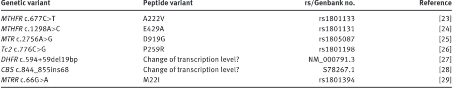
Genomic DNA prepared from peripheral leukocytes was used for genotyping by PCR amplifi cation and, where applicable, subsequent restriction analysis of the seven genetic variants of homocysteine me-tabolism ( Table 1 ).
Statistical analysis
Plasma levels of homocysteine plasma and serum levels of folate and vitamin B12 were tested for normal distribution by the Kolmogorov-Smirnov test. The distribution of genotypes was tested with a chi-square goodness-of-fi t test (Pearson). Bivariate Pearson ’ s correlation was used to analyze correlations between folate, vitamin B12 and homocysteine levels. To analyze associations between the diff erent genotypes and folate and vitamin B12 levels and folate and vitamin B12 tertiles, univariate analysis of variance (ANOVA) and Pearson ’ s χ 2 -tests were used, respectively. To analyze independent associations
with homocysteine plasma level as the primary parameter of inter-est, we applied multivariate linear regression analysis with homo-cysteine plasma level as the dependent variable and with the genetic variants, folate and vitamin B12 plasma levels and age and sex as covariables. One-way ANOVA was used for exploratory comparison of homocysteine plasma levels between patients with the MTHFR c.677C > T genotype treated with either carbamazepine or phenytoin. The threshold was defi ned as two-sided α = 0.05.
Results
Demographic, biochemical and genetic data from the 498
patients (51.4% male) enrolled in this study are shown
in
Table 2
. Genotyping succeeded for all genetic
vari-ants. Genotype distributions did not deviate from
Hardy-Weinberg equilibrium. Homocysteine plasma levels as
well as folate and vitamin B12 serum levels were within
the normal distribution. Thus, the data were not
log-trans-formed. First, we evaluated the relationships between
folate and vitamin B12 levels and homocysteine level by
uni-variate analysis and found negative correlations between
homocysteine and folate (Pearson
= − 0.334; p < 0.001)
and homocysteine and vitamin B12 (Pearson
= − 0.236;
p
= 0.001). Therefore, we included folate and vitamin B12
plasma levels along with age, sex and all seven genetic
variants as covariables for multivariate analysis of
inde-pendent associations with homocysteine plasma level as
the dependent variable. The associations between folate
and vitamin B12 with homocysteine level were confirmed,
but none of the genotypes showed an association with
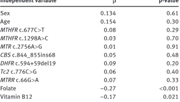
homocysteine level ( Table 3 ). In addition, none of the
gen-otypes was associated with folate or vitamin B12 serum
level (data not shown). However,
MTHFR c.677C > T was
associated with folate tertiles; i.e., patients with the TT
genotype had a higher likelihood of having folate serum
Demographic/biochemical data Mean SD Age, years 40.0 14.0 Vitamin B12, pmol/L 268 137 Folate, nmol/L 11.6 7.9 Homocysteine, μ mol/L 15.5 8.6
Sex, n (%) Male Female
256 (51.4%) 242 (48.6%) Frequency of genotype MTHFR c.677C > T CC CT TT n (%) 219 (44%) 224 (45%) 55 (11%) MTHFR c.1298A > C AA AC CC n (%) 214 (43%) 229 (46%) 55 (11%) MTR c.2756A > G AA AG GG n (%) 349 (70%) 134 (27%) 15 (3%) TC2 c.776 C > G CC CG GG n (%) 149 (30%) 229 (46%) 120 (24%) DHFR c.594 + 59del19 dd di ii n (%) 90 (18%) 244 (49%) 164 (33%) CBS c.844_855ins68 dd di ii n (%) 418 (84%) 75 (15%) 5 (1%) MTRR G > A GG GA AA n (%) 100 (20%) 259 (52%) 139 (28%) Table 2 Demographic and biochemical data and frequency of genotypes in the study population (n = 498).
All genotypes were in Hardy-Weinberg equilibrium. SD, standard deviation.
Genetic variant Peptide variant rs/Genbank no. Reference
MTHFR c.677C > T A222V rs1801133 [ 23 ]
MTHFR c.1298A > C E429A rs1801131 [ 24 ]
MTR c.2756A > G D919G rs1805087 [ 25 ]
Tc2 c.776C > G P259R rs1801198 [ 26 ]
DHFR c.594 + 59del19bp Change of transcription level? NM_000791.3 [ 27 ]
CBS c.844_855ins68 Change of transcription level? S78267.1 [ 28 ]
MTRR c.66G > A M22I rs1801394 [ 29 ]
levels in the lowest tertile (
χ
2= 3.1; p = 0.011). Next, we
con-ducted an exploratory analysis of patients who received
carbamazepine or phenytoin monotherapy (n = 76),
looking for an association between
MTHFR c.677C > T and
homocysteine plasma level, which has been reported by
previous studies [ 18 – 22 ]. However, we observed no
signifi-cant differences (ANOVA: F = 2.5; p = 0.091).
Discussion
In our study cohort of 498 AED-treated epilepsy patients,
we observed no associations between any of seven genetic
variants of homocysteine metabolism and homocysteine
plasma level. Only folate and vitamin B12 serum levels
were associated with homocysteine plasma level. This
indicates that hyperhomocysteinemia during chronic AED
treatment is driven by decreased folate and vitamin B12
levels and not by a pharmacogenetic risk profile.
This is surprising, because genetic variants of
homo-cysteine metabolism are firmly established risk factors for
hyperhomocysteinemia in the general population; e.g.,
MTHFR c.677C > T influences homocysteine plasma levels,
with differences of approximately 2
μ mol/L (15% – 20%)
between homozygous carriers of the wild-type C versus the
mutant T allele [ 23 ]. In addition, the T variant also
influ-ences folate metabolism, resulting in lower total folate
levels [ 30 ]. In our study, the association with folate level
was weak and was significant only with folate tertiles. We
speculate that the effects of the AEDs on folate and
homo-cysteine levels overcame the weaker effects of the genetic
variants in our patient population. This is in contrast to
previous studies describing genetic risk factors for
hyper-homocysteinemia during AED treatment – principally the
T allele of
MTHFR c.677 C > T and the C allele of MTHFR
c.1298A
> C [ 18 – 22 ] – and may be explained by the differing
study populations. For example, Yoo et al. described an
association between the TT genotype of
MTHFR c.677 C > T
and higher homocysteine plasma levels in AED-treated
patients [ 21 ]. However, the subjects enrolled in that study
were from Korea and were younger (27.5
± 8.5 years) and
had lower mean homocysteine plasma levels (11.2
± 1.5
μ
mol/L), higher folate (18.8
± 10.2 nmol/L) and higher
vitamin B12 serum levels (630
± 252 pmol/L) than the
patients in our study ( Table 2 ). Thus, we cannot exclude
the possibility that the small subgroup sizes of that study
or demographic differences between the populations
con-tributed to the conflicting results.
In conclusion, patients undergoing chronic AED
treat-ment should be screened for folate, vitamin B12 deficiency
and hyperhomocysteinemia on a regular basis and any
vitamin deficiency should be corrected when necessary
[ 4 ]. Screening for genetic variants is not feasible for the
detection of patients at risk and should not be included in
the clinical work-up.
Conflict of interest statement
Authors ’ conflict of interest disclosure : The authors stated that there are no conflicts of interest regarding the publication of this article. Research funding : None declared.
Employment or leadership : None declared. Honorarium : None declared.
Received September 15, 2012; accepted December 26, 2012; previously published online February 1, 2013
References
1. Sener U, Zorlu Y, Karaguzel O, Ozdamar O, Coker I, Topbas M. Effects of common anti-epileptic drug monotherapy on serum levels of homocysteine, vitamin B12, folic acid and vitamin B6. Seizure 2006;15:79 – 85.
2. Gidal BE, Tamura T, Hammer A, Vuong A. Blood homocysteine, folate and vitamin B-12 concentrations in patients with
epilepsy receiving lamotrigine or sodium valproate for initial monotherapy. Epilepsy Res 2005;64:161 – 6.
3. Karabiber H, Sonmezgoz E, Ozerol E, Yakinci C, Otlu B, Yologlu S. Effects of valproate and carbamazepine on serum levels of homocysteine, vitamin B12, and folic acid. Brain Dev 2003;25:113 – 5.
Independent variable β p-Value
Sex 0.134 0.61 Age 0.154 0.30 MTHFR c.677C > T 0.08 0.29 MTHFR c.1298A > C 0.03 0.70 MTR c.2756A > G 0.01 0.91 CBS c.844_855ins68 0.05 0.48 DHFR c.594 + 59del19 0.09 0.20 Tc2 c.776C > G 0.06 0.40 MTRR c.66G > A 0.07 0.33 Folate − 0.27 < 0.001 Vitamin B12 − 0.17 0.021
Table 3 Multiple logistic regression analysis (R 2 = 0.249) with
homocysteine plasma level as the dependent variable and age, sex, folate, vitamin B12 plasma level and all tested genetic variants as covariables.
4. Linnebank M, Moskau S, Semmler A, Widman G, Stoffel-Wagner B, Weller M, et al. Antiepileptic drugs interact with folate and vitamin B12 serum levels. Ann Neurol 2011;69:352 – 9. 5. Homocysteine and risk of ischemic heart disease and stroke: a
meta-analysis. J Am Med Assoc 2002;288:2015 – 22.
6. de Ruijter W, Westendorp RG, Assendelft WJ, den Elzen WP, de Craen AJ, le Cessie S, et al. Use of Framingham risk score and new biomarkers to predict cardiovascular mortality in older people: population based observational cohort study. Br Med J 2009;338:a3083.
7. Faux NG, Ellis KA, Porter L, Fowler CJ, Laws SM, Martins RN, et al. Homocysteine, vitamin B12, and folic acid levels in Alzheimer ’ s disease, mild cognitive impairment, and healthy elderly: baseline characteristics in subjects of the Australian Imaging Biomarker Lifestyle study. J Alzheimers Dis 2011;27:909 – 22.
8. Hooshmand B, Solomon A, Kareholt I, Leiviska J, Rusanen M, Ahtiluoto S, et al. Homocysteine and holotranscobalamin and the risk of Alzheimer disease: a longitudinal study. Neurology 2010;75:1408 – 14.
9. Stanger O, Fowler B, Piertzik K, Huemer M, Haschke-Becher E, Semmler A, et al. Homocysteine, folate and vitamin B12 in neuropsychiatric diseases: review and treatment recommen-dations. Expert Rev Neurother 2009;9:1393 – 412.
10. Gaitatzis A, Carroll K, Majeed A, Sander JW. The epidemiology of the comorbidity of epilepsy in the general population. Epilepsia 2004;45:1613 – 22.
11. Gaitatzis A, Johnson AL, Chadwick DW, Shorvon SD, Sander JW. Life expectancy in people with newly diagnosed epilepsy. Brain 2004;127:2427 – 32.
12. Aurlien D, Larsen JP, Gjerstad L, Tauboll E. Comorbid and underlying diseases – major determinants of excess mortality in epilepsy. Seizure 2012;21:573 – 7.
13. Nilsson L, Tomson T, Farahmand BY, Diwan V, Persson PG. Cause-specific mortality in epilepsy: a cohort study of more than 9,000 patients once hospitalized for epilepsy. Epilepsia 1997;38:1062 – 8.
14. Baldelli E, Leo G, Andreoli N, Fuxe K, Biagini G, Agnati LF. Homocysteine potentiates seizures and cell loss induced by pilocarpine treatment. Neuromolecular Med 2010;12:248 – 59. 15. Bleich S, Bayerlein K, Hillemacher T, Degner D, Kornhuber J,
Frieling H. An assessment of the potential value of elevated homocysteine in predicting alcohol-withdrawal seizures. Epilepsia 2006;47:934 – 8.
16. Stover PJ. Polymorphisms in 1-carbon metabolism, epigenetics and folate-related pathologies. J Nutrigenet Nutrigenomics 2011;4:293 – 305.
17. Mudd SH, Levy HL, Kraus JP. Disorders of transsulfuration. In: Scriver CR, Beaudet AL, Sly WS, Valle D, Childs B, Kinzler K, et al., editors. The metabolic and molecular bases of inherited disease. New York: Mc Graw-Hill, 2001:2007 – 56.
18. Ono H, Sakamoto A, Mizoguchi N, Sakura N. The C677T mutation in the methylenetetrahydrofolate reductase gene contributes
to hyperhomocysteinemia in patients taking anticonvulsants. Brain Dev 2002;24:223 – 6.
19. Belcastro V, Gaetano G, Italiano D, Oteri G, Caccamo D, Pisani LR, et al. Antiepileptic drugs and MTHFR polymorphisms influence hyper-homocysteinemia recurrence in epileptic patients. Epilepsia 2007;48:1990 – 4.
20. Caccamo D, Condello S, Gorgone G, Crisafulli G, Belcastro V, Gennaro S, et al. Screening for C677T and A1298C MTHFR polymorphisms in patients with epilepsy and risk of hyperho-mocysteinemia. Neuromolecular Med 2004;6:117 – 26. 21. Yoo JH, Hong SB. A common mutation in the
methyl-enetetrahydrofolate reductase gene is a determinant of hyperhomocysteinemia in epileptic patients receiving anticon-vulsants. Metabolism 1999;48:1047 – 51.
22. Sniezawska A, Dorszewska J, Rozycka A, Przedpelska-Ober E, Lianeri M, Jagodzinski PP, et al. MTHFR, MTR, and MTHFD1 gene polymorphisms compared to homocysteine and asymmetric dimethylarginine concentrations and their metabolites in epileptic patients treated with antiepileptic drugs. Seizure 2011;20:533 – 40.
23. Frosst P, Blom HJ, Milos R, Goyette P, Sheppard CA, Matthews RG, et al. A candidate genetic risk factor for vascular disease: a common mutation in methylenetetrahydrofolate reductase. Nat Genet 1995;10:111 – 3.
24. van der Put NM, Gabreels F, Stevens EM, Smeitink JA, Trijbels FJ, Eskes TK, et al. A second common mutation in the methylene-tetrahydrofolate reductase gene: an additional risk factor for neural-tube defects? Am J Hum Genet 1998;62:1044 – 51. 25. Leclerc D, Campeau E, Goyette P, Adjalla CE, Christensen B,
Ross M, et al. Human methionine synthase: cDNA cloning and identification of mutations in patients of the cblG comple-mentation group of folate/cobalamin disorders. Hum Mol Genet 1996;5:1867 – 74.
26. Afman LA, Lievers KJ, van der Put NM, Trijbels FJ, Blom HJ. Single nucleotide polymorphisms in the transcobalamin gene: relationship with transcobalamin concentrations and risk for neural tube defects. Eur J Hum Genet 2002;10:433 – 8. 27. Johnson WG, Stenroos ES, Spychala JR, Chatkupt S, Ming
SX, Buyske S. New 19 bp deletion polymorphism in intron-1 of dihydrofolate reductase (DHFR): a risk factor for spina bifida acting in mothers during pregnancy? Am J Med Genet A 2004;124:339 – 45.
28. Tsai MY, Bignell M, Schwichtenberg K, Hanson NQ. High prevalence of a mutation in the cystathionine beta-synthase gene. Am J Hum Genet 1996;59:1262 – 7.
29. Wilson A, Leclerc D, Rosenblatt DS, Gravel RA. Molecular basis for methionine synthase reductase deficiency in patients belonging to the cblE complementation group of disorders in folate/cobalamin metabolism. Hum Mol Genet 1999;8:2009 – 16. 30. Jacques PF, Bostom AG, Williams RR, Ellison RC, Eckfeldt JH,
Rosenberg IH, et al. Relation between folate status, a common mutation in methylenetetrahydrofolate reductase, and plasma homocysteine concentrations. Circulation 1996;93:7 – 9.