Recent advances in biochemical and molecular diagnostics for the rapid detection
of antibiotic-resistant Enterobacteriaceae: a focus on ß-lactam resistance
Jean-Winoc Decousser
a,b, Laurent Poirel
c,d,eand Patrice Nordmann
c,d,e,faDepartment of Virology, Bacteriology - Infection Control, Parasitology - Mycology, Assistance Publique - Hôpitaux de Paris, University Hospital
Henri Mondor, Créteil, France;bIAME, UMR 1137, INSERM, Paris, France;cEmerging Antibiotic Resistance Unit, Medical and Molecular Microbiology,
Department of Medicine, University of Fribourg, Fribourg, Switzerland;dFrench INSERM European Unit, University of Fribourg (LEA-IAME), Fribourg,
Switzerland;eNational Reference Center for Emerging Antibiotic Resistance (Switzerland);fUniversity of Lausanne and University hospital Center,
Lausanne, Switzerland
ABSTRACT
Introduction: Drug resistance among bacteria is a scourge to patients and infectious disease and infection control specialists. The rapid detection of resistance is a challenge for clinical microbiologists who wish to prevent deleterious individual and collective consequences such as (i) delaying efficient antibiotic therapy, which worsens the survival rate of the most severely ill patients, or (ii) delaying the isolation of the carriers of multidrug-resistant bacteria and promoting outbreaks; this last consequence is of special concern, and there are an increasing number of approaches and market-based solutions in response.
Areas covered: β-lactams are the cornerstone of numerous empirical and definitive antimicrobial strategies. From simple, cheap biochemical tests to whole-genome sequencing, clinical microbiologists must select the most adequate phenotypic and genotypic tools to promptly detect and confirm β-lactam resistance from cultivated bacteria or from clinical specimens. Here, the authors review the published literature from the last 5 years about the primary technical approaches and commercial laboratory reagents for these purposes, including molecular, biochemical and immune assays. Furthermore, the authors discuss their intrinsic and relative performance, and we challenge their putative clinical impact.
Expert commentary: Until the availability of fully automated wet and dry whole genome sequencing solutions, microbiologists should focus on inexpensive biochemical tests for cultured isolates or mono-microbial clinical specimen and on using the expensive molecular PCR-based strategies for the targeted screening of complex biological environments (such as stool or respiratory tract clinical specimens).
KEYWORDS
Antimicrobial resistance; gene detection; ß-lactamase; ESBL; MALDI-TOF; whole genome sequencing; real-time PCR; microarray assay; electrospray ionization mass spectrometry
1. Introduction
1.1. General statementsSeveral factors are key elements with respect to the emer-gence of new bacterial agents, such as the acquisition of virulence factors, an increasing capacity for rapid spreading, and the development of antimicrobial resistance [1]. This last property has broken the‘antibiotic dream’ that occurred after the discovery of penicillin, and it defies the 20th century belief that natural, semi-synthetic,and synthetic drugs might defini-tively treat any type of bacterial infection [2]. In the USA, the number of patients who are infected with antibiotic-resistant bacteria has reached 2 million people each year, 23,000 of whom had fatal outcomes [3]. ß-Lactams remain the corner-stone of antibacterial treatments [4]. We are now facing a continuous race between the marketing of new ß-lactam compounds and the in vivo selection of ß-lactamases with increased or wider hydrolytic activities, particularly in gram-negative bacteria [1,4]. Enterobacteriaceae and,more specifi-cally, Escherichia coli, include the most important sources of
severe bacterial infections in humans for illnesses such as bloodstream infections [5]. From a human perspective, our ability to detect these resistant bacteria at the bedside of infected patients and to optimize antibiotic stewardship are among the essential steps in controlling the vicious circle of antimicrobial resistance. Here, we focus on advances in the rapid detection of ß-lactam resistance amongst the Enterobacteriaceae.
1.2. What is rapid detection testing?
A rapid detection test should provide results within a two-hour turnaround time (TAT) according to the workflow observed in many clinical laboratories [6]. We can distinguish between the standard analytical time and the current TAT. Beyond the bench procedure in the laboratory, the TAT includes the time between the prescription of the test and the availability of the results to clinicians. Point-of-care tests (POCTs) that deliver results within 30minof specimen collec-tion could be performed at the patient’s bedside by a
CONTACTJean-Winoc Decousser jean-winoc.decousser@hmn.aphp.fr Department of Microbiology, Assistance Publique–Hôpitaux de Paris, University Hospital Henri Mondor, Créteil 94000, France
http://doc.rero.ch
Published in "Expert Review of Molecular Diagnostics 17(4): 327–350, 2017"
which should be cited to refer to this work.
physician or nurse. As a reminder, the culture-based techni-ques used as a reference here have a TAT of 18 hor longer. Moreover, the complete process of a culture-based approach requires selective screening media, antimicrobial susceptibility testing, and confirmatory tests; each of these steps lasts 16–20h, which corresponds to the need to obtain a bacterial culture. Consequently, all the methods that appear in the literature as‘detection tests’ but require overnight growth to produce results are outside the scope of this work. This is the case, for example, for the Carbapenemase Detection Set
®
used to detect carbapenemase-producing bacteria (MAST-CDS, Mast Group, Merseyside, U.K.) [7]. Beyond carbapene-mase detection, the use of specific inhibitors allows for the identification of the different enzyme subgroups on the same day or on the following day at a low additional cost [8]. As further reported, the ongoing development of automated digital analysis could accelerate the detection of bacterial growth and may decrease the TAT of these methods [9].1.3. Relevance of the subject
1.3.1. Contribution of rapid detection tests to antimicrobial stewardship
Next to infection control measures, antimicrobial stewardship is the cornerstone of the fight against multidrug-resistant microorganisms (MDRO). The implementation of an antimicro-bial stewardship program is based on a series of measures such as the elaboration of empirical antibiotic therapy proce-dures, skilled expertise among infectious disease consultants, control-of-last-resort antimicrobial compounds,and the avail-ability of laboratory-based tools to confirm the bacterial origin of sepsis (e.g. the procalcitonin dosage) and to detect resis-tance-related traits as soon as possible [10]. This last segment of the laboratory’s contribution to antimicrobial stewardship allows for a narrower empirical antimicrobial spectrum and the de-escalation (i.e. the reduction of the antimicrobial spec-trum) of the ultimate targeted therapy. Furthermore, the rapid detection of a resistant trait may allow us to upgrade the antibiotic strategy in case of inadequate empirical therapy. At present, this detection is a hot topic and a top priority for clinical microbiologists [6]. Rapid detection tests clearly belong to this armamentarium.
1.3.2. The key role ofβ-lactam antibiotics in antimicrobial chemotherapy
According to their intrinsic properties, such as bactericidal activity, low toxicity and few side effects, a wide spectrum of activity, excellent pharmacokinetic parameters, and relatively low price, β-lactams are among the most frequently pre-scribed antimicrobial agents, ranking first in human medicine and second in veterinary medicine [11,12]. The fact that they are prescribed for treating community-acquired infections as well as severe nosocomial infections is due to their wide spectrum of antibacterial activity.
1.3.3. The evolving world of ß-lactamases in Enterobacteriaceae: an endless race?
The diversity ofβ-lactamases identified in Enterobacteriaceae is high in contrast to the diversity of clinically relevant,
multidrug-resistant gram-positive bacteria such as vancomy-cin-resistant Enterococci (VRE) or methicillin-resistant Staphylococcus aureus (MRSA) in which the variety of resistant genes responsible for key antimicrobial compounds (i.e. ß-lactams or glycopeptides) is limited, including one or two genes (mecA/C, vanA/B) [4]. Regarding Staphylococci, it is nota-ble that a significant percentage of the clinical strains (40– 80%) produce penicillinase, of which different types have been described. The Ambler scheme classifiesβ-lactamases into four classes (A, B, C, and D) according to the protein homology of enzymes. Considering the current challenges in antimicrobial therapy and infection control, there will be a focus on the two families of ß-lactamases found in Enterobacteriaceae, the class A extended-spectrumβ-lactamases (ESBL) and the carbapene-mases that gather enzymes from the class A, B,and D groups [4]. Plasmid-encoded AmpC-type β-lactamases belong to Ambler class C and are less relevant than ESBL and carbape-nemase in terms of public health. Nevertheless, this class should be monitored with caution. ESBLs inactivate the pri-mary first-line therapy for gram-negative bacteria, namely the expanded cephalosporins (cefotaxime, ceftriaxone, ceftazi-dime, and cefepime). The most commonly identified ESBLs in Enterobacteriaceae to date are the CTX-M enzymes, followed by the TEM and SHV derivatives. Carbapenemases inactivate carbapenems, and they may either possess a serine residue in their active site (class A and class D β-lactamases) or require zinc ions in their active sites to be functional (class B ß-lactamases, which are also named metallo-β-lactamases). The most commonly identified carbapenemases in Enterobacteriaceae are the class A ß-lactamase KPC, the class B IMP-, VIM-, and NDM- enzymes, and the class D OXA-48-like enzymes. The biochemical diversity of carbapenemases leads to a degree of variability in the hydrolysis profiles; it constitu-tes an additional challenge for their detection. The hydrolysis and inactivation of carbapenems, which are the primary last-resort antimicrobial compounds, constitute a real threat to public health [13].
1.3.4. Impact of ß-lactam resistance detection on clinical success: the individual point of view
Resistance toβ-lactams is a risk factor for therapeutic failure, and, for some special situations, it is a risk factor for death. With regard to empirical therapy, the delayed administration of an effective therapy has a negative effect on the clinical outcome such as the clinical cure, length of hospitalization, or, for the most severe infections or the weakest patients, the survival rate [14]. This relationship is clearly established parti-cularly for septic shock. Considering the definitive therapeutic option, resistance to β-lactams is associated with poor out-come, with infections caused by ESBL- or carbapenemase-producing strains associated with a higher mortality rate. For example, the mortality caused by Klebsiella pneumoniae bac-teraemia is significantly increased if the given strains are resistant to carbapenems [15,16].
1.3.5. Impact of MDRO detection on infection control policy success: the collective point of view
The efficacy of the ‘search and destroy’ policy is strongly associated with the rapid implementation of the appropriate
infection control measures such as patient isolation and dedi-cated health-care staff [17]. In 2014, Fournier and colleagues showed that the implementation of extensive infection control measures during the first 48 h of the hospitalization of an MDRO carrier is statistically associated with fewer secondary cases compared to the results of delayed strategies [18]. The multiplicity of patients requiring isolation measures is asso-ciated with decreased hygiene compliance [19].
1.4. Practical aspects of the topic
The rapid detection of antibiotic resistance can be considered at the different steps of the microbiological diagnosis process, either directly from clinical specimens or after a primary cul-ture step (from broth culcul-tures such as blood culcul-tures or agar-based cultures), from carriers or infected patients. The putative contribution of these tests is summarized inTable 1.
Some issues that are discussedlatercould be extended to the management of other MDROs, including gram-positive bacteria (VRE and MRSA) and non-fermentative Pseudomonas aeruginosa and Acinetobacter baumannii. Nevertheless, the impact of the rapid detection of those two bacteria at the species level and in terms of resistance traits is primarily limited to intensive care units. Beyond the focus on a single antibiotic class, i.e. the ß-lactams, we targeted a particular resistance mechanism, which is the production of β-lacta-mases, the enzymes that inactivate the β-lactam ring that is responsible for antimicrobial activity. Along with the produc-tion of β-lactamases, additional mechanisms have been described to be responsible for β-lactam resistance such as efflux pumps, penicillin-binding protein modifications, and the decreased production or loss of porins. However, the most significant mechanism of ß-lactam resistance either clinically or epidemiologically in Enterobacteriaceae remains the pro-duction of ß-lactamases. Since the genes that encode β-lacta-mases might be transferable, facilitating their detection to improve their control is of paramount importance for control-ling their spread. Among the very large panel of β-lactam compounds that includes penicillin, cephalosporins, monobac-tams, and carbapenems, we focused on two categories of
antibiotics, the broad-spectrum third-generation cephalospor-ins (3GC) and the carbapenems. Due to their low cost, great clinical efficiency,and global availability inside and outside of hospitals, the 3GC constitutes a first-line choice for empirical and definitive therapy in many cases. However, carbapenems constitute last-resort options for which the clinical efficiency and tolerability are higher than that of alternative anti-biotherapies such as polymyxins or tigecycline.
2. Biochemical diagnostics for rapid detection of
antibiotic-resistant enterobacteriaceae
2.1. What is the gold standard for the biochemical detection ofβ-lactam-resistant Enterobacteriaceae?
The definition of the gold standard in antimicrobial testing for clinical purposes is one of the most important issues for microbiologists and clinicians. From a biochemical point of view, the gold standard for the biochemical characterization of aβ-lactamase requires the lysis of the cell, the purification of the proteins, the analytic isoelectric focusing of the extract, and the identification of its hydrolytic activity using a penicillin such as Nitrocefin
®
(Oxoid, Hampshire, U.K.). Beyond this basic characterization, the hydrolytic activity spectrum should be measured using an UV spectrophotometer against a panel of β-lactam compounds and inhibitors; the specific activity and kinetics parameters could then be established. Clearly, this method may take days and may not be implemented as part of a clinical approach that requires prompt results to optimize the therapeutic options.2.2. Recent advances in biochemical diagnostics for the rapid detection of antibiotic-resistant
Enterobacteriaceae
The identification of broad-spectrum ß-lactamase activity is the cornerstone of the biochemical approach to rapidly detecting broad-spectrum β-lactam resistance. Due to the high diversity and prevalence of β-lactamases among Enterobacteriaceae, the biochemical approach should be more selective, focusing on ESBL and carbapenemase
produc-Table 1.Key challenges in antimicrobial chemotherapy and putative contribution of the rapid diagnostic techniques in clinical laboratory. Key challenge
Responses
At the individual level At the community level
-To identify the carriers of bacteria resistant to antibiotics
-To rapidly identify the carriers of MDROaand to
prevent cross transmission by implementing isolation measures
-To identify risk factors for resistant bacteria carriage as history of antibiotic consumption or hospitalization
-To identify history of MDRO carriage
-To detect MDRO carriage using selective culture-based approach and to confirm the presence of resistance traits
-To detect resistance traits directly from clinical specimens -To identify resistant traits
among bacteria responsible for sepsis
-To limit the use of empiric broad range antimicrobial therapy
-To perform culture-based diagnosis of bacteria responsible for sepsis
and to perform antimicrobial susceptibility testing/to detect resistant traits from cultivated bacteria
-To avoid extensive impact onto the natural microbiota, especially the intestinal microbiota, that promote the acquisition of Clostridium
difficile or drug-resistant
bacteria
-To detect resistance traits directly from biological specimen
a
Multidrug-resistant organisms.
tion. Two approaches have been developed for this detection, the colorimetric approach and the use ofmatrix-assisted laser desorption ionization-time of flight mass spectrometry (MALDI-TOF)technology.
2.2.1. The colorimetric approach
The colorimetric approach consists in obtaining a variation in the color of the reagent medium resulting from a hydrolytic activity that modifies the chemical composition of the med-ium. This variation could be detected by eye or it could be measured by a spectrophotometer.
2.2.1.1. The use of a chromogenic substrate to detect the ß-lactam hydrolysis capacity of bacteria. The principle of this approach is to specifically select a substrate for the tar-geted enzymes; the hydrolysis of this enzyme would lead to the production of a product that shows a different color relative to the initial color of the substrate. The ancestral example of this approach was the Cefinase
®
test that was marketed in the early 1970s to detect the penicillinase of S. aureus. Nitrocefin (Oxoid, Hampshire, U.K.) was reported in 1972 as a chromogenic cephalosporin that acted as a sub-strate for any type ofβ-lactamases [20]. This substrate cannot differentiate between narrow- and extended-spectrum β-lac-tamases (ESBLs) in Enterobacteriaceae. Some authors have attempted to improve this test by addingβ-lactamase-specific inhibitors and optical density measurements (i.e. the Penta-well test), but neither ESBLs nor carbapenemases could be accurately identified [21].2.2.1.1.1. The detection of ESBL activity. The β-Lacta test
®
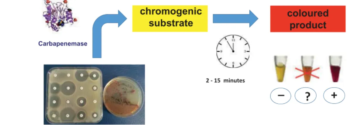
(Bio-Rad, Marnes-La-Coquette, France) is the first commercial kit for using this approach to detect ESBL-producing Enterobacteriaceae [22]. It consistsofa chromogenic substrate (HMRZ-86) that was discovered more than10 years ago. This substrate resists narrow-spectrumβ-lactamases but is hydro-lyzed in the presence of an ESBL, yielding a red product; an orange color should be considered an equivocal, undeter-mined result (Figure 1) [23]. Theβ-Lacta test®
was first used with isolated colonies and was then conducted directly from clinical specimens such as urine or blood cultures [24–26]. The characteristics and performance of this test are reported in Table 2. Theβ-Lacta test®
was initially developed to detect allthe broad-spectrum ß-lactamases that hydrolyze cephalospor-ins. This test not only detects ESBLs but also some hyperpro-duced cephalosporinases and some carbapenemases with activity toward expanded-spectrum cephalosporins [27]. The β-Lacta test
®
may be performed in parallel with a rapid iden-tification method, such as MALDI-TOF MS [24,25]. Although the sensitivity of this test for detecting ESBL-producing Enterobacteriaceae is high, the positive predictive value is rather low because this test also detects AmpC hyperprodu-cers, the class A carbapenemase KPC, the OXA-48-like class D carbapenemases, and, at a lower efficacy, the class B carbape-nemase producers [27].2.2.1.1.2. Detection of carbapenemase activity. Theβ-CARBA test
®
(Bio-Rad, Marnes-La-Coquette, France) has recently been marketed for the detection of carbapenemase among freshly isolated Enterobacteriaceae colonies; it employs a chromogenic substrate ( http://www.bio-rad.com/fr-fr/sku/68260-beta-carba-test). The color change in the reactive medium must be visua-lized by eye within 30min. The bacterial culture must be fresh (<24 h). The manufacturer of the β-CARBA test®
reports a sensitivity of 100% (207/207) when testing colonies that were obtained on Columbia agar containing 5% sheep blood, and they also report a specificity of 97.8% (89/81). The sensitivity decreases significantly if the colonies are picked from a chromogenic (98.6%) or Drigalski (77.6%) medium. The false-positive non-carbapenemase-producing strains were AmpC producers. Recently, Compain et al. tested the β-CARBA test®
against a collection of 42 carbapenem-resistant Enterobacteriaceae, with 30 out of 42 being carbapenemase producers [29]. Within the 30minof incubation recommended by the manufacturer, the test was positive for 26 of the carbapenemase-producing strains, missing four OXA-48-pro-ducing E. coli; these missed strains yielded a positive test result if the incubation was prolonged to 1 h. No false-positive results were obtained. Since only a single published study is available, those data will be treated with caution, and they require further clinical validation.2.2.1.2. The use of a non-chromogenic substrate in which hydrolysis leads to a color change in the medium
2.2.1.2.1. Detection of ESBLs. The newly marketed Rapid ESBL NP Test
®
(Senova GmbH, Weimar, Germany) is anCarbapenemase
chromogenic
substrate
2 - 15 minutescoloured
product
_
?
+
Figure 1.Theβ -lacta Test
®
.upgraded version of the ESBL NDP test that was first devel-oped as a home-made test for detecting the hydrolysis of cefotaxime (and any other broad-spectrum cephalosporins) in less than 20minby using a pH indicator (phenol red) [30]. In comparison to a negative control well without antibiotic, the reactive well containing cefotaxime and the pH indicator experiences a color change from red to yellow if ESBL pro-duces some carboxyl-acid groups resulting from cefotaxime hydrolysis. The same reaction occurs in the presence of a penicillinase inhibitor, namely tazobactam, which inhibits the hydrolysis reaction, thereby helping to identify the ESBL nat-ure of the ß-lactamase (Figure 2). The performance of this home-made test has been evaluated with either cultured bacteria (sensitivity, 92.6%; specificity, 100%) or directly from urine (sensitivity, 98%; specificity, 99.8%) or positive blood culture samples (sensitivity, 100%; specificity, 100%). Its sensi-tivity is excellent, particularly for detecting CTX-M producers (100%) [30–32]. A copy of the ESBL NDP test has been mar-keted (the Rapid ESBL Screen kit
®
, Rosco-Diagnostica, Tasstrup, Denmark); neither the specific characteristics nor performance results are published. A performance comparison of three tests for detecting the ESBL-producing bacteria (the rapid ESBL NDP test, the Rapid ESBL Screen kit®
, and the β-Lacta test®
) was recently performed using the exact same collection of strains [28]. The ESBL NDP test reached high sensitivity and specificity (95% and 100%, respectively). The limits of the β-Lacta test®
in selectively detecting the ESBL-producing bacteria among cephalosporin-resistantEnterobacteriaceae were again highlighted here (specificity, 71%). Regarding the Rapid ESBL Screen kit
®
, the protocol appeared to be time-consuming (2 h) and the results varied considerably according to the reading time. The sensitivity and specificity of the Rapid ESBL Screen kit were lower than those of the ESBL DP test.2.2.1.2.2. Carbapenemase detection. The first commercial test was the RAPIDEC® Carba NP test (bioMérieux, La Balme-les-Grottes, France). The marketed version and its prototypes (seelater) are the most frequently evaluated methods for the rapid detection of carbapenemase activity, with more than 60 published references, primarily in highly rated journals. This test is an identical version of the original Carba NP test that took advantage of several improvements. Notably, the Carba NP test is now recommended as a first-line test for screening carbapenemase activity by the Clinical and Laboratory Standards Institute in the US [35]. This test targets any type of carbapenemase activity by detecting the hydrolysis of imi-penem. Originally, the Carba NP test was developed as an in-house technique using imipenem as substrate, with the prin-ciple of the test based on the acidification of the reaction medium when the β-lactam ring of the imipenem molecule is opened because of the hydrolytic activity of carbapene-mases [36]. The modification of the pH of the reacting med-ium is revealed by a color shift in the pH indicator, namely the phenol red that turns from red to yellow with acidification (Figure 3). Another version of the in-house Carba NP test, the Carba NP II test, has been developed, and it includes
Table 2.Principle and performances of rapid biochemical tests for the detection of clinically relevantβ-lactam-resistant Enterobacteriaceae. Principle/name of the test
Targeted enzymes
Required additional
supplies
Delay for first/ definitive results Performances on cultured bacteria Performances on clinical specimen References Colorimetric–chromogenic substrate
ß-Lacta test
®
ESBLb None 15 min Sensitivity: 88%Specificity: 71%
Urines: sensitivity: 94%, specificity:100% (positive blood culture: sensitivity: 95.7%, specificity: 100%)
[22–28]
β-CARBA test
®
Carbapenemase None 30 min No direct comparison:sensibility 87%, specificity 100%
[29]
Colorimetric–non-chromogenic substrate
Rapid ESBL NP test
®
ESBLb None 20 min Sensitivity: 95%Specificity: 100%
Urines: sensitivity 98%, specificity 99.8%, positive blood culture: sensitivity 100%, specificity 100%
[28,30–32]
Rapid ESBL Screen kit
®
ESBLb None 30 min/2 h Sensitivity: 92%Specificity: 83%
[28]
Rapidec
®
Carba NP test Carbapenemase None 30 min/2 h Sensitivity: 99%Specificity: 100%
Positive blood culture: preliminary experimental data
[33]
Rapid CARB Screen
®
Carbapenemase None 5 min/2h Sensitivity: 89.5%Specificity: 70.9%
[33]
Rapid Carb Blue kit
®
Carbapenemase None 15 min/1h No direct comparison:sensitivity 100%, specificity 100%
[34]
Mass spectrometry detection
Pending marketed kits from
MALDI-TOF MSaplatforms manufacturers Carbapenemase MALDI-TOFMSa
platforms
1–4h Sensitivity: 72.5%–
100%
Specificity:98%–100%
Positive blood culture: preliminary experimental data
aMatrix-assisted laser desorption ionization-time of flight mass spectrometry.
b
Extended-spectrum ß-lactamase.
c
Nonevaluated.
Indications in brackets are not validated by the manufacturers.
Performances in boldareprovide from direct comparisons (as the corresponding reference).
additional wells with class-specific β-lactamase inhibitors [37,38]. This version allows investigators to screen and to more precisely characterize the carbapenemase activity of a given strain, allowing them to differentiate between enzymes for which the activity is inhibited by clavulanic acid (class A), by EDTA (class B), or by neither of those two (class D). To date, the Carba NP II test has not been developed industrially. Interestingly, the hydrolytic activity of metallo-carbapene-mases is boosted by reagent medium that includes zinc, a
basic element that is essential for metallo-β-lactamases. The global use of the Carba NP test allowed for some improve-ment of its performance, either for the recommended inocu-lum particularly for mucoid strains, the nature of the imipenem, or the type of agar medium from which tested colonies are picked [39–43]. The sensitivity of the carbapene-mase detection could be negatively influenced by several culture media such as the Drigalski or MacConkey agar plates, considering that colored pigments may interfere with the N O R COOH S-R Carbapenem Imipenem H2N R COOH S-R O HO Production of acid
pH
Colorimetric detection 1 2 1 2 Carbapenemase production No carbapenemase Red Orange-Yellow1: Control solution without imipenem
2 : Solution with imipenem
Carbapenemase
Figure 3.The Carba NP
®
Test.Cephalosporin
cefotaxime Hydrolysis
pH
Production of acid
Red Orange-Yellow
Extended-spectrum ß-lactamase (ESBL)
+
ß-lactamase inhibitor tazobactam No hydrolysis Red RedFigure 2.The ESBL NP
®
Test.reading of the test [44]. This in-house method has been improved several times and then industrially developed, and it has been on the market since early 2015 under the trade name RAPIDEC® Carba NP test (Figure 4) [45–50].
The ability of the RAPIDEC
®
Carba NP test to detect carba-penemase activity depends at least in part on the level of enzyme production and the enzyme’s capacity to hydrolyze the substrate and the tested inoculum (seelater). The perfor-mance of the RAPIDEC®
Carba NP makes this biochemical test appropriate for the rapid (30 min to 2 h) and convenient screening of carbapenemase activity in gram-negative bac-teria. Its sensitivity and specificity reached 96% in the original study, subsequently increasing to 97.8% and 98.5%, respec-tively, during independent evaluations [45–47]. Some publica-tions reported relatively low sensitivities for in-house and RAPIDEC®
Carba NP tests for detecting the OXA-48-type pro-ducers [42,49,50]. Those results may be explained by the weaker carbapenemase activity of the OXA-48-like carbapene-mases, or the use of old cultures or too little inoculum. Notably, the use of the correct inoculum (one full 10-μl loop for the Carba NP test) and a proper culture plate are critical for obtaining good sensitivity. The zinc concentration and/or the color of the colonies on chromogenic medium must be opti-mal for detecting the enzymatic activity with enough sensitiv-ity [49]. Since the CarbaNP test®
was successfully challenged directly against positive blood cultures, ongoing studies are evaluating the performance of the RAPIDEC®
Carba NP directly from clinical specimens (Pr Nordmann, personal com-munication) [51].Commercial alternatives to the RAPIDEC Carba NP test have been developed to screen for carbapenemase activity, such as the Neo-CARB kit
®
, which was formerly the Rapid CARB Screen®
or the Rapid Carb Blue kit®
(Rosco Diagnostica A/S, Taastrup, Denmark). The former is a copy of the RAPIDEC®
Carba NP and the latter test is identical to the RAPIDEC®
Carba NP, with the exception of the color indicator, which is bro-mothymol blue instead of phenol red [34,39,52–54]. Furthermore, and beyond the performance of these tests in the original publications, their direct comparisons against the Carba NP test and/or the RAPIDEC®
Carba NP showed super-iority in terms of performance (Table 2) [33,55–57].2.2.1.3. Advantages of the colorimetric approach. The col-orimetric approachfulfillsthe requirements of an optimal test for β-lactamase detection in that it is reliable, rapid, cheap, and requires no or very limited additional supplies. The bio-chemical detection of ESBL or carbapenemase activities per-mits researchers to identify any type of enzyme, whereas molecular-based techniques only detect the genes that are included in the corresponding test.
One primary advantage of colorimetric approaches is that they can be applied directly to colonies that are grown on selective media for the rapid detection of multidrug-resistant strains. For the detection of either ESBL- or carbapenemase-producing bacteria, the ESBL NDP and the Carba NP tests are perfectly suitable. The screening of MDRO carriers can there-fore be optimized in terms of effectiveness, combining excel-lent sensitivity and rapidity, which are two critical features in infection control policy.
2.2.1.4. Pitfalls of the colorimetric approach. The expected performance of colorimetric methods is based on their capa-city to detect the hydrolysis activity of a broad range of biochemically diverse enzymes (and therefore it has high sen-sitivity) without amplifying nonspecific activities (therefore providing highly specific results). The sensitivity of a biochem-ical test with the aim of detecting an enzymatbiochem-ically mediated mechanism of antibiotic resistance depends on different factors such as (i) the amount of enzyme produced, which relies on the level of expression of the corresponding gene and the bacterial inoculum; (ii) the ability of the enzyme to hydrolyze the substrate, which relies on the extraction proto-col; (iii) the affinity of the enzyme for the substrate, which relies on the pharmacokinetics properties of the enzyme/sub-strate couple; and (iv) the sensitivity of the revelation step, which relies on the indicator properties and the mode of measurement (by naked eye or photometric means).
In addition to the lack of sensitivity, phenotypic colori-metric tests might run into some interference from the unde-sirable hydrolytic activities of nontargeted enzymes. This result is found for intrinsic AmpC-type cephalosporinases from dif-ferent enterobacterial species or the K1 penicillinase of Klebsiella oxytoca, which hydrolyzes broad-spectrum cephalos-porins when they are overproduced (ß-Lacta test
®
). Therefore, determining an accurate threshold for phenotypic colorimetric methods may be a critical issue. It is notable that the TAT of some assays could exceed 2h. Additionally, those tests are not suitable for rectal screening due to the lack of specific recog-nition of their ESBLs or carbapenemases among all the micro-biome bacterial activity.2.2.2. MALDI-TOF MS approach
2.2.2.1. Principle of the MALDI-TOF MS approach. The implementation of MALDI-TOF MS technology in clinical laboratories has significantly modified bacterial diagnostics. Combined with the automation and implementation of a user-friendly interface, the identification of bacteria by mass spectrometry opens up a new area for microbiologists in terms of accuracy and TAT to deliver results [58]. This technology identifies the bacteria at the species and/or genus level from single isolated colonies on solid media, but it is also applied directly to positive blood and urine [58]. After a first rapid preparation step including sample application to a slide and the addition of an organic matrix solution, the identification can be performed in less than 1 min. If a clinical fluid or a positive blood culture is directly tested, a pre-processing step has to be performed. The MALDI-TOF MS principle is based on the ionization, with a laser source, of bacterial biomolecules containing proteins embedded in a matrix. Once they are ionized and sublimated into a gas phase, these molecules are accelerated into an electric field and projected onto a detector. They can then be separated and analyzed according to their time of flight. This delay between the ionization process and the final impact depends on their mass-to-charge ratio.
Commercial MALDI-TOF technology focuses on the identi-fication of proteins or DNA molecules [58]. Several systems are on the market including different machines and databases; the
three primary commercial products are the Bruker Biotyper
®
(Bruker Daltonics, Wissenbourg, France), the bioMérieux Vitek MS System®
(bioMérieux, Marcy l’Etoile, France), and the Andromas®
(Beckman Coulter, Villepinte, France) system, the database of which can be implemented in different machines. Before bacterial identification using libraries of protein profiles via the MALDI-TOF concept, MS technology was developed to identify chemical compounds, including antibiotics. When coupled with liquid or gas chromatography, MS is the refer-ence method for detecting and dosing antibiotics in clinical samples. Due to its cost and lack of user friendliness, this approach was replaced by immunological techniques such as ELISA. Nevertheless, MS maintains the capacity to detect anti-microbial molecules and, interestingly in the context of β-lactam resistance, the products of the antibiotic after hydro-lysis. Therefore, MALDI-TOF MS has great potential for the detection of resistance traits among bacteria strains in two particular contexts, i.e. (i) the detection of the enzyme and (ii) the detection of a substrate degradation replaced by the signal of its product after a variable incubation time.When dealing with β-lactamases, the identification of a discriminatory peak corresponding to a protein with signifi-cant hydrolysis activity was first considered, but it was rapidly abandoned in favor of observing the degradation product [58]. MALDI-TOF MS is used to identify both the β-lactam molecule and its degradation product. As an example, the hydrolysis of imipenem is interpreted as the disappearance/ appearance of specific peaks (300 Da and 254 Da, respectively) when this antibiotic is added to a bacterial suspension, and the MS spectrum is measured at the time of substrate addition and after 20 min of incubation. The TAT of this process is approximately 30 min [59]. Some other protocols are more time-consuming, requiring an incubation time from 1 to 4 h [60–62]. The performance of this approach depends on the metabolite/imipenem ratio but also the metabolite cut-off. For example, in Knox et al., the MALDI-TOF MS approach was compared to the Carba NP test; the breakpoint was deter-mined to be a disappearance of 95% of the area under the curve for the test compared to the control replicate [60]. In this study, the performance of the two methods was equiva-lent. Beyond the various published home-made protocols with differences in the substrate (meropenem/imipenem/ertape-nem/faropenem), the lysis conditions, the incubation time, and the parameters/breakpoints, a specific module should be marketed in the very near future (MALDI Biotyper STAR BL
®
, Bruker Daltonics, Wissenbourg, France) [63].Additionally, the chemical properties of the enzyme may be indirectly investigated by adding specific inhibitors (such as the ß-lactamase inhibitor clavulanic acid) that hinder the β-lactamase hydrolytic activity and therefore slow the disappear-ance of the ß-lactam native peak. Several companies have developed kits for that purpose, and they have produced protocols that are about to be marketed. Some authors have attempted to adapt the protocol to positive blood cultures, and it is much more time-consuming (3–4 h) since a pre-culture is needed to amplify the amount of bacteria in the specimen [64,65]. Next toβ-lactamase production, the loss of a porin that allows the entry of the antibiotic inside the bacterial cell constitutes a clinically and epidemiologically relevant
mechanism of resistance in Enterobacteriaceae. The MALDI-TOF MS technology could replace cumbersome and time-con-suming reference methods such as sodium dodecyl sulf ate-polyacrylamide gel electrophoresis (SDS-PAGE), which is labor-ious to use routinely. For example, the loss of porins (OmpK35, OmpK36) from Klebsiella species may lead to carbapenem resistance The SDS-PAGE technique is the reference method for identifying the presence/absence of these porins [66]. After the identification of the corresponding peaks, MALDI-TOF MS permits the localization of these porins and objectivizes the loss of OmpK36 [66]. As previously reported, the infection control consequences and particularly the risk of an epidemic spread of the genetic support underlying a resistance trait are associated with its mobility and its capacity to transfer from one bacterium to another. In the present case, the genetic trait is the truncation of the chromosomal gene that cannot be horizontally transferred. Although it is technically reliable, this potential application has not been included in the panel of MALDI-TOF MS kits that are about to be marketed.
2.2.2.2. Advantages of the MALDI-TOF MS approach. The hydrolytic activity is determined regardless of the genetic basis of the resistance and the type of ß-lactamase. The sensi-tivity of this technology seems to be good regardless of the nature of theβ-lactamase, including for OXA-48-like enzymes [67]. The theoretical cost in consumables for this approach is low (less than 0.1 euro, excluding the cost of acquiring the machines that are now very common in clinical laboratories) as is the cost associated with technician time. To date, the real cost of the commercial kits remains unknown.
2.2.2.3. Pitfalls of the MALDI-TOF MS approach. A direct comparison of the MALDI-TOF MS and Carba NP test results highlights some discrepancies. For unresolved reasons, several NDM- or VIM-producing Proteus spp. and Morganella spp. are not detected by the MALDI-TOF MS approach [60]. The authors suggested that there was a problem related to enzyme avail-ability for the MALDI-TOF method, which did not include a cell lysis step. Another explanation could be the type of carbapenem compound under testing. In fact, no false-negative result was obtained in the two other studies including M. morganii and Providencia sp. strains, but they tested the degradation of erta-penem or meroerta-penem instead of imierta-penem [61,62]. Moreover, for some OXA-48 variants (OXA-204) that exhibited a very low increase in the carbapenem minimal inhibitory concentrations (MIC), a false-negative result could be found [59]. However, the hyperproduction of cephalosporinase could lead to a false- posi-tive result [59]. An additional complex spectral comparison did not improve the performance of the challenged protocol. The standardization of the experimental conditions through the mar-keting of kits may help to solve these problems. Notably, a positive signal when using the MALDI-TOF MS that indicates β-lactamase activity cannot be translated into an estimation of a MIC that might still be below the resistance breakpoint. In other words, extrapolating whether the strain is resistant to the tested antibiotic is not possible. However, since infection control guide-lines are based on the presence of certain enzymes (e.g.ESBL or carbapenemases) and preferably but not systematically to the exact MIC value, this approach makes clinical sense. Finally, if
using MALDI-TOF MS technology directly on positive blood cul-tures, the same problems arise as the problems of using it for identification purposes, including decreased sensitivity and time-consuming pretreatment requirements. To date, using those types of techniques for detecting multidrug-resistant strains within a mixed flora (gut) remains impossible.
3. Antigenic rapid detection of antibiotic-resistant
Enterobacteriaceae
Recently, a new category of test was marketed on the basis of the immunological detection of some specificβ-lactamases as the KPC and OXA-48 enzymes [68–70]. The performance of these immunochromatographic tests (OXA-48/KPC K-set
®
, Coris BioConcept, Gembloux, Belgium) that are easy to perform and to interpret seems to be higher. This lateral flow immunochro-matographic assay uses monoclonal antibodies and colloidal gold nanoparticles bound to a nitrocellulose membrane, and it provides results from a single colony in 15min. Its performance reaches 100% specificity and sensitivity, with a limit of detection of 106 CFU/ml [69]. A recent study that employed the whole genome sequencing approach as the reference method showed the complete concordance between those two methods [70]. To the best of our knowledge, no corresponding assay exists for the detection of ESBLs, and the high diversity of enzymes that must be detected likely prevents further developments.3.1. Advantages of the immunological approach
Theoretically, the immunological approach could be as sensi-tive and specific as the enzymatic approach. In fact, both methods depend on the level of protein production, with the immunological approach revealing the presence of this protein and the enzymatic one revealing its biochemical activ-ity. In published studies, these two approaches were strictly equivalent for detecting blaKPC genes [71]; with regard to
blaOXA-48 carbapenemases, the immunological method
detected a few additional cases that were missed or that yielded equivocal results with the enzymatic method (3 out of 100 in the study by Dortet et al., 7 out of 130 in Glupczynski et al.) [71,72]. The OXA-48 variants that do not possess any significant carbapenemase activity such as 163 and OXA-405 have been reported by the manufacturers as nonreacting variants. The small number of clinical strains (n = 3) producing those types of enzymes that were tested in three published studies unfortunately prevent any valid conclusions. This high level of specificity among the large number of OXA-48 variants is clearly a positive aspect of the immunological approach. Some authors have tested the OXA-48K-set
®
directly from spiked blood cultures (Aerobic FA Plus 30-ml bottles, BacT/ Alert system, bioMérieux, Durham, supplemented with 10 ml of heparinized horse blood). The testing of a lysate after an 18-h incubation time yielded excellent results [69].3.2. Drawbacks of the immunological approach
Targeting a specific type of enzyme alone as the first step is an approach that cannot be used as a screening test. Its price (ca.
10€ depending on the country) and the narrow panel of available targets limit its use as a first-line strategy. It could be of interest only in some specific settings, or in countries with a high predominance of a single type of carbapenemase, or during the course of an investigation of an outbreak con-text that generates secondary cases that are presumably related to strains that produce well-identified carbapene-mases. Beyond the remaining doubts about the functionality of detecting specific proteins that have been extensively acknowledged, we must be aware of the risk of emerging specific enzyme variants for which the targeted antigenic site might be modified, and they consequently would not be detected by the initially designed antibody. Regarding the continuously increasing number of variants, a thorough mon-itoring of the performance of these tests must be completed. The most recent data are quite reassuring: all the OXA-48 variants with carbapenemase activity, such as 181, OXA-204, OXA-232, OXA-244, OXA-245, OXA-436, and OXA-484, were successfully detected [70]. Nevertheless, the OXA-48K-set
®
detected the naturally occurring OXA-48-like enzyme from Shewanella spp., which is a shortcoming [70,73]. Although the prevalence of this bacterial genus in human specimens seems to be rare, we support the presumptive identification of suspected colonies with an oxidase test (Shewanella spp. as an oxidase-positive bacterium as opposed to Enterobacteriaceae) or a MALDI-TOF identification. Due to the difference in terms of the infection control measures that should be implemented, a positive result should be inter-preted according to an unambiguous genus identification. Notably, this technique cannot be used directly from clinical specimens. In conclusion, the ICT method could be of interest for detecting carbapenemase from isolated colonies in some specific settings when the two targeted enzymes are highly prevalent among carbapenemase-producing strains.4. Recent advances in molecular diagnostics for the
rapid detection of antibiotic-resistant
Enterobacteriaceae
For two decades, molecular techniques have taken up an increasing and now prominent place in clinical laboratories. The primary technologies that are now implemented as rou-tine methods arepolymerase chain reaction (PCR), including real-time PCR for the detection of pathogens and their asso-ciated virulence or resistance genes, PCR hybridization, includ-ing the microarray, to identify molecular sequences, and sequencing (in particular, the so-called ‘next generation sequencing’ (NGS)) that will likely be considered as routine techniques within a few years. Numerous reviews have been published recently about the contribution of nonphenotypic and/or molecular tests.
4.1. The DNA-targeted PCR-based approach
The PCR-based approach was the earliest molecular method to be implemented in clinical laboratories to detect ß-lactam resistance genes among Enterobacteriaceae. Single end-point PCRs were first described in combination with a confirmation step using the enzymatic restriction of amplified fragments
and/or size evaluation by agar electrophoresis, or DNA–DNA hybridization. This multiplex process was laborious and was subject to contamination at each step in the process. Further improvements were implemented, such as the multiplexing and miniaturization of the hybridization step. Real-time PCR permitted the combination of the amplification and detection steps in a single step, limiting the risks of environmental contamination by amplicons. Home-made PCRs were progres-sively replaced by commercial kits including sound protocols and quality controls. A summary of the primary commercially available kits is reported in Table 3. After specific multiplex PCR amplification, the amplified DNA fragments are revealed by hybridization with a panel of specific probes. The support of this hybridization could be the reactive medium when using real-time PCR or a solid surface. The number of probes actually increases in parallel with the miniaturization of the support, as with the microarray. The detection of resistance genes could be performed independently, or it could be included in multiplexed panels for the diagnosis of infections, for example, in the context of bloodstream or respiratory infections.
4.1.1. The whole automated real-time PCR platform: the example of the Xpert
®
systemThe Xpert
®
system is the first fully automated PCR instrument to allow for the implementation of a PCR diagnostic method in a very large panel of clinical laboratories. This technology requires neither technical competency nor skilled technolo-gists, allowing its use over a broader period during open laboratory hours. This type of nucleic acid amplification test might be considered as a molecular diagnostic POCT. The different steps are performed in a single compact cassette after the introduction of the specimen, combining DNA extrac-tion, amplification,and revelation. An internal positive control is included in the multiplexed targets. The Carba-R assay®
(Cepheid, Sunnyvale, CA, USA) runs on the Gen Expert®
plat-form, which is suitable for the detection and quantification of numerous bacterial or viral species, in addition to human tumor targets. This test is marketed to detect several carbape-nemase genes directly from a rectal sample; a derived protocol is provided by the manufacturer to use this assay on cultured bacteria [95,96].4.1.2. The Check-Direct assay
®
The Check assays
®
(Checks points, Wageningen, The Netherlands) include a large panel of different multiplex real-time PCR kits using different probes, with one of them target-ing an internal control. The DNA extraction step must be performed outside the real-time PCR platform. The number and type of targeted genes varyaccording to the kit, including narrow- and/or broad-spectrum β-lactamase genes (see Table 3). Different PCR platforms may be used. Although they are thought to be cumbersome, expensive, and time-consuming, the Check assays®
are used as a reference tech-nique to characterize β-lactamase genes from isolated colo-nies. Recently, a new multiplex PCR panel was developed to detect the four primary types of carbapenemase genes directly from rectal swabs, i.e. the Check-direct CPE®
[74,77,97]. Furthermore, successful attempts to shorten the duration of the DNA extraction step were recently pub-lished [74].
4.1.3. Other systems
Numerous commercial systems can detect and/or confirm the presence of resistance genes (see Table 3). They differ from one another in their theoretical principles and their panel of targeted genes. The most interesting features of those tests would be their potential to identify both the bacteria and their resistance traits as a whole, particularly in blood cultures. The primary representative of these ‘broad-range multiplex PCR panels’ are the Filmarray
®
(bioMérieux, Marcy l’Etoile, France), the Verigene®
(Nanosphere, Northbrook, US), the ePlex®
(GenMark Dx, Zug, Switzerland), the Hyplex®
superBug ID (Amplex Diagnostics GmbH, Gars-Bahnhof, Germany), and the Unyvero®
(Curetis AG, Holzgerlingen, Germany), with the two first panels being the most widely tested ones. Regarding the Xpert®
system, these PCR panels do not require extended skills and could theoretically be used 24 h per day. Interestingly, some assays seem to be valuable for the detection of resistant genes in other clinical specimens of high interest to diagnose respiratory, urinary tract, or joint infections due to resistant bacteria [82,86,98]. Regarding bloodstream infections, some of these assays could be positive earlier in comparison to blood culture systems [83]. Beyond their analytical performance, the adequacy of the targeted gene panel for local epidemiology and their costs (approxi-mately 150 euros per test) are still considered as a limitation to their development (seelater).Other assays have been developed to characterize the β-lactamase content of a given isolate to decrease the cost and improve the detection of multiple allele variants in a single isolate. The Luminex
®
(Luminex Corporation, Austin,TX,USA) technology is a well-established detection approach that relies on colored microsphere-based flow cytometry assays. This test allows for the detection of specific alleles, antibodies,or pep-tides, and it has been already marketed for other purposes such as HLA typing, seroprevalence studies,or the detection of a broad panel of microorganisms directly from clinical specimens such as stools. Recently, a Luminex xTAG®
assay was developed to detect ESBLs, plasmid-mediated cephalos-porinases, and carbapenemases [94]. The modular multiplex oligonucleotide ligation-PCR procedure allows for the detec-tion of β-lactamase genes and their variants for less than 5 euros per sample and a TAT of 5 h. Regarding the first pub-lished study, the sensitivity and specificity are excellent (100% and 99.4%); the different variants of the same β-lactamase genes (as blaTEM) present in the same isolate could besepa-rated. The subtyping of the blaCTX-M gene is also performed.
Additional data are required, but this approach seems to be promising.
4.1.4. Performance
4.1.4.1. Sensitivity. The performance of the molecular meth-ods, and particularly the commercial multiplexed PCR assays, must be considered in view of different clinical or technical settings. The most favorable setting is the characterization of
Table 3. Examples of commercial molecular tests, including targeted genes, material requirement , and turnaround times. Test name (manufacturer) Targeted genes Specimen Technology/material requirement/instrumentation Turnaround time (manufacturer ’s specification) Reference Carba-R assay
®
(Cepheid, Sunnyvale, CA, USA) bla KPC , bla NDM , bla VIM , bla OXA-48 , bla IMP-1 -like new version of the assay includes bla OXA-181 Rectal swab (colonies) a One-step multiplex real-time PCR using specific probes – dedicated platform (GenExpert®
) <1 h [ 74 ,75 , 76 ] Check-Direct CPE®
assay (checks points, Wageningen, The Netherlands) ● Check-ESBL®
● Check-KPC-ESBL®
● Check-MDR CT101®
● Check-MDR 102®
● MDR Carba®
● Check-MDR 103XL®
● Check-Direct CPE assay®
bla SHV , bla TEM , bla CTX-M including ESBL variants, bla KPC , bla CMY , bla DHA , bla ACT/MIR , bla ACC ,bla NDM ,bla VIM , bla IMP , bla OXA-48 -like, bla GIM ,bla SPM , bla GES including carbapemase variants, bla OXA-23 , bla OXA-40 and bla OXA-58 , bla BEL , bla PER and bla VEB Strains, stool (positive blood culture) Two-step ligation-mediated multiplex real -time PCR amplification – micro array hybridization, a nondedicated real -time PCR platform as the LightCycler®
(Roche, Brussels, Belgium), the BD Max®
(Becton Dickinson, Oxford, U . K . ) , or an Applied Biosystems®
Real Time PCR. The performances of the test in distinguishing separately the different positive signals may vary according to the PCR platform 2 – 6 h [ 74 , 75 , 77 – 79 ] Verigene Gram-negative blood culture nucleic acid test (Nanosphere, Northbrook, IL, USA) bla CTX-M , bla IMP , bla KPC , bla NDM , bla VIM , bla OXA-23 , bla OXA-40 , bla OXA-48 , bla OXA-58 Positive blood culture Micro-array hybridization-dedicated platform (Verigene Processor SP®
and Verigene Reader®
) 2– 2.5 h [ 80 , 81 ] FilmArray®
blood culture identification Panel (bioMérieux, Marcy l’Etoile, France) bla KPC Positive blood culture (biofilms from prosthetic joint infection, sterile fluids injected in blood culture bottle) Nested-PCR and melting curve analysis, dedicated platform 1 h [ 80 , 82 – 85 ] ePlex®
Blood Culture Identification Gram Negative Panel (GenMark Dx, Zug, Switzerland) bla CTX-M, bla OXA , bla KPC , bla VIM , bla IMP , bla NDM Positive blood culture Multiplexed nucleic acid amplification test and a electrochemical detection technology with a dedicated platform 90 min Manufacturer data. No published study Unyvero HPN Pneumonia, i60 ITI Implant Tissue Infection, and BCU Blood Culture (Curetis, Holzgerlingen, Germany) bla TEM , bla SHV , bla NDM , bla VIM , bla IMP , bla KPC , bla OXA-48 , bla CTX-M Respiratory specimen, sputum, lavage, tracheal aspirate, blood cultures, liquid samples (exudate, pus, etc . ) Two-step multiplex PCR – hybridization with a dedicated platform 4– 5 h [ 86 – 88 ] CarbDetect AS-2 Kit, Identibac AMR-ve assays (Alere Technologies GmbH, Jena, Germany) b bla TEM , bla CTX-M , bla SHV , bla OXA , bla CME , bla CMY , bla ACC , bla PER , bla LEN , bla VEB , bla MOX , bla SPM , bla NDM , bla IMP , bla VIM , bla KPC , bla BIC , bla DIM , bla GES , bla GIM , bla GOB , bla IMI , bla SIM , bla IND , bla KHM , bla PAM , bla SFH , bla SMB , bla SME , bla TMB , bla OXA-48 -like Strains Two-step multiplex PCR – microarray hybridization with a dedicated platform 5– 8 h [ 89 ] Hyplex SuperBug ID assays (Amplex Diagnostics, Gars, Germany) bla KPC , bla IMP , bla VIM , bla NDM , bla OXA-48 -like (including bla OXA-181 , bla OXA -162 , bla OXA -204 , and bla OXA -244 ) Strains (positive blood culture) 2 steps multiplex PCR – Hybridization /ELISA 2.5 – 4 h [ 90 ] eazyplex®
Superbug complete A kit (Amplex, Gie β en, Germany) bla KPC-2 to KPC-15 , bla NDM-1 to NDM-7 , bla VIM-1 to VIM-37 , bla OXA-48 , bla CTX-M-1 and CTX-M-9 . The new version of the assay includes bla OXA-181 Strains, positive blood culture (urines) Loop-mediated isothermal amplification technology and a non dedicated real-time PCR platform as the Genie (Optigene, Horsham, U . K . ) <30 min [ 75 , 91 , 92 ] VAPChip®
(Eppendorf Array Technologies, Namur, Belgium) bla TEM , bla SHV , bla CTX-M , bla OXA-23, bla OXA-24/40, bla OXA -48, bla OXA-58, bla KPC , bla VIM , bla IMP-7,-13, IMP-1-like (The test can identify most of the narrow-and extended-spectrum bla TEM and bla SHV genes) Respiratory samples Real-time PCR and microarray detection, on a dedicated RAP-ID platform®
4 h [ 93 ] Luminex xTAG®
assay (Luminex Corporation, Austin, TX, USA) bla TEM , bla CTX-M , bla SHV , bla PER , bla LEN , bla VEB , bla BEL , bla OXA , bla GES , bla CMY , bla ACC , bla MOX , bla FOX , bla DHA , bla MIR/ACT , bla SIM , bla NDM , bla IMP , bla IMI , bla VIM , bla KPC , bla BIC , bla DIM , bla GES , bla OXA-48-like , bla OXA-23-like , bla OXA-51-like Strains Multiplex oligonucleotide ligation PCR and detection using colo red microsphere-based flow cytometric assays on a dedicated platform that could be shared by different biological domains 5 h [ 94 ] aT he use of the test on colonies was not supported by the CE Marketing but a protocol is provided by the supplier . bT he kit is unable to distinguish between ESBLs and non ESBL variant among bla TEM and bla SHV . Indications in brackets are not validated by the manufacturers. R f i b ld id i b t t d t thttp://doc.rero.ch
the β-lactamase content of bacterial cultures, and the most difficult setting is the detection of resistance genes in a highly diverse and DNA-rich environment such as the gut flora.
4.1.4.1.1. Sensitivity with pure bacterial cultures. In this case, the sensitivity of the assay relies only on the size of the targeted gene panel and not on the quantity of available DNA material. According to the increasing number of targets, including in the most recent commercial kits, the performance is excellent. For example, the Check-MDR CT103 XL array is a new product that allows for the amplification and identifica-tion of a large panel ofβ-lactamases, including ESBLs, cepha-losporinases, and carbapenemases. Its sensitivity against a sample of 223 strains was 100% [79]. Moreover, the commer-cial assays include an internal control that detects the pre-sence of Taq polymerase inhibitors.
4.1.4.2. Sensitivity with clinical specimens
4.1.4.2.1. Clinical specimens with poor bacterial diversity (positive blood culture, urine). The sensitivity ofβ-lactamase gene detection directly from clinical specimens such as urines or positive blood cultures, with blood culture being an ‘artifi-cially boosted’ clinical specimen, relies solely on the correla-tion between the targeted genes and the local epidemiological setting. For example, the FilmArray
®
blood culture identification panel that targets only the blaKPCcarba-penemase gene could be of interest in areas where this gene is highly prevalent (Italy, Greece, etc.). Frequently, assays tar-geting the clinical specimen mix the identification of the causative bacteria and the most relevant associated resistance genes. This tendency could at least decrease the possibility of multiplexing the resistance genes for technical limitations. However, in considering the culture as the reference techni-que, the performances of those molecular tests are excellent if the resistance gene is included in the test panel, with the sensitivity being 100% [97]. Comparative studies basically showed similar and excellent performances for the different commercial kits [80].
4.1.4.2.2. Clinical specimens with a high diversity of bacteria (stools, rectal swabs). Compared to the culture-based method, the performance of molecular methods, and particu-larly their respective sensitivities, is still a matter of debate. In fact, the culture-based method is considered the gold stan-dard in termsof sensitivity, especially if combined with a pre-enrichment step. Recently, data that were provided aboutNGS technologies, particularly shotgun metagenome sequencing, question the validity of culture-based techniques as the gold standard [99,see discussion also later].
The detection limit of culture-based approaches for ESBL-producing or carbapenem-resistant Enterobacteriaceae in pure culture when using the best selective plates is approximately 10 CFU/ml [93,100].
Performance characteristics from molecular methods vary significantly according to the type of assay; the limits of detection for clinical specimens vary from 10 CFU/ml to 1,000 CFU/ml [78,93]. Global performance should be adjusted to the bacterial DNA extraction rate, which may vary according to the nature of the strain,the putative inhibitory properties of some specimens (stool, blood, etc.), and the number of resis-tance genes. Some home-made real-time PCR assays reach
performances as high as those of culture-based methods, with detection limits evaluated at 100 CFU/ml of feces. In some cases, the molecular method is 10- to 100-fold more sensitive than the culture-based strategy [77,93,101,102]. The significance and interpretation of a molecular positive-culture negative screening test still constitutes a challenge to clinical microbiology [77,78,97]. Overall, a home-made PCR targeting a single β-lactamase seems to be more sensitive than multi-plexed commercial assays; the competitive and technical requirements for administrative agreement lead to increased values in the limits of detection [93].
Several studies have reported suboptimal sensitivities when dealing with mixed gram-negative blood cultures, or when a single strain carried several resistance genes [80,103]. Beyond the previously reported difficulties in including all the variants of a large family of β-lactamases, for example, in blaCTX-M,
competition may occur when different targets must be ampli-fied from the same specimen [80]. This pitfall may lead to false-negative results and could have clinical consequences. Obviously, the risk of false detection results for polymicrobial specimens is shared by the different methods (culture, bio-chemical testing, MALDI-TOF analysis,and molecular screen-ing). Microbiologists should modulate their confidence in the results according to the nature of the specimen, whether it is a pure subcultured isolate, primoculture,or noncultured clinical specimen. Some recent assays seem to solve this issue, such as the Luminex
®
assay [94].4.1.4.3. Specificity
4.1.4.3.1. Specificity for pure culture. Once the panel is suffi-ciently large and adapted to local epidemiology, the real remaining challenge for molecular methods is the specificity, or the capacity of an assay to separate the natural chromoso-mally or plasmid-encoded narrow-spectrumβ-lactamases from the extended-spectrum, acquired ones. This issue is well established for cephalosporinase coding genes such as blaAmpC, blaACC, or blaDHA and constitutes a challenge for
some molecular methods [104]. Moreover, among the variety ofβ-lactamases identified in Enterobacteriaceae, a high num-ber of variants from the same class have sometimes been described; for example, more than 300 members of OXA enzymes have been reported. In some cases, and as noted earlierfor OXA-48 variants, the acquisition of a single or few mutations may instead lead to an expanded or narrowed hydrolytic activity [87]. This phenomenon is also known in ESBLs that are derived from their narrow spectrum counter-parts in the TEM and SHV families. This issue definitely con-stitutes a real challenge for the design of specific primers and probes [80]. For example, the Check-ESBL assay (Check-Points Health) correctly differentiates among the narrow-spectrum blaTEM and blaSHV-encoding genes from the variants that
encode ESBLs. However, the same technology fails to distin-guish some acquired and plasmid-borne blaAmpCgenes from
their intrinsic and chromosomally encoded counterparts. This challenge was clearly addressed by the most recent assays [79,94]. For some assays, the subtyping of the β-lactamase is available, as it is for the blaCTX-Mfamilies [94]. Unfortunately,
the detection of overexpressed natural enzymes through a
mutation in the regulator genes, as is the case for blaAmpC,
could not be detected without a sequencing approach.
4.1.4.3.2. Specificity for clinical specimens. The vast majority of the PCR methods identify their targets (the genes) without identifying the surrounding genetic environment. This draw-back was previously reported with MRSA and the confounding coinfection or co-colonization with methicillin-resistant coagu-lase-negative Staphylococci and methicillin-susceptible S. aur-eus. Additional targeted genes have been shown to overwhelm these pitfalls, for example, in the Xpert
®
MRSA/ SA BC or SSTI that included an additional target corresponding to the junction between the staphylococcal cassette chromo-some mec gene (SSCmec) and the part of the S. aureus genome where the SSCmec is supposed to be inserted, namely the SCCmec-orfX junction. Furthermore, the detection of a gene without phenotypic correspondence constitutes a well-known pitfall of DNA-based strategies [81]. Combined with low pre-valence for the targets, the positive predictive value of the molecular test may dramatically decrease to as low as 5–10% [17]. This drawback is particularly relevant when rich micro-biota, such as that of the digestive tract, is screened for CPE. As previously described for the immunological methods, this type of pitfall has been identified recently for the detection of ß-lactamase genes as follows: nonpathogenic Shewanella spe-cies harboring a blaOXA-48-like gene (its natural progenitor) ledto a false-positive molecular result in a patient with a history of blaOXA-48K. pneumoniae [73]. Moreover, the increasing
pre-valence of nonfermentative gram-negative bacteria harboring carbapenemase genes such as blaVIM-positive P. aeruginosa is
a source of confounding results when a screening program is implemented for infection control purposes [105]. Thus, in a large screening cohort of 3644 patients and 16,296 samples, the number of positive CPE patients was low (n = 43), and 4 patients carried a VIM-positive P. aeruginosa. Ten percent (4/ 43) of patients were initially and falsely identified as CPE-positive, leading to confounding isolating measures and mes-sages toward patients, their families,and health-care workers [106]. Moreover, the positive predictive value of the screening was only 86% [106]. A lower positive predictive value of 50% for the screening was reported by Simnet et al., with PCR-positive culture-negative cases exhibiting mostly high thresh-old cycle values (CT >39) [107]. The authors suspected
low-level colonization below the limit of detection for the culture-based screening method or nonspecific signal. This finding contrasts with in vitro studies that support a higher sensitivity in bacterial culture. Regarding the clinical specimen that required a semiquantitative interpretation of the cultures, for instance, the respiratory tract specimens, the cut-off loads (103 for a protected distal specimen, 104 for a bronchoalveolar specimen, and 106 for sputum) are significantly higher than the limits of detection for the molecular method. This differ-ence leads to confusing discrepancies [104]. Nevertheless, the follow-up of these PCR-positive-culture-negative patients is questionable (seelater).
4.2. The RNA-targeted molecular approach
One of the primary disadvantages of the DNA-targeted approach corresponds to a lack of differentiation between
silent and expressed genes or between dead and living bac-teria [81]. The replacement of DNA by RNA targets overcomes this problem and diminishes the putative gaps between genetic and phenotypic results. One manufacturer has devel-oped a kit for the detection of blaKPC variants (NucliSENS EasyQKPC test
®
, bioMérieux), and its TAT is 2 h [108].4.3. The PCR electrospray ionization mass spectrometry (PCR/ESI MS) approach
This technology is based on the ability of some very powerful mass spectrometry instruments to accurately measure the exact molecular masses of small PCR products at less than 500 bp [109]. After that, advanced software reconstructs the sequence of the DNA fragments, allowing for their accurate identification. Given the example of the former Septifast
®
assay (Roche Diagnostics, Basel, Switzerland), these approaches do not require a culture step, and they are time-and cost-effective. A fully automated system is also available (PLEX-ID; Abbott Bioscience), with the corresponding first-pub-lished data about bacterial identifications being quite encouraging, and the TAT from clinical samples being 4–6 h. Preliminary results for resistance gene analysis support the use of this technology to identify the blaKPC genes [110].Nevertheless, the cost of the system (ca. 200,000 USD) remains prohibitively high for a large majority of clinical laboratories.
4.4. The clinical impact of the molecular approach 4.4.1. The individual impact
Beyond theoretical and intermediary results such as the TAT, the cost savings, and the delayed optimization of antimicro-bial therapy, the impact of molecular methods on the clinical outcome is poorly established. A recent review and meta-analysis showed that (i) there was a relatively low number of studies pertaining to clinical outcomes, (ii) the one combined cohort showing a significant reduction in mortality when rapid molecular testing is associated with direct communication, and (iii) there was a lack of significant impact on mortality for rapid phenotypic techniques associated with direct com-munication [6]. More specifically, the molecular assay was a peptide nucleic acid fluorescent in situ hybridization that was performed directly on a positive blood culture in comparison with a Gram stain, and the phenotypic approaches that failed to reduce the mortality consisted of rapid antimicrobial sus-ceptibility testing with Vitek
®
(bioMérieux, Marcy l’Etoile, France) or Microscan®
(Beckman Coulter, Sacramento, CA, USA).4.4.2. The collective impact
Although it seems like common sense, the impact of decreased antibiotic pressure in response to a rapid molecular testing strategy was not established. Moreover, the impact of this approach in terms of infection control, i.e. the decrease of the occurrence or the duration of outbreaks has not been concluded; an intermediate positive impact such as the time until contact isolation has nonetheless been reported [111].