. . . .
. . . .
Non-invasive nuclear myocardial perfusion
imaging improves the diagnostic yield of invasive
coronary angiography
Ronny R. Buechel
1, Beat A. Kaufmann
2, Daniel Tobler
2, Damian Wild
3,
and Michael J. Zellweger
1*
1
Department of Nuclear Medicine, University Hospital Zurich, Switzerland;2
Department of Cardiology, University Hospital Basel, Switzerland; and3
Department of Radiology, Division of Nuclear Medicine, University Hospital Basel, Switzerland
Received 23 December 2014; accepted after revision 19 March 2015; online publish-ahead-of-print 27 April 2015
Aims Several studies reported on the moderate diagnostic yield of elective invasive coronary angiography (ICA) regarding the presence of coronary artery disease (CAD), but limited data are available on how prior testing for ischaemia may contribute to improve the diagnostic yield in an every-day clinical setting. This study aimed to assess the value and use of cardiac myocardial perfusion single photon emission computed tomography (MPS) in patient selection prior to elective ICA.
Methods and results
The rate of MPS within 90 days prior to elective ICA was assessed and the non-invasive test results were correlated with the presence of obstructive CAD on ICA (defined as stenosis of≥50% of a major epicardial coronary vessel). Multivariate logistic regression analysis was performed to identify predictors of obstructive CAD. A total of 7530 consecutive patients were included. At catheterization, 3819 (50.7%) were diagnosed as having obstructive CAD. Patients with a positive result on MPS (performed in 23.5% of patients) were significantly more likely to have obstructive CAD as assessed by ICA than those who did not undergo non-invasive testing (74.4 vs. 45.6%, P , 0.001). Furthermore, a pathological MPS result was a strong, independent predictor for CAD findings among traditional risk factors and symptoms.
Conclusion In an every-day clinical setting, the use of MPS substantially increases the diagnostic yield of elective ICA and provides incremental value over clinical risk factors and symptoms in predicting obstructive CAD, thus emphasizing its importance in the decision-making process leading to the use of diagnostic catheterization.
-Keywords cardiac imaging † myocardial perfusion SPECT † diagnostic yield † invasive coronary † angiography
Introduction
Current guidelines recommend comprehensive risk stratification for patients undergoing assessment for coronary artery disease (CAD). Direct referral for invasive coronary angiography (ICA) is recom-mended only for patients with high pre-test probability of CAD, whereas patients at intermediate risk are to undergo non-invasive ischemia testing.1The importance of adherence to these guidelines is underscored by studies demonstrating that stress testing prior to ICA and percutaneous coronary intervention (PCI) has been asso-ciated with lower overall diagnostic costs, shorter hospital stays, and lower rates of revascularization, without adverse effects on cardiac death or myocardial infarction.2,3The fact that the application of risk scores leads to a substantial fraction of patients being accredited
to a low-to-moderate risk further corroborates the importance of non-invasive stress testing prior to elective ICA. In reality, however, only a minority of patients undergoing ICA are referred to a stress test prior to the procedure.4Hence, the lack of
non-invasive testing of patients at low-to-moderate risk for CAD may contribute to the reported low diagnostic yield of ICA.5
Conversely, against this background, it may be hypothesized that the application of non-invasive stress testing according to guidelines may potentially contribute to improving the diagnostic yield of ICA while simultaneously reducing the rate of normal and thus unneces-sary invasive procedures. However, while numerous studies have reported on the diagnostic performance of individual non-invasive stress tests such as myocardial perfusion single photon emission com-puted tomography (MPS),6 it remains to be elucidated how MPS
*Corresponding author. Tel:+41 61 265 44 44; Fax: +41 61 265 45 98, Email: michael.zellweger@usb.ch
performs in a real-world clinical setting with regard to its ability to correctly triage patients prior to elective ICA.
Thus, this retrospective study aimed to assess the role and value of cardiac imaging (MPS) prior to elective ICA with regard to its ability to improve the diagnostic yield of ICA and thus to improve insight into indication quality at our institution.
Methods
Patient population and data sources
We retrospectively identified patients without known prior CAD under-going elective ICA over the course of 10 years (between 1 April 2000 and 31 March 2010) from the database of the patient information system of the Cardiology Department of the University Hospital Basel, Switzerland. Patients with an indication for emergency or urgent ICA were excluded, as were patients who did undergo ICA as part of pre-operative assessment and those with a history of valvular surgery or cardiac transplantation. Information on demographic characteristics, symptoms (i.e. typical and atypical chest pain, dyspnoea, or a general intolerance to physical effort) and clinical risk factors were recorded for each patient, as well as the results of MPS prior to ICA. A modified Framingham Risk Score was calculated based on the available clinical data as previously described:5A score of one point was assigned for the presence of documented dyslipidaemia or the use of statins and for a history of hypertension or the use of any antihypertensive medication if no values for blood lipids or blood pressure were available.
Cardiac imaging
The MPS results performed within 90 days prior to ICA were recorded for each patient.
MPS was performed using a 1-day gated single isotope99mTc-Sestamibi
stress/rest protocol or a 1-day gated dual-isotope 201Tl-rest/99m Tc-Sestamibi stress protocol as suggested by the guidelines of the European Association of Nuclear Medicine7and as previously reported.8,9 When-ever possible, ergometry using standard protocols was performed for stress imaging. If physical exercise was not possible or insufficient, a pharmacological stimulation (using adenosine with a dose of 140 mg/kg per min for 6 min) was used either combined with physical stress or alone.
99mTc-sestamibi was injected after 3 min of adenosine infusion. Images
were scored by an experienced nuclear cardiologist using a 20-segment model with a 5-point scale (ranging from 0 ¼ normal to 4 ¼ no uptake). A summed stress score (SSS) was obtained by adding the scores of the 20 segments of the stress images, and a summed rest score (SRS) by adding the scores of the 20 segments on the rest images.7To assess defect reversibility, a summed difference score (SDS) was calculated by subtracting SRS from SSS, reflecting the severity and extent of ischaemia. An SSS≥4 and/or an SDS ≥2 was considered pathological.8Of note,
MPS interpretation was consistently performed and recorded prior to ICA.
Obstructive coronary artery disease
ICA was performed according to the modified Judkin’s technique.10 Obstructive CAD was defined as the angiographic presence of at least one stenosis of 50% or more in any major epicardial vessel or branch vessel. The degree of stenosis was estimated from a comparison with the diameter of the normal reference vessel proximal to the lesion and defined by the invasive cardiologist conducting ICA.
Statistical analysis
Continuous variables are presented as medians and interquartile ranges, and categorical variables as percentages. Wilcoxon – Mann – Whitney or
Student’s t-test was used to compare continuous variables, while a x2 test was used to compare categorical variables. Multivariate logistic-regression analysis was performed to identify factors predicting obstruct-ive CAD with the following variables included into analysis: traditional cardiovascular risk factors, Framingham Risk Score, cardiac symptoms (i.e. dyspnoea, atypical and typical angina), and a positive MPS. Further-more, four separate models were built to assess the relative value of these factors in predicting obstructive CAD; we started with a model for predicting risk for obstructive CAD as assessed with the use of the Fra-mingham Risk Score alone, then added into the model clinical risk factors, followed by documented symptoms, and finally the results of non-invasive stress testing. The predictive value of each model entered stepwise into the Cox proportional hazard model is represented by the respective global x2value. Significance levels for increments in the global x2values were calculated. P-values of , 0.05 were considered statistically signifi-cant for all tests. SPSS 18 (SPSS Inc.) was used for statistical testing.
Results
Study population
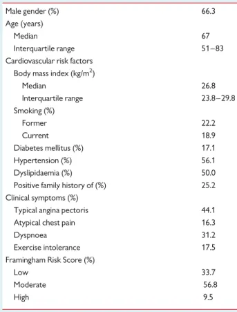
A total of 13 143 consecutive patients that underwent elective ICA were screened. Of these, 5613 (42.7%) patients were excluded because they met one or more exclusion criteria: 3413 (26.0%) had a known history of myocardial infarction, 694 (5.3%) had under-gone cardiac surgery, 1758 (13.4%) underwent ICA as part of preoperative assessment, and 1304 (9.9%) had a history of prior per-cutaneous coronary intervention. Thus, a total of 7530 (57.3%) patients were included into the final analysis for the present study. Patient baseline characteristics for the overall population and strati-fied by whether or not MPS was performed prior to ICA are given in Table1and2, respectively. Of note, patients in whom MPS was per-formed differed significantly from those without prior MPS with regard to all clinical characteristics except age, current smoking status, and the ratio of patients with a high Framingham risk score.
Angiographic findings and cardiac imaging
ICA revealed obstructive CAD in 3819 (50.7%) patients, of whom 528 (11.9%) had multi-vessel disease. MPS was performed within 90 days prior to ICA in a total of 1773 (23.5%) patients (18.9% among low-risk patients, 25.5% among moderate-risk patients, and 28.5% among patients presenting with a high risk for CAD according to the modified Framingham Risk Score).
Patients with a positive MPS result had a significantly higher rate of obstructive CAD than those who did not undergo MPS (74.4 vs. 45.6%, P , 0.001). Figure1shows that this finding holds true for all Framingham Risk Score categories with constant differences in rates of obstructive CAD findings for low risk, for those at moderate risk, and for patients at high risk for obstructive CAD.
Figure2demonstrates the results of four separate models for the prediction of a finding of obstructive CAD by ICA represented by the global x2value for each model: Model (1) including only the modi-fied Framingham Risk Score (global x2331.7, P , 0.001); Model (2) including clinical risk factors (global x2643.7, P , 0.001); Model (3) adding clinical symptoms (global x21043.0, P , 0.001); Model (4) in-clusion of the results of MPS (global x21225.2, P , 0.001).
Expectedly, as shown in Figure3, in a population at low risk for CAD, the addition of MPS provided only very limited additional value over clinical symptoms. By contrast, in patients at moderate
and high risk, MPS substantially increased the model’s predictive ability as shown in Figure3.
Predictors of obstructive coronary artery
disease
Independent predictors of obstructive CAD were male gender, typical angina pectoris, a positive MPS, diabetes, smoking, having a positive family history, and increasing age (Table3).
Discussion
Our study demonstrates that MPS independently predicts obstruct-ive CAD findings by ICA in an unselected population without known CAD irrespective of traditional risk factors and symptoms. Substan-tial increase in obstructive CAD findings at catheterization was observed if ICA was performed after objective evidence of ischaemia by MPS (74.4 vs. 45.6%, P , 0.0001). In our study cohort, the overall diagnostic yield of invasive cardiac catheterization was low with only about half of patients (50.7%) having obstructive CAD.
These finding are essentially in line with previous results addressing the rate of CAD findings at ICA.11A recently published large study by Patel et al.5reported that only a minority (38%) of the 400 000 patients who underwent elective ICA at 663 hospitals in the USA had obstructive CAD. However, a positive result on non-invasive testing only moderately increased the diagnostic yield (41.0 vs. 35.0%) in their study.5In contrast, our data demonstrate a substantial incremental value of prior MPS in predicting obstructive CAD find-ings by ICA over clinical risk factors and symptoms. The discrepancy
. . . . Table 2 Patient baseline characteristics by groups
Characteristic MPS performed n 5 1773 No MPS performed n 5 5757 P-value Male gender (%) 72.2 64.5 ,0.001 Age (years) NS Median 67 67 Interquartile range 51 – 83 51 – 83 Cardiovascular risk factors
Body mass index (kg/m2) ,0.001 Median 27.1 26.7 Interquartile range 21.1 – 33.1 21.7 – 32.7 Smoking (%) Former 34.5 18.4 ,0.005 Current 20.4 18.4 NS Diabetes mellitus (%) 21.0 15.8 ,0.005 Hypertension (%) 67.7 52.5 ,0.001 Dyslipidaemia (%) 57.6 47.6 ,0.001 Positive family history (%) 32.7 22.8 ,0.001 Clinical symptoms (%)
Typical angina pectoris 51.0 41.9 ,0.001 Atypical chest pain 20.9 14.9 ,0.001 Dyspnoea 36.1 30.2 ,0.001 Exercise intolerance 24.8 15.2 ,0.001 Framingham Risk Score (%)
Low 27.1 35.8 ,0.001 Moderate 61.3 55.3 ,0.005
High 11.5 8.9 NS
CAD, coronary artery disease; MPS, myocardial perfusion single-photon-emission computed tomography; NS, not significant.
Table 1 Patient baseline characteristics (n 5 7530)
Male gender (%) 66.3 Age (years)
Median 67
Interquartile range 51 – 83 Cardiovascular risk factors
Body mass index (kg/m2)
Median 26.8 Interquartile range 23.8 – 29.8 Smoking (%) Former 22.2 Current 18.9 Diabetes mellitus (%) 17.1 Hypertension (%) 56.1 Dyslipidaemia (%) 50.0 Positive family history of (%) 25.2 Clinical symptoms (%)
Typical angina pectoris 44.1 Atypical chest pain 16.3
Dyspnoea 31.2
Exercise intolerance 17.5 Framingham Risk Score (%)
Low 33.7
Moderate 56.8
High 9.5
Figure 1 Comparison of rates of obstructive CAD as identified by invasive coronary angiography between patients without MPS imaging prior to elective invasive coronary angiography (black boxes) and patients with a prior positive stress test (white boxes) stratified by risk groups. While rates of obstructive CAD findings increased with higher risk, the difference in rates for both groups remained constant throughout all Framingham risk groups.
with respect to the findings by Patel et al. may be mainly due to the fact that our analysis is based on cardiac imaging (MPS), whereas resting electrocardiogram or resting echocardiography were not regarded as tests suitable to predict ischaemia and consecutively the presence of obstructive CAD.
In fact, in the present study, non-invasive MPS had an incremental value in predicting obstructive CAD over clinical risk factors and symptoms regardless of the pre-test probability as defined by the Fra-mingham Risk Score. This further corroborates the clinical value of cardiac imaging (MPS) in an every-day clinical population. While it has been previously demonstrated that MPS plays an important role as a gatekeeper by preventing unnecessary invasive proce-dures,12,13the present data extend this knowledge by demonstrating that MPS also increases the diagnostic yield of invasive ICA, shifting the focus more on its role as a gatekeeper for revascularization.14
This study also explores patterns of risk stratification prior to ICA. Our study demonstrates a low rate of only 23.5% of patients having undergone non-invasive MPS prior to the invasive procedure. Of note, the majority particularly of patients at moderate risk for CAD did not undergo MPS prior to ICA although documentation of ischaemia is recommended by current guidelines particularly for this population.15,16
Interestingly, in view of the above, the patient population in which MPS was performed differed from the patients who did not undergo MPS prior to ICA: in fact, with the exception of age, current smoking status, and the ratio of patients at a high cardiovascular risk, all other baseline characteristics differed significantly. Of note, the ratio of patients at a low cardiovascular risk was higher in the group where no MPS was performed while by contrast, the ratio of patients at high risk did not differ statistically between the two groups. This finding is surprising, as implementation of the current guidelines would lead one to expect an opposite distribution.
Previous studies have demonstrated obstructive CAD findings at catheterization of up to 60% but have applied more lenient inclusion criteria.17Thus, the low rate of obstructive CAD in the present study may to some extent be perceived as a reflection of stricter inclusion criteria as patients with a known history of CAD or those undergoing emergency procedures and preoperative assessment were excluded. This is of importance with regard to the context and impact of our
Figure 2 Incremental value of information obtained before inva-sive coronary angiography to predict obstructive coronary artery disease. Model 1 included the Framingham Risk Score. Clinical risk factors were added to Model 2, symptoms to Model 3, and the results of non-invasive stress testing to Model 4.
Figure 3 Incremental value of information obtained before inva-sive coronary angiography to predict obstructive coronary artery disease in different risk populations. Clinical risk factors were included in Model 1, symptoms were added to Model 2, and the results of non-invasive stress testing to Model 3.
. . . . Table 3 Independent predictors of coronary artery disease (n 5 7530)
Variable Odds ratio (95% CI) P-value
Male gender 3.07 (2.72 – 3.47) ,0.001 Typical angina pectoris 2.95 (2.63 – 3.30) ,0.001 Positive MPS 2.74 (2.36 – 3.19) ,0.001 Diabetes 1.80 (1.56 – 2.08) ,0.001 Smoking 1.38 (1.20 – 1.56) ,0.001 Positive family history 1.41 (1.24 – 1.60) ,0.001 Age (per year) 1.04 (1.03 – 1.04) ,0.001
CI, confidence interval; MPS, myocardial perfusion single photon emission computed tomography.
findings on clinical decision making as our findings apply to more than two-thirds of all performed elective cardiac catheterizations of our study population.
Although it has been shown that percutaneous coronary interven-tion in addiinterven-tion to optimal medical treatment is not superior to medical therapy alone in terms of prognosis and quality of life in patients with stable CAD in general populations,18,19it remains an important treatment option for obstructive CAD and its use has increased several fold over the last decade.20Although the incidences of periprocedural morbidity and mortality are low, diagnostic coron-ary angiography may cause serious complications and, hence, the benefits must justify the risks and appropriate patient selection is crucial. This is particularly true in patients with a low-to-moderate risk for obstructive CAD where the risk-to-benefit ratio is easily shifted towards the unfavourable side of the balance.
With regard to the risk-to-benefit ratio, it is important to note that aside from being non-invasive, nuclear cardiac imaging has recently seen revolutionary milestone in technical innovation with the intro-duction of cadmium-zinc-telluride detectors and sophisticated iterative reconstruction algorithms, as this has led to a massive in-crease in system sensitivity.21This, in turn, allows either substantially shortening the time needed for image acquisition22or reduction of radiotracer activity. The latter results in a radiation dose exposure of around 5 mSv23and even below.24Such a reduction in radiation dose exposure addresses valid concerns about radiation dose from medical imaging and, importantly, places MPS at the same level with invasive angiography.
It is against the abovementioned background that current recom-mendations call for documentation of ischaemia by non-invasive testing prior to ICA. However, while the present data document the ability of MPS to improve the diagnostic yield of coronary angiography, it also reveals that such testing is not performed consistently and thus highlights room for improvement in the clinical decision-making process that leads to the diagnostic use of cardiac catheterization.
Limitations
Aside from the limitations that are inherent for any retrospective study, we acknowledge a number of particular limitations of the present work: the rate of non-invasive testing in our study is lower than those reported in previous studies2,4and is possibly underestimated as we may have missed patients that underwent ICA only after unsuc-cessful optimal medical therapy because of the 90-day time window chosen in our study. Furthermore, we cannot comment on the amount of non-invasive stress testing performed outside of our clinic. While it is reasonable to assume that the amount is negligible for MPS due to limited availability (our division being the only one per-forming nuclear cardiac imaging within the average referral area of our institution), the same might not be true for other forms of stress testing such as exercise stress testing or stress echocardiography. However, against the background of the low rate of CAD findings during the in-vasive procedure, we assume that this holds true only for a minority of patients as a direct referral to ICA would only take place in the event of a clearly pathological finding which would in turn expectedly again in-crease the rate of CAD findings to some extent.
In the present study, ICA did not reveal at least one coronary lesion with≥50% luminal diameter narrowing in 25.6% of patients with an
abnormal MPS result. Several aspects may to some extent explain the discrepant findings: for one, we cannot comment on the presence of any angiographic borderline stenosis or sequential coronary lesions, which may have led to ischaemia an identifiable culprit lesion. Con-versely, we cannot comment on whether MPS borderline findings (e.g. a SDS of 2) were overrepresented in these patients. More im-portantly, however, the current work suffers from the methodol-ogical limitation inherent to all studies comparing a functional vs. a morphological test due to the well-known incongruence of function and morphology.25
Finally, performance of the Framingham Risk Scores may be under-estimated as we substituted values for unknown lipid levels and blood pressure.
Conclusions
In an every-day clinical setting, non-invasive MPS has the ability to sub-stantially increase the diagnostic yield of elective ICA. Furthermore, MPS provides incremental value over clinical risk factors and symp-toms in predicting obstructive CAD findings, thus emphasizing its im-portance in the decision-making process that leads to the use of cardiac catheterization.
Conflict of interest: none declared.
References
1. Montalescot G, Sechtem U, Achenbach S, Andreotti F, Arden C, Budaj A et al. 2013 ESC guidelines on the management of stable coronary artery disease: the Task Force on the management of stable coronary artery disease of the European Society of Cardiology. Eur Heart J 2013;34:2949 – 3003.
2. Topol EJ, Ellis SG, Cosgrove DM, Bates ER, Muller DW, Schork NJ et al. Analysis of coronary angioplasty practice in the United States with an insurance-claims data base. Circulation 1993;87:1489 – 97.
3. Shaw LJ, Hachamovitch R, Berman DS, Marwick TH, Lauer MS, Heller GV et al. The economic consequences of available diagnostic and prognostic strategies for the evaluation of stable angina patients: an observational assessment of the value of pre-catheterization ischemia. Economics of Noninvasive Diagnosis (END) Multicenter Study Group. J Am Coll Cardiol 1999;33:661 – 9.
4. Lin GA, Dudley RA, Lucas FL, Malenka DJ, Vittinghoff E, Redberg RF. Frequency of stress testing to document ischemia prior to elective percutaneous coronary inter-vention. JAMA 2008;300:1765 – 73.
5. Patel MR, Peterson ED, Dai D, Brennan JM, Redberg RF, Anderson HV et al. Low diag-nostic yield of elective coronary angiography. N Engl J Med 2010;362:886 – 95. 6. Loong CY, Anagnostopoulos C. Diagnosis of coronary artery disease by
radio-nuclide myocardial perfusion imaging. Heart 2004;90(Suppl. 5):v2 – 9.
7. Hesse B, Tagil K, Cuocolo A, Anagnostopoulos C, Bardies M, Bax J et al. EANM/ESC procedural guidelines for myocardial perfusion imaging in nuclear cardiology. Eur J Nucl Med Mol Imaging 2005;32:855 – 97.
8. Zellweger MJ, Weinbacher M, Zutter AW, Jeger RV, Mueller-Brand J, Kaiser C et al. Long-term outcome of patients with silent versus symptomatic ischemia six months after percutaneous coronary intervention and stenting. J Am Coll Cardiol 2003;42: 33 – 40.
9. Zellweger MJ, Maraun M, Osterhues HH, Keller U, Muller-Brand J, Jeger R et al. Pro-gression to overt or silent CAD in asymptomatic patients with diabetes mellitus at high coronary risk: main findings of the prospective multicenter BARDOT Trial with a pilot randomized treatment substudy. JACC Cardiovasc Imaging 2014;7:1001 – 10. 10. Oldroyd KG, Phadke KV, Phillips R, Carson PH, Clarke M, Davis JA. Cardiac
catheter-isation by the Judkins technique as an outpatient procedure. BMJ 1989;298:875 – 6. 11. Moschovitis A, Cook S, Meier B. Percutaneous coronary interventions in Europe in
2006. EuroIntervention 2010;6:189 – 94.
12. Hoilund-Carlsen PF, Johansen A, Christensen HW, Vach W, Moldrup M, Bartram P et al. Potential impact of myocardial perfusion scintigraphy as gatekeeper for invasive examination and treatment in patients with stable angina pectoris: observational study without post-test referral bias. Eur Heart J 2006;27:29 – 34.
13. Miller TD, Hodge DO, Milavetz JJ, Gibbons RJ. A normal stress SPECT scan is an ef-fective gatekeeper for coronary angiography. J Nucl Cardiol 2007;14:187 – 93.
14. Wijns W, De Bruyne B, Vanhoenacker PK. What does the clinical cardiologist need from noninvasive cardiac imaging: is it time to adjust practices to meet evolving demands? J Nucl Cardiol 2007;14:366 – 70.
15. Gibbons RJ, Abrams J, Chatterjee K, Daley J, Deedwania PC, Douglas JS et al. ACC/AHA 2002 guideline update for the management of patients with chronic stable angina--summary article: a report of the American College of Cardiology/ American Heart Association Task Force on practice guidelines (Committee on the Management of Patients With Chronic Stable Angina). J Am Coll Cardiol 2003; 41:159 – 68.
16. Wijns W, Kolh P, Danchin N, Di Mario C, Falk V, Folliguet T et al. Guidelines on myo-cardial revascularization. Eur Heart J 2010;31:2501 – 55.
17. Shaw LJ, Shaw RE, Merz CN, Brindis RG, Klein LW, Nallamothu B et al. Impact of eth-nicity and gender differences on angiographic coronary artery disease prevalence and in-hospital mortality in the American College of Cardiology-National Cardio-vascular Data Registry. Circulation 2008;117:1787 – 801.
18. Boden WE, O’Rourke RA, Teo KK, Hartigan PM, Maron DJ, Kostuk WJ et al. Optimal medical therapy with or without PCI for stable coronary disease. N Engl J Med 2007; 356:1503 – 16.
19. Weintraub WS, Spertus JA, Kolm P, Maron DJ, Zhang Z, Jurkovitz C et al. Effect of PCI on quality of life in patients with stable coronary disease. N Engl J Med 2008;359: 677 – 87.
20. Roger VL, Go AS, Lloyd-Jones DM, Adams RJ, Berry JD, Brown TM et al. Heart disease and stroke statistics--2011 update: a report from the American Heart Asso-ciation. Circulation 2011;123:e18 – e209.
21. Gambhir SS, Berman DS, Ziffer J, Nagler M, Sandler M, Patton J et al. A novel high-sensitivity rapid-acquisition single-photon cardiac imaging camera. J Nucl Med 2009;50:635 – 43.
22. Buechel RR, Herzog BA, Husmann L, Burger IA, Pazhenkottil AP, Treyer V et al. Ultrafast nuclear myocardial perfusion imaging on a new gamma camera with semi-conductor detector technique: first clinical validation. Eur J Nucl Med Mol Imaging 2010;37:773 – 8.
23. Duvall WL, Croft LB, Ginsberg ES, Einstein AJ, Guma KA, George T et al. Reduced isotope dose and imaging time with a high-efficiency CZT SPECT camera. J Nucl Cardiol 2011;18:847 – 57.
24. Einstein AJ, Blankstein R, Andrews H, Fish M, Padgett R, Hayes SW et al. Comparison of image quality, myocardial perfusion, and left ventricular function between stand-ard imaging and single-injection ultra-low-dose imaging using a high-efficiency SPECT camera: the MILLISIEVERT study. J Nucl Med 2014;55:1430 – 7.
25. Tonino PA, Fearon WF, De Bruyne B, Oldroyd KG, Leesar MA, Ver Lee PN et al. Angiographic versus functional severity of coronary artery stenoses in the FAME study fractional flow reserve versus angiography in multivessel evaluation. J Am Coll Cardiol 2010;55:2816 – 21.
IMAGE FOCUS
. . . .
doi:10.1093/ehjci/jev077
Online publish-ahead-of-print 7 April 2015
Aortic forward flow in aortic atresia via ventriculo-coronary arterial
connections
Yasunobu Hayabuchi*, Miho Sakata, and Shoji Kagami
Department of Pediatrics, Tokushima University, Kuramoto-cho-3, Tokushima 770-8503, Japan
*Corresponding author. Tel:+81 886 33 7135; Fax: +81 886 31 8697. E-mail: hayabuchi@tokushima-u.ac.jp; hayabuchi@clin.med.tokushima-u.ac.jp
A full-term male newborn weighing 2.7 kg was eval-uated because of suspected cyanotic congenital heart disease on the day of birth. The echocardio-graphic study showed corrected transposition of the great arteries, severe tricuspid stenosis, right ventricular (RV) hypoplasia, aortic atresia, intact ventricular septum, and ventriculo-coronary arter-ial connections (VCACs) (Panel A, yellow arrow). RV contraction created reverse flow in the right coronary and left anterior descending coronary ar-teries via the VCACs, resulting in forward flow in the ascending aorta (Panels B and C, see Supplementary data online, Video S1). Computed tomographic angi-ography on the third day of life also demonstrated
the VCACs between the hypoplastic RV and ascending aorta (Panels D and E). The patient was maintained on prostaglandin E1, and bilateral
pulmonary arterial banding was performed on the 14th day of life. On the ninth postoperative day, echocardiography demonstrated a re-strictive patent foramen ovale with accelerated flow velocity. Therefore, balloon atrial septostomy and right ventriculography were per-formed. The examination demonstrated the presence of VCACs consistent with the prior echocardiographic findings (Panels F and G). The patient underwent the Norwood procedure with an LV-PA shunt on the 55th day of life. This is the first report of this anatomic variant. Coronary insufficiency and critical ventricular dysfunction in the presence of VCACs is a well-described phenomenon in hypoplastic right and left hearts. The present case had a definitively different blood flow pattern in the ascending aorta and coronary arteries compared with these two representative anomalies with hypoplastic left or right ventricles.
Conflict of interest: None declared.
Supplementary data are available at European Heart Journal – Cardiovascular Imaging online.