Identification of CD19-B220+c-Kit+Flt3/Flk-2+cells as early B lymphoid precursors before pre-B-I cells in juvenile mouse bone marrow
12
0
0
Texte intégral
(2) 314 c-Kit and Flt3/Flk-2 on early B progenitors in vitro (13). These bipotential progenitors express a C1qRPrelated glycosylated transmembrane protein recognized by the mAb AA4.1 (14). In fact, a series of molecules expressed in and on progenitors have been employed to characterize and FACS purify early B-lineage progenitors and precursors in the bone marrow of adult mice. Initially these progenitors and precursors were found by Hardy et al. (15) in the fraction termed ‘A’ which was found to be B220 (CD45R)⫹CD43⫹CD24 [heat-stable antigen (HSA)]loBP-1–. Four subpopulations were characterized within this fraction A by Rolink et al. (16), which were all found not to express the B-lineage-specific marker CD19: (i) NK1.1⫹ precursors of NK cells, (ii) CD4⫹ cells not developing into B-lineage cells upon transplantation or tissue culture, (iii) MHC class II⫹ cells again not developing into B-lineage cells and (iv) marker-negative cells which could develop into Blineage cells. In vitro, these marker-negative cells within fraction A developed into CD19⫹ cells, which proliferated long term on stromal cells in the presence of IL-7. Thereafter, Li et al. (17) characterized B-lineage progenitors within Hardy’s fraction A further as CD19–AA4.1⫹, partly CD4⫹ cells which express components of the pre-B cell receptor (Igα, Igβ, λ5), early B-lineage related transcription factors (Pax-5, E12, E47), as well as sterile transcripts of the µ heavy chain locus. Recently, Allman et al. (18) further characterized the three subpopulations of fraction A, referred as A0, A1 and A2, in terms of expression of stem cell-related markers, potential to give rise to multiple lineages of hematopoietic cells and frequency of DHJH-rearranged alleles. Their results suggested that fraction A1 and A2 sorted from adult bone marrow contains only B-lineage committed progenitors which have germline configuration of IgH alleles. Another interesting phenotype of these progenitors compared to CD19⫹ pro-B-I or fraction ‘B’ cells is the absence of expression of c-Kit receptor tyrosine kinase on the surface, implying that c-Kit is once downregulated in the course of B cell differentiation from HSC in the bone marrow of adult mice. Although c-Kit has been implicated in proliferation of HSC (19–21), the role of c-Kit in B cell development is still controversial. Despite indications that c-Kit is an essential molecule for B cell development in vitro (22), it was also shown that B cells can be generated in vivo in the absence of c-Kit (23–25). B cell development in the bone marrow of normal adult mice was not inhibited by injection of an antagonistic anti-c-Kit mAb (19). This inconsistency could be partially due to the existence of other receptor tyrosine kinases like Flt3/Flk-2, which may compensate for the loss of c-Kit function. The ligands for c-Kit and Flt3/Flk-2, the stem cell factor (SCF) and FL, are cofactors for the growth of progenitor cells in vitro (26–29) (for review, see 30,31). Targeted disruption of the flt3/ flk-2 gene causes deficiency of early differentiation of B lymphocytes in mouse bone marrow (32). A severely enhanced phenotype was observed in juvenile mice when the disrupted allele was brought into the W/Wv background which have defects in the c-kit gene, suggesting that c-Kit and Flt3/Flk-2 might be involved in early B cell development in juvenile mice. The role of c-Kit and Flt3/Flk-2 in the adult bone marrow cannot be examined in the compound mice because of their lethality. However, co-injection of antagonistic mAb against c-Kit and Flt3/Flk-2 into normal adult mice did. not affect B cell development in the bone marrow (33), suggesting that these receptor tyrosine kinases are indispensable for B cell development in the bone marrow of juvenile mice but not of adult mice. In order to identify early B cell progenitors which express c-Kit and Flt3/Flk-2 in the bone marrow of juvenile mice, we combined the analysis of the expression of c-Kit, Flt3/Flk-2, B220, CD19, CD43, CD24, BP-1, NK1.1, Sca-1, CD4, AA4.1 and λ5 of surrogate light chain on sIg– progenitors of 1-weekold mouse bone marrow with an in vitro analysis of their growth properties in medium containing SCF, FL and IL-7, their clonability on stromal cells in the presence of IL-7, and their capacity to differentiate to CD19⫹c-Kit– B-lineage cells. We identified three B-lineage progenitors which express both c-Kit and Flt3/Flk-2 before the stage of a pre-B-I cell. Singlecell PCR analysis of the rearrangement status of the IgH alleles (34) allows us to order them in their B-lineage pathway of development in juvenile mouse bone marrow. Methods Mice C57BL/6 mice of various ages were purchased from Biological Research Laboratories (Fu¨ llinsdorf, Switzerland). RAG2-deficient mice obtained from Dr F. Alt (The Children’s Hospital, Boston, MA) and 5⬘λ5–huCD25 transgenic mice developed by Dr I.-L. Mårtensson (Lund University, Lund, Sweden) (35) were bred in our animal facilities. Antibodies The FITC- and phycoerythrin (PE)-labeled mAb 1D3 (antiCD19), FITC- and biotin-conjugated mAb RA3-6B2 (antiCD45R, B220), biotin-conjugated mAb RM4-5 (anti-CD4), E13-161.7 (anti-Ly-6A/E, Sca-1) and PK136 (anti-NK1.1), and unlabeled mAb M1/70 (anti-CD11b, Mac-1) and TER-119 (anti-TER119) were purchased from PharMingen (San Diego, CA). The FITC-labeled mAb 2A3 (anti-human CD25) was purchased from Becton Dickinson (San Jose, CA). The mAb AA4.1 (anti-AA4.1), A2F10 (anti-Flt3/Flk-2) (33) and ACK4 (anti-c-Kit) were purified from hybridoma culture supernatants on a Protein G–Sepharose column (Pharmacia, Uppsala, Sweden) as recommended in the instruction manual. They were labeled with biotin, PE or allophycocyanin (APC) by standard methods. Flow cytometry Bone marrow cells derived from 1-week-old mice were incubated with unlabeled anti-Mac-1 and anti-TER119 mAb. Cells were then depleted of positive cells by using magnetic beads conjugated with anti-rat IgG (Dynal, Oslo, Norway) according to the instruction manual. The Mac-1–TER119– cells were stained with FITC-labeled anti-CD19, PE-labeled anti-Flt3/Flk-2, APC-labeled anti-c-Kit and biotin-conjugated mAb, which were revealed by streptavidin–Red613 (Gibco/BRL, Life Technologies, Paisley, UK). Stained cells were resuspended in HBSS (Gibco/BRL) containing 1% BSA and 200 nM TO-PRO-1 iodide (Molecular Probes, Eugene, OR) to exclude dead cells by gating in FL1 (FITC range). In some experiments, streptavidin–PharRed (PharMingen) and propidium iodide.
(3) c-Kit and Flt3/Flk-2 on early B progenitors 315 (Sigma, St Louis, MO) were used in place of streptavidin– Red613 and TO-PRO-1 iodide. Cells were analyzed by a FACStar Plus (Becton Dickinson, Mountain View, CA), which was calibrated by using 2 µm microspheres (Polysciences, Warrington, PA), and data was printed out by using software Lysys II or CellQuest (Becton Dickinson). For cell sorting, bone marrow cells were stained with FITC-labeled anti-CD19 together with unlabeled anti-Mac-1 and anti-TER119. Cells were depleted of positive cells and re-stained as described above. Cell sorting was performed by a FACStar Plus . The CD19–B220⫹ cells were gated first, and c-KithiFlt3/Flk-2lo and c-KitloFlt3/Flk-2hi cells in this fraction were sorted by using two overlapping sorting gates. Cells that fell into the overlapping region were logically rejected during sorting so that contamination of the two close populations was minimized. Sorted cell populations were routinely reanalyzed and showed ⬎95% purity. In some experiments, FITC (CD19)-negative cells sorted in the first round were re-stained with FITC–anti-human CD25, and positive and negative cells were re-sorted. For single-cell PCR analysis, cells were sorted into 96-well plates at the density of 1 cell/well by using an automatic cell deposition unit (ACDU). Cell culture on a stromal cell layer For limiting dilution assay of B cell progenitors, a ST2 cell (36) monolayer was allowed to form in 96-well plates (Falcon, Oxnard, CA). Sorted CD19–B220⫹c-Kit⫹Flt3/Flk-2⫹ cells were diluted in various concentrations and inoculated into the plates. Cells were cultured in IMDM (Gibco/BRL) supplemented with 2% FCS (Gibco/BRL), 1⫻non-essential amino acids, 5⫻10–5 M β-mercaptoethanol (Fluka, Buchs, Switzerland), 5 µg/ml insulin (Sigma) and 0.03% Primatone RL (Quest International, Naarden, The Netherlands) in the presence of 100 U/ml IL-7. IL-7 was derived from culture supernatant of J558L cells transfected with murine IL-7 cDNA. After 10 days of culture, outgrowth of B lymphocytes was surveyed under a microscope. In some experiments, cultured cells were harvested by gentle pipetting, stained with several mAb and analyzed by flow cytometry. Culture with recombinant ligands Sorted cells were inoculated into 96-well plates at the density of 3.5⫻103 cells/well containing 200 µl culture medium described above. Recombinant murine c-Kit ligand (SCF), human Flt3/ Flk-2 ligand (FL) and murine IL-7 were added at a concentration of 100 ng/ml. The recombinant factors were purchased from R & D Systems Europe (Oxford, UK). After 1 week of culture, harvested cells were stained with FITC–anti-B220 and PE–antiCD19, and analyzed by flow cytometry. For the short-term culture in the absence of ligands, 2⫻104 cells of sorted population were inoculated in a well of a 24-well plate and cultured for 24 h without the soluble ligands. Harvested cells did not retain mAb used for sorting on the surface after the culture period. Cells were re-stained with mAb and analyzed by flow cytometry. In vitro colony formation assay Cells were incubated in 1 ml of α-MEM (Gibco/BRL) containing 1.2% methylcellulose (Muromachi Kagaku Kogyo, Tokyo, Japan), 30% FCS (Gibco/BRL), 1% deionized BSA (Sigma), 50 µM β-mercaptoethanol, in the presence of 100 U/ml murine IL-3, 10 ng/ml murine SCF and 2 U/ml murine erythropoietin. (Boehringer Mannheim, Mannheim, Germany). After 7 days of culture period, aggregates consisting of ⬎40 cells were differentially scored as colonies. RT-PCR Total RNA was prepared from sorted cells using RNAzol B (Biotecx, Houston, TX). RNA was reverse transcribed by using Superscript II reverse transcriptase (Life Technologies, Gaithersburg, MD) and oligo(dT)12–18 primer (Life Technologies) according to the instruction manual. PCR assays were performed in the reaction mixture containing 1⫻PCR buffer (Life Technologies), 200 µM dNTPs (Pharmacia), 2.5 mM MgCl2, TaqStart antibody (Clontech, Palo Alt, CA), AmpliTaq DNA polymerase (Roche, Basel, Switzerland) and 1 µM of the following primers: β-actin, 5⬘-CCT AAG GCC AAC CGT GAA AAG-3⬘, 5⬘-TCT TCA TGG TGC TAG GAG CCA-3⬘; λ5, 5⬘-CTT GAG GGT CAA TGA AGC TCA GAG TA 3⬘, 5⬘-CTT GGG CTG ACC TAG GAT TG-3⬘; Mb-1, 5⬘-GCC AGG GGG TCT AGA AGC3⬘, 5⬘-TCA CTT GGC ACC CAG TAC AA-3⬘; µ0, 5⬘-AAC ATC TGA GTT TCT GAG GCT TGG-3⬘, 5⬘-TCA TCT GAA CCT TCA AGG ATG CTC-3⬘. Amplification of the cDNA was carried out with 1 cycle at 95°C for 30 s followed by 30 cycles at 95°C for 30 s, 57°C for 30 s, 72°C for 60 s and one additional cycle at 72°C for 10 min. RT-PCR products were electrophoresed through 1.5% agarose gel and transferred to Zeta-Probe GT membrane (BioRad, Hercules, CA) with 0.4 M NaOH. The membranes were hybridized with 32P-labeled specific probes and washed according to the manufacturer’s recommendations and exposed to X-ray films (Eastman Kodak, Rochester, NY). Single-cell PCR for DHJH rearrangement Single-cell PCR was carried out according to the method previously described (34,37). Cells were sorted into 96-well plates containing 10 mM Tris–HCl, pH 7.4 at a density of 1 cell/well by using ACDU. Cells were treated with 1 mg/ml proteinase K (Boehringer Mannheim) at 55°C for 60 min followed by inactivation at 95°C for 10 min. DNA amplification was performed in two rounds of PCR using a Hybaid Omnigene PCR machine (Hybaid, Middlesex, UK). PCR conditions and sequences of the primers were described before (34). The first round of amplification contained two different 5⬘ DH primers (DFL/SP and DQ52) and a 5⬘ JH1 primer in combination with two 3⬘ primers downstream of JH2 and JH4. The 3⬘ JH2 primer largely enhanced detectability of germline alleles. In the second round of PCR, 1 µl of the first PCR amplification was reamplified with a nested 5⬘ JH1 primer and the 3⬘ JH2 primer for germline configuration, or nested 5⬘ DH primers and either the 3⬘ JH2 or JH4 primer for DHJH-rearranged alleles. The second PCR products were analyzed on 1.5% agarose gels stained with ethidium bromide. The 3⬘ JH2 primer: 5⬘-AGG TGT CCC TAG TCC TTC ATG ACC TG-3⬘; the 5⬘ JH1 primer (2nd round): 5⬘-GAG GCA GAA CAG AGA CTG TGC TAC TGG-3⬘. Results The expression of c-Kit and Flt3/Flk-2 on mouse bone marrow cells In order to identify early B cell precursors which may coexpress c-Kit and Flt3/Flk-2, we first analyzed the expression.
(4) 316 c-Kit and Flt3/Flk-2 on early B progenitors. Fig. 2. Proliferative capacity of the c-Kit⫹Flt3/Flk-2⫹ cells in response to stromal cells and IL-7. The CD19–B220⫹c-KithiFlt3/Flk-2lo (s) and CD19–B220⫹c-KitloFlt3/Flk-2hi (d) cells sorted from bone marrow of 1-week-old B6 mice were diluted in various concentrations and inoculated into 96-well plates which were pre-seeded with stromal cells. Cells were cultured for 10 days in the presence of IL-7 and growth of B lymphocytes was monitored under a microscope. The data points represent the fraction of wells showing no growth of B lymphocytes out of 96 wells seeded.. Fig. 1. FACS analysis and sorting of B220⫹CD19–c-Kit⫹Flt3/Flk-2⫹ bone marrow cells from 1-week-old normal B6 mice. (A) Cells were stained with anti-Mac-1 and anti-TER-119 mAb, and depleted of positive cells by using immunomagnetic beads. Cells were then stained with FITC–anti-CD19, PE–anti-Flt3/Flk-2, APC–anti-c-Kit and biotinylated anti-B220 (CD45R) which was revealed with Red613– streptavidin. Dead cells were excluded by staining with TO-PRO-1 iodide which was detected in FL1 (FITC range). (B) B220⫹CD19– cells were gated as indicated in (A), and expression of c-Kit and Flt3/Flk-2 on this population was analyzed. Two overlapping sorting gates are shown. (C and D) Reanalysis of sorted c-KithiFlt3/Flk2lo cell fraction. (E and F) Reanalysis of sorted c-KitloFlt3/Flk-2hi cell fraction.. of c-Kit and Flt3/Flk-2 on the CD19–B220⫹ cell population in the bone marrow of a very young, i.e. 1-week-old, B6 mouse by using specific mAb. CD19–B220⫹ cells comprised 11.6 ⫾ 2.5% of the bone marrow cells depleted of Mac-1⫹ and TER119⫹ cells (Fig. 1A). In the CD19–B220⫹ cell population, we detected cells which co-express c-Kit and Flt3/Flk-2 (Fig. 1B). These cells comprise 38.0 ⫾ 2.8% of the CD19–B220⫹ cell population (7⫻103 cells/femur). They could be further subdivided into two fractions according to the expression level of c-Kit and Flt3/Flk-2, i.e. CD19–B220⫹c-KithiFlt3/Flk-2lo and CD19–B220⫹c-KitloFlt3/Flk-2hi. In this paper, we refer to these fractions as c-KithiFlt3/Flk-2lo and c-KitloFlt3/Flk-2hi fractions respectively. These fractions were also detected at the same frequency in the bone marrow of age-matched RAG2-deficient mice, suggesting that rearrangements of Ig or TCR gene loci are not required for the development of these cells (data not shown).. Proliferative capacity of the c-Kit⫹Flt3/Flk-2⫹ cells in response to stromal cells In order to examine whether the c-KithiFlt3/Flk-2lo and c-KitloFlt3/Flk-2hi fractions contain any B lymphoid progenitors, these fractions were sorted by FACS from bone marrow cells of 1-week-old B6 mice. Two overlapping gates were used for sorting to minimize possible contamination of the two close populations, as the cell sorter logically rejects cells that fall into the overlapping region (Fig. 1B). Although both populations were B220⫹, the reanalyses of the sorted fractions revealed that the intensity of B220 fluorescence was higher in the c-KitloFlt3/Flk-2hi cells than in c-KithiFlt3/Flk-2lo cells (Fig. 1C–F). The sorted cells were cultured in the presence of stromal cells and IL-7. Both fractions gave rise to proliferation of CD19⫹c-Kit⫹ as well as CD19⫹c-Kit– cells which contain surface IgM⫹ B cells under bulk culture conditions (data not shown). To determine the frequency of B cell progenitors which are clonable in the presence of stromal cells and IL-7, sorted fractions were cultured under limiting dilution conditions. Figure 2 shows that around one in five cells in the c-KitloFlt3/ Flk-2hi fraction gave rise to a B cell colony in this culture condition, whereas the c-KithiFlt3/Flk-2lo fractions contained less B cell progenitors (one in 30 cells). FACS analysis of the colonies derived from each fraction confirmed that they consisted of CD19⫹ B lymphocytes (data not shown). We also examined the frequency of myeloid and erythroid progenitor cells in the fractions sorted from 1-week-old mouse bone marrow by in vitro colony formation analysis with recombinant cytokines. In contrast to the frequency of B cell progenitors, the c-KithiFlt3/Flk-2lo fraction contained high.
(5) c-Kit and Flt3/Flk-2 on early B progenitors 317. Fig. 3. Ontogenic restriction of the c-KitloFlt3/Flk-2hi cell fraction. The fetal liver and bone marrow cells of different ages were stained with FITC–anti-CD19, non-labeled anti-Mac-1 and anti-TER119 mAb, and depleted of positive cells by using immunomagnetic beads. Cells were then stained with FITC–anti-CD19, PE–anti-Flt3/Flk-2, APC–anti-c-Kit and biotinylated anti-B220 (CD45R) which was revealed with streptavidin– PharRed. Dead cells were excluded by staining with propidium iodide. B220⫹CD19– cells were gated as indicated in upper panels, and expression of c-Kit and Flt3/Flk-2 on this population was analyzed (lower panels). The numbers indicate the percentage of cells that appeared in each area.. Table 1. Colony formation of CD19–B220⫹ subfractions in response to IL-3, SCF and erythropoetin Cell fractions. c-KithiFlt3/Flk-2lo c-KitloFlt3/Flk-2hi. No. of colonies per disha (frequency) CFU-M. CFU-G. CFU-GM. BFU-Eb. 134.0 ⫾ 12.7 (1/4) 5.5 ⫾ 1.3 (1/91). 48.5 ⫾ 2.1 (1/10) 0.0 ⫾ 0.0 (⬍1/500). 32.5 ⫾ 3.5 (1/15) 1.3 ⫾ 0.5 (1/385). 0.0 ⫾ 0.0 (⬍1/500) 0.0 ⫾ 0.0 (⬍1/500). Bone marrow cells sorted from 1-week-old B6 mice were cultured in a semi-solid medium containing recombinant murine IL-3, SCF and erythropoetin. The inoculum size was 500 cells/dish. Colonies were differentially counted after 7 days of culture period. aMean ⫾ SD of triplicate cultures. bLin–c-Kit⫹ cells, analyzed as a positive control, gave rise to erythroid colonies at the frequency of 1/152 cells.. frequencies of myeloid progenitors (one in two to three cells), whereas the c-KitloFlt3/Flk-2hi fraction contained less myeloid progenitors (one in 70 cells) (Table 1). As B lymphoid progenitor was highly enriched in the c-KitloFlt3/Flk-2hi fraction, further analyses were mainly performed on this fraction. Ontogenic restriction of the c-KitloFlt3/Flk-2hi cell fraction In 1-week-old mice, B220⫹ cells were abundantly found in the CD19–Mac-1–TER119–c-Kit⫹Flt3/Flk-2⫹ cell population (⬎50% in c-KithiFlt3/Flk-2lo cells and ⬎90% in c-KitloFlt3/Flk2hi cells). In contrast, the same population in the bone marrow of adult mice contained few, if any, B220⫹ cells (data not shown). We detected only few B cell progenitors in the CD19– c-KithiFlt3/Flk-2lo and CD19–c-KitloFlt3/Flk-2hi fractions sorted from adult bone marrow (less than one in 100 cells). To investigate time course for the development of the c-KitloFlt3/Flk-2hi cells during ontogeny, we examined presence of this fraction in the fetal liver and bone marrow of different ages by flow cytometry (Fig. 3). In the fetal liver, committed B cell precursors emerge at day 14 of gestation. (38). At this time point, significant numbers of CD19–B220⫹ cells were present in the fetal liver and the c-KitloFlt3/Flk-2hi cells were detectable in the B220⫹ population. During neonatal ontogeny, the c-KitloFlt3/Flk-2hi cells were consistently present in the bone marrow of 1-day-old and 1-week-old mice (Fig. 3 and data not shown). In the bone marrow of 2-weekold mice, this cell fraction decreased in number and became almost undetectable at 12 weeks of age. This result indicates that the presence of the CD19–B220⫹c-KitloFlt3/Flk-2hi fraction is restricted to the fetal liver and bone marrow of juvenile mice, and is not detectable in the adult bone marrow. Cellular phenotype of the c-KitloFlt3/Flk-2hi fraction The surface character of the c-KitloFlt3/Flk-2hi fraction in the bone marrow of 1-week-old mice was further examined with additional mAb (Fig. 4). Weak expression of CD4 and Sca-1 was found on half of the c-KitloFlt3/Flk-2hi cells, whereas none of them expressed NK1.1. Interestingly, about one-third of the c-KitloFlt3/Flk-2hi cells showed expression of AA4.1. The expression of AA4.1 was not detected in the c-KithiFlt3/Flk-2lo.
(6) 318 c-Kit and Flt3/Flk-2 on early B progenitors. Fig. 4. Cell surface phenotype of the CD19–B220⫹c-KitloFlt3/Flk-2hi cells. Bone marrow cells from 1-week-old B6 mice were stained with FITC–anti-CD19, PE–anti-Flt3/Flk-2, APC–anti-c-Kit and biotinylated mAb with different specificity. CD19–c-KitloFlt3/Flk-2hi cells were gated and re-plotted for staining of anti-c-Kit and biotinylated mAb which were revealed by streptavidin–Red613.. Fig. 5. Expression of mRNA for λ5, Mb-1, β-actin and µ0 transcripts in sorted CD19⫹ and CD19–B220⫹c-KitloFlt3/Flk-2hi cells from bone marrow of 1-week-old B6 mice. Different dilution of cDNA prepared from the sorted fractions were subjected to PCR amplification specific for β-actin, λ5, Mb-1 and µ0 transcripts. PCR products were separated on 1.5% agarose gel and transferred to membranes and hybridized with specific probes.. fraction (data not shown). Li et al. reported that only the AA4.1⫹ cells in Hardy’s fraction A were able to proliferate on a stromal cell layer (17). In agreement with their result, we observed ⬎4 times higher frequency of B cell progenitors in the c-KitloFlt3/Flk-2hiAA4.1⫹ cells than in the c-KitloFlt3/Flk2hiAA4.1– cells (data not shown). This allows us to estimate that 80% of the B cell progenitors in the c-KitloFlt3/Flk-2hi fraction express AA4.1. We also analyzed the transcription of B cell-specific genes by RT-PCR (Fig. 5). In the c-KitloFlt3/Flk-2hi fraction, we detected transcripts of the λ5 and mb-1 genes as well as a sterile µ heavy chain transcripts (µ0), although the expression levels of λ5 and mb-1 were lower than that detected in the CD19⫹ bone marrow cells. Progressive differentiation of c-KitloFlt3/Flk-2hi B cell progenitors Only 10% of the CD19⫹c-Kit⫹ pre-B-I cells in the bone marrow of juvenile mice weakly express Flt3/Flk-2 (data not shown). This fact prompted us to investigate how the expression of the tyrosine kinase receptors changes upon differentiation of c-KitloFlt3/Flk-2hi B cell progenitors. A short-term (24 h) culture of sorted c-KitloFlt3/Flk-2hi cells in the absence of stromal cells and soluble ligands revealed that the cells rapidly down-regulate the expression of Flt3/Flk-2 but not c-Kit upon differentiation to the CD19⫹ stage with an apparently intermediate stage of CD19⫹c-KitloFlt3/Flk-2lo cells (Fig. 6). The frequency of B cell progenitors which are reactive to stromal cells and IL-7 in CD19⫹c-KitloFlt3/Flk-2lo and CD19⫹c-KitloFlt3/ Flk-2– cells freshly sorted from bone marrow of juvenile mice were comparably high (1:3 and 1:6 respectively, data not shown). In addition to the CD19⫹c-KitloFlt3/Flk-2lo cells,. Fig. 6. Differentiation of CD19⫹c-KitloFlt3/Flk-2lo cells from CD19–cKitloFlt3/Flk-2hi B cell progenitors. The B220⫹CD19–c-KitloFlt3/Flk-2hi cells sorted from bone marrow of 1-week-old B6 mice (upper panels) were cultured for 24 h in the absence of stromal cells and soluble ligands. Cells were re-stained with FITC–anti-CD19, PE–anti-Flt3/ Flk-2 and APC–anti-c-Kit mAb, and analyzed by flow cytometry (lower panels).. another population which is CD19–c-Kit–Flt3/Flk-2hi also appeared in the short-term culture of the c-KitloFlt3/Flk-2hi cells (Fig. 6). Although we have not further examined this population, we propose that this might represent another.
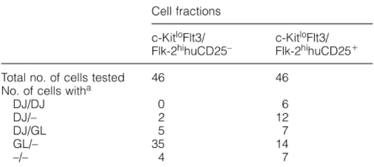
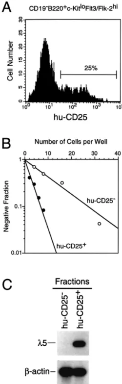
(7) c-Kit and Flt3/Flk-2 on early B progenitors 319 Table 2. Single-cell PCR analysis of DHJH gene configuration in the c-KitloFlt3/Flk-2hihuCD25⫹ and c-KitloFlt3/Flk-2hihu CD25– cells Cell fractions. Total no. of cells tested No. of cells witha DJ/DJ DJ/– DJ/GL GL/– –/–. Fig. 7. Growth requirement of B220⫹CD19–c-KitloFlt3/Flk-2hi cells in vitro. B220⫹CD19–c-KitloFlt3/Flk-2hi cells sorted from bone marrow of 1-week-old B6 mice were inoculated into 96-well plates at the density of 3.5⫻103 cells/well. Cells were cultured for 1 week in the presence of SCF, FL and IL-7. Cultured cells were stained with FITC– anti-B220 and PE–anti-CD19 mAb, and analyzed by flow cytometry. The data represent the number of cells recovered per well.. subpopulation in fraction A, B220⫹CD19–CD4⫹c-Kit–Flt3/Flk2⫹ cells which do not show any progenitor activities (33). Growth factor requirement of c-KitloFlt3/Flk-2hi B cell progenitors We next examined growth factor requirements for the early B cell progenitors in vitro. The cells in the c-KitloFlt3/Flk-2hi fraction which were sorted from 1-week-old mice were cultured in the presence of recombinant soluble ligands (Fig. 7). Neither c-Kit ligand (SCF), Flt3/Flk-2 ligand (FL) nor IL-7 alone could support the growth of the cells in this fraction. On the other hand, the combined presence of IL-7 and either SCF or FL promoted the growth of B220⫹CD19⫹ cells from the same cell population. PCR analysis of the status of rearrangements in the IgH locus in the B220⫹CD19⫹ cells showed that most of the cells had DHJH-rearranged IgH alleles (data not shown). These results suggest that IL-7 and one of the receptor tyrosine kinases are required for proliferative expansion and differentiation of CD19⫹ cells from early CD19– B cell precursors in vitro. Recovery of the CD19⫹ cells was highest when the cells were cultured in the presence of all three factors, indicating a synergistic effect of SCF and FL. One out of four cells in the c-KitloFlt3/Flk-2hi fraction gave rise to a colony which consists of CD19⫹ cells in a methylcellulose culture containing SCF, FL and IL-7, and the frequency was consistent with that obtained on the stromal cell layer (data not shown). The c-KitloFlt3/Flk-2hi B cell progenitors in 5⬘λ5–huCD25 transgenic mice In order to investigate the expression of surrogate light chain in the c-KitloFlt3/Flk-2hi fraction which is enriched for B cell progenitors, we took advantage of a transgenic mouse in which the human CD25 gene (encoding the IL-2 receptor α chain) has been introduced as a transgene under the regulation of the mouse λ5 promoter. Previous analysis of these. c-KitloFlt3/ Flk-2hihuCD25–. c-KitloFlt3/ Flk-2hihuCD25⫹. 46. 46. 0 2 5 35 4. 6 12 7 14 7. aDJ/–, only one D J -rearranged allele was detected; GL/–, only H H germline allele was detected; –/–, no PCR band was detected.. transgenic mice has shown that the human CD25 gene is expressed in parallel to surrogate light chain (35). Hence, human CD25 expression in these mice is a marker for endogenous λ5 expression. Flow cytometry analysis of bone marrow cells of 1-week-old transgenic mice showed that 25% of the cells in the c-KitloFlt3/Flk-2hi fraction expressed the huCD25 antigen (Fig. 8A). We next subdivided the c-KitloFlt3/Flk-2hi fraction into huCD25⫹ and huCD25– populations, and measured the frequency of stromal cell-dependent B cell precursors (Fig. 8B). One in three cells in the huCD25⫹ population produced B cell colonies on a stromal cell layer. Surprisingly, the huCD25– population also contained significant number of B cell progenitors (one in 10 cells). This frequency suggests that at least half of B cell progenitors in the c-KitloFlt3/Flk-2hi fraction does not express the huCD25 transgene. Colonies derived from the huCD25– population were composed of CD19⫹huCD25⫹ cells, indicating that the huCD25– progenitors are proper precursors for the B lymphoid lineage (data not shown). Interestingly, colonies derived from huCD25– cells contained, on average, 10 times more cells than colonies derived from huCD25⫹ cells (5.5 ⫾ 3.4⫻104 versus 0.5 ⫾ 0.4⫻104 on average of 14 colonies after 9 days of culture). This suggests that huCD25– progenitors are more immature than huCD25⫹ progenitors and have a strong proliferative capacity. We examined expression of the endogenous λ5 gene in the c-KitloFlt3/Flk-2hihuCD25⫹ and huCD25– cells by RT-PCR. λ5 transcript was observed in the huCD25⫹ population but not in the huCD25– population, indicating the specific expression of the transgene (Fig. 8C). Thus, the results suggest that the huCD25– fraction contains early B lymphoid precursors which do not yet express λ5. DHJH gene configuration of the c-KitloFlt3/Flk-2hi fraction Finally, we analyzed the configuration of the µ heavy chain loci of the c-KitloFlt3/Flk-2hihuCD25⫹ and c-KitloFlt3/Flk2hihuCD25– cells on a single-cell level by PCR. DHJHrearranged alleles were amplified in the presence of primers specific to 5⬘ sequences of DFL16, DSP2, DQ52 and JH1, and 3⬘ sequences of JH2 and JH4. The result summarized in.
(8) 320 c-Kit and Flt3/Flk-2 on early B progenitors Table 2 shows striking differences in the number of cells with germline versus DHJH-rearranged IgH alleles. In the c-KitloFlt3/ Flk-2hihuCD25– fraction, 15% of the cells contained at least one DHJH-rearranged allele whereas, in the c-KitloFlt3/Flk2hihuCD25⫹ fraction, approximately half did. By comparison, ⬎95% of all CD19⫹c-Kit⫹ pre-B-I cells have both IgH alleles in DHJH-rearranged configuration (34). These analyses allow us to order the B-lineage progenitors in the following. sequence: (i) B220⫹CD19–c-KitloFlt3/Flk-2hi(huCD25–)λ5–, (ii) B220⫹CD19–c-KitloFlt3/Flk-2hi(huCD25⫹)λ5⫹, (iii) B220⫹ CD19⫹c-KitloFlt3/Flk-2lo(huCD25⫹)λ5⫹ and (iv) B220⫹CD19⫹ c-KitloFlt3/Flk-2–(huCD25⫹)λ5⫹ (pre-B-I). It was also suggested that B cell progenitors in the c-KitloFlt3/Flk-2hi fraction have already started DHJH gene joining and this may take place prior to λ5 expression. Discussion The B220⫹CD19⫹ committed B lymphoid progenitor in the bone marrow of adult mice, which is referred as the pre-B-I cell, is known to express low levels of a receptor tyrosine kinase c-Kit (39,40). The proliferation of this progenitor in tissue culture has been found to be influenced by the ligand for c-Kit, SCF (22,39,41). Flt3/Flk-2, another receptor tyrosine kinase with close structural similarities to c-Kit, has been found also to be expressed on early hematopoietic progenitors (42,43) as well as cell populations which contain B lymphoid progenitors (44,45). The ligand for Flt3/Flk-2 (FL) influences proliferation and differentiation of B-lineage cells from early hematopoietic progenitor cells in vitro (27). However, the role of the receptor tyrosine kinases on B lymphoid progenitor cells in bone marrow of adult mice is controversial. No deficiencies in B lymphopoiesis have been shown in the bone marrow of W/Wv mice that have defects in the c-kit gene (23). Injection of c-Kit-specific mAb into normal adult mice did not inhibit, as in vitro (39,40), but actually enhanced B lymphopoiesis (19). Mice defective for Flt3/Flk-2 showed only partial defects in B lymphopoiesis in adult bone marrow (32). The frequency of stromal cell-dependent B lymphoid progenitors in the B220⫹Flt3/Flk-2⫹ fraction sorted from adult bone marrow was almost comparable to that detected in whole B220⫹ cells (33). Simultaneous treatment of normal adult mice with Flt3/Flk-2-specific mAb and c-Kit-specific mAb did not affect B lymphopoiesis (33). These findings suggested that the receptor tyrosine kinases have a minimal role, if any, on B cell progenitors in adult bone marrow. The moderate defect of pro-B cells in Flt3/Flk-2-deficient mice is already observed at the age of 3 weeks (32). Interestingly, 3-week-old mice deficient for both c-Kit and Flt3/Flk-2 exhibit a more severe defect in B cell development (32). B. Fig. 8. B220⫹CD19–c-KitloFlt3/Flk-2hi B cell progenitors in the bone marrow of 5⬘λ5–huCD25 transgenic mice. (A) Expression of the human CD25 transgene in the B220⫹CD19–c-KitloFlt3/Flk-2hi cells from bone marrow of 1-week-old 5⬘λ5–huCD25 transgenic mice. B220⫹CD19–c-Kitloflt3/Flk-2hi cells were sorted from bone marrow of the transgenic mice as described in the legend of Fig. 1. Sorted cells were re-stained with FITC–anti-huCD25 mAb and analyzed by flow cytometry. The huCD25⫹ and huCD25– cells were re-sorted for further analyses. (B) Limiting dilution analysis of the c-KitloFlt3/ Flk-2hihuCD25⫹ (d) and c-KitloFlt3/Flk-2hihuCD25– (s) cells. The frequency of stromal cell/IL-7-dependent B cell progenitors in the sorted cells was determined as described in the legend of Fig. 2. (C) Expression of mRNA for λ5 and β-actin in sorted c-KitloFlt3/Flk2hihuCD25– and c-KitloFlt3/Flk-2hihuCD25⫹ cells. cDNA prepared from the sorted fractions were subjected to PCR amplification specific for β-actin and λ5 transcripts. PCR products were analyzed as described in the legend of Fig. 5..
(9) c-Kit and Flt3/Flk-2 on early B progenitors 321. Fig. 9. A model of early developmental stages of B cell lineage in the bone marrow of juvenile mice. *Percent of cell population in total B220⫹ cells in the bone marrow at 1 week of age. ‡Relative clone size at day 9 of in vitro culture (see Results).. cell progenitors isolated from fetal liver and bone marrow of young mice were shown to proliferate in vitro in response to IL-7 and either of SCF and FL (26,29). Therefore, the receptor tyrosine kinases might have a distinct role on B cell precursors at different ages. In this paper, we studied the expression of the two tyrosine kinase receptors in early B lymphoid progenitor populations in the bone marrow of 1-week-old mice, collectively identified by their expression of B220 (CD45R). Our results show that the differential expression pattern of these two receptors and the λ5 component of the surrogate light chain are valuable additional markers to allow further characterization of early B-lineage progenitors in juvenile mice. All these B220⫹ progenitors are defined as committed to B-lineage development by their capacity to develop, under the stimulatory influence of IL-7 and stromal cells (11), into clones of further differentiated CD19⫹ B-lineage cells (Figs 2 and 8). Together with the analyses of other markers used in previous studies by other laboratories for a definition of B-lineage-committed progenitors and precursors (15,40,46) (Fig. 4), and with the help of a single-cell PCR analysis of the rearrangement status of the IgH loci in these cells (34) (Table 2), we propose a sequence of progenitors with phenotypes shown in Fig. 9. In our scheme, B220⫹CD19– B lymphoid progenitors express both c-Kit and Flt3/Flk-2. Furthermore, these progenitors respond optimally in vitro to the combined activities of SCF, FL and IL-7 (Fig. 7), suggesting that the two receptors act together on and in one progenitor. The expression level of c-Kit does not change until progenitors differentiate to preB-II cells, whereas Flt3/Flk-2 is rapidly down-regulated upon differentiation into pre-B-I cells (Fig. 6). Down-regulation of Flt3/Flk-2 upon differentiation of murine B cell precursors is consistent with the situation in human B lymphopoiesis (47), although the expression of FLT3 (CD135) on B cell progenitors persists longer in the human bone marrow where FLT3 is expressed on a part of CD34⫹CD19⫹ pre-B-I cells and CD34– CD10⫹ pre-B-II cells (47,48).. We have assumed, and illustrated in Fig. 9, that only one line of B cell development exists in juvenile bone marrow at these early stages. It is, however, still possible that other parallel lineages of B cell progenitors exist, which co-express c-Kit and Flt3/Flk-2. Our analysis estimated that 80% of the B cell progenitors in the c-KitloFlt3/Flk-2hi fraction express AA4.1. Although this agrees with previous reports by others showing that most of early B cell progenitors are found in the AA4.1⫹ cell fraction (13,17,18), significant number of B cell progenitors are still present in the c-KitloFlt3/Flk-2hiAA4.1– fraction. We also detected a fraction of CD19–B220loc-KithiFlt3/Flk-2lo cells which comprised 3% of all B220⫹ cells (Fig. 1). These cells are again AA4.1– (data not shown), whereas one in 30 of them yielded a colony of CD19⫹ B-lineage cells (Fig. 2). The relationship of these fractions with the major developmental pathway of early B lymphoid precursors proposed in Fig. 9 remains to be clarified. The bone marrow of a juvenile mouse has ⬎0.5% of all B220⫹ cells as c-KitloFlt3/Flk-2hi B-lineage progenitors which respond to stromal cells and IL-7 (Figs 1 and 2). On the other hand, the same population was hardly detected in adult mice (Fig. 3). Although it remains to be investigated whether this is a consequence of a decrease in the total number of the early progenitors with age or a change in the phenotype of early B progenitors, it should be remembered that the frequency of early precursors which are clonable on stromal cells and IL-7 has previously been shown to decrease during 8 months of life (49,50). Therefore, we consider it likely that the early B-lineage progenitors identified in this paper are found in young, but much less frequently in adult mice. This might account for the previous result that simultaneous treatment of mAb specific to c-Kit and Flt3/Flk-2 did not affect B lymphopoiesis in the bone marrow of normal adult mice (33). Changes in frequency of B lymphoid precursors in mice are reminiscent of the situation in human B lymphopoiesis where, again only young but, much less, old individuals have early progenitors in their bone marrow (48,51)..
(10) 322 c-Kit and Flt3/Flk-2 on early B progenitors We showed that the CD19–B220⫹c-KitloFlt3/Flk-2hi cell fraction was detectable in the fetal liver by day 14 of gestation (Fig. 3). This population was present in the bone marrow of neonatal to 2 weeks-old mice but not in the adult mice (Fig. 3). This is consistent with previous report showing that pro-B cells which belong to fractions A1 and A2 in adult bone marrow do not express c-Kit (18). It is well known that there are several differences between adult and fetal B lymphopoiesis. Pro-B cells in the fetal liver but not in the adult bone marrow reconstitute CD5⫹ (B1) B cells (12,52), indicating different developmental pathways of the B cell lineage during ontogeny. Terminal deoxynucleotidyl transferase is absent in the fetal liver, which results in the lack of N segment addition during V–D–J joining in fetal and neonatal life (53). More significantly, pre-B and immature IgM⫹ B cells generated in the fetal liver initially lack MHC class II expression, whereas B-lineage cells in the adult bone marrow express class II antigen from pre-B cell stage (54,55). This finding suggested that the fetal- and adult-type B cell lineages can be distinguished by a difference in cell surface phenotype. The adulttype lineage begins to emerge in the bone marrow by 8 days of postnatal age and gradually replaces the fetal-type lineage which predominates in the bone marrow at birth (54). The fetal-type lineage is characterized by class II– pre-B and immature B cells, whereas the c-KitloFlt3/Flk-2hi fraction reported here belongs to pro-B cells (Table 2). Taking this into account, kinetics of the c-KitloFlt3/Flk-2hi fraction in juvenile bone marrow might be compatible with that of the fetal-type lineage and this may suggest that the c-KitloFlt3/Flk-2hi cells correspond to the fetal-type B cell progenitors. If this is the case, expression of the genes for the receptor tyrosine kinases is differently regulated in the process of fetal- and adult-type B cell development. Alternatively, the c-KitloFlt3/Flk-2hi fraction may represent a differentiation pathway which is not simply confined to the fetal-type lineage but also exists in the adulttype lineage. During neonatal ontogeny, rapid proliferation of B lymphoid progenitors would be required until the adult-type immune system reaches a state of homeostasis (50). We would, therefore, suggest that c-Kit and Flt3/Flk-2 might play a distinct role in the formation of a pro-B cell pool which is more prominent in neonatal mice than in adult mice. Li et al. have reported that the λ5 gene expression becomes detectable in the cells which belong to fraction A2 before DHJH gene rearrangement commences (17,18,56). This has been further confirmed by Mårtensson et al. (35) by using the 5⬘λ5-huCD25 transgenic mice. In contrast to these results, our experiments showed that the commencement of DHJH rearrangement in the B lymphoid progenitors of juvenile mice precedes λ5 expression (Table 2). The λ5 expression has been found on B cell progenitors in RAG2-deficient mice (57), and DHJH gene rearrangements have been shown to occur normally in B cell precursors of λ5-deficient mice (58). Taken together, DHJH recombination and the λ5 gene expression might take place independently and in different timing. The frequency of the cells containing at least one DHJH-rearranged allele was comparable to that of clonable B lymphoid progenitors in the c-KitloFlt3/Flk-2hiλ5(huCD25)– and c-KitloFlt3/Flk2hiλ5(huCD25)⫹ fractions (Fig. 8 and Table 2). Therefore, it is likely that most of the clonable progenitors in these fractions have started DHJH recombination. Allman et al. (18) proposed. that commitment to the B lymphoid lineage occurs before DHJH recombination. If this is also the case in juvenile mice, there should be some intermediate steps to be identified between the earliest diverging point to B lymphoid lineage of development and the c-KitloFlt3/Flk-2hiλ5– pro-B cell stage. In conclusion, the differential expression of the two receptor tyrosine kinases c-Kit and Flt3/Flk-2 on early B-lineage progenitors is a valuable tool which should help to clarify the molecular controls underlying early stages of B lymphopoiesis.. Acknowledgments We thank Drs Hans-Reimer Rodewald and Jan Andersson for critical reading of this manuscript. We are grateful to Gholam Reza Dastoornikoo for technical assistance, and Mark Dessing and Annette Pickert for cell sorting. The Basel Institute for Immunology was founded and is supported by F. Hoffmann-La Roche Ltd, Basel, Switzerland.. Abbreviations ACDU APC FL HSA HSC PE SCF. automatic cell deposition unit allophycocyanin Flt3/Flk-2 ligand heat-stable antigen hematopoietic stem cells phycoerythrin stem cell factor. References 1 Visser, J. W. and van Bekkum, D. W. 1990. Purification of pluripotent hemopoietic stem cells: past and present. Exp. Hematol. 18:248. 2 Morrison, S. J., Uchida, N. and Weissman, I. L. 1995. The biology of hematopoietic stem cells. Annu. Rev. Cell Dev. Biol. 11:35. 3 Spangrude, G. J., Heimfeld, S. and Weissman, I. L. 1988. Purification and characterization of mouse hematopoietic stem cells. Science 241:58. 4 Ikuta, K. and Weissman, I. L. 1992. Evidence that hematopoietic stem cells express mouse c-kit but do not depend on steel factor for their generation. Proc. Natl Acad. Sci. USA 89:1502. 5 Osawa, M., Hanada, K., Hamada, H. and Nakauchi, H. 1996. Long-term lympho-hematopoietic reconstitution by a single CD34low/negative hematopoietic stem cell. Science 273:242. 6 Morrison, S. J. and Weissman, I. L. 1994. The long-term repopulating subset of hematopoietic stem cells is deterministic and isolatable by phenotype. Immunity 1:661. 7 Kondo, M., Weissman, I. L. and Akashi, K. 1997. Identification of clonogenic common lymphoid progenitors in mouse bone marrow. Cell 91:661. 8 Rolink, A., Karasuyama, H., Haasner, D., Grawunder, U., Martensson, I. L., Kudo, A. and Melchers, F. 1994. Two pathways of B-lymphocyte development in mouse bone marrow and the role of surrogate L chain in this development. Immunol. Rev. 137:185. 9 Melchers, F., Rolink, A., Grawunder, U., Winkler, T. H., Karasuyama, H., Ghia, P. and Andersson, J. 1995. Positive and negative selection events during B lymphopoiesis. Curr. Opin. Immunol. 7:214. 10 Melchers, F., Haasner, D., Grawunder, U., Kalberer, C., Karasuyama, H., Winkler, T. and Rolink, A. 1994. Roles of IgH and L chains and of surrogate H and L chains in the development of cells of the B lymphocyte lineage. Annu. Rev. Immunol. 12:209. 11 Rolink, A., Kudo, A., Karasuyama, H., Kikuchi, Y. and Melchers, F. 1991. Long-term proliferating early pre-B cell lines and clones with the potential to develop to surface Ig-positive, mitogenreactive B cells in vitro and in vivo. EMBO J. 10:327. 12 Hardy, R. R. and Hayakawa, K. 1991. A developmental switch in B lymphopoiesis. Proc. Natl Acad. Sci. USA 88:11550..
(11) c-Kit and Flt3/Flk-2 on early B progenitors 323 13 Cumano, A., Paige, C. J., Iscove, N. N. and Brady, G. 1992. Bipotential precursors of B cells and macrophages in murine fetal liver. Nature 356:612. 14 Petrenko, O., Beavis, A., Klaine, M., Kittappa, R., Godin, I. and Lemischka, I. R. 1999. The molecular characterization of the fetal stem cell marker AA4. Immunity 10:691. 15 Hardy, R. R., Carmack, C. E., Shinton, S. A., Kemp, J. D. and Hayakawa, K. 1991. Resolution and characterization of pro-B and pre-pro-B cell stage in normal mouse bone marrow. J. Exp. Med. 173:1213. 16 Rolink, A., ten Boekel, E., Melchers, F., Fearon, D. T., Krop, I. and Andersson, J. 1996. A subpopulation of B220⫹ cells in murine bone marrow does not express CD19 and contains natural killer cell progenitors. J. Exp. Med. 183:187. 17 Li, Y.-S., Wasserman, R., Hayakawa, K. and Hardy, R. R. 1996. Identification of the earliest B lineage stage in mouse bone marrow. Immunity 5:527. 18 Allman, D., Li, J. and Hardy, R. R. 1999. Commitment to the B lymphoid lineage occurs before DH–JH recombination. J. Exp. Med. 189:735. 19 Ogawa, M., Matsuzaki, Y., Nishikawa, S., Hayashi, S.-I., Kunisada, T., Sudo, T., Kina, T., Nakauchi, H. and Nishikawa, S.-I. 1991. Expression and function of c-kit in hematopoietic progenitor cells. J. Exp. Med. 174:63. 20 Okada, S., Nakauchi, H., Nagayoshi, K., Nishikawa, S., Nishikawa, S.-I., Miura, Y. and Suda, T. 1991. Enrichment and characterization of murine hematopoietic stem cells that express c-kit molecule. Blood 78:1706. 21 Ogawa, M. 1993. Differentiation and proliferation of hematopoietic stem cells. Blood 81:2844. 22 Era, T., Nishikawa, S., Sudo, T., Wang, F. H., Ogawa, M., Kunisada, T., Hayashi, S. and Nishikawa, S. 1994. How Bprecursor cells are driven to cycle. Immunol. Rev. 137:35. 23 Landreth, K. S., Kincade, P. W., Lee, G. and Harrison, D. E. 1984. B lymphocyte precursors in embryonic and adult W anemic mice. J. Immunol. 132:2724. 24 Takeda, S., Shimizu, T. and Rodewald, H. R. 1997. Interactions between c-kit and stem cell factor are not required for B-cell development in vivo. Blood 89:518. 25 Rodewald, H. R., Ogawa, M., Haller, C., Waskow, C. and DiSanto, J. P. 1997. Pro-thymocyte expansion by c-kit and the common cytokine receptor gamma chain is essential for repertoire formation. Immunity 6:265. 26 Hunte, B. E., Hudak, S., Campbell, D., Xu, Y. and Rennick, D. 1995. flk2/flt3 ligand is a potent cofactor for the growth of primitive B cell progenitors. J. Immunol. 156:489. 27 Hirayama, F., Lyman, S. D., Clark, S. C. and Ogawa, M. 1995. The flt3 ligand supports proliferation of lymphohematopoietic progenitors and early B-lymphoid progenitors. Blood 85:1762. 28 Namikawa, R., Muench, M. O., de Vries, J. E. and Roncarolo, M. G. 1996. The FLK2/FLT3 ligand synergizes with interleukin-7 in promoting stromal-cell-independent expansion and differentiation of human fetal pro-B cells in vitro. Blood 87:1881. 29 Ray, R. J., Paige, C. J., Furlonger, C., Lyman, S. D. and Rottapel, R. 1996. Flt3 ligand supports the differentiation of early B cell progenitors in the presence of interleukin-11 and interleukin-7. Eur. J. Immunol. 26:1504. 30 Hirayama, F. and Ogawa, M. 1996. Cytokine regulation of early lymphohematopoietic development. Stem Cells 14:369. 31 Lyman, S. D. and Jacobsen, E. W. 1998. c-kit ligand and flt3 ligand: stem/progenitor cell factors with overlapping yet distinct activities. Blood 91:1101. 32 Mackarehtschian, K., Hardin, J. D., Moore, K. A., Boast, S., Goff, S. P. and Lemischka, I. R. 1995. Targeted disruption of the flk2/flt3 gene leads to deficiencies in primitive hematopoietic progenitors. Immunity 3:147. 33 Ogawa, M., Sugawara, S., Kunisada, T., Sudo, T., Hayashi, S. I., Nishikawa, S., Kodama, H. and Nishikawa, S.-I. 1998. Flt3/Flk-2 and c-Kit are not essential for the proliferation of B lymphoid progenitor cells in the bone marrow of the adult mouse. Exp. Hematol. 26:478.. 34 ten Boekel, E., Melchers, F. and Rolink, A. 1995. The status of Ig loci rearrangements in single cells from different stages of B cell development. Int. Immunol. 7:1013. 35 Mårtensson, I.-L., Melchers, F. and Winkler, T. H. 1997. A transgenic marker for mouse B lymphoid precursors. J. Exp. Med. 185:653. 36 Ogawa, M., Nishikawa, S., Ikuta, K., Yamamura, F., Naito, M., Takahashi, K. and Nishikawa, S. 1988. B cell ontogeny in murine embryo studied by a culture system with the monolayer of a stromal cell clone, ST2: B cell progenitor develops first in the embryonal body rather than in the yolk sac. EMBO J. 7:1337. 37 ten Boekel, E., Melchers, F. and Rolink, A. G. 1997. Changes in the V(H) gene repertoire of developing precursor B lymphocytes in mouse bone marrow mediated by the pre-B cell receptor. Immunity 7:357. 38 Delassus, S. and Cumano, A. 1996. Circulation of hematopoietic progenitors in the mouse embryo. Immunity 4:97. 39 Rolink, A., Streb, M., Nishikawa, S. and Melchers, F. 1991. The c-kit-encoded tyrosine kinase regulates the proliferation of early pre-B cells. Eur. J. Immunol. 21:2609. 40 Nishikawa, S. I., Era, T., Ogawa, M., Nishikawa, S., Ohno, N., Hayashi, S. I. and Kunisada, T. 1992. Control of intramarrow B-cell genesis by stromal cell-derived molecules. Curr. Top. Microbiol. Immunol. 182:27. 41 Yasunaga, M., Wang, F., Kunisada, T., Nishikawa, S. and Nishikawa, S. 1995. Cell cycle control of c-kit⫹IL-7R⫹ B precursor cells by two distinct signals derived from IL-7 receptor and c-kit in a fully defined medium. J. Exp. Med. 182:315. 42 Matthews, W., Jordan, C. T., Wiegand, G. W., Pardoll, D. and Lemischka, I. R. 1991. A receptor tyrosine kinase specific to hematopoietic stem and progenitor cell-enriched populations. Cell 65:1143. 43 Zeigler, F. C., Bennett, B. D., Jordan, C. T., Spencer, S. D., Baumhueter, S., Carroll, K. J., Hooley, J., Bauer, K. and Matthews, W. 1994. Cellular and molecular characterization of the role of the flk-2/flt-3 receptor tyrosine kinase in hematopoietic stem cells. Blood 84:2422. 44 Rasko, J. E., Metcalf, D., Rossner, M. T., Begley, C. G. and Nicola, N. A. 1995. The flt3/flk-2 ligand: receptor distribution and action on murine haemopoietic cell survival and proliferation. Leukemia 9:2058. 45 Wasserman, R., Li, Y. S. and Hardy, R. R. 1995. Differential expression of the blk and ret tyrosine kinases during B lineage development is dependent on Ig rearrangement. J. Immunol. 155:644. 46 Rolink, A., Ghia, P., Grawunder, U., Haasner, D., Karasuyama, H., Kalberer, C., Winkler, T. and Melchers, F. 1995. In-vitro analyses of mechanisms of B-cell development. Semin. Immunol. 7:155. 47 Rappold, I., Ziegler, B. L., Kohler, I., Marchetto, S., Rosnet, O., Birnbaum, D., Simmons, P. J., Zannettino, A. C., Hill, B., Neu, S., Knapp, W., Alitalo, R., Alitalo, K., Ullrich, A., Kanz, L. and Buhring, H. J. 1997. Functional and phenotypic characterization of cord blood and bone marrow subsets expressing FLT3 (CD135) receptor tyrosine kinase. Blood 90:111. 48 Ghia, P., ten Boekel, E., Sanz, E., de la Hera, A., Rolink, A. and Melchers, F. 1996. Ordering of human bone marrow B lymphocyte precursors by single-cell polymerase chain reaction analyses of the rearrangement status of the immunoglobulin H and L chain gene loci. J. Exp. Med. 184:2217. 49 Rolink, A., Haasner, D., Nishikawa, S. and Melchers, F. 1993. Changes in frequencies of clonable pre B cells during life in different lymphoid organs of mice. Blood 81:2290. 50 Moscatello, K. M., Biber, K. L., Dempsey, D. C., Chervenak, R. and Wolcott, R. M. 1998. Characterization of a B cell progenitor present in neonatal bone marrow and spleen but not in adult bone marrow and spleen. J. Immunol. 161:5391. 51 Nunez, C., Nishimoto, N., Gartland, G. L., Billips, L. G., Burrows, P. D., Kubagawa, H. and Cooper, M. D. 1996. B cells are generated throughout life in humans. J. Immunol. 156:866. 52 Kantor, A. B., Stall, A. M., Adams, S., Herzenberg, L. A. and Herzenberg, L. A. 1992. Differential development of progenitor activity for three B-cell lineages. Proc. Natl Acad. Sci. USA 89:3320..
(12) 324 c-Kit and Flt3/Flk-2 on early B progenitors 53 Feeney, A. J. 1992. Comparison of junctional diversity in the neonatal and adult immunoglobulin repertoires. Int. Rev. Immunol. 8:113. 54 Lam, K. P. and Stall, A. M. 1994. Major histocompatibility complex class II expression distinguishes two distinct B cell developmental pathways during ontogeny. J. Exp. Med. 180:507. 55 Hayakawa, K., Tarlinton, D. and Hardy, R. R. 1994. Absence of MHC class II expression distinguishes fetal from adult B lymphopoiesis in mice. J. Immunol. 152:4801. 56 Li, Y. S., Hayakawa, K. and Hardy, R. R. 1993. The regulated. expression of B lineage associated genes during B cell differentiation in bone marrow and fetal liver. J. Exp. Med. 178:951. 57 Karasuyama, H., Rolink, A., Shinkai, Y., Young, F., Alt, F. W. and Melchers, F. 1994. The expression of Vpre-B/lambda 5 surrogate light chain in early bone marrow precursor B cells of normal and B cell-deficient mutant mice. Cell 77:133. 58 Kitamura, D., Kudo, A., Schaal, S., Muller, W., Melchers, F. and Rajewsky, K. 1992. A critical role of lambda 5 protein in B cell development. Cell 69:823..
(13)
Figure


+3

Documents relatifs
Biological abnormalities associated with B lymphocytes in SS are also a hallmark of the disease and these abnormalities are characterized by the presence of rheumatoid factor (RF),
2) A l’aide d’un algorithme, donner, au jour près, le temps nécessaire pour que le plant de maïs atteigne une hauteur supérieure à 1, 5 m.. 3) On s’intéresse à la vitesse
Normal erythropoiesis is simulated in two dimensions, and the influence on the output of the model of some parameters involved in cell fate (differentiation, self-renewal, and death
This paper presents a robust and accurate novel method for processing WBC (leukocyte) using a combination of ideas. The segmentation of cells is achieved in two phases, the first is
The bone marrow haematopoietic cell cultures supplemented with either GM-CSF or Flt3L differentiated into large cells with dendritic morphology within 10 to 12 days (termed GM-BMDC
Using this debugging process and several other real ontologies debugging one, we found out that in several occasions domain experts were just changing axioms from the original
Here we describe ex vivo expansion of murine myeloid cells in response to Flt3 ligand (FL) and investigate their response to cytokines inducing differentiation towards
(A) Light micrographs of ZL-, ZDF-BMMSCs, and HUVECs (Human Umbilical Vein Endothelial Cells) cultured in an angiogenic medium for a 14- day period and then on a Matrigel for 24
