. . . .
. . . .
Whole-heart dynamic three-dimensional
magnetic resonance perfusion imaging
for the detection of coronary artery disease
defined by fractional flow reserve: determination
of volumetric myocardial ischaemic burden
and coronary lesion location
Robert Manka
1,2*
†, Ingo Paetsch
3†, Sebastian Kozerke
1, Marco Moccetti
2,
Rainer Hoffmann
3, Joerg Schroeder
3, Sebastian Reith
3, Bernhard Schnackenburg
3,
Oliver Gaemperli
2, Lukas Wissmann
1, Christophe A. Wyss
2, Philipp A. Kaufmann
2,
Roberto Corti
2, Peter Boesiger
1, Nikolaus Marx
3, Thomas F. Lu¨scher
2,
and Cosima Jahnke
31
Institute for Biomedical Engineering, University and ETH Zurich, Zurich, Switzerland;2
Department of Cardiology, University Hospital Zurich, Zurich, Switzerland; and3
Department of Cardiology, University RWTH Aachen, Aachen, Germany
Received 9 March 2012; revised 6 May 2012; accepted 21 May 2012; online publish-ahead-of-print 7 June 2012
This paper was guest edited by Prof. Udo Sechtem, Department of Cardiology, Robert-Bosch-Medical Center, Stuttgart, Germany
Aims Dynamic three-dimensional-cardiac magnetic resonance (3D-CMR) perfusion proved highly diagnostic for the detec-tion of angiographically defined coronary artery disease (CAD) and has been used to assess the efficacy of coronary stenting procedures. The present study aimed to relate significant coronary lesions as assessed by fractional flow reserve (FFR) to the volume of myocardial hypoenhancement on 3D-CMR adenosine stress perfusion imaging and to define the inter-study reproducibility of stress inducible 3D-CMR hypoperfusion.
Methods and results
A total of 120 patients with known or suspected CAD were examined in two CMR centres using 1.5 T systems. The protocol included cine imaging, 3D-CMR perfusion during adenosine infusion, and at rest followed by delayed en-hancement (DE) imaging. Fractional flow reserve was recorded in epicardial coronary arteries and side branches with≥2 mm luminal diameter and .40% severity stenosis (pathologic FFR , 0.75). Twenty-five patients underwent an identical repeat CMR examination for the determination of inter-study reproducibility of 3D-CMR perfusion def-icits induced by adenosine. Three-dimensional CMR perfusion scans were visually classified as pathologic if one or more segments showed an inducible perfusion deficit in the absence of DE. Myocardial ischaemic burden (MIB) was measured by segmentation of the area of inducible hypoenhancement and normalized to left ventricular myo-cardial volume (MIB, %). Three-dimensional CMR perfusion resulted in a sensitivity, specificity, and diagnostic accur-acy of 90, 82, and 87%, respectively. Substantial concordance was found for inter-study reproducibility [Lin’s correlation coefficient: 0.98 (95% confidence interval: 0.96 – 0.99)].
Conclusion Three-dimensional CMR stress perfusion provided high diagnostic accuracy for the detection of functionally signifi-cant CAD. Myocardial ischaemic burden measurements were highly reproducible and allowed the assessment of CAD severity.
-*Corresponding author. Tel:+41 44 2551251, Fax: +41 44 2554506, Email:robert.manka@usz.ch
†These authors equally contributed to this work.
Keywords Cardiac magnetic resonance † Fractional flow reserve † Adenosine stress † Three-dimensional cardiac MR perfusion † Myocardial ischaemic burden
Introduction
Cardiac magnetic resonance (CMR) imaging enables non-invasive assessment of myocardial perfusion in patients with known and suspected coronary artery disease (CAD).1–5 Two-dimensional multislice CMR perfusion techniques yield high diagnostic accuracy, but the limited spatial coverage does not permit quantification of ischaemic tissue volume to guide therapy as recommended by the recent European guidelines on myocardial revascularization.6 Therefore, radiation-based nuclear imaging methods have become the methods of choice for quantifying ventricular ischaemia.7,8 Besides exposure of the patient with ionizing radiation, the limited spatial resolution provided by single photon and positron emission tomography may be regarded as a drawback particularly when aiming at the detection of subendocardial ischaemic reactions.
In order to address the limited, non-contiguous coverage of two-dimensional, multislice CMR perfusion techniques, three-dimensional (3D-CMR) methods have been developed.9–11 Whole-heart coverage is achieved by employing data under sam-pling strategies in conjunction with appropriate image reconstruc-tion techniques.12 In a particular implementation, dynamic 3D-CMR perfusion imaging utilizes under-sampling in k-space and time (k-t) including sensitivity encoding (SENSE).11,13,14 This ap-proach proved highly diagnostic for the detection of angiographi-cally defined CAD based on quantitative angiographic measurements and has been successfully employed to assess the efficacy of coronary stenting procedures using volumetry of myo-cardial hypoenhancement.15However, quantitative coronary angi-ography (QCA) is of limited value for the characterization of the functional severity of coronary artery narrowing especially at inter-mediate degrees of stenosis and thus has been recognized as a poor indicator of functionally significant CAD when compared with pressure-derived fractional flow reserve (FFR).16,17
Consequently, the objectives of the present dual-centre study were: (i) to evaluate the diagnostic performance of dynamic 3D-CMR perfusion for the detection of functionally significant CAD as defined by FFR, (ii) to relate the volume of myocardial hypoenhancement to the extent and anatomical localization of the disease, and (iii) to assess the inter-study reproducibility of stress inducible 3D-CMR hypoperfusion.
Methods
Study population
The present prospective study was conducted at two centres (Univer-sity Hospital Zurich, Switzerland, and Univer(Univer-sity Hospital RWTH Aachen, Germany). A total of 120 patients (n ¼ 31 from Zurich, n ¼ 89 from Aachen; n ¼ 90 males; mean age 63.7 + 11.9 years, range 42 – 90 years) scheduled for diagnostic coronary angiography for the evaluation of known or suspected CAD, but without prior coronary
bypass grafting, were consecutively recruited between July 2010 and January 2011. All patients gave written informed consent and the study was approved by the local ethics review boards. Patients were instructed to refrain from caffeine-containing substances 24 h prior to the examination. Patients were not considered for study inclusion if they had typical contraindications for CMR imaging (e.g. incompatible metallic implants and claustrophobia) or administration of adenosine.
In a subgroup of 25 patients, a repeat stress 3D-CMR perfusion examination was performed within 24 h prior to invasive coronary angiography to assess inter-study reproducibility.
Cardiac magnetic resonance study
Cardiac magnetic resonance imaging was performed with the patient in the supine position using a 1.5 T MR scanner (Philips Achieva, Best, The Netherlands). A five-element cardiac synergy coil was used for signal reception and cardiac synchronization was performed with a vector-ECG.
After the acquisition of standard cine scans for the assessment of left ventricular function, 3D-CMR perfusion imaging data were acquired. Adenosine was administered intravenously at a dose of 140 mg/kg/ min under continuous monitoring of heart rate and blood pressure. After at least 3 min of adenosine infusion, 3D-CMR stress first-pass perfusion imaging (i.v. bolus application of 0.1 mmol/kg of a gadolinium-based contrast agent, Magnevist Bayer Schering, Berlin, Germany; injection rate 4.0 mL/s followed by 20 mL saline flush) was performed in short-axis geometry with full left ventricular coverage. After a 10 min waiting period for equilibration of the contrast agent within the myocardium, the identical 3D-CMR perfusion scan was repeated at rest. Finally, following another 10 min waiting period, delayed enhancement (DE) imaging was done in the identical short-axis geometry with a 3D inversion prepared spoiled gradient-echo sequence.
Three-dimensional cardiac magnetic
resonance perfusion imaging technique
The perfusion imaging protocol consisted of a saturation-recovery gradient-echo pulse sequence (TR/TE/flip angle 1.9 ms/0.8 ms/158, sat-uration prepulse delay 150 ms, partial Fourier acquisition, FOV 350× 350 mm, measured voxel size 2.3× 2.3 × 10.0 mm3reconstructed to 2.0× 2.0× × 5.0 mm3, number of slices 16). Ten-fold data undersam-pling was employed resulting in a net seven-fold acceleration relative to standard CMR imaging techniques, thereby permitting acquisition of a volume with 16 slices of 5.0 mm thickness each in every heartbeat.11Visual assessment of dynamic
three-dimensional cardiac magnetic
resonance perfusion scans
Cardiac magnetic resonance examinations were analysed visually by a single observer fully blinded to clinical and angiographic patient data on a dedicated workstation (Extended Work Space, Philips Medical Systems). All short-axis slices with clearly identifiable left ventricular cavity enhancement during contrast agent first-pass and with .75% circumferential left ventricular myocardium were selected. The short-axis slices were divided into six equally distributed segments and
evaluated visually. Perfusion defects in any segment with≥25% trans-murality persisting for three or more consecutive dynamics not being visible on the rest perfusion scan and in the absence of DE were con-sidered to be pathologic.
Measurement of myocardial ischaemic
burden
Quantification of myocardial hypoenhancement was performed in all patients using GT Volume (version 1.3.16, GyroTools, Zurich, Switzerland). For the determination of myocardial hypoenhancement, the single dynamic slice images of the stress perfusion scan showing the maximum extent of regional hypoenhancement during peak signal en-hancement of remote myocardium were chosen. In the presence of extensive ischaemia-related hypoenhancement (e.g. high-grade triple-vessel disease), the remote myocardium either represented an entire myocardial segment or its subepicardial layer. The observer manually traced left ventricular endo- and epicardial borders in all slices. Segmentation of the left ventricular myocardium was done on a pixel-by-pixel basis by adjusting the signal intensity threshold of .2 standard deviations (SD) below the signal of the remote myocardium, and the area of hypoenhancement was measured. Volumes of total left ventricular myocardium and total myocardial hypoenhancement were calculated using the disk summation method. Myocardial ischaemic burden (MIB) was defined by the volume of hypoenhancement nor-malized to total left ventricular myocardial volume and expressed in per cent.
In the presence of myocardial scar, the amount was quantified on DE images by manual segmentation based on a signal intensity thresh-old of .2 SD above the average signal of the remote myocardium and subtracted from the volume of hypoenhancement.
Inter-study reproducibility
In 25 randomly chosen patients, 3D-CMR perfusion imaging was per-formed twice prior to invasive coronary angiography for the determin-ation of inter-study reproducibility. Evaludetermin-ation of MIB of repeat examinations was conducted by the same reader following a time period of ≥4 weeks with the reader being fully blinded to clinical and angiographic patient data.
Fractional flow reserve measurements
and quantitative coronary angiography
Coronary angiography was performed by standard techniques. At least two orthogonal views of every major coronary vessel and its side branches were acquired. QCA (Philips Inturis CardioView, QCA V3.3, Pie Medical Imaging) was performed off-line by an independent observer being unaware of the results of CMR imaging. FFR was measured using standard methods17with a 0.014 in. coronary pressure sensor-tip wire(Volcano Therapeutics, San Diego, CA, USA, and Pressure-Wire Certus, St Jude Medical Systems AB, Uppsala, Sweden) in all vessels with a≥40% diameter stenosis in two orthogonal views in vessels with ≥2 mm luminal diameter. For the purposes of the study, smaller diameter stenoses were considered non-significant. Coronary lesions with an FFR value of ,0.75 were classified as haemodynamically relevant. Total or subtotal vessel occlusions were assigned an FFR value of 0.5.
Statistical analysis
Statistical data analysis was performed using SPSS for Windows 17.0.0 (2008, Chicago, IL, USA). Continuous variables are expressed as mean + SD; categorical variables are expressed as proportions. The paired Student’s t-test was used to assess statistical significance of con-tinuous variables between rest and stress. All tests were two tailed;
P , 0.05 was considered significant. To determine the cut-off value of MIB being predictive of the presence or absence of significant cor-onary artery disease as defined by FFR , 0.75, receiver-operator char-acteristic (ROC) curve analysis was performed and the area under the curve was calculated. Sensitivity and specificity with corresponding 95% confidence intervals (95% CI) were calculated according to stand-ard definitions. In addition, the Bland – Altman analysis was carried out to assess the instudy reproducibility. To address clustering of ter-ritories within patients, linear regressions with MIB as a dependent variable and robust standard error with patient ID as a cluster were performed. Independent variables were dummy variables for ischaemic vs. non-ischaemic territories and for distal, medial, proximal, and side branch territory, respectively, Stata 11.2 (StataCorp, College Station, TX, USA) was used.
Results
Patient characteristics
All 120 patients successfully completed the CMR protocol and the invasive procedures and constituted the final population for data analysis. Tables1and2provide the clinical baseline characteristics of the patient population and the haemodynamic data recorded during the CMR examination, respectively.
Diagnostic performance
The prevalence of CAD as defined by FFR , 0.75 was 57.5% (69 of 120 patients). Fifty-three patients had single-vessel disease,
. . . .
. . . .
. . . .
Table 1 Patient demographics and clinical data
Parameter n 5 120 patients
Male, n (%) 90 (75) Age (years) 63.7 + 11.9
Range 42 – 90
BMI (kg/m2) 27.0 + 3.4 Cardiovascular risk factors, n (%)
Arterial hypertension 87 (72.5) Diabetes mellitus 31 (25.8) Hyperlipoproteinaemia 68 (56.7)
Smoking 53 (44.2)
Family history of CAD 33 (27.5)
Coronary artery disease
Known CAD 66 (55.0) Single-vessel diseasea 53 (44.2) Multi-vessel diseasea 16 (13.3)
Left ventricular function
LVEF (%) 54.8 + 9.8 LVEDV (mL) 157.4 + 55.9 LVESV (mL) 74.4 + 48.0
Data are n (%) or mean + SD. BMI, body mass index; CAD, coronary artery disease; LVEF, left ventricular ejection fraction; LVEDV, left ventricular end-diastolic volume; LVESV, left ventricular end-systolic volume.
a
11 patients had double-vessel disease, and 5 patients had triple-vessel disease. Visual analysis of 3D-CMR perfusion on a per-patient basis yielded a sensitivity of 90% [95% confidence interval (CI): 82 – 98], specificity of 82% (95% CI: 71 – 94), and diagnostic accuracy of 87% (95% CI: 80 – 93). For the detection of single-and multi- (i.e. double single-and triple) vessel disease, sensitivity was 89% (47 of 53) and 94% (15 of 16), respectively.
The prevalence of CAD as defined by QCA≥ 50% was 54.2% (65 of 120 patients). Visual analysis of 3D-CMR perfusion on a per-patient basis with QCA as the reference standard yielded a sensi-tivity of 88% (95% CI: 77 – 95), specificity of 75% (95% CI: 61 – 83), and diagnostic accuracy of 81% (95% CI: 73 – 88).
Representative CMR imaging examples are shown in Figures1–3
with corresponding movie files (see Supplementary material online, Movies S1 – S5).
. . . .
. . . .
. . . .
Table 2 Haemodynamic parameters
Parameter n 5 120 patients
Heart rate (b.p.m.)
At rest 68.1 + 10.9
Adenosine stress 84.1 + 14.8*
Systolic blood pressure (mmHg)
At rest 130.2 + 21.7 Adenosine stress 127.7 + 20.3
Heart rate pressure product (b.p.m.× mmHg)
At rest 8904 + 2311
Adenosine stress 10780 + 2812*
Data are mean + SD.
*P , 0.001 for rest vs. stress (paired Student’s t-test).
Figure 1 (A) Three-dimensional cardiac magnetic resonance perfusion scans during adenosine stress (a) and at rest (b). A strictly inducible perfusion deficit in anterior/anteroseptal segments extending from apical to mid-ventricular slices can be clearly delineated (myocardial ischae-mic burden 18.8% of the left ventricular myocardium). Invasive X-ray coronary angiogram (c and d) demonstrated high-grade single-vessel disease with subtotal occlusion of the left anterior descending artery (LAD; see also Supplementary material online, Movies S1 – S3). (B) Three-dimensional delayed enhancement cardiac magnetic resonance imaging proved the absence of myocardial scar tissue.
Assessment of myocardial ischaemic
burden
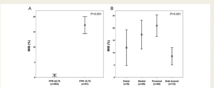
In all patients MIB averaged to 13.8 + 17.2% (range, 0 – 79.2%). The mean MIB per coronary territory with and without haemodynam-ically significant stenosis as defined by FFR is displayed in Figure4A.
The relation between lesion localization within the coronary arter-ial tree (i.e. proximal, medarter-ial, distal, or side branch stenosis) and the corresponding amount of MIB is illustrated in Figure 4B and confirmed the gradual increase in MIB with more proximal lesion location.
Figure 2 (A) Three-dimensional cardiac magnetic resonance perfusion scans during adenosine stress (a) and at rest (b). An extensive indu-cible perfusion deficit in the anterior/anteroseptal and inferolateral segments can be appreciated (myocardial ischaemic burden 33.6% of the left ventricular myocardium). Invasive X-ray coronary angiogram demonstrated a double-vessel disease of the left anterior descending artery (LAD, c) with a fractional flow reserve (FFR) of 0.73 and the intermediate branch (d) with an FFR value of 0.46 (see also Supplementary material online, Movie S4). (B) Three-dimensional delayed enhancement cardiac magnetic resonance imaging proved the absence of myocardial scar tissue.
In order to identify the amount of MIB leading to most accurate identification of FFR-defined coronary artery disease, ROC analysis was carried out and revealed an MIB cut-off value of 4.4% with a sensitivity and specificity of 85.5 and 86.3%, respectively (Figure5).
Inter-study reproducibility of myocardial
ischaemic burden
Inter-study reproducibility of MIB measurements showed a high correlation (Pearson’s correlation coefficient, 0.98, P , 0.0001) and substantial concordance [Lin’s correlation coefficient, 0.98 (95% CI: 0.96 – 0.99)]. The Bland – Altman plot is shown in Figure6; the mean bias for inter-study measurements was 3.0 + 1.4% (95% CI: 2.4 – 3.6).
Figure 7 shows a representative imaging example for first and repeat 3D-CMR examinations.
Discussion
The present study evaluated the diagnostic performance and inter-study reproducibility of 3D-CMR perfusion for the detection of functionally significant CAD as defined by FFR measurements. The main findings of the study were as follows: (i) 3D-CMR perfu-sion imaging at 1.5 T was successfully performed in a large, con-secutive patient population in two independent cardiology departments; (ii) the diagnostic accuracy of 3D-CMR perfusion imaging was high and within the range of previously published data on 2D multislice CMR perfusion imaging using FFR as the
Figure 3 (A) Three-dimensional cardiac magnetic resonance perfusion scans during adenosine stress (a) and at rest (b). An extensive indu-cible perfusion deficit in the anterior/anteroseptal/anterolateral and in the inferior/inferoseptal/inferolateral segments can be appreciated (myo-cardial ischaemic burden 79.2% of the left ventricular myocardium). Invasive X-ray coronary angiogram demonstrated high-grade left main (LM, c) and right coronary artery (RCA, d) stenoses (see Supplementary material online, Movie S5). (B) Three-dimensional delayed enhancement cardiac magnetic resonance imaging proved the presence of subendocardial scar tissue in basal inferior segments only (white arrows).
reference standard18–20; (iii) the diagnostic accuracy was equally high in patients with single- and multi-vessel coronary artery disease; (iv) the amount of MIB gradually increased with more proximal anatomical localization of coronary lesions; and (v) MIB measurement showed high inter-study reproducibility.
The present dual-centre 3D-CMR perfusion study with FFR as the standard of reference yielded similar sensitivity but improved specificity (90 vs. 92% and 82 vs. 74%, respectively) compared
with previously published data on 3D-CMR perfusion using angio-graphic stenosis quantification as the standard of reference.15The improved specificity may have been due to a lower rate of false-positive examinations of intermediate degrees of coronary luminal narrowing on QCA, which are not functionally flow limiting.
The diagnostic performance of 3D-CMR perfusion encountered in the current study was similar to previous smaller single-centre studies comparing conventional20and high-resolution182D multi-slice CMR perfusion against FFR measurements (reported sensitiv-ity of 91 and 82% and specificsensitiv-ity of 94 and 94%, respectively).
Figure 4 (A) Error bar charts of myocardial ischaemic burden (MIB) per coronary territory showing the mean values and corresponding 95% confidence intervals for coronary arteries with and without haemodynamically significant stenosis as defined by fractional flow reserve. Differ-ences between groups were statistically significant (P , 0.001). (B) Error bar charts of myocardial ischaemic burden per coronary territory according to the location of a haemodynamically significant stenosis in the coronary arterial tree as defined by fractional flow reserve. Differ-ences between groups were statistically significant (P , 0.001).
Figure 5 Receiver-operator characteristic analysis to deter-mine the cut-off value of myocardial ischaemic burden being pre-dictive of the presence or absence of significant coronary artery disease as defined by fractional flow reserve ,0.75. A cut-off value of myocardial ischaemic burden ¼ 4.4% resulted in a sensi-tivity and specificity of 85.5 and 86.3%, respectively. AUC, area under the curve.
Figure 6 The Bland – Altman plot demonstrating the agree-ment of volumetric myocardial ischemic burden (MIB) measure-ments in the same patient. In each plot, the central horizontal line (solid) indicates the mean absolute difference; upper and lower lines (dashed) represent the 95% confidence intervals.
A key advantage of 3D-CMR perfusion imaging over 2D acquisi-tion techniques relates to the volumetric assessment of MIB with a high inter- and intra-reader reproducibility.15The high inter-study reproducibility of 3D-CMR perfusion demonstrated in the present study further corroborated its robustness and clinical potential to monitor disease progression or the effectiveness of anti-ischaemic therapeutic strategies. Of note, the inter-study reproducibility of MIB was within the range of previously published data on nuclear perfusion imaging (3.0 vs. 7.2%).21In addition, the high re-producibility may result in a lower number of patients needed to achieve sufficient statistical power in anti-ischaemic trials.22
According to the current guidelines, the decision to perform coronary revascularization procedures should be based on an ob-jective documentation of myocardial ischaemia preferably together with its anatomical localization and amount. Three-dimensional CMR perfusion imaging proved useful to derive such information since MIB and coronary stenosis localization/distribution were readily available. Knowingly, proximal coronary artery disease confers a worse cardiac prognosis. In addition, the amount of myo-cardial ischaemia has been successfully employed to guide treat-ment decisions: revascularization has been recommended in the case of an MIB exceeding 10%6of the total left ventricular myocar-dium, but these cut-off values were derived from myocardial nuclear studies.7,8 Whether a similar cut-off value may apply to 3D-CMR perfusion imaging is currently unknown and shall be addressed in future studies.
Recently, the invasive assessment of the functional significance of coronary lesions has become the cornerstone of therapeutic decision-making,23,24though FFR measurements are invasive, time-consuming, and associated with radiation exposure rendering the method less attractive for serial assessments.
Finally, 3D-CMR perfusion imaging may be considered a non-invasive approach to stratify patients according to guidelines before coronary angiography and therefore might reduce unneces-sary invasive diagnostic examinations.
Study limitations
Myocardial ischaemic burden as defined by 3D-CMR perfusion does not provide a measure of quantitative myocardial blood flow but rather represents a snapshot of relative myocardial perfu-sion distribution at a specific time point during a dynamic perfuperfu-sion measurement. Consequently, MIB needs to be considered a volu-metric measure of per cent ischaemic myocardium in the individual patient and, thus, may be used to assist in adequate treatment se-lection as recommended by the current guidelines on myocardial revascularization.6
We found that multi-vessel disease was reliably detected by 3D-CMR stress perfusion which mainly resulted from the inherent-ly high in-plane spatial resolution of the applied 3D-CMR perfusion technique: an overall myocardial signal suppression (so-called ‘balanced ischemia’) as reported for scintigraphic techniques was not encountered. However, taking into account the relatively low prevalence of multi-vessel disease in the current patient popu-lation (13 and 4% for multi- and triple-vessel disease, respectively), future studies are necessary to further research and corroborate this observation.
Finally, in line with the current guidelines and in order to minim-ize complications of FFR measurements, haemodynamic assess-ments were only performed in vessels with luminal stenosis of greater than 40% at angiography.
Conclusion
Three-dimensional CMR stress perfusion imaging proved to be highly accurate and reproducible in the evaluation of functionally significant coronary artery disease as defined by FFR. Consequent-ly, 3D-CMR stress perfusion imaging may be regarded as a clinically useful, non-invasive diagnostic tool for stratification of patients with suspected and known coronary artery disease based on the assessment of MIB and lesion localization.
Figure 7 Three-dimensional cardiac magnetic resonance adenosine stress perfusion scans during the first and second examination of the same patient for determination of inter-study reproducibility: identical extent of inducible perfusion deficits in anterior/anteroseptal and infer-olateral segments can be appreciated (myocardial ischaemic burden of first vs. second examination amounted to 33.6 and 30.2%, respectively).
Supplementary material
Supplementary material is available at European Heart Journal online.
Acknowledgements
We are grateful to Anja Struwe and Christina Heep for their tech-nical support in the CMR studies.
Funding
Swiss National Science Foundation, grant #CR3213_132671/1. Conflict of interest: none declared.
References
1. Cheng AS, Pegg TJ, Karamitsos TD, Searle N, Jerosch-Herold M, Choudhury RP, Banning AP, Neubauer S, Robson MD, Selvanayagam JB. Cardiovascular magnetic resonance perfusion imaging at 3-tesla for the detection of coronary artery disease: a comparison with 1.5-tesla. J Am Coll Cardiol 2007;49:2440 – 2449. 2. Greenwood JP, Maredia N, Younger JF, Brown JM, Nixon J, Everett CC,
Bijsterveld P, Ridgway JP, Radjenovic A, Dickinson CJ, Ball SG, Plein S. Cardiovas-cular magnetic resonance and single-photon emission computed tomography for diagnosis of coronary heart disease (CE-MARC): a prospective trial. Lancet 2012; 379:453 – 460.
3. Nagel E, Klein C, Paetsch I, Hettwer S, Schnackenburg B, Wegscheider K, Fleck E. Magnetic resonance perfusion measurements for the noninvasive detection of coronary artery disease. Circulation 2003;108:432 – 437.
4. Paetsch I, Jahnke C, Wahl A, Gebker R, Neuss M, Fleck E, Nagel E. Comparison of dobutamine stress magnetic resonance, adenosine stress magnetic resonance, and adenosine stress magnetic resonance perfusion. Circulation 2004;110:835 – 842. 5. Schwitter J, Wacker CM, van Rossum AC, Lombardi M, Al-Saadi N, Ahlstrom H,
Dill T, Larsson HB, Flamm SD, Marquardt M, Johansson L. MR-IMPACT: compari-son of perfusion-cardiac magnetic recompari-sonance with single-photon emission com-puted tomography for the detection of coronary artery disease in a multicentre, multivendor, randomized trial. Eur Heart J 2008;29:480 – 489. 6. Wijns W, Kolh P, Danchin N, Di Mario C, Falk V, Folliguet T, Garg S, Huber K,
James S, Knuuti J, Lopez-Sendon J, Marco J, Menicanti L, Ostojic M, Piepoli MF, Pirlet C, Pomar JL, Reifart N, Ribichini FL, Schalij MJ, Sergeant P, Serruys PW, Silber S, Sousa Uva M, Taggart D, Vahanian A, Auricchio A, Bax J, Ceconi C, Dean V, Filippatos G, Funck-Brentano C, Hobbs R, Kearney P, McDonagh T, Popescu BA, Reiner Z, Sechtem U, Sirnes PA, Tendera M, Vardas PE, Widimsky P, Alfieri O, Dunning J, Elia S, Kappetein P, Lockowandt U, Sarris G, Vouhe P, von Segesser L, Agewall S, Aladashvili A, Alexopoulos D, Antunes MJ, Atalar E, Brutel de la Riviere A, Doganov A, Eha J, Fajadet J, Ferreira R, Garot J, Halcox J, Hasin Y, Janssens S, Kervinen K, Laufer G, Legrand V, Nashef SA, Neumann FJ, Niemela K, Nihoyannopoulos P, Noc M, Piek JJ, Pirk J, Rozenman Y, Sabate M, Starc R, Thielmann M, Wheatley DJ, Windecker S, Zembala M. Guidelines on myocardial revascularization: The Task Force on Myo-cardial Revascularization of the European Society of Cardiology (ESC) and the European Association for Cardio-Thoracic Surgery (EACTS). Eur Heart J 2010; 31:2501 – 2555.
7. Hachamovitch R, Hayes SW, Friedman JD, Cohen I, Berman DS. Comparison of the short-term survival benefit associated with revascularization compared with medical therapy in patients with no prior coronary artery disease undergoing stress myocardial perfusion single photon emission computed tomography. Circu-lation 2003;107:2900 – 2907.
8. Shaw LJ, Berman DS, Maron DJ, Mancini GB, Hayes SW, Hartigan PM, Weintraub WS, O’Rourke RA, Dada M, Spertus JA, Chaitman BR, Friedman J, Slomka P, Heller GV, Germano G, Gosselin G, Berger P, Kostuk WJ, Schwartz RG, Knudtson M, Veledar E, Bates ER, McCallister B, Teo KK,
Boden WE. Optimal medical therapy with or without percutaneous coronary intervention to reduce ischemic burden: results from the Clinical Outcomes Util-izing Revascularization and Aggressive Drug Evaluation (COURAGE) trial nuclear sub study. Circulation 2008;117:1283 – 1291.
9. Kellman P, Derbyshire JA, Agyeman KO, McVeigh ER, Arai AE. Extended coverage first-pass perfusion imaging using slice-interleaved TSENSE. Magn Reson Med 2004;51:200 – 204.
10. Shin T, Hu HH, Pohost GM, Nayak KS. Three dimensional first-pass myocardial perfusion imaging at 3T: feasibility study. J Cardiovasc Magn Reson 2008;10:57. 11. Vitanis V, Manka R, Giese D, Pedersen H, Plein S, Boesiger P, Kozerke S. High
resolution three-dimensional cardiac perfusion imaging using compartment-based k-t principal component analysis. Magn Reson Med 2011;65:575 – 587. 12. Kozerke S, Plein S. Accelerated CMR using zonal, parallel and prior knowledge
driven imaging methods. J Cardiovasc Magn Reson 2008;10:29.
13. Plein S, Ryf S, Schwitter J, Radjenovic A, Boesiger P, Kozerke S. Dynamic contrast-enhanced myocardial perfusion MRI accelerated with k-t sense. Magn Reson Med 2007;58:777 – 785.
14. Tsao J, Boesiger P, Pruessmann KP. k-t BLAST and k-t SENSE: dynamic MRI with high frame rate exploiting spatiotemporal correlations. Magn Reson Med 2003;50: 1031 – 1042.
15. Manka R, Jahnke C, Kozerke S, Vitanis V, Crelier G, Gebker R, Schnackenburg B, Boesiger P, Fleck E, Paetsch I. Dynamic 3-dimensional stress cardiac magnetic resonance perfusion imaging: detection of coronary artery disease and volumetry of myocardial hypoenhancement before and after coronary stenting. J Am Coll Cardiol 2011;57:437 – 444.
16. Tonino PA, De Bruyne B, Pijls NH, Siebert U, Ikeno F, van’ t Veer M, Klauss V, Manoharan G, Engstrom T, Oldroyd KG, Ver Lee PN, MacCarthy PA, Fearon WF. Fractional flow reserve versus angiography for guiding percutaneous coronary intervention. N Engl J Med 2009;360:213 – 224.
17. Pijls NH, De Bruyne B, Peels K, Van Der Voort PH, Bonnier HJ, Bartunek JKJJ, Koolen JJ. Measurement of fractional flow reserve to assess the functional severity of coronary-artery stenoses. N Engl J Med 1996;334:1703 – 1708.
18. Lockie T, Ishida M, Perera D, Chiribiri A, De Silva K, Kozerke S, Marber M, Nagel E, Rezavi R, Redwood S, Plein S. High-resolution magnetic resonance myo-cardial perfusion imaging at 3.0-Tesla to detect hemodynamically significant cor-onary stenoses as determined by fractional flow reserve. J Am Coll Cardiol 2011;57: 70 – 75.
19. Rieber J, Huber A, Erhard I, Mueller S, Schweyer M, Koenig A, Schiele TM, Theisen K, Siebert U, Schoenberg SO, Reiser M, Klauss V. Cardiac magnetic res-onance perfusion imaging for the functional assessment of coronary artery disease: a comparison with coronary angiography and fractional flow reserve. Eur Heart J 2006;27:1465 – 1471.
20. Watkins S, McGeoch R, Lyne J, Steedman T, Good R, McLaughlin MJ, Cunningham T, Bezlyak V, Ford I, Dargie HJ, Oldroyd KG. Validation of magnetic resonance myocardial perfusion imaging with fractional flow reserve for the de-tection of significant coronary heart disease. Circulation 2009;120:2207 – 2213. 21. Prigent FM, Berman DS, Elashoff J, Rozanski A, Maddahi J, Friedman J, Dwyer JH.
Reproducibility of stress redistribution thallium-201 SPECT quantitative indexes of hypoperfused myocardium secondary to coronary artery disease. Am J Cardiol 1992;70:1255 – 1263.
22. Bellenger NG, Davies LC, Francis JM, Coats AJ, Pennell DJ. Reduction in sample size for studies of remodeling in heart failure by the use of cardiovascular magnet-ic resonance. J Cardiovasc Magn Reson 2000;2:271 – 278.
23. Pijls NH, Fearon WF, Tonino PA, Siebert U, Ikeno F, Bornschein B, van’t Veer M, Klauss V, Manoharan G, Engstrom T, Oldroyd KG, Ver Lee PN, MacCarthy PA, De Bruyne B. Fractional flow reserve versus angiography for guiding percutaneous coronary intervention in patients with multivessel coronary artery disease: 2-year follow-up of the FAME (Fractional Flow Reserve Versus Angiography for Multi-vessel Evaluation) study. J Am Coll Cardiol 2010;56:177 – 184.
24. Tonino PA, Fearon WF, De Bruyne B, Oldroyd KG, Leesar MA, Ver Lee PN, Maccarthy PA, Van’t Veer M, Pijls NH. Angiographic versus functional severity of coronary artery stenoses in the FAME study fractional flow reserve versus angi-ography in multivessel evaluation. J Am Coll Cardiol 2010;55:2816 – 2821.