HAL Id: hal-01146927
https://hal-univ-rennes1.archives-ouvertes.fr/hal-01146927
Submitted on 17 Nov 2015HAL is a multi-disciplinary open access archive for the deposit and dissemination of sci-entific research documents, whether they are pub-lished or not. The documents may come from teaching and research institutions in France or abroad, or from public or private research centers.
L’archive ouverte pluridisciplinaire HAL, est destinée au dépôt et à la diffusion de documents scientifiques de niveau recherche, publiés ou non, émanant des établissements d’enseignement et de recherche français ou étrangers, des laboratoires publics ou privés.
radioembolization with systemic chemotherapy is a
promising method for the downstaging to surgery of
initially unresectable huge intrahepatic
cholangiocarcinoma
M Rayar, Laurent Sulpice, J Edeline, Etienne Garin, G B Levi Sandri,
Bernard Meunier, Eveline Boucher, Karim Boudjema
To cite this version:
M Rayar, Laurent Sulpice, J Edeline, Etienne Garin, G B Levi Sandri, et al.. The combination of intra-arterial yttrium-90 radioembolization with systemic chemotherapy is a promising method for the downstaging to surgery of initially unresectable huge intrahepatic cholangiocarcinoma. Annals of Surgical Oncology, Springer Verlag, 2015, 22 (9), pp.3102-8. �10.1245/s10434-014-4365-3�. �hal-01146927�
The combination of intra-arterial yttrium-90 radioembolization with systemic chemotherapy
is a promising method for the downstaging to surgery of initially unresectable huge
intrahepatic cholangiocarcinoma
Running title: Cholangiocarcinoma downstaging with Ytt90
M. Rayar MD1, L. Sulpice1 MD PhD, J. Edeline2 MD, E. Garin3 MD PhD, GB Levi Sandri1
MD, B. Meunier1 MD, E. Boucher2 MD, and K. Boudjema1 MD PhD.
1 Service de Chirurgie Hépatobiliaire et Digestive. Hôpital Pontchaillou, Centre Hospitalier
Universitaire de Rennes, Université de Rennes 1, France
2 Service d’Oncologie Digestive, Centre Régional de Lutte contre le Cancer, Rennes, France
3 Service de Médecine Nucléaire, Centre Régional de Lutte contre le Cancer, Rennes, France
Correspondence:
Pr Karim Boudjema. Service de Chirurgie Hépatobiliaire et Digestive. Hôpital Pontchaillou,
Centre Hospitalier Universitaire de Rennes, France
karim.boudjema@chu-rennes.fr
Conflicts of Interest and Source of Funding:
E.Garin is consultant for BTG. For the remaining authors none were declared.
Synopsis:
Surgical resection is the only potential curative treatment of intra-hepatic cholangiocarcinoma
(ICC). However, few patients are resectable and curently no treatments have proved efficacy
for the downstagging. We report our experience of 8 initially unresectable ICC, downstaged
Abstract
Purpose: To evaluate the downstaging efficacy of yttrium-90 radioembolization
(Ytt-90)-associated with chemotherapy and the results of surgery for initially unresectable huge ICC.
Methods: Between January 2008 and October 2013, unresectable ICC were treated with
chemotherapy and Ytt-90. Patients with unique tumors localized to non-cirrhotic livers and
without extra-hepatic metastasis were considered as potentially resectable and were evaluated
every 2 months for possible secondary resection.
Results: Forty-five patients were treated for unresectable ICCs; 10 had potentially resectable
tumors, and 8 underwent surgery. Initial unresectability was due to the involvement of the
hepatic veins or portal vein of the future liver remnant in 7 and 1 cases, respectively.
Pre-operative treatment induced significant decreases in tumor volume (295 versus 168 ml,
p=0.02) and allowed for R0 resection in all cases. Three (37.5%) patients had Clavien-Dindo grade ≥ 3 complications, including two postoperative deaths. The median follow-ups were 15.6 [4 – 40.7] months after medical treatment initiation and 7.2 [0.13 - 36.4] months
post-surgery. At the end of the study period, five patients were still alive with one patient still alive
40 months after medical treatment initiation (36.4 months post-surgery), two patients
experienced recurrences.
Conclusions: For initially unresectable huge ICCs, chemotherapy with Ytt-90
INTRODUCTION:
Although rare, intrahepatic cholangiocarcinomas (ICC) are the second most frequent primary
liver cancer after hepatocellular carcinomas (HCC). 1, 2 ICC account for 5 to 10% of primary
liver malignancies,1 and their incidence has significantly increased over the last two decades
3-5.
Complete surgical resection remains the only potentially curative treatment 6, 7. However,
because these tumors remain clinically silent, patients are generally diagnosed when the
tumors are huge. Resection is therefore frequently impossible or incomplete,7 and recurrence
rates remain high.8-10
For patients with huge ICC, non-resectability results from the vascular involvement (i.e.,
glissonian vessels and hepatic veins) of the potential future liver remnant (FLR). In these
patients, tumor downsizing can improve the resectability and eventually lead to curative
treatment, regardless of the method used to achieve downsizing. Currently, no preoperative
treatment has proven to be efficientin this setting.
Radioembolization using yttrium-90 (Ytt-90) has demonstrated its efficacy in the palliative
treatment of HCC 11, 12 and ICC.13-16 Our team has previously reported that, in HCC patients,
Ytt-90 radioembolization is effective in reducing tumor volumes and simultaneously inducing
hypertrophy of the liver contralateral to the tumor. 17 This combined effect might allow for
secondary resection in patients who are initially unresectable.
The aim of this study was to report our experience with initially unresectable patients with
huge ICC who were first treated with a combination of intra-arterial Ytt-90 radioembolization
PATIENTS AND METHODS
Patients
Between January 2008 and October 2013, all patients with mass-forming type ICC that were
judged to be unresectable by a multidisciplinary committee including oncologists, radiologists
and senior surgeons were offered a combination of intra-arterial Ytt-90 radioembolization and
systemic chemotherapy as a palliative treatment. The inclusion criteria were a biopsy-proven
ICC, an Eastern Cooperation Oncology Group (ECOG) performance status ≤ 2,18and
adequate liver function (i.e., a normal bilirubin blood level). The treated patients were closely
monitored with physical examinations, biology tests, evaluations of the side effects after each
chemotherapy cycle, and CT-scans every 2 months.
This retrospective study received approval from the local ethical committee (authorization
number 14.28).
Chemotherapy and Ytt-90 radioembolization
Systemic chemotherapy was first initiated using gemcitabine and/or platinum salts.
Gemcitabine was not administered in the cycles before and after each radioembolization to
avoid possible increases in toxicity.
The Ytt-90 radioembolizations were administered few weeks after the initiation of the
chemotherapy as previously described.17 Briefly, a supra-selective diagnostic angiography
with Tc-99m macro-aggregated albumin was first performed to evaluate pulmonary shunting
and the absence of digestive uptake and to calculate the Ytt-90 activity according to target
volume. Therapeutic arteriography was performed 8 days later using glass microspheres
(Therasphere®). When necessary, a second injection was administered 1 month later to treat
Tumor response and resectability criteria:
All patients underwent CT-scan prior to the initiation of chemotherapy and every 2 months
thereafter with evaluation of the tumor responses by the multidisciplinary committee. Patients
with solitary tumors on non-cirrhotic livers and an absence of extra-hepatic metastases were
considered for potential resection. Resectability was defined as the possible complete removal
of the tumor regardless of the margin width and regardless of the technique (i.e., in situ or ex
vivo resections) and with the use of complex vascular reconstruction when necessary. When
tumor resectability was possible in terms of the anatomical criteria, but the future liver
remnant volume (FLRV) represented less than 30% of the total liver volume (TLV),
additional portal vein embolization was performed. The liver resections included hilar
lymphadenectomy in all cases. The extrahepatic bile duct was resected when necessary, and
bilioenteric anastomosis using a Roux-en-Y was performed. The time between the last
chemotherapy cycle and the surgery was at least one month.
Survival analyses
The overall survivals (OS) were calculated from the time of the initiation of chemotherapy
and from the time of surgery.
Disease-free survivals (DFS) were calculated from time of surgery.
Statistical analyses
The quantitative variables are expressed as the medians with ranges. Categorical variables are
expressed as numbers and percentages. Volumetric values were compared with paired
Survival analyses were performed using the Kaplan-Meier method. Statistical analyses were
performed with the R statistical software version 3.0.0. Volumetric analyses were performed
RESULTS
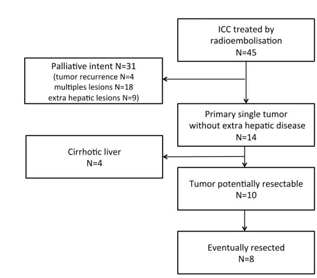
During the study period, 45 patients were treated with a combination of systemic
chemotherapy and intra-arterial Ytt-90 radioembolization for unresectable ICCs.
Twenty-seven (60%) patients had multiples lesions and/or extra-hepatic disease and were treated in a
palliative intent. Four (9%) patients were treated for tumor recurrence. Among the 14 (31%)
patients remaining, 4 had liver cirrhosis with portal hypertension. Eventually, 10 patients had
single huge ICCs that developed non-cirrhotic liver without extrahepatic disease and were
considered for potential resection. Ultimately, 8 patients (5 women and 3 men) underwent
surgical resection with curative intent and represent our study population. The 2 remaining
patients were not resected due to tumoral progression. (Figure 1)
Among these eight resected patients, the initial unresectabilities were due to tumoral
involvement of the hepatic veins (HV) of the FLR in seven cases and tumoral involvement of
the portal pedicle of the FLR in the remaining case. The demographic data and tumor
characteristics of the resected patients are reported in table 1.
Pre-operative treatment:
All eight patients received a median of seven cycles [range: 3 - 18] of systemic chemotherapy.
Chemotherapy associated gemcitabine (six cases) or 5-fluorouracil (two cases) with a
platinum salts (oxaliplatin in five cases, and cisplatin in three cases).
Intra-arterial Ytt-90 was administered with a median injected activity of 2 GBq [1.5-3.2]. Two
patients underwent two successive radioembolization procedures, in order to treat the entire
tumoral volume. The median duration of chemo-radioembolization therapy (i.e., the time
from the initiation of chemotherapy until surgery) was 7.6 months [3.4 – 16.7]. Tolerance was
Tumor downstaging and FLRV hypertrophy:
There was no difference between the pre- and post-treatment median TLVs (1530 ml
[1200-3580] versus 1372 ml [1255 – 2000]; p=0.16), and the tumor volume downsizing was
significant (295 ml [90-1250] versus 168 ml [46 – 535]; p=0.02). Figures 2 show three
representative cases of tumor shrinkage after treatment.
There was no significant difference between the pre- and post-treatment median FLRVs (467
ml [185 - 917] versus 507.5 ml [310 -795], respectively; p=0.46), while the median
FLRV/TLV ratio significantly increased (27.2% vs. 35.9%, p=0.04) and was greater than 30%
in 7 cases [30.0% – 49.7%]. The remaining patient had a pre-treatment FLRV/TLV ratio of
23.8%, which reached 38.5 % after PVE.
Surgical procedure:
The resections were performed in situ with the exception of one patient who underwent an ex
vivo surgery because complex vascular reconstruction was required. Extended hepatectomies
(i.e., resection of at least five segments) were performed in all cases including right
hepatectomies extended to segment 1 in two cases (25%), right trisectionectomy extended to
segment 1 in five cases (62.5%) and a left trisectionectomy in one case (12.5%).19
The biliary confluence and the extrahepatic biliary tree were resected in two cases with
bilio-enteric reconstruction. The retro-hepatic inferior vena cava (IVC) was resected in four cases
(laterally in two cases and totally in two cases with vascular prosthetic replacement). The
median duration of surgery was 210 min [150-600]. All in situ resections were performed
under continuous simultaneous clamping of the portal pedicle and the infrahepatic IVC. Three
(37.5%) patients required blood transfusion.
Postoperative course (table 2):
Three (37.5%) patients experienced Clavien-Dindo complication of grade 3 or more during
their hospital stays. Among these three patients, two died. One patient had a sudden massive
stroke on postoperative day 9 (1 day before planned discharge). The second patient had
undergone an ex vivo resection of the tumor with arterial, portal and suprahepatic vein
reconstructions and developed severe liver failure due to thrombosis of both the hepatic artery
and portal vein. Urgent liver revascularization did not prevent the fatal evolution at
postoperative day 4.
None of the remaining patients exhibited bile leakage or post-operative hemorrhage.
The median hospital stay duration was 9.5 days [4 – 21].
Midterm evolution and survival analysis
The median follow up time of the entire serie was 15.6 months [4 – 40.7] from the initiation
of medical treatment and 7.2 months [0.13 - 36.4] from the date of surgery.
Among the six patients who survived the postoperative period, one patient (aged 80 years old)
died 6.5 months after surgery (14 months after the initiation of treatment) without evidence of
initial disease recurrence, and five patients were still alive (figure 3). The median follow-up
for these six patients was 16.9 [15.2 – 40.7] months after the initiation of medical treatment
(8.2 [2 – 36.4] months after surgery).
Two patients experienced recurrences. One patient exhibited lung metastasis 19 months after
surgery and received chemotherapy and stereotaxic radiotherapy and is still alive 36.4 month
after the surgery (40.7 months after medical treatment initiation). The other patient exhibited
(15.1 month after the initiation of medical treatment). The median disease-free survival was
Discussion
ICC is a primary liver tumor whose incidence and mortality are increasing in western
countries.5 Complete surgical resection is the only available treatment that allows for
prolonged survival. For unresectable patients, survival without treatment is under one year.20
As the lesion remains clinically silent, ICCs are generally diagnosed at an advanced stage, and
only a minority of patients can be resected. Indeed, Tan et al.7 reported a global resection rate
of 12 %, with only 37% of the potentially resectable patients (defined by single unilobar
tumors without evidence of vascular invasion) who were eventually resected.
In the present study, of the 10 patients who were eligible for surgery, eight (80%) patients
with single localized ICCs who were initially considered unresectable were offered a
complete resection after the induction treatment of combined chemotherapy and Ytt-90
radioembolization.
For unresectable ICCs, systemic chemotherapy (associating Gemcitabine and platinum salt)
produces a response rate of 20-35% and a median overall survival of 10 - 15 month. 21-25 Such
low efficiency is also associated with a high rate of grade 3 - 4 toxicity.21 Ytt-90
radioembolization is a novel and promising therapy for the palliative treatment of ICC, and, to
our knowledge, five studies (two from the same team) have reported median overall survivals
of 9.3 to 22 months after treatment.13-16, 26 In a study by Ibrahim et al., 13 one patient was
downstaged to resection, and another patient was downstaged to liver transplantation. This
study was expanded to 46 patients,16 and three additional patients were eventually resected or
transplanted. Saxena et al.14 also reported one patient who was downstaged to surgery.
Reported tolerances of Ytt-90 radioembolization were excellent and usually better than other
intra-arterial treatment.27, 28 In our study, no patients presented severe toxicity (grade III or
was observed, we could consider that Ytt-90 radioembolization is a suitable treatment before
liver resection, as liver parenchyma seemed not to be altered even after multiple procedures.
Recently, Servajean et al.29 reported a case of a huge ICC with involvement of the three
hepatic veins that was initially non-resectable. After Ytt-90 radioembolization with resin
microspheres (SIRS-spheres®) combined with chemotherapy (eight cycles before and four
cycles after radioembolization), the patient underwent a left hepatectomy that was enlarged to
segment 8 with a free margin between the tumor and the right hepatic vein.
In our study, 8/10 patients were eventually resected. This high percentage compared to the
published results might have been because only solitary tumors and non-cirrhotic livers were
considered for surgery. Indeed, we did not offer extended liver resection to patients with
cirrhotic liver even when the FLRV was greater than 30%. For such patients, liver
transplantation might be a potential therapeutic option. Indeed, we believe that liver
transplantation was not an appropriate option for these patients because, in our series,
recurrence after surgery was primarily extra hepatic (i.e., lung localizations in the two
patients), which suggests that extra hepatic microscopic disease might have been present at
the time of surgery.
Although pre-operative treatment induced significant tumor volume reductions, R0 resection
could only be achieved through extended hepatectomy with the removal of at least five
segments, and half of the patients also had complex vascular reconstructions (including IVC
resection in 4 cases and ex-vivo surgery in 1 case). These factors might explain our
postoperative mortality rate. However, the mortality rate should be interpreted in light of Wang et al.’s nomogram, 30 which predicts 3-year survival rates below < 15% among patients
with similar huge ICCs with vascular involvements. Moreover, it should be noted that one of
After complete resection, ICC’s recurrence rate remained high, and recurrence typically
occurred within the first year after surgery. 8, 31 In our series, two of the six patients who were
alive after the postoperative course exhibited recurrence, but no deaths related to tumor
recurrence were observed. These findings might suggest that recurrences were managed more
efficiently after removal of the primary lesion, and a parallel could be drawn with
management of unresectable liver metastases from colorectal cancer.32
Recently, Ytt-90 radioembolization was also proposed as an alternative to PVE in HCC
cirrhotic patients.17 Our results support these findings because only one patient required PVE
prior to surgery to increase the FLRV/TLV ratio above 30%. However, in our series, the
increase in the FLR/TLV ratio was primarily due to liver tumor shrinkage rather than
hypertrophy of the FLRV.
Our series is currently the largest to report on secondarily resected patients with ICC after
downstaging with Ytt-90 radioembolization plus systemic chemotherapy but has some
limitations. First, our downstaging rate of 80% was biased by our patient’s selection criteria
as we only considered patients with unique tumor on non-cirrhotic liver and without
extra-hepatic disease, which represents only 10/45 (22%) patients. Second, our population included
only eight patients, and the median follow-up time remains limited. Therefore, we cannot yet
outline the oncological interest of the method. However, considering the time from the
initiation of chemotherapy, nearly all patients were followed up for more than 1 year, and one
patient remained alive 3 years after surgery. We intentionally did not compare the survival of
the 8 resected patients with the 37 remaining patients since these 2 groups were different
(unresected patients had generally multiple hepatic tumours and/or extrahepatic metastases).
In conclusion, our experiences is the largest serie that support the notion that Ytt-90
downstage huge, unique and initially non-resectable ICCs for secondary resections. Larger
numbers of patients and longer follow-up times are obviously required to further validate this
Acknowledgments :
M. Rayar contributed to study design, manuscript writing, data collection, statistical analysis
and manuscript reviewing.
L.Sulpice contributed to study design, manuscript writing and manuscript reviewing.
J.Edeline contributed to data collection and manuscript reviewing.
E.Boucher contributed to data collection and manuscript reviewing.
E.Garin contributed to data collection and manuscript reviewing.
G.B. Levi Sandri contributed to data collection and statistical analysis.
B. Meunier contribute to data collection and manuscript reviewing
K. Boudjema contribute to study design, manuscript writing, data collection and manuscript
REFERENCES
1. Endo I, Gonen M, Yopp AC, et al. Intrahepatic cholangiocarcinoma: rising frequency, improved survival, and determinants of outcome after resection. Ann Surg 2008; 248:84-96.
2. Rizvi S, Gores GJ. Pathogenesis, diagnosis, and management of cholangiocarcinoma. Gastroenterology 2013; 145:1215-29.
3. Siegel R, Ma J, Zou Z, Jemal A. Cancer statistics, 2014. CA Cancer J Clin 2014; 64:9-29. 4. Dodson RM, Weiss MJ, Cosgrove D, et al. Intrahepatic cholangiocarcinoma: management
options and emerging therapies. J Am Coll Surg 2013; 217:736-750 e4.
5. Patel T. Increasing incidence and mortality of primary intrahepatic cholangiocarcinoma in the United States. Hepatology 2001; 33:1353-7.
6. Farges O, Fuks D. Clinical presentation and management of intrahepatic cholangiocarcinoma. Gastroenterol Clin Biol 2010; 34:191-9.
7. Tan JC, Coburn NG, Baxter NN, et al. Surgical management of intrahepatic cholangiocarcinoma--a population-based study. Ann Surg Oncol 2008; 15:600-8.
8. Sulpice L, Rayar M, Boucher E, et al. Treatment of recurrent intrahepatic cholangiocarcinoma. Br J Surg 2012; 99:1711-7.
9. Nathan H, Pawlik TM, Wolfgang CL, et al. Trends in survival after surgery for
cholangiocarcinoma: a 30-year population-based SEER database analysis. J Gastrointest Surg 2007; 11:1488-96; discussion 1496-7.
10. Ohtsuka M, Ito H, Kimura F, et al. Results of surgical treatment for intrahepatic
cholangiocarcinoma and clinicopathological factors influencing survival. Br J Surg 2002; 89:1525-31.
11. Geschwind JF, Salem R, Carr BI, et al. Yttrium-90 microspheres for the treatment of hepatocellular carcinoma. Gastroenterology 2004; 127(5 Suppl 1):S194-205.
12. Vente MA, Wondergem M, van der Tweel I, et al. Yttrium-90 microsphere radioembolization for the treatment of liver malignancies: a structured meta-analysis. Eur Radiol 2009; 19:951-9. 13. Ibrahim SM, Mulcahy MF, Lewandowski RJ, et al. Treatment of unresectable
cholangiocarcinoma using yttrium-90 microspheres: results from a pilot study. Cancer 2008; 113:2119-28.
14. Saxena A, Bester L, Chua TC, et al. Yttrium-90 radiotherapy for unresectable intrahepatic cholangiocarcinoma: a preliminary assessment of this novel treatment option. Ann Surg Oncol 2010; 17:484-91.
15. Rafi S, Piduru SM, El-Rayes B, et al. Yttrium-90 radioembolization for unresectable standard-chemorefractory intrahepatic cholangiocarcinoma: survival, efficacy, and safety study. Cardiovasc Intervent Radiol 2013; 36:440-8.
16. Mouli S, Memon K, Baker T, et al. Yttrium-90 radioembolization for intrahepatic
cholangiocarcinoma: safety, response, and survival analysis. J Vasc Interv Radiol 2013; 24:1227-34.
17. Edeline J, Lenoir L, Boudjema K, et al. Volumetric changes after (90)y radioembolization for hepatocellular carcinoma in cirrhosis: an option to portal vein embolization in a preoperative setting? Ann Surg Oncol 2013; 20:2518-25.
18. Oken MM, Creech RH, Tormey DC, et al. Toxicity and response criteria of the Eastern Cooperative Oncology Group. Am J Clin Oncol 1982; 5:649-55.
19. Strasberg SM, Phillips C. Use and dissemination of the brisbane 2000 nomenclature of liver anatomy and resections. Ann Surg 2013; 257:377-82.
20. Chou FF, Sheen-Chen SM, Chen YS, et al. Surgical treatment of cholangiocarcinoma. Hepatogastroenterology 1997; 44:760-5.
21. Valle J, Wasan H, Palmer DH, et al. Cisplatin plus gemcitabine versus gemcitabine for biliary tract cancer. N Engl J Med 2010; 362:1273-81.
22. Kiba T, Nishimura T, Matsumoto S, et al. Single-agent gemcitabine for biliary tract cancers. Study outcomes and systematic review of the literature. Oncology 2006; 70:358-65.
23. Lee GW, Kang JH, Kim HG, et al. Combination chemotherapy with gemcitabine and cisplatin as first-line treatment for immunohistochemically proven cholangiocarcinoma. Am J Clin Oncol 2006; 29:127-31.
24. Knox JJ, Hedley D, Oza A, et al. Combining gemcitabine and capecitabine in patients with advanced biliary cancer: a phase II trial. J Clin Oncol 2005; 23:2332-8.
25. Andre T, Tournigand C, Rosmorduc O, et al. Gemcitabine combined with oxaliplatin (GEMOX) in advanced biliary tract adenocarcinoma: a GERCOR study. Ann Oncol 2004; 15:1339-43. 26. Hoffmann RT, Paprottka PM, Schon A, et al. Transarterial hepatic yttrium-90 radioembolization
in patients with unresectable intrahepatic cholangiocarcinoma: factors associated with prolonged survival. Cardiovasc Intervent Radiol; 35:105-16.
27. Hyder O, Marsh JW, Salem R, et al. Intra-arterial therapy for advanced intrahepatic cholangiocarcinoma: a multi-institutional analysis. Ann Surg Oncol 2013; 20:3779-86. 28. Benson AB, 3rd, Geschwind JF, Mulcahy MF, et al. Radioembolisation for liver metastases:
results from a prospective 151 patient multi-institutional phase II study. Eur J Cancer; 49:3122-30.
29. Servajean C, Gilabert M, Piana G, et al. One case of intrahepatic cholangiocarcinoma amenable to resection after radioembolization. World J Gastroenterol; 20:5131-4.
30. Wang Y, Li J, Xia Y, et al. Prognostic nomogram for intrahepatic cholangiocarcinoma after partial hepatectomy. J Clin Oncol; 31:1188-95.
31. Mavros MN, Economopoulos KP, Alexiou VG, Pawlik TM. Treatment and Prognosis for Patients With Intrahepatic Cholangiocarcinoma: Systematic Review and Meta-analysis. JAMA Surg 2014.
32. de Mestier L, Manceau G, Neuzillet C, et al. Primary tumor resection in colorectal cancer with unresectable synchronous metastases: A review. World J Gastrointest Oncol 2014; 6:156-69.
Figure titles and legends: Figure 1
Figure 1: Flow chart of the patients with unresectable ICCs who were treated with Ytt-90 radioembolization and resected in our institution.
Figure 2
Les paramètres nécessaires sont manquants ou erronés.Initial
CTscan Preop. CTscan Preop. Treatment 5Fluorouracyl + Cisplatin (6 cycles) Yttrium-90 (2.24 Bq) Time interval: 3 m Gemcitabine + Cisplatin (4 cycles) Yttrium-90 (2.0 Bq) Time interval : 5 m Capecitabine + Cisplatin (4 cycles) Yttrium-90 (1.95 Bq) Time interval:2 m A B C
Figure 2: Three representative cases of tumor downstaging from our series.
A: A 66-year-old woman with a 9-cm lesion. Initially, the tumor deeply infiltrated the right anterior section up to the median hepatic vein. After treatment, tumor downstaging and left liver hypertrophy allowed for the safe performance of a right hepatectomy that extended to segment 1 with partial resection of inferior retro-hepatic vena cava.
B: A 39-year-old man with a 20-cm lesion. Initially, the tumor was in contact with the origin of the left portal pedicle and involved the three hepatic veins. After treatment, significant downstaging of the tumor and hypertrophy of the left liver allowed for the performance of a R0 right trisectionectomy with resection of the inferior retro-hepatic vena cava.
C: A 70-year-old woman with a 6.5-cm-diameter central lesion. Initially, the tumor seemed to infiltrate the left portal pedicle. After treatment, the tumor shrinkage and liver remnant hypertrophy allowed for the performance of a right trisectionectomy that extended to segment 1 with biliary confluence resection.
Figure 3:
A
Figure 3: Overall survival of the resected patients. A: Overall survival from the day of surgery.
Table 1: Clinical features of the resected patients. Variables Age (years) * 68 [39 - 79] Sex ratio (M/F) 0.6 (3/5) BMI* 23 [19 - 27] ASA score 0 1 2 2 (25%) 4 (50%) 2 (25%) ECOG status 0 1 2 4 (50%) 4 (50%) 0 CA 19-9 level (KU/l) at time of diagnosis* 18.5 [10 – 1125] Tumor size* before treatment (cm) 9.2 [6.5 – 20] Vascular involvement of the FLR
Artery Portal vein Hepatic veins
Retro hepatic inferior vena cava Biliary confluence
Lymph node invasion hilar distant 0 1 (12.5%) 7 (87.5%) 4 (50%) 2 (25%) 3 (37.5%) 3 (37.5%) 2 (25%) Tumor distribution Unilobar Bilobar 3 (37.5%) 5 (62.5%) * median value [range].
Table 2: Operative and post-operative parameters. Variables Major resection right-side hepatectomy left-side hepatectomy 8 (100%) 7 (87.5%) 1 (12.5%) Number of segments resected* 6 [5 – 6] Duration (min)* 210 (150 -600) Transfusion 3 (37.5%) Bile leakage Liver failure Postoperative hemorrhage Ascitis Reoperation 0 2 (25%) 0 3 (37.5%) 2 (25%) Clavien classification: <3 3 4 5 5 (62.5%) 1 (12.5%) 0 2 (25%) Hospital stay duration (days)* 9.5 [4-21]