HAL Id: inserm-01322542
https://www.hal.inserm.fr/inserm-01322542
Submitted on 27 May 2016
HAL is a multi-disciplinary open access
archive for the deposit and dissemination of
sci-entific research documents, whether they are
pub-lished or not. The documents may come from
teaching and research institutions in France or
abroad, or from public or private research centers.
L’archive ouverte pluridisciplinaire HAL, est
destinée au dépôt et à la diffusion de documents
scientifiques de niveau recherche, publiés ou non,
émanant des établissements d’enseignement et de
recherche français ou étrangers, des laboratoires
publics ou privés.
Human lung fibroblasts may modulate dendritic cell
phenotype and function: results from a pilot in vitro
study
Olivia Freynet, Joëlle Marchal-Sommé, Francette Jean-Louis, Arnaud
Mailleux, Bruno Crestani, Paul Soler, Laurence Michel
To cite this version:
Olivia Freynet, Joëlle Marchal-Sommé, Francette Jean-Louis, Arnaud Mailleux, Bruno Crestani, et al..
Human lung fibroblasts may modulate dendritic cell phenotype and function: results from a pilot in
vitro study. Respiratory Research, BioMed Central, 2015, 17 (1), pp.36. �10.1186/s12931-016-0345-4�.
�inserm-01322542�
L E T T E R T O T H E E D I T O R
Open Access
Human lung fibroblasts may modulate
dendritic cell phenotype and function:
results from a pilot
in vitro study
Olivia Freynet
1,2,3,4†, Joëlle Marchal-Sommé
1,2,3†, Francette Jean-Louis
5, Arnaud Mailleux
1,2,3, Bruno Crestani
1,2,3,4,6*,
Paul Soler
1,2,3and Laurence Michel
5,7*Abstract
In human lung fibrotic lesions, fibroblasts were shown to be closely associated with immature dendritic cell (DC) accumulation. The aim of the present pilot study was to characterize the role of pulmonary fibroblasts on DC phenotype and function, using co-culture of lung fibroblasts from patients with idiopathic pulmonary fibrosis (IPF) and from control patients, with a DC cell line MUTZ-3. We observed that co-culture of lung control and IPF fibroblasts with DCs reduced the expression of specific DC markers and down-regulated their T-cell stimulatory activity. This suggests that pulmonary fibroblasts might sustain chronic inflammation in the fibrotic lung by maintaining in situ a pool of immature DCs.
Keywords: Lung, Fibroblasts, Dendritic cells, Fibrosis Findings
Background
We previously reported that in human fibrotic intersti-tial lung disease, immature DC accumulate in large numbers in areas of established fibrosis, in the close vicinity of fibroblasts expressing the main chemokines involved in the recruitment of immune cells [1–3]. Accumulating evidence indicates that the stromal micro-environment plays an important role in the regulation of the trafficking and phenotype and function regulation of immune cells, including DC [4, 5].
Fibroblasts and other stromal cells from various tissues have been shown to modulate DC differentiation and activation [6–8] and may induce the development/differen-tiation of DCs from blood monocytes or CD34+ hematopoietic precursors [9, 10]. Kitamura et al [11] reported that fibroblasts have a crucial role in regulating both fibrotic and immune responses in the lung by chemo-kine secretion, favoring dendritic cell trafficking, through
TGF-β αvβ8-mediated activation. Dermal fibroblasts were also reported to act as potent immunoregulatory cells able to modulate the maturation of DCs [12–14].
Herein, we aimed to characterizein vitro the influence of pulmonary fibroblasts on DC phenotype and function, using co-culture of human pulmonary fibroblasts obtained from patients with idiopathic pulmonary fibrosis (IPF) and from control patients, with immature DCs developed from the precursor cell line MUTZ-3 [15].
Materials
Primary fibroblasts
Primary fibroblasts were derived in Dulbecco’s modified Eagle’s medium (DMEM, Life Technologies-Invitrogen, Courtaboeuf, France) supplemented with 10 % FCS (Life Technologies-Invitrogen) from lung tissue biopsies ob-tained from 6 patients with IPF (age = 65.4 ± 10.6, 2 F), and from 6 patients with removal of a primary lung tumor without any fibrosis (control patients, age = 53 ± 14, 3 F), according to previous detailed procedures [16]. Fibroblasts were used at passage 5. No significant mor-phological and phenotypic differences were observed between IPF and control fibroblasts. Informed consent was obtained before lung sampling. The protocol was
* Correspondence:[email protected];[email protected] Bruno Crestani, Paul Soler and Laurence Michel are co-seniors.
†Equal contributors
1Inserm U 1152, 46, rue Henri Huchard, Paris 75018, France
5Inserm UMR-S 976, Université Paris Diderot, Sorbonne Paris Cité, Hôpital
Saint Louis, Paris, France
Full list of author information is available at the end of the article
© 2016 Freynet et al. Open Access This article is distributed under the terms of the Creative Commons Attribution 4.0 International License (http://creativecommons.org/licenses/by/4.0/), which permits unrestricted use, distribution, and reproduction in any medium, provided you give appropriate credit to the original author(s) and the source, provide a link to the Creative Commons license, and indicate if changes were made. The Creative Commons Public Domain Dedication waiver (http://creativecommons.org/publicdomain/zero/1.0/) applies to the data made available in this article, unless otherwise stated.
approved by the ethical committee of Paris-Ile de France 1.
MUTZ-3 dendritic cells
The CD34+ human MUTZ-3 progenitor cell line (ob-tained from DSMZ, Braunschweig, Germany) main(ob-tained in α-MEM (Life Technologies) supplemented with 20 % FCS and 0.25 ng/ml rhGM-CSF (Gentaur, Bruxelles, Belgium) express CD34 for 80 % and HLA-DR+for 70 %, as previously reported [17]. Generation of dendritic-like cells MUTZ-3 DCs was obtained by culturing MUTZ-3 progenitor (4×10⁵cells/ml) for 7 days in α-MEM with 20 % FCS, 10 ng/ml rhGM-CSF, 10 ng/ml rhIL-4 (Gentaur), and 0.5 ng/ml TNF-α (R&D Systems Europe, Lille, France) (MUTZ− 3 DC medium) [17].
Co-cultures
MUTZ-3 DCs (1×10⁶) were co-cultured with fibroblast monolayers (80–90 % confluence) for 48 h in MUTZ-3 DC medium either in 6-well tissue culture plates for direct contact or in Transwell® support. As controls, MUTZ-3 DCs and fibroblasts alone were cultured for 48 h under the same conditions.
Mixed Leukocyte Reaction
For mixed leukocyte reaction (MLR), human peripheral blood mononuclear cells were isolated from one leucocyte-enriched buffycoat, obtained from Etablisse-ment Français du Sang (Paris, France), by Lymphocyte Separation MediumM (GE Healthcare, PAA, Austria) density gradient centrifugation, cultured in RPMI 1640 (Gibco, Life Technologies) with 10 % heat inactivated human AB serum (RPMI complete medium) for 2 h in order to eliminate adherent mononuclear cells and re-suspended at 1×10⁶ cells/ml RPMI complete medium. MUTZ-3 DCs cultured alone or in the presence of fibro-blasts as indicated above were collected, washed once and resuspended in RPMI complete medium also. 2×10⁴ DC were cultured with 1×10⁵ allogeneic T cells in 96-well round-bottom plates, in a final volume of 200μl for each well. All determinations were performed in sixpli-cate. Cell proliferation was assessed for 6 days, with during the last 18 h of the culture period, addition of 1μCi of3H-thymidine (Perkin-Elmer Wallac, Courtabeuf, France) to each well. The cells were then harvested onto FilterMat filters (Wallac) by using the Harvester 96 cell Tomtec harvester, and incorporated radioactivity was determined using a MicroBeta plate reader (Wallac Trilux 1450 MicroBeta Liquid Scintillation Counter & Luminometer, WS-Trilux 1450).
Flow Cytometry
Cell surface staining was performed in phosphate-buffered saline (PBS, Life Technologies) (without calcium and
magnesium) containing 1 % bovine serum albumin (BSA) (Sigma-Aldrich) using fluorochrome-conjugated monoclo-nal antibodies recognizing MHC class II and DC-specific molecules: HLA-DR-PE (Pharmingen-BD, L’Isle d’Abeau, France), CD1a-PE, CD34-FITC, CD83-PE, CD86-PE, and CD209-PE (Immunotech-Beckman Coulter, Marseille, France) for 20 min at 4 °C. After two washes in PBS/BSA, the cells were fixed in formaldehyde 1 % and subsequently analyzed using an Epics cytometer (Beckman Coulter). Appropriate gating was used to exclude cell debris. Gating on isotype control staining was used to identify the per-centage of cells expressing DC-specific markers. Data were analyzed with FlowJo software (Tree Star, Inc., San Carlos, CA). Analysis of each epitope staining was assessed using both the percentage of positive cells (%) and the mean fluorescence intensity (MFI) of DCs.
Cytokine Production
Supernatants from MUTZ-3 DC and fibroblasts either cul-tured alone or in co-culture were centrifuged (1600 rpm, 10 min, 4 °C) and frozen at−70 °C until assayed using spe-cific enzyme-linked immunosorbent assays (ELISAs) from R&D Systems (Quantikine, R&D Systems, Abingdon, UK).
Statistical analysis
Data are presented as means (± SEM), or as individual values and median. Comparisons were performed with Wilcoxon paired nonparametric test for group compari-sons or Mann–Whitney U test. P values < 0.05 were considered significant. Statistical analysis was performed with Prism 5 (Graphpad Software Inc., La Jolla, CA USA).
Results
MUTZ-3 DCs obtained after a 7 day-differentiation culture (n = 12) were characterized by a low CD34 expression (<15 %), and the expression of CD209 (33.7 ± 1.6 %, mean ± SD) (marker of immaturity), HLA-DR (66.7 ± 2.8 %) and CD86 (59.5 ± 1 %). Few cells, if any, expressed the maturation marker CD83 (0.68 ± 0.23 %). These immature cells can undergo significant matur-ation by stimulmatur-ation with 25 ng/ml TNF-α during 48 h as detected by an increased expression of HLA-DR (88.6 ± 1.3 %p ≤ 0.001) and the induction of the matur-ation marker CD83 (47.2 ± 2.1 %).
Co-culture of lung fibroblasts with immature MUTZ3-DCs for 48 h induced a 20 % decrease of HLA-DR expres-sion and a 40 % decrease in CD83 and CD86 expresexpres-sion by DCs (Fig. 1). CD209 expression by DC was not signifi-cantly altered by co-culture (data not shown). It is note-worthy that IPF and control fibroblasts had a similar inhibitory effect on DCs.
In accordance with the decreased expression of activa-tion/maturation markers, co-culture of lung fibroblasts from either control or IPF patients and MUTZ-3 DCs
induced a significant 30 % reduction of lymphocyte allostimulatory activity as compared to that observed for MUTZ-3 DCs cultured alone (Table 1).
Lung fibroblasts might alter MUTZ-3 DC phenotype by either direct cell-cell contact or soluble factors. Of interest, MUTZ-3 DCs co-cultured with fibroblasts using modified Boyden chambers (Transwell® support) to prevent direct cell-cell contact did also show significant decrease in DC CD86 and CD83 markers (Fig. 1blower panels), decreases that were not significantly different from those ob-served in cell-cell contact conditions (upper panels).
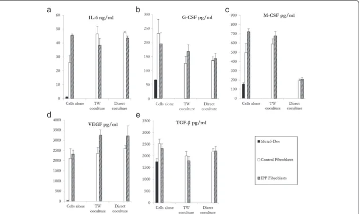
Numerous cytokines such as IL-6, G-CSF, M-CSF, VEGF, and TGF-β [18, 19] are produced by fibroblasts in the microenvironment and may inhibit MUTZ-3 DC maturation and function. Accordingly, we detected measurable levels of these cytokines in the culture supernatants of control and IPF fibroblasts, either alone or in co-culture (Fig. 2). Although IL6 production was lower in control fibroblasts alone compared to IPF ones, no differences were observed between the two fibroblast types when co-cultured with MUTZ3-DC, either in con-tact or not. Noteworthy, a significant decrease in MCSF production was detected for both populations when cultured in contact with MUTZ-3 cells.
Discussion
The present study demonstrates that lung fibroblasts co-cultured with DCs, differentiated from CD34+ MUTZ-3 precursors and closely resembling monocyte-derived DCs in terms of morphology, phenotype and function [15, 17, 20–22], induced a reduced MHC classII, CD86, and CD83 expression by DCs, with a decrease in their T-cell stimulatory activity. These findings support the notion that lung fibroblasts are effective in maintaining DCs in an immature state within fibrotic lesions. Our results are in line with re-cent data showing that human dermal fibroblasts are potent immunoregulatory cells inhibiting allogeneic T-cell activation by DCs [11]. Moreover, dermal fibro-blasts inhibited T-cell proliferation in response to DCs with similar potency to bone marrow-derived mesenchymal stem cells (MSC), suggesting that im-munosuppression is a general property of stromal cells [11, 23].
The absence of difference between IPF and control fibroblasts in our study suggests that the inhibitory effect of lung fibroblasts on DC differentiation and function is an intrinsic property of this cell type, independently of the physiological context. This possibility, which fits well with the recently documented immunoregulatory function of
(See figure on previous page.)
Fig. 1 Phenotype of MUTZ-3 DCs alone or cultured with control or IPF fibroblasts for 48 h. Immature MUTZ-3 DCs (1×10⁶) were directly added to fibroblasts monolayers grown to confluence in 6-well tissue culture plates (direct contact) or fibroblasts (low compartment) and MUTZ-3 DCs (high compartment) were separated using modified Boyden chambers (Transwell® permeable support-0.4μm) to prevent direct cell-cell contact, for 48 h in MUTZ-3 DC medium (Transwell condition). Controls were MUTZ-3 DCs cultured alone for 48 h in the same condition. Flow Cytometry analysis was assessed on DCs stained with monoclonal antibodies HLA-DR-PE (Pharmingen-BD), CD34-FITC, CD83-PE, CD86-PE, and CD209-PE (Immunotech-Beckman Coulter), and acquired with an Epics cytometer (Beckman Coulter). a One representative experiment or (b) percentage of positive cells for HLA-DR, CD86, and CD83 expression, represented as mean (dark horizontal bars) and individual values (n = 10) for direct contact co-cultures (upper panels) and Transwell® co-cultures (lower panels). MFI: Mean fluorescence intensity. **p < 0.01, ***p < 0.001. Statistical comparisons were performed with Wilcoxon paired nonparametric test for group comparisons or Kruskal-Wallis for unpaired nonparametric test, using Prism 5 (Graphpad Software Inc.)
Table 1 Mixed leukocyte reaction (MLR)
Conditions MUTZ-3 DC medium Contact Co-culture
medium Control Fibro IPF Fibro Lymphocyte Proliferation (mean cpm ± SD,n = 3) 56,870 ± 6110 40,236 ± 6047 34,322 ± 2843
p value (versus MUTZ-3 DC) 0.02 0.01
Conditions MUTZ-3 DC medium Transwell Co-culture
medium Control Fibro IPF Fibro Lymphocyte Proliferation (mean cpm ± SD,n = 3) 36,967 ± 4942 29,328 ± 3215 26,890 ± 6564
p value (versus MUTZ-3 DC) 0.04 0.05
MLR was performed by culturing with 100 000 lymphocytes (depleted of adherent mononuclear cells by a 2 h-lasting culture) and 20 000 MUTZ-3 DCs previously cultured alone (medium) or co-cultured with control or IPF fibroblasts for 48 h (as in Fig.1) in 200μl RPMI complete medium (n = 6 for each condition) for 6 days During the last 18 h, 1μCi of 3H-thymidine (Perkin-Elmer Wallac) was added per well. Incorporated radioactivity was determined using a MicroBeta plate reader (Wallac WS-Trilux 1450
Results are presented as mean (±SD) lymphocyte proliferation induced by MUTZ-3 DCs previously alone (medium) or co-cultured with control or IPF fibroblasts either in direct contact or in transwell conditions during 48 h before MLR
3H-thymidine incorporation of 105 lymphocytes alone was 605 ± 436 cpm and 3H-thymidine incorporation of 20 000 MUTZ3 DC alone was 138 ± 59 cpm, (mean ± SD,n = 6)
MSC [24], needs to be further assessed with fibroblasts isolated from various other tissues.
Conclusion
In conclusion, our findings suggest that lung fibroblasts by their ability to modulate DC differentiation and func-tion, are able to participate in pulmonary inflammatory and immune responses. Whereas their role in produc-tion of extracellular matrix proteins is a key factor in healing and remodeling, their likely interaction with lung DCs might be also considered as critical in inflammatory lung disorders.
Abbreviations
DC:Dendritic cells; IPF: Idiopathic pulmonary fibrosis; MSC: Mesenchymal stem cells.
Competing interests
The authors declare that they have no competing interests.
Authors’ contributions
OF conceived and performed the experiments, revised the manuscript. JM-S conceived and performed the experiments, analyzed the results, revised the manuscript. FJ-L performed the experiments. AM, BC, PS and LM conceived the experiments, analyzed the results, revised the manuscript. All authors read and approved the final manuscript.
Acknowledgements
This work was supported by the European Commission (FP7 project #202224, European IPF Network), by the Agence Nationale de la Recherche (ANR Physio 2006, FIBROPNEUMO) and by the Chancellerie des Universités de Paris (Legs Poix). Paul Soler was the recipient of a Contrat d’Interface Inserm - Assistance Publique/Hôpitaux de Paris. Olivia Freynet was supported by a grant from the Société de Pneumologie de Langue Française (SPLF).
Author details
1
Inserm U 1152, 46, rue Henri Huchard, Paris 75018, France.2Université Paris Diderot, Sorbonne Paris Cité, Paris, France.3DHU FIRE, Paris, France. 4Assistance Publique-Hôpitaux de Paris, Hôpital Bichat, Service de
Pneumologie A, Paris, France.5Inserm UMR-S 976, Université Paris Diderot,
Sorbonne Paris Cité, Hôpital Saint Louis, Paris, France.6Service de Pneumologie, Hôpital Bichat, 46, rue Henri Huchard, Paris cedex 18 75018, France.7Inserm UMR-S 976, Hôpital Saint-Louis, 1 avenue Claude Vellefaux,
75475, Paris 75010, France.
Received: 4 September 2015 Accepted: 9 March 2016
References
1. Lipscomb MF, Masten BJ. Dendritic cells: immune regulators in health and disease. Physiol Rev. 2002;82:97–130.
2. Marchal-Somme J, Uzunhan Y, Marchand-Adam S, Kambouchner M, Valeyre D, et al. Dendritic cells accumulate in human fibrotic interstitial lung disease. Am J Respir Crit Care Med. 2007;176:1007–14.
3. Marchal-Somme J, Uzunhan Y, Marchand-Adam S, Valeyre D, Soumelis V, et al. Cutting edge: nonproliferating mature immune cells form a novel type of organized lymphoid structure in idiopathic pulmonary fibrosis. J Immunol. 2006;176:5735–9.
4. Parsonage G, Filer AD, Haworth O, Nash GB, Rainger GE, et al. A stromal address code defined by fibroblasts. Trends Immunol. 2005;26:150–6.
a
b
d
e
c
Fig. 2 Production of the cytokines IL-6, G-CSF, M-CSF, VEGF and TGF-β by MUTZ-3 DC (dark), control fibroblasts (white) or IPF (dashed) fibroblasts, cultured either alone or in direct contact or using Transwell (TW) support. Mean levels (± SEM,n = 6) of (a) IL-6, (b) G-CSF, (c) M-CSF, (d) VEGF, and (e) TGF-β, assessed by ELISA after 48 h of both culture types. Cytokine levels were analysed using the normality test. The Mann-Whitney U and Studentt test were used to compare the mean values of cytokines between patients and control subjects. TW: transwell. *p < 0.05
5. Svensson M, Kaye PM. Stromal-cell regulation of dendritic-cell differentiation and function. Trends Immunol. 2006;27:580–7.
6. Berthier R, Rizzitelli A, Martinon-Ego C, Laharie AM, Collin V, et al. Fibroblasts inhibit the production of interleukin-12p70 by murine dendritic cells. Immunology. 2003;108:391–400.
7. Butler M, Ng CY, van Heel DA, Lombardi G, Lechler R, et al. Modulation of dendritic cell phenotype and function in an in vitro model of the intestinal epithelium. Eur J Immunol. 2006;36:864–74.
8. Zhang M, Tang H, Guo Z, An H, Zhu X, et al. Splenic stroma drives mature dendritic cells to differentiate into regulatory dendritic cells. Nat Immunol. 2004;5:1124–33.
9. Guironnet G, Dezutter-Dambuyant C, Gaudillere A, Marechal S, Schmitt D, et al. Phenotypic and functional outcome of human monocytes or monocyte-derived dendritic cells in a dermal equivalent. J Invest Dermatol. 2001;116:933–9.
10. Mollah ZU, Aiba S, Manome H, Yoshino Y, Tagami H. Cord blood CD34+ cells differentiate into dermal dendritic cells in co-culture with cutaneous fibroblasts or stromal cells. J Invest Dermatol. 2002;118:450–60.
11. Kitamura H, Cambier S, Somanath S, Barker T, Minagawa S, et al. Mouse and human lung fibroblasts regulate dendritic cell trafficking, airway inflammation, and fibrosis through integrinαvβ8-mediated activation of TGF-β. J Clin Invest. 2011;121:2863–75.
12. Haniffa MA, Wang XN, Holtick U, Rae M, Isaacs JD, et al. Adult human fibroblasts are potent immunoregulatory cells and functionally equivalent to mesenchymal stem cells. J Immunol. 2007;179:1595–604.
13. Saalbach A, Klein C, Sleeman J, Sack U, Kauer F, et al. Dermal fibroblasts induce maturation of dendritic cells. J Immunol. 2007;178:4966–74. 14. Saalbach A, Klein C, Schirmer C, Briest W, Anderegg U, et al. Dermal fibroblasts
promote the migration of dendritic cells. J Invest Dermatol. 2010;130:444–54. 15. van Helden SF, van Leeuwen FN, Figdor CG. Human and murine model cell
lines for dendritic cell biology evaluated. Immunol Lett. 2008;117:191–7. 16. Marchand-Adam S, Fabre A, Mailleux A, Marchal J, Quesnel C, et al.
Defect of pro-hepatocyte growth factor activation by fibroblasts in idiopathic pulmonary fibrosis. Am J Respir Crit Care Med. 2006;174:58–66. 17. Larsson K, Lindstedt M, Borrebaeck CA. Functional and transcriptional profiling
of MUTZ-3, a myeloid cell line acting as a model for dendritic cells. Immunology. 2006;117:156–66.
18. Gabrilovich D. Mechanisms and functional significance of tumour-induced dendritic-cell defects. Nat Rev Immunol. 2004;4:941–52.
19. Li MO, Wan YY, Sanjabi S, Robertson AK, Flavell RA. Transforming growth factor-beta regulation of immune responses. Annu Rev Immunol. 2006;24:99–146. 20. Masterson AJ, Sombroek CC, De Gruijl TD, Graus YM, van der Vliet HJ, et al.
MUTZ-3, a human cell line model for the cytokine-induced differentiation of dendritic cells from CD34+ precursors. Blood. 2002;100:701–703.21. 21. Santegoets SJ, Schreurs MW, Masterson AJ, Liu YP, Goletz S, et al. In vitro
priming of tumor-specific cytotoxic T lymphocytes using allogeneic dendritic cells derived from the human MUTZ-3 cell line. Cancer Immunol Immunother. 2006;2006(55):1480–90.
22. Lundberg K, Albrekt AS, Nelissen I, Santegoets S, de Gruijl TD, et al. Transcriptional profiling of human dendritic cell populations and models–unique profiles of in vitro dendritic cells and implications on functionality and applicability. PLoS One. 2013;8(1):e52875.
23. Li Q, Guo Z, Xu X, Xia S, Cao X. Pulmonary stromal cells induce the generation of regulatory DC attenuating T-cell-mediated lung inflammation. Eur J Immunol. 2008;38:2751–61.
24. Uccelli A, Moretta L, Pistoia V. Immunoregulatory function of mesenchymal stem cells. Eur J Immunol. 2006;36:2566–73.
• We accept pre-submission inquiries
• Our selector tool helps you to find the most relevant journal • We provide round the clock customer support
• Convenient online submission • Thorough peer review
• Inclusion in PubMed and all major indexing services • Maximum visibility for your research
Submit your manuscript at www.biomedcentral.com/submit