HAL Id: hal-02262531
https://hal-amu.archives-ouvertes.fr/hal-02262531
Submitted on 5 Aug 2019
HAL is a multi-disciplinary open access
archive for the deposit and dissemination of
sci-entific research documents, whether they are
pub-lished or not. The documents may come from
teaching and research institutions in France or
abroad, or from public or private research centers.
L’archive ouverte pluridisciplinaire HAL, est
destinée au dépôt et à la diffusion de documents
scientifiques de niveau recherche, publiés ou non,
émanant des établissements d’enseignement et de
recherche français ou étrangers, des laboratoires
publics ou privés.
Distributed under a Creative Commons Attribution| 4.0 International License
Coxiella burnetii
Soraya Mezouar, Joana Vitte, Laurent Gorvel, Amira Ben Amara, Benoit
Desnues, Jean-Louis Mege
To cite this version:
Soraya Mezouar, Joana Vitte, Laurent Gorvel, Amira Ben Amara, Benoit Desnues, et al.. Mast Cell
Cytonemes as a Defense Mechanism against Coxiella burnetii. mBio, American Society for
Microbiol-ogy, 2019, 10 (2), pii: e02669-18. �10.1128/mBio.02669-18�. �hal-02262531�
Mast Cell Cytonemes as a Defense Mechanism against Coxiella
burnetii
Soraya Mezouar,
aJoana Vitte,
a,bLaurent Gorvel,
cAmira Ben Amara,
cBenoit Desnues,
aJean-Louis Mege
a,b aAix-Marseille Université, IRD, APHM, MEPHI, IHU-Méditerranée Infection, Marseille, FrancebAPHM, IHU-Méditerranée Infection, UF Immunologie, Marseille, France
cCRCM, CNRS UMR7258, INSERM U1068, Institut Paoli-Calmettes, Aix-Marseille Université, UM105, Marseille, France
ABSTRACT
Mast cells (MCs) are critical mediators of inflammation; however, their
microbicidal activity against invading pathogens remains largely unknown. Here, we
describe a nonpreviously reported antibacterial mechanism used by MCs against
Coxiella burnetii, the agent of Q fever. We show that C. burnetii interaction with MCs
does not result in bacterial uptake but rather induces the formation of extracellular
actin filaments named cytonemes. MC cytonemes express cathelicidin and
neutro-phil elastase and mediate the capture and destruction of entrapped bacteria. We
provide evidence that MC cytoneme formation and microbicidal activity are
depen-dent on the cooperation of the scavenger receptor CD36 and Toll-like receptor 4.
Taken together, our results suggest that MCs use an extracellular sophisticated
mechanism of defense to eliminate intracellular pathogens, such as C. burnetii,
be-fore their entry into host cells.
IMPORTANCE
Mast cells (MCs) are found in tissues that are in close contact with
ex-ternal environment, such as skin, lungs, or intestinal mucosa but also in the placenta
during pregnancy. If their role in mediating allergic conditions is established, several
studies now highlight their importance during infection with extracellular
patho-gens. This study showed a new and effective antimicrobial mechanism of MCs
against Coxiella burnetii, an intracellular bacterium whose infection during pregnancy
is associated with abortion, preterm labor, and stillbirth. The data reveal that in
re-sponse to C. burnetii, MCs release extracellular actin filaments that contain
antimicro-bial agents and are capable to trap and kill bacteria. We show that this mechanism
is dependent on the cooperation of two membrane receptors, CD36 and Toll-like
re-ceptor 4, and may occur in the placenta during pregnancy by using ex vivo
placen-tal MCs. Overall, this study reports an unexpected role for MCs during infection with
intracellular bacteria and suggests that MC response to C. burnetii infection is a
pro-tective defense mechanism during pregnancy.
KEYWORDS
CD36, Coxiella burnetii, mast cells, TLR4, cytonemes
M
ast cells (MCs) are hematopoietic cells residing in tissues that occupy a strategic
position at host-environment interfaces, such as skin and mucosae. They are
characterized by a high content of electron-dense secretory granules containing
high-performance mediators, such as histamine, amines, serotonin, and proteases, such as
tryptase and chymase, and express CD117 (c-kit) and IgE (Fc
R1) receptors (1). They are
well-known as immune effectors of anaphylaxis, but their role in defending against
pathogens is emerging. MCs contribute to antibacterial immunity via multiple
mech-anisms. They are equipped with microbial sensors, such as Toll-like receptors (TLRs),
including TLR2 and TLR4, and are involved in the recognition of Gram-positive and
-negative bacteria (2–4). In response to lipopolysaccharide (LPS) or bacterial pathogens,
MCs release inflammatory cytokines that mediate the recruitment of immune cells and
Citation Mezouar S, Vitte J, Gorvel L, Ben
Amara A, Desnues B, Mege J-L. 2019. Mast cell cytonemes as a defense mechanism against
Coxiella burnetii. mBio 10:e02669-18.https://doi .org/10.1128/mBio.02669-18.
Editor Jon P. Boyle, University of Pittsburgh Copyright © 2019 Mezouar et al. This is an
open-access article distributed under the terms of theCreative Commons Attribution 4.0 International license.
Address correspondence to Jean-Louis Mege, jean-louis.mege@univ-amu.fr. Received 30 November 2018 Accepted 1 March 2019 Published 16 April 2019
RESEARCH ARTICLE
Host-Microbe Biology
crossm
®on August 5, 2019 by guest
http://mbio.asm.org/
Downloaded from
secrete antimicrobial agents, including cathelicidin (LL-37) and neutrophil elastase (5).
MCs are able to ingest and kill extracellular bacteria, such as Staphylococcus aureus,
through an endosome-lysosome pathway (6) or to use an extracellular antimicrobial
mechanism consisting of the release of extracellular traps (ETs) (7). Similarly to those
formed by neutrophils, these ETs are composed of antimicrobial peptide-lined DNA
projections, allowing the rapid immobilization and killing of microorganisms (7).
Coxiella burnetii is an intracellular bacterium responsible for Q fever, an acute
infectious disease that may become persistent in specific clinical contexts (8). In
myeloid cells, such as monocytes/macrophages and dendritic cells, C. burnetii survives
and replicates, but it has nevertheless been shown to infect other cell types, including
trophoblasts and adipocytes (9–12). C. burnetii is recognized by myeloid cells through
␣v3 integrin and TLR2/TLR4-dependent mechanism, leading to cytokine production
and cytoskeleton reorganization (13, 14). C. burnetii is also able to subvert immune
responses by interfering with uptake mechanisms and phagosome biogenesis (15),
stimulating the production of interleukin-10 (IL-10), and promoting the expansion of
regulatory T cells (13).
In the present study, we report that MCs were microbicidal for the intracellular
pathogen C. burnetii through a nonpreviously reported mechanism. Indeed, MCs have
released cytonemes consisting of long extensions of F-actin enriched with cathelicidin
and neutrophil elastase. These structures trapped and killed C. burnetii organisms.
Cytoneme-mediated killing of C. burnetii was under the control of the cross talk
between TLR4 and CD36. These results suggest that MCs have shaped an extracellular
sophisticated mechanism of defense to eliminate intracellular pathogens before their
entry in immune cells.
RESULTS
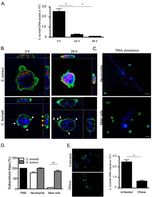
MCs kill C. burnetii through an extracellular mechanism. HMC-1.2 cells were
incubated with C. burnetii (50 bacteria per cell) for different periods of time, and the
number of bacterial DNA copies was determined by qPCR. After 3 h of infection, more
than 10 (7) C. burnetii DNA copies were detected. This number markedly decreased by
90% after 24 and 48 h (Fig. 1A). We questioned whether the decrease in the number of
bacterial DNA copies reflected the uptake and elimination of bacteria by MCs. We
assessed the uptake of C. burnetii by confocal microscopy, using S. aureus as the control.
S. aureus organisms were found within MCs at 3 h postinfection (p.i.), and the bacterial
burden increased at 24 h p.i. (Fig. 1B). In contrast, no C. burnetii organisms were found
within MCs at 3 and 24 h p.i, as opposed to monocytes which are permissive cells for
C. burnetii (see Fig. S1 in the supplemental material) as we previously described (16). It
is noteworthy that some C. burnetii organisms were observed at the surfaces of MCs
with intense F-actin rearrangements (Fig. 1B). The decreased number of C. burnetii DNA
copies and the defective uptake of bacteria suggested that an extracellular
antimicro-bial mechanism was employed by MCs to eliminate C. burnetii. Since ETs are used by
neutrophils and MCs to eliminate different types of bacteria (7), we quantified the
release of ETs in response to C. burnetii and S. aureus and used PMA as a positive
control. In neutrophils, C. burnetii and S. aureus induced a release of ETs similar to that
induced by PMA (Fig. 1C). In MCs, S. aureus triggered intense formation of ETs as in
neutrophils, whereas C. burnetii induced the release of few ETs (Fig. 1D). Therefore, we
wondered whether these rare traps were sufficient to eliminate C. burnetii organisms.
For that purpose, we treated C. burnetii-incubated MCs with DNase, which is known to
disrupt ETs (17). This treatment did not increase the number of intracellular C. burnetii
DNA copies, suggesting that another extracellular mechanism is involved in the
trap-ping and elimination of C. burnetii by MCs (Fig. 1E). Taken together, these results
suggested that MCs use an ET-independent extracellular mechanism to kill C. burnetii.
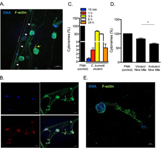
C. burnetii induces the formation of cytonemes by MCs. As C. burnetii markedly
remodeled MC cytoskeleton, we analyzed the F-actin rearrangements induced by
bacteria. We observed the release of extracellular thread-like membrane actin filaments
(Fig. 2A). These structures were predominantly linear and distinct from pseudopods.
on August 5, 2019 by guest
http://mbio.asm.org/
FIG 1 Extracellular killing of C. burnetii. HMC-1.2 cells (1⫻ 106) were incubated with C. burnetii (50 bacteria per cell) for different
periods of time. (A) After the cells were washed, the number of C. burnetii DNA copies was determined by qPCR. (B) Confocal sections of MCs incubated with S. aureus (top panel) or C. burnetii (bottom panel). Bacteria (red), F-actin (green), nucleus (blue), and cytoskeletal reorganization (white arrows) are indicated. Bacteria located on the MC membrane are represented by yellow arrows. (C and D) The extracellular traps released by neutrophils or MCs incubated with PMA, C. burnetii, or S. aureus for 3 h were observed (DNA in blue and F-actin in green) (C) and quantified by evaluating the release of fluorescent DNA (D). The results, expressed as relative to PMA-stimulated cells, are the means plus standard deviations (SD) (error bars) for triplicate samples from at least three independent experiments.**, Pⱕ 0.01. (E) The number of C. burnetii DNA copies was determined by qPCR in MCs incubated with bacteria for 3 h and then treated with 50 U/ml DNase for 10 min to eliminate ETs. The results are expressed as the means plus SD for triplicate samples from at least three independent experiments.*, Pⱕ 0.05.
Mast Cell Cytonemes and Coxiella burnetii Clearance ®
on August 5, 2019 by guest
http://mbio.asm.org/
They measured up to 200
m in length and were composed of F-actin and tubulin
(Fig. 2B), suggesting that they were similar to the cytonemes produced by MCs as
previously described (18). The cytonemes appeared 15 min after C. burnetii stimulation;
the number of cytonemes reached a maximum between 3 and 6 h (
ⱖ50%) and
decreased thereafter (Fig. 2C). Several strains of C. burnetii have been described: they
include the Nine Mile strain (the reference strain) and Guyana strain (the most virulent).
We previously observed intense cell projections from monocytes stimulated by virulent
C. burnetii compared to the avirulent variant (19). We wondered whether cytoneme
formation was related to the virulence of C. burnetii. Avirulent variants of the Nine Mile
strain poorly induced the formation of cytonemes compared to the virulent Nine Mile
strain (Fig. 2D). Interestingly, the Guyana strain induced a significant increase of
cytonemes formation compared to the PMA control (Fig. S2).
As the formation of cytonemes was observed with a MC line, we wondered whether
primary MCs responded similarly to C. burnetii. Therefore, we purified MCs from
placenta, a tissue for which C. burnetii has a strong tropism (20). The placental MCs
(pMCs) were identified by flow cytometry using Fc
R1
⫹/CD117
⫹(Fig. S3A) and tryptase
staining (Fig. S3B). These pMCs were characterized by an ovoid nucleus, an irregular
membrane, a metachromatic staining of granules, and the presence of several
tryptase-FIG 2 C. burnetii-induced formation of cytonemes. (A) Cytonemes (white arrows, distance 208.4m) and pseudopods (yellow arrow, distance 11.1m) stained with indicated markers were observed by confocal microscopy in MCs stimulated for 3 h. (B) Tubulin (red), F-actin (green), and DNA (blue) staining were evaluated on cytonemes. (C) The formation of cytonemes by MCs was quantified after stimulation with C. burnetii and expressed as a percentage relative to PMA as the positive control. (D) The formation of cytonemes was quantified after a 3-h incubation of MCs with virulent (Nine Mile) and avirulent (Nine Mile variant) bacteria. The results are expressed as a percentage relative to PMA stimulation. (E) The release of cytonemes by pMCs stimulated by C. burnetii for 3 h was observed by confocal microscopy, stained with the indicated markers. The results are expressed as the means plus SD for triplicate samples from four independent experiments.*, Pⱕ 0.05.
on August 5, 2019 by guest
http://mbio.asm.org/
positive cytoplasmic granules using MGG and toluidine blue (Fig. S3C), electron
mi-croscopy (Fig. S3D) and immunofluorescence (Fig. S3E). As observed for the MC cell line,
C. burnetii also induced the formation of cytonemes in primary MCs (Fig. 2E).
Alto-gether, these findings show that C. burnetii induces the formation of cytonemes by MCs
and suggest that this phenotype depends, at least in part, on the virulence of the
bacteria.
MC cytonemes capture and kill bacteria. In order to understand the role of
cytonemes in the MC response to C. burnetii, we incubated MCs with bacteria. We found
that organisms were entrapped in cytonemes (Fig. 3A). After 3 h of contact between
MCs and bacteria, approximately 20% of C. burnetii organisms were already dead and
this number reached 80% after 6 h with the use of propidium iodide staining (Fig. 3B
and C), suggesting that cytonemes were involved in C. burnetii killing. As MCs are
known to secrete several antimicrobial products, we investigated their presence in
cytonemes. MCs were incubated with C. burnetii for 3 h, and the distribution of
antimicrobial agents, such as cathelicidin or neutrophil elastase, and extracellular
F-actin, was studied by confocal microscopy. We found that both cathelicidin and
FIG 3 Cytonemes trap and kill C. burnetii. HMC-1.2 cells were incubated with C. burnetii (Nine Mile strain) for 3 h. (A) Bacteria (white arrows), colocalizing with cytonemes, appeared in yellow. (B) Bacteria entrapped in cytonemes were indicated with white arrows. Their viability was studied by immunofluo-rescence microscopy: Live bacteria were stained with Sytox 9 and appeared in green, whereas dead bacteria were stained with propidium staining and appeared in red. (C) The viability of bacteria entrapped in cytonemes was quantified at 3 and 6 h p.i. (D) Cathelicidin and neutrophil elastase were stained with specific Abs and were observed in red on MC cytonemes infected by C. burnetii with F-actin labeled in green and DNA in blue. (E) The percentage of cytonemes induced by heat-inactivated bacteria is expressed relative to the number of cytonemes induced by living bacteria. (F) The heat-inactivated bacteria were labeled in red within MCs (white arrows). F-actin and DNA are shown in green and blue, respectively. Data are the means plus SD for triplicate samples from three independent experiments.**, Pⱕ 0.01.Mast Cell Cytonemes and Coxiella burnetii Clearance ®
on August 5, 2019 by guest
http://mbio.asm.org/
neutrophil elastase colocalized with cytonemes (Fig. 3D), demonstrating that
cy-tonemes were armed to kill entrapped bacteria.
Second, we wondered whether cytonemes may protect MCs by avoiding the
internalization of virulent organisms. When MCs were incubated with heat-inactivated
C. burnetii instead of living bacteria, cytoneme formation was significantly (P
⫽ 0.0042)
reduced (Fig. 3E). Interestingly, we found that heat-inactivated bacteria were found
inside MCs (Fig. 3F), suggesting that cytoneme formation was associated with restricted
uptake of bacterial pathogens. Taken together, these results highlight the role of
cytonemes in killing virulent C. burnetii and represent another way other than
phago-cytosis in MCs.
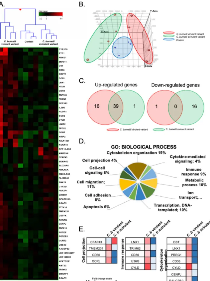
Specific transcriptomic signature of C. burnetii-stimulated MCs. To understand
the molecular pathways involved in the formation of MC cytonemes, we studied the
transcriptional signature of MCs stimulated with C. burnetii by whole-genome
microar-ray. Hierarchical clustering revealed a specific pattern for MCs stimulated with virulent
bacteria that induced the formation of cytonemes, whereas MCs stimulated with an
avirulent variant clustered with unstimulated MCs (Fig. 4A). Principal-component
anal-ysis confirmed that the signatures of MCs, stimulated with virulent and avirulent
bacteria, were clearly distinct (Fig. 4B). We found that 56 genes were differentially
expressed in response to C. burnetii, and most of these genes were upregulated in
response to virulent organisms, whereas downregulated genes were prominent in
response to the avirulent variant (Fig. 4C). Genes involved in several biological
pro-cesses were enriched using the Gene Ontology (GO) Consortium approach. They
included genes involved in cytoskeleton organization, cytokine-mediated signaling,
immune response, metabolic process, ion transport, transcription, apoptosis, cell
ad-hesion, cell-cell signaling, and cell projection (Fig. 4D). Therefore, to validate our
findings, we selected 10 genes (TMEM231, OCRL, CYLD, IL36G, TRIM62, LNX1, DST, PRRG1,
CENPJ, and RALGPS2) for which we assessed the modulation by qRT-PCR (see Table S2
in the supplemental material).
Interaction of CD36 with TLR4 in C. burnetii cell infection. Among the modulated
genes, we found that the gene encoding CD36 was found in three GO terms, including
cell projection, immune response, and cytoskeleton organization, and its expression
was upregulated in response to virulent bacteria compared to the avirulent variant
(Fig. 4E). The analysis of CD36 expression by qRT-PCR and flow cytometry confirmed
microarray data and showed that CD36 expression was increased only in MCs
stimu-lated with virulent bacteria (Fig. 5A and B). The confocal microscopy analysis revealed
that CD36 was overexpressed as membrane clusters in MCs stimulated with C. burnetii
(Fig. 5C). In addition, CD36 colocalized with C. burnetii at the surfaces of MCs (Fig. 5D).
As CD36 is known to cooperate with TLRs to clear microbial infection (21) and TLR4 has
been involved in actin remodeling in myeloid cells during C. burnetii infection (22), we
investigated the expression of TLR2 and TLR4 in stimulated MCs. The expression of the
TLR4 gene, but not that of the TLR2 gene, was dramatically upregulated in response to
C. burnetii, as measured by qRT-PCR (Fig. 5E) and flow cytometry (Fig. 5F). In a second
step, we wondered whether CD36 and TLR4 were associated in MCs stimulated by C.
burnetii. Image overlay obtained by confocal microscopy showed that membrane CD36
and TLR4 colocalized at the surfaces of MCs incubated with C. burnetii (Fig. 5G). The
direct interaction between CD36 and TLR4 was then assessed by immunoprecipitation
experiment. We found that CD36 immunoprecipitated with TLR4 with a maximal
intensity 1 h after incubation of MCs with C. burnetii (Fig. 5H). The cross talk between
CD36 and TLR4 was further confirmed by inhibition experiments. First, inhibition of
CD36 with blocking Abs (Fig. 5J) or transfection of MCs with a siRNA directed
against CD36 (Fig. S4), significantly reduced the expression of TLR4 in stimulated MCs
(Fig. 5I). In addition, polymyxin B, an inhibitor of LPS binding, reduced TLR4 and CD36
expression of C. burnetii-stimulated cells. Finally, in TLR4-deficient BMdMCs stimulated
by C. burnetii, the expression of CD36 was severely impaired compared to wild-type
on August 5, 2019 by guest
http://mbio.asm.org/
FIG 4 Transcriptional signature of MCs in response to C. burnetii. MCs were stimulated with C. burnetii (50 bacteria per cell) or left untreated for 8 h, and the total RNA was extracted prior to microarray analysis. (A) Up- and downregulated genes are indicated in red and green, respectively. (B) The
(Continued on next page)
Mast Cell Cytonemes and Coxiella burnetii Clearance ®
on August 5, 2019 by guest
http://mbio.asm.org/
BMdMCs (Fig. 5K). These results indicated a direct cooperation between TLR4 and CD36
in the response of MCs to C. burnetii.
Role of CD36/TLR4 complex in cytoneme formation. Since the CD36/TLR4
coop-eration is initiated by the interaction of C. burnetii with MCs, we wondered whether the
CD36/TLR4 complex was involved in the formation of cytonemes. We therefore
mea-sured the cytoneme formation in response to C. burnetii in the presence of CD36
inhibitors. CD36 siRNA or CD36 blocking Abs inhibited cytoneme formation (Fig. 6A).
Similarly, polymyxin B that inhibited CD36 expression (Fig. 5I) also prevented the
formation of cytoneme. Finally, in TLR4-deficient BMdMCs stimulated by C. burnetii, an
inhibition of 85% of cytoneme formation was observed (Fig. 6B). We then confirmed
these results in primary MCs and showed that the treatment of pMCs with polymyxin
B or CD36 blocking Abs inhibited cytoneme formation (Fig. 6C). Taken together, these
results indicate that the release of cytoneme by MCs depends on cooperation between
CD36 and TLR4 in response to the challenge of C. burnetii.
DISCUSSION
We have demonstrated here an antibacterial mechanism of MCs based on
cy-tonemes that had never been reported before. It is established that bacteria are killed
by innate immune cells via numerous intra- and extracellular mechanisms. The former
are based on phagocytosis and phagosomal maturation, whereas the latter involve the
formation of ETs and the release of bactericidal compounds (23). Microorganisms,
including intracellular bacteria, have developed various strategies to subvert innate
immune responses. Indeed, C. burnetii successfully infects macrophages by controlling
phagocytosis and phagosome biogenesis (15, 24). We observed that MCs were able to
kill C. burnetii without internalizing it, suggesting an extracellular microbicidal
mecha-nism. This result is markedly distinct from S. aureus, which is killed by MCs via
internalization and ET formation (6). While C. burnetii induces the formation of ETs as
efficiently as S. aureus in neutrophils, C. burnetii is a poor inducer of ETs in MCs, which
appeared insufficient to kill bacteria.
We also provided evidence that MCs use cytonemes for a microbicidal effect toward
C. burnetii. Cytonemes were initially associated with cell-cell communication (25, 26).
Although these cytoskeletal structures may participate in host defense, their specific
antimicrobial role has not been described thus far. Cytonemes have been implicated in
cell-to-cell spreading of virions, such as human immunodeficiency virus type 1 and
human T cell leukemia virus type 1 (HTLV-1) (27, 28, 29). Recently, Hashimoto et al.
reported that macrophage cytonemes enable the rapid transfer of HTLV-1 to
surround-ing cells (30). In contrast, neutrophil cytonemes are involved in the tethersurround-ing of
bacteria, such as S. aureus, Salmonella enterica serovar Typhimurium, and Helicobacter
pylori. This capture of bacteria by neutrophil cytonemes allows internalization and
subsequent intracellular destruction (31). The microbicidal activity of neutrophil
cy-tonemes is probably based on the release of bactericidal molecules, such as lactoferrin,
lipocalin, myeloperoxidase, defensins, and cathepsin G (32). We reported here that
cytonemes entrapped C. burnetii and reduced its viability. The latter response is likely
mediated by elastase and cathelicidin, which colocalized with bacteria within
cy-tonemes. We also discovered that cytoneme-mediated killing of C. burnetii is associated
with the absence of phagocytosis, whereas cytonemes and phagocytosis are
associ-ated, during the interaction of S. aureus, with MCs. The dissociation of cytoneme
formation from phagocytosis was related to bacterial viability, since inactivated bacteria
were internalized and did not induce significant cytoneme formation. The prevention
FIG 4 Legend (Continued)
relative distance between MCs stimulated with virulent C. burnetii (red), avirulent bacteria (green), or resting cells (blue) was assessed using principal-component analysis. (C) Venn diagrams showed the distribution of upregulated (left) and downregulated (right) genes in MCs stimulated with virulent C. burnetii (red) or avirulent variants (green). (D) Transcriptional analysis of modulated genes revealed several GO terms of biological process. (E) A modular analysis of the cell projection, immune response, and cytoskeletal organization of GO terms showed the genes involved and their modulation (up- and downregulation in red and blue, respectively).
on August 5, 2019 by guest
http://mbio.asm.org/
FIG 5 Involvement of CD36 and TLR4 in the response of MCs to C. burnetii. MCs were stimulated with C. burnetii or left untreated, and the modulation of the CD36 and TLR4 genes or encoded proteins was determined. (A) The expression of the gene encoding CD36 was evaluated in MCs stimulated by C. burnetii for 8 h and normalized to the value for unstimulated cells. (B) The expression of CD36 protein was determined by flow cytometry with FITC-conjugated anti-CD36 on MCs stimulated with C. burnetii virulent and avirulent variants for 3 h. The results are expressed as mean fluorescence intensity. (C) Confocal microscopy revealed that the expression of CD36 (green) was essentially expressed at the membrane. (D) The staining of CD36 (green) and C. burnetii (red) showed that they colocalized (in yellow) at the MC membrane. (E) The expression of TLR2 and TLR4 transcripts was evaluated in MCs stimulated by C. burnetii for 8 h by qRT-PCR and normalized to the value for unstimulated cells. (F) The modulated expression of TLR4 protein in response to C. burnetii was determined by flow cytometry and expressed as mean fluorescence intensity. (G) Confocal microscopy showed that TLR4 (red) and CD36 (green) colocalized (yellow) in C. burnetii-stimulated MCs (DNA appeared in blue). (H) MCs were stimulated with C. burnetii for different periods. Immunoprecipitations were immunoblotted with TLR4, CD36, or irrelevant Abs. (I and J) The expression of the CD36 (I) and TLR4 (J) genes was evaluated by qRT-PCR in MCs stimulated with C. burnetii for 8 h and treated with CD36 blocking Abs or polymyxin B or after siRNACD36 transfection. The results are normalized to the values for untreated MCs. (K) The expression of CD36 gene expression was assessed in TLR4⫺/⫺BMdMCs and normalized to the value
(Continued on next page)
Mast Cell Cytonemes and Coxiella burnetii Clearance ®
on August 5, 2019 by guest
http://mbio.asm.org/
of phagocytosis in MCs is an active process reminiscent of what we reported in
macrophages and monocytes infected with virulent C. burnetii (19, 33). Indeed, C.
burnetii induced cytoskeletal reorganization of macrophages, such as F-actin
protru-sions, which was associated with phagocytosis prevention (10, 22). Manipulation of the
cytoskeleton organization in macrophages restores phagocytosis, thus establishing a
direct link between cytoskeleton modulation and phagocytosis interference (33, 34).
We hypothesize that C. burnetii-induced cytonemes alert the immune system since it
has been reported that externalized F-actin acts as a danger signal (35). Finally, in line
with Manfredi et al. who proposed a choice between phagocytosis and generation of
ETs for neutrophils during inflammation and infection (36), we think that MCs make a
choice between MC formation and phagocytosis in the context of C. burnetii infection.
Our study also described that a functional cooperation between CD36 and TLRs is
necessary for cytoneme formation. Indeed, we provide evidence that CD36 associates
with TLR4, thus forming a molecular complex that is involved in the production of
cytonemes and entrapping C. burnetii. This is consistent with previous reports in which
CD36 mediated signal transduction for TLR4 (37, 38, 39). The role of CD36 in the
immune response to microorganisms remains unclear. Some studies evoked a direct
role of CD36 in inflammatory response or in pathogen recognition (40, 41). Indeed,
Stuart et al. reported that CD36 may be a bacterial receptor for S. aureus, its cytoplasmic
C-terminal extremity being involved in bacterial internalization (42). The transfection of
CD36 into HeLa cells enhances the uptake of bacteria, including Escherichia coli,
Klebsiella pneumoniae, S. Typhimurium, S. aureus, and Enterococcus faecalis (40). In
addition, CD36 is involved in the internalization of LPS by endothelial or MCs (25, 43,
FIG 5 Legend (Continued)
for wild-type BMdMCs. Data are the means plus SD for triplicate samples from four independent experiments. *, Pⱕ 0.05; **, P ⱕ 0.01.
FIG 6 CD36/TLR4 cooperation. MCs were stimulated with C. burnetii for 3 h. (A) The percentage of cytonemes was calculated after treatment of stimulated MCs during 10 min with MDC, polymyxin B, CD36 blocking Abs, or after siRNA CD36 transfection compared to untreated cells. (B) The percentage of cytonemes was calculated in TLR4-deficient BMdMCs compared to wild-type BMdMCs. (C) Placental MCs from healthy donors were collected after enzymatic digestion, Percoll cushion procedure, and magnetic selection. The release of cytonemes by pMCs stimulated by C. burnetii for 3 h were quantified after polymyxin B sulfate or CD36 blocking antibody treatments. Data are the means⫾ SD for triplicate samples and are representative of four independent experiments. **, Pⱕ 0.01.
on August 5, 2019 by guest
http://mbio.asm.org/
44). Future studies are required to address the precise mechanism underlying CD36/
TLR4 cross talk during C. burnetii infection.
The formation of cytonemes in response to C. burnetii was not restricted to MC lines
or murine MCs derived from bone marrow. It was also observed in primary MCs isolated
from the human placenta. C. burnetii is known for its tropism for placenta tissue, and
Q fever is a major risk for pregnancy and impaired fetal development (20). We
previously described that C. burnetii interacts with dendritic cells from placenta and
replicates in trophoblasts (11, 45). Here, we report that MCs from placenta present an
extracellular mechanism to kill bacteria in the early phases of C. burnetii infection. The
role of MCs in promoting host resistance in bacterial infection is documented (46–48).
Thus, we can speculate that during C. burnetii infection, MCs play a role of protection
at the placenta level by intercepting extracellular pathogens and limiting abortion. This
finding may also explain why trophoblasts constitute a niche for C. burnetii (11).
However, these hypotheses have to be clarified by careful examination of organism
distribution in the naturally infected placenta in order to provide insights into the roles
of individual cell types in abortion during Q fever.
In conclusion, our data described a new extracellular bactericidal mechanism based
on the release of cytonemes by MCs. Cytonemes were involved in the capture of
virulent C. burnetii and the destruction of entrapped bacteria mediated by antimicrobial
peptides. We also showed that the formation of cytonemes requires the CD36/TLR4
complex. This report opens new perspectives in the antimicrobial activity of MCs and
provides new insights into the role of cytonemes in the immune response.
MATERIALS AND METHODS
Cells. The human mast cell line HMC-1.2 was generously provided by M. Arock (Paris, France) and cultured in Iscove’s modified Dulbecco’s medium (IMDM) (Life Technologies) supplemented with 10% fetal bovine serum (FBS), 100 IU/ml penicillin, and 50g/ml streptomycin (Life Technologies) at 37°C. In some experiments, placental MCs (pMCs) were isolated. Placenta from at-term healthy women were included after providing written informed consent and after approval was granted from the Comité d’Ethique d’Aix Marseille Université (number 08-012). Placenta tissue was digested with trypsin, and the cell suspension was deposited on a 25 to 60% Percoll cushion and centrifuged as previously described (49). Placental cells were collected, and pMCs were enriched by positive selection after FcR1/CD117 staining (Miltenyi Biotec). In some experiments, bone marrow-derived MCs (BMdMCs) were differentiated as previously described (50). Briefly, wild-type mice were obtained from Charles River Laboratories, and TLR4-deficient mice were generously provided by L. Alexopoulou (Marseille, France). Bone marrow cells were flushed and incubated with IMDM containing 15% FBS, 10 ng/ml IL-3, and 10 ng/ml stem cell factor (Miltenyi Biotec). After 4 weeks of culture, the differentiation into BMdMCs was checked by flow cytometry using CD117 and FcR1 as specific markers. About 98% of cultured cells were BMdMCs (data not shown). Human neutrophils and monocytes were isolated from blood samples from three healthy donors (Établissement Français du Sang, Marseille, France) using Percoll or Ficoll gradient, respectively, and incubated in RPMI 1640 (Life Technologies), as previously described (17).
Bacteria. Bacteria (Nine Mile and Guyana strains of C. burnetii) were prepared as previously reported (12). Avirulent variants of Nine Mile bacteria were obtained after repeated passages in L929 cells. Bacteria were stored at⫺80°C, their concentration was determined by Gimenez staining, and bacterial viability was assessed using the live/dead BacLight bacterial viability kit (Molecular Probes, Life Technologies) (51). Bacteria were inactivated at 95°C for 30 min. S. aureus (ATCC 25923) bacteria were grown on blood agar plates (bioMérieux) and quantified by flow cytometry (FACS BD Fortessa).
Cell stimulation. MCs and neutrophils (1⫻ 106cells/well) were incubated in 24-well plates
pre-treated with fibronectin (1 mg/well; Life Technologies) for 3 h. Adherent cells were stimulated with 100g/ml LPS, 25 nM phorbol-12-myristate-13-acetate (PMA) (MP Biomedicals), or bacteria (bacterium-to-cell ratio of 25 and 50 bacteria per cell for S. aureus and C. burnetii, respectively) at 37°C. The roles of CD36 and TLR4 were studied using MCs pretreated with 5g/ml CD36-blocking antibodies (Abs) (mouse IgG2a; Thermo Fisher Scientific) or 10g/ml of the TLR4 inhibitor polymyxin B sulfate (Sigma-Aldrich) for 10 min.
Bacterial detection. C. burnetii organisms were detected by quantitative PCR (qPCR) and immuno-fluorescence as previously described (12). The total DNA was extracted using the NucleoSpin kit (Macherey-Nagel). Quantitative PCR was performed using Sybr Green Technologies using a CFX (Bio-Rad) with 5l of DNA and specific primers targeting C. burnetii com1 gene: sense (5=-GCACTATTTTTAGCCG GAACCTT-3=) and antisense (5=-TTGAGGAGAAAAACTGGATTGAGA-3=). C. burnetii organisms were also detected by immunofluorescence. In brief, MCs were fixed with 3% paraformaldehyde, permeabilized with 0.1% Triton X-100 for 5 min, and incubated with a 1/100 dilution of Q fever patient serum (11). After washing, MCs were incubated with Alexa Fluor 647-conjugated Abs. S. aureus was labeled with the fluorochrome DID (4-(4-(dihexadecylamino)styryl)-N-methylpyridinium iodide) (Thermo Fisher Scientific) for 20 min at 37°C.
Mast Cell Cytonemes and Coxiella burnetii Clearance ®
on August 5, 2019 by guest
http://mbio.asm.org/
Cytonemes and extracellular traps. The quantification of ET release was based on the evaluation of the area of labeled extracellular DNA filaments using an Axio Scan coupled with Hamamatsu sCOMS Flash 4 camera (Zeiss). The area of extracellular DNA of ETs was quantified on five different fields as previously described (52, 53). The results are expressed as a percentage relative to the PMA-stimulated cells as a positive control (cells stimulated with PMA) (54). Similarly, the evaluation of cytoneme formation over time was realized by assessment of area of extracellular F-actin filaments and expressed as a percentage relative to the PMA control, as depicted in Fig. S5 in the supplemental material. In some experiments, stimulated MCs were treated with 50 U/ml DNase (Sigma-Aldrich) for 30 min at the end of the experiment to disrupt ETs as previously described (17). The cytonemes were quantified using a similar method based on the release of extracellular F-actin labeled with phalloidin-488. To inhibit the formation of cytonemes, MCs were pretreated with 10 mM methyl--D-cyclodextrin (MDC) (Sigma-Aldrich) for 10 min as previously described (18).
MC phenotyping and cytoneme staining. MCs were incubated with Abs directed against CD36 (mouse IgG1; Beckman Coulter), CD117 (mouse IgG1; Beckman Coulter), FcR1 (mouse IgG1; Bühlmann Laboratories), TLR4 (mouse IgG1; BD Pharmigen), tryptase (mouse IgG1; Thermo Fisher Scientific), neutrophil elastase (rabbit IgG; Abcam), cathelicidin (rabbit IgG; Thermo Fisher Scientific), tubulin (mouse IgG1; Thermo Fischer Scientific), or appropriate isotype controls for 1 h. After the cells were washed, secondary Abs conjugated to Alexa Fluor 647 goat anti-rabbit or anti-mouse IgG1 (Life Technologies) were added to MCs for 30 min. Stained cells were then analyzed by confocal microscopy using an LSM 800 Airyscan confocal microscope (Zeiss) or by flow cytometry (10,000 events/acquisition) using a FACS BD Fortessa flow cytometer (BD Biosciences). The results of flow cytometry are expressed in mean fluorescence intensity (MFI), as calculated by the FlowJo software vX.0.7.
Microarray and data analysis. MCs (1⫻ 106cells/well) were stimulated or not stimulated with C.
burnetii for 8 h, and the total RNA was extracted using a RNeasy Mini kit followed by DNase treatment (Qiagen) to perform microarray experiment as described above (11). The 4X44K Human Whole Genome G4112F microarrays (Agilent Technologies), representing 45,000 probes, were used. Sample labeling and hybridization were performed using one-color microarray-based gene expression analysis. Four samples per experimental condition were included in the analysis. Slides were scanned with a pixel size of 5-m resolution with a G2505C DNA microarray scanner (Agilent Technologies), and data were analyzed with Feature Extraction Software 10.5.1. The selected probes were filtered for differentially expressed genes using an absolute fold change (FC) ofⱖ1.5. The functional annotation was performed using DAVID Bioinformatics Resources (55, 56). The modulation of some genes was confirmed by quantitative reverse transcription-PCR (qRT-PCR) using the MMLV-RT kit (Life Technologies) and SYBR Green Fast Master Mix (Roche Diagnostics). Confirmation experiments were conducted using specific primers designed with Primer3 software (Table S1). The results were normalized using the housekeeping gene actb gene encoding-actin and are expressed as the mean of FC⫽ 2⫺ΔΔCtin which ΔΔCt⫽ (Ct
Target⫺ CtActin)assay⫺ (CtTarget⫺ CtActin)control. The threshold cycle
(Ct) was defined as the number of cycles required to detect the fluorescent signal. The expression of genes was considered modulated when the FC wasⱖ1.5.
Small interference RNA transfection (siRNA). siRNAs directed against CD36 were purchased from Ambion (Life Technologies) and constructed with the following target sequences: sense (5=-CACUAUC AGUUGGAACAGAtt-3=) and antisense (5=-UCUGUUCCAACUGAUAGUaa-3=). HMC-1.2 cell line (1⫻ 106
cells/well) were grown to 80% confluence and transfected with 5 nM CD36 siRNA for 6 h using Lipofectamine 2000 (Life Technologies), according to the manufacturer’s instructions (Life Technologies). Immunoprecipitation. HMC-1.2 cells (1⫻ 107cells) were treated with C. burnetii (50 bacteria per cell)
for the indicated periods, washed in ice-cold phosphate-buffered saline and lysed in 20 mM Tris-HCl (pH 7.4), 200 mM NaCl, 1 mM EDTA, 1% Triton X-100 with protease inhibitors as previously described (41). Protein lysate was incubated with 4g of anti-human CD36 (Thermo Fisher Scientific) or control IgG (mouse IgG2a; Beckman Coulter) overnight at 4°C and then incubated with protein G-Sepharose beads (Sigma-Aldrich) for 3 h at 4°C. Immunoblotting was performed on 10% polyacrylamide gels using anti-CD36 (rabbit IgG; Thermo Fisher Scientific) and anti-TLR4 (rabbit IgG; Thermo Fisher Scientific) Abs, and the signal were recorded using the ECL Plus reagent (Thermo Fisher Scientific).
Statistical analysis. Data were analyzed with GraphPad Prism 5.0c and Student’s t test. A P value of ⬍0.05 was considered statistically significant.
Accession number(s). The data have been deposited in NCBI’s Gene Expression Omnibus (57) and are accessible though GEO series accession numberGSE111971.
SUPPLEMENTAL MATERIAL
Supplemental material for this article may be found at
https://doi.org/10.1128/mBio
.02669-18
.
FIG S1, PDF file, 1.9 MB.
FIG S2, PDF file, 0.03 MB.
FIG S3, PDF file, 1.2 MB.
FIG S4, PDF file, 0.03 MB.
FIG S5, PDF file, 0.03 MB.
TABLE S1, PDF file, 0.01 MB.
TABLE S2, PDF file, 0.03 MB.
on August 5, 2019 by guest
http://mbio.asm.org/
Downloaded from
ACKNOWLEDGMENTS
We thank Pascal Weber for his assistance with confocal microscopy, Jacques Bou
Khalil and Jean-Pierre Baudoin for their contributions to electron microscopy
experi-ments, and Malgorzata Kowalczewska for her contributions to immunoblot assays. We
are grateful to Lena Alexopoulou (CIML, Marseille, France) and Michel Arock (Paris,
France) for providing us with the TLR4 knockout mice and HMC-1.2 cell line,
respec-tively. We thank Christian Capo for his help and advice in writing the manuscript.
Soraya Mezouar was supported by a personal grant from the “Fondation pour la
Recherche Médicale” (FRM) postdoctoral fellowship (SPF20151234951).
This work was supported by technical support, including the French Government
under the “Investissements d’avenir” (Investments for the Future) program managed by
the “Agence Nationale de la Recherche” (reference Méditerranée Infection 10-IAHU-03)
and the Région Provence-Alpes-Côte d’Azur and the European funding FEDER PRIMI.
S.M. and J.-L.M. conceived and designed the experiments. S.M., L.G., B.D., and A.B.A.
performed experiments, and S.M., L.G., and A.B.A. analyzed the data. S.M., J.V., B.D., and
J.-L.M. wrote the paper.
We declare that we have no competing interests.
REFERENCES
1. Sayed BA, Christy A, Quirion MR, Brown MA. 2008. The master switch: the role of mast cells in autoimmunity and tolerance. Annu Rev Immunol 26:705–739.https://doi.org/10.1146/annurev.immunol.26.021607.090320. 2. Abraham SN, St John AL. 2010. Mast cell-orchestrated immunity to
pathogens. Nat Rev Immunol 10:440 – 452.https://doi.org/10.1038/nri 2782.
3. Sandig H, Bulfone-Paus S. 2012. TLR signaling in mast cells: common and unique features. Front Immunol 3:185.https://doi.org/10.3389/fimmu .2012.00185.
4. Kawasaki T, Kawai T. 2014. Toll-like receptor signaling pathways. Front Immunol 5:461.https://doi.org/10.3389/fimmu.2014.00461.
5. Frank BT, Rossall JC, Caughey GH, Fang KC. 2001. Mast cell tissue inhibitor of metalloproteinase-1 is cleaved and inactivated extracellu-larly by␣-chymase. J Immunol 166:2783–2792.https://doi.org/10.4049/ jimmunol.166.4.2783.
6. Malaviya R, Twesten NJ, Ross EA, Abraham SN, Pfeifer JD. 1996. Mast cells process bacterial Ags through a phagocytic route for class I MHC pre-sentation to T cells. J Immunol 156:1490 –1496.
7. von Köckritz-Blickwede M, Goldmann O, Thulin P, Heinemann K, Norrby-Teglund A, Rohde M, Medina E. 2008. Phagocytosis-independent anti-microbial activity of mast cells by means of extracellular trap formation. Blood 111:3070 –3080.https://doi.org/10.1182/blood-2007-07-104018. 8. Eldin C, Mélenotte C, Mediannikov O, Ghigo E, Million M, Edouard S,
Mege J-L, Maurin M, Raoult D. 2017. From Q fever to Coxiella burnetii infection: a paradigm change. Clin Microbiol Rev 30:115–190.https://doi .org/10.1128/CMR.00045-16.
9. Shannon JG, Howe D, Heinzen RA. 2005. Virulent Coxiella burnetii does not activate human dendritic cells: role of lipopolysaccharide as a shield-ing molecule. Proc Natl Acad Sci U S A 102:8722– 8727.https://doi.org/ 10.1073/pnas.0501863102.
10. Benoit M, Barbarat B, Bernard A, Olive D, Mege J-L. 2008. Coxiella burnetii, the agent of Q fever, stimulates an atypical M2 activation program in human macrophages. Eur J Immunol 38:1065–1070.https:// doi.org/10.1002/eji.200738067.
11. Ben Amara A, Ghigo E, Le Priol Y, Lépolard C, Salcedo SP, Lemichez E, Bretelle F, Capo C, Mege J-L. 2010. Coxiella burnetii, the agent of Q fever, replicates within trophoblasts and induces a unique transcrip-tional response. PLoS One 5:e15315.https://doi.org/10.1371/journal .pone.0015315.
12. Bechah Y, Verneau J, Ben Amara A, Barry AO, Lépolard C, Achard V, Panicot-Dubois L, Textoris J, Capo C, Ghigo E, Mege J-L. 2014. Persis-tence of Coxiella burnetii, the agent of Q fever, in murine adipose tissue. PLoS One 9:e97503.https://doi.org/10.1371/journal.pone.0097503. 13. Amara AB, Bechah Y, Mege J-L. 2012. Immune response and Coxiella
burnetii invasion. Adv Exp Med Biol 984:287–298. https://doi.org/10 .1007/978-94-007-4315-1_15.
14. Capo C, Lindberg FP, Meconi S, Zaffran Y, Tardei G, Brown EJ, Raoult D,
Mege J-L. 1999. Subversion of monocyte functions by Coxiella burnetii: impairment of the cross-talk between␣v3 integrin and CR3. J Immunol 163:6078 – 6085.
15. Abnave P, Muracciole X, Ghigo E. 2017. Coxiella burnetii lipopolysaccharide: what do we know? Int J Mol Sci 18:E2509.https://doi.org/10.3390/ ijms18122509.
16. Dellacasagrande J, Ghigo E, Hammami SM, Toman R, Raoult D, Capo C, Mege JL. 2000. alpha(v)beta(3) integrin and bacterial lipopolysaccharide are involved in Coxiella burnetii-stimulated production of tumor necro-sis factor by human monocytes. Infect Immun 68:5673–5678.https://doi .org/10.1128/IAI.68.10.5673-5678.2000.
17. Thomas GM, Brill A, Mezouar S, Crescence L, Gallant M, Dubois C, Wagner DD. 2015. Tissue factor expressed by circulating cancer cell-derived microparticles drastically increases the incidence of deep vein thrombosis in mice. J Thromb Haemost 13:1310 –1319.https://doi.org/ 10.1111/jth.13002.
18. Fifadara NH, Beer F, Ono S, Ono SJ. 2010. Interaction between activated chemokine receptor 1 and FcepsilonRI at membrane rafts promotes communication and F-actin-rich cytoneme extensions between mast cells. Int Immunol 22:113–128.https://doi.org/10.1093/intimm/dxp118. 19. Meconi S, Jacomo V, Boquet P, Raoult D, Mege JL, Capo C. 1998. Coxiella
burnetii induces reorganization of the actin cytoskeleton in human monocytes. Infect Immun 66:5527–5533.
20. Nielsen SY, Mølbak K, Henriksen TB, Krogfelt KA, Larsen CS, Villumsen S. 2014. Adverse pregnancy outcomes and Coxiella burnetii antibodies in pregnant women, Denmark. Emerg Infect Dis 20:925–931.https://doi .org/10.3201/eid2006.130584.
21. Canton J, Neculai D, Grinstein S. 2013. Scavenger receptors in homeo-stasis and immunity. Nat Rev Immunol 13:621– 634.https://doi.org/10 .1038/nri3515.
22. Honstettre A, Ghigo E, Moynault A, Capo C, Toman R, Akira S, Takeuchi O, Lepidi H, Raoult D, Mege J-L. 2004. Lipopolysaccharide from Coxiella burnetii is involved in bacterial phagocytosis, filamentous actin reorga-nization, and inflammatory responses through Toll-like receptor 4. J Immunol 172:3695–3703.https://doi.org/10.4049/jimmunol.172.6.3695. 23. Agier J, Brzezin´ska-Błaszczyk E. 2016. Cathelicidins and defensins
regu-late mast cell antimicrobial activity. Postepy Hig Med Dosw (Online) 70:618 – 636.https://doi.org/10.5604/17322693.1205357.
24. Abnave P, Mottola G, Gimenez G, Boucherit N, Trouplin V, Torre C, Conti F, Ben Amara A, Lepolard C, Djian B, Hamaoui D, Mettouchi A, Kumar A, Pagnotta S, Bonatti S, Lepidi H, Salvetti A, Abi-Rached L, Lemichez E, Mege J-L, Ghigo E. 2014. Screening in planarians identifies MORN2 as a key component in LC3-associated phagocytosis and resistance to bac-terial infection. Cell Host Microbe 16:338 –350.https://doi.org/10.1016/j .chom.2014.08.002.
25. Marshall JS. 2004. Mast-cell responses to pathogens. Nat Rev Immunol 4:787–799.https://doi.org/10.1038/nri1460.
Mast Cell Cytonemes and Coxiella burnetii Clearance ®
on August 5, 2019 by guest
http://mbio.asm.org/
26. Sherer NM, Mothes W. 2008. Cytonemes and tunneling nanotubules in cell– cell communication and viral pathogenesis. Trends Cell Biol 18: 414 – 420.https://doi.org/10.1016/j.tcb.2008.07.003.
27. Sherer NM, Lehmann MJ, Jimenez-Soto LF, Horensavitz C, Pypaert M, Mothes W. 2007. Retroviruses can establish filopodial bridges for effi-cient cell-to-cell transmission. Nat Cell Biol 9:310 –315.https://doi.org/ 10.1038/ncb1544.
28. Alfsen A, Yu H, Magérus-Chatinet A, Schmitt A, Bomsel M. 2005. HIV-1-infected blood mononuclear cells form an integrin- and agrin-dependent viral synapse to induce efficient HIV-1 transcytosis across epithelial cell monolayer. Mol Biol Cell 16:4267– 4279.https://doi.org/10 .1091/mbc.e05-03-0192.
29. Sowinski S, Jolly C, Berninghausen O, Purbhoo MA, Chauveau A, Köhler K, Oddos S, Eissmann P, Brodsky FM, Hopkins C, Onfelt B, Sattentau Q, Davis DM. 2008. Membrane nanotubes physically connect T cells over long distances presenting a novel route for HIV-1 transmission. Nat Cell Biol 10:211–219.https://doi.org/10.1038/ncb1682.
30. Hashimoto M, Bhuyan F, Hiyoshi M, Noyori O, Nasser H, Miyazaki M, Saito T, Kondoh Y, Osada H, Kimura S, Hase K, Ohno H, Suzu S. 2016. Potential role of the formation of tunneling nanotubes in HIV-1 spread in macro-phages. J Immunol 196:1832–1841. https://doi.org/10.4049/jimmunol .1500845.
31. Corriden R, Self T, Akong-Moore K, Nizet V, Kellam B, Briddon SJ, Hill SJ. 2013. Adenosine-A3 receptors in neutrophil microdomains promote the formation of bacteria-tethering cytonemes. EMBO Rep 14:726 –732.
https://doi.org/10.1038/embor.2013.89.
32. Galkina SI, Fedorova NV, Serebryakova MV, Romanova JM, Golyshev SA, Stadnichuk VI, Baratova LA, Sud’ina GF, Klein T. 2012. Proteome analysis identified human neutrophil membrane tubulovesicular extensions (cy-tonemes, membrane tethers) as bactericide trafficking. Biochim Biophys Acta 1820:1705–1714.https://doi.org/10.1016/j.bbagen.2012.06.016. 33. Meconi S, Capo C, Remacle-Bonnet M, Pommier G, Raoult D, Mege J-L.
2001. Activation of protein tyrosine kinases by Coxiella burnetii: role in actin cytoskeleton reorganization and bacterial phagocytosis. Infect Im-mun 69:2520 –2526.https://doi.org/10.1128/IAI.69.4.2520-2526.2001. 34. Conti F, Boucherit N, Baldassarre V, Trouplin V, Toman R, Mottola G,
Mege J-L, Ghigo E. 2015. Coxiella burnetii lipopolysaccharide blocks p38␣-MAPK activation through the disruption of TLR-2 and TLR-4 asso-ciation. Front Cell Infect Microbiol 4:182.https://doi.org/10.3389/fcimb .2014.00182.
35. Mostowy S, Shenoy AR. 2015. The cytoskeleton in cell-autonomous immunity: structural determinants of host defence. Nat Rev Immunol 15:559 –573.https://doi.org/10.1038/nri3877.
36. Manfredi AA, Ramirez GA, Rovere-Querini P, Maugeri N. 2018. The neu-trophil’s choice: phagocytose vs make neutrophil extracellular traps. Front Immunol 9:288.https://doi.org/10.3389/fimmu.2018.00288. 37. Hoebe K, Georgel P, Rutschmann S, Du X, Mudd S, Crozat K, Sovath S,
Shamel L, Hartung T, Zähringer U, Beutler B. 2005. CD36 is a sensor of diacylglycerides. Nature 433:523–527.https://doi.org/10.1038/nature 03253.
38. Triantafilou M, Gamper FGJ, Haston RM, Mouratis MA, Morath S, Hartung T, Triantafilou K. 2006. Membrane sorting of Toll-like receptor (TLR)-2/6 and TLR2/1 heterodimers at the cell surface determines heterotypic associations with CD36 and intracellular targeting. J Biol Chem 281: 31002–31011.https://doi.org/10.1074/jbc.M602794200.
39. Wright SD, Ramos RA, Tobias PS, Ulevitch RJ, Mathison JC. 1990. CD14, a receptor for complexes of lipopolysaccharide (LPS) and LPS binding protein. Science 249:1431–1433.https://doi.org/10.1126/science.1698311. 40. Baranova IN, Kurlander R, Bocharov AV, Vishnyakova TG, Chen Z,
Rema-ley AT, Csako G, Patterson AP, Eggerman TL. 2008. Role of human CD36 in bacterial recognition, phagocytosis, and pathogen-induced JNK-mediated signaling. J Immunol 181:7147–7156.https://doi.org/10.4049/ jimmunol.181.10.7147.
41. Cao D, Luo J, Chen D, Xu H, Shi H, Jing X, Zang W. 2016. CD36 regulates lipopolysaccharide-induced signaling pathways and mediates the inter-nalization of Escherichia coli in cooperation with TLR4 in goat mammary gland epithelial cells. Sci Rep 6:23132. https://doi.org/10.1038/srep 23132.
42. Stuart LM, Deng J, Silver JM, Takahashi K, Tseng AA, Hennessy EJ, Ezekowitz RAB, Moore KJ. 2005. Response to Staphylococcus aureus requires CD36-mediated phagocytosis triggered by the COOH-terminal cytoplasmic domain. J Cell Biol 170:477– 485.https://doi.org/10.1083/jcb .200501113.
43. Hampton RY, Golenbock DT, Penman M, Krieger M, Raetz CR. 1991. Recognition and plasma clearance of endotoxin by scavenger receptors. Nature 352:342–344.https://doi.org/10.1038/352342a0.
44. Vishnyakova TG, Bocharov AV, Baranova IN, Chen Z, Remaley AT, Csako G, Eggerman TL, Patterson AP. 2003. Binding and internalization of lipopolysaccharide by Cla-1, a human orthologue of rodent scavenger receptor B1. J Biol Chem 278:22771–22780.https://doi.org/10.1074/jbc .M211032200.
45. Gorvel L, Ben Amara A, Ka MB, Textoris J, Gorvel JP, Mege JL. 2014. Myeloid decidual dendritic cells and immunoregulation of pregnancy: defective responsiveness to Coxiella burnetii and Brucella abortus. Front Cell Infect Microbiol 4:179.https://doi.org/10.3389/fcimb.2014.00179. 46. Piliponsky AM, Chen C-C, Grimbaldeston MA, Burns-Guydish SM, Hardy
J, Kalesnikoff J, Contag CH, Tsai M, Galli SJ. 2010. Mast cell-derived TNF can exacerbate mortality during severe bacterial infections in C57BL/6-KitW-sh/W-sh mice. Am J Pathol 176:926 –938.https://doi.org/10.2353/ ajpath.2010.090342.
47. Thakurdas SM, Melicoff E, Sansores-Garcia L, Moreira DC, Petrova Y, Stevens RL, Adachi R. 2007. The mast cell-restricted tryptase mMCP-6 has a critical immunoprotective role in bacterial infections. J Biol Chem 282:20809 –20815.https://doi.org/10.1074/jbc.M611842200.
48. Kalesnikoff J, Galli SJ. 2008. New developments in mast cell biology. Nat Immunol 9:1215–1223.https://doi.org/10.1038/ni.f.216.
49. Mezouar S, Ben Amara A, Vitte J, Mege J-L. 2018. Isolation of human placental mast cells. Curr Protoc Cell Biol 80:e52. https://doi.org/10 .1002/cpcb.52.
50. Abel J, Goldmann O, Ziegler C, Höltje C, Smeltzer MS, Cheung AL, Bruhn D, Rohde M, Medina E. 2011. Staphylococcus aureus evades the extra-cellular antimicrobial activity of mast cells by promoting its own uptake. J Innate Immun 3:495–507.https://doi.org/10.1159/000327714. 51. Ka MB, Mezouar S, Ben Amara A, Raoult D, Ghigo E, Olive D, Mege JL.
2016. Coxiella burnetii induces inflammatory interferon-like signature in plasmacytoid dendritic cells: a new feature of immune response in Q fever. Front Cell Infect Microbiol 6:70. https://doi.org/10.3389/fcimb .2016.00070.
52. Kraaij T, Cowling RM, van Wilgen BW, Rikhotso DR, Difford M. 2017. Vegetation responses to season of fire in an aseasonal, fire-prone fynbos shrubland. PeerJ 5:e3591.https://doi.org/10.7717/peerj.3591.
53. Rebernick R, Fahmy L, Glover C, Bawadekar M, Shim D, Holmes CL, Rademacher N, Potluri H, Bartels CM, Shelef MA. 2018. DNA Area and NETosis Analysis (DANA): a high-throughput method to quantify neu-trophil extracellular traps in fluorescent microscope images. Biol Proced Online 20:7.https://doi.org/10.1186/s12575-018-0072-y.
54. White PC, Chicca IJ, Ling MR, Wright HJ, Cooper PR, Milward MR, Chapple ILC. 2017. Characterization, quantification, and visualization of neutro-phil extracellular traps. Methods Mol Biol 1537:481– 497.https://doi.org/ 10.1007/978-1-4939-6685-1_29.
55. Larkin JE, Frank BC, Gavras H, Sultana R, Quackenbush J. 2005. Indepen-dence and reproducibility across microarray platforms. Nat Methods 2:337–344.https://doi.org/10.1038/nmeth757.
56. Bammler T, Beyer RP, Bhattacharya S, Boorman GA, Boyles A, Bradford BU, Bumgarner RE, Bushel PR, Chaturvedi K, Choi D, Cunningham ML, Deng S, Dressman HK, Fannin RD, Farin FM, Freedman JH, Fry RC, Harper A, Humble MC, Hurban P, Kavanagh TJ, Kaufmann WK, Kerr KF, Jing L, Lapidus JA, Lasarev MR, Li J, Li Y-J, Lobenhofer EK, Lu X, Malek RL, Milton S, Nagalla SR, O’Malley JP, Palmer VS, Pattee P, Paules RS, Perou CM, Phillips K, Qin L-X, Qiu Y, Quigley SD, Rodland M, Rusyn I, Samson LD, Schwartz DA, Shi Y, Shin J-L, Sieber SO, Slifer S, Speer MC, et al. 2005. Standardizing global gene expression analysis between laboratories and across platforms. Nat Methods 2:351–356.
57. Barrett T, Edgar R. 2006. Gene expression omnibus: microarray data storage, submission, retrieval, and analysis. Methods Enzymol 411: 352–369.https://doi.org/10.1016/S0076-6879(06)11019-8.