HAL Id: hal-01382516
https://hal.sorbonne-universite.fr/hal-01382516
Submitted on 17 Oct 2016
HAL is a multi-disciplinary open access
archive for the deposit and dissemination of
sci-entific research documents, whether they are
pub-lished or not. The documents may come from
teaching and research institutions in France or
abroad, or from public or private research centers.
L’archive ouverte pluridisciplinaire HAL, est
destinée au dépôt et à la diffusion de documents
scientifiques de niveau recherche, publiés ou non,
émanant des établissements d’enseignement et de
recherche français ou étrangers, des laboratoires
publics ou privés.
Distributed under a Creative Commons Attribution| 4.0 International License
N. Philippi, V. Noblet, E. Duron, B. Cretin, C. Boully, I. Wisniewski, M. L.
Seux, C. Martin-Hunyadi, E. Chaussade, C. Demuynck, et al.
To cite this version:
N. Philippi, V. Noblet, E. Duron, B. Cretin, C. Boully, et al.. Exploring anterograde memory: a
volumetric MRI study in patients with mild cognitive impairment. Alzheimer’s Research and Therapy,
BioMed Central, 2016, 8, pp.26. �10.1186/s13195-016-0190-1�. �hal-01382516�
R E S E A R C H
Open Access
Exploring anterograde memory: a
volumetric MRI study in patients with mild
cognitive impairment
N. Philippi
1,2,3,4*, V. Noblet
2, E. Duron
5, B. Cretin
1,3, C. Boully
5, I. Wisniewski
2, M. L. Seux
5, C. Martin-Hunyadi
3,4,
E. Chaussade
5, C. Demuynck
3,4, S. Kremer
2,6, S. Lehéricy
7,8, D. Gounot
2, J. P. Armspach
2, O. Hanon
5,9and
F. Blanc
1,2,3,4Abstract
Background: The aim of this volumetric study was to explore the neuroanatomical correlates of the Free and Cued Selective Reminding Test (FCSRT) and the Delayed Matching-to-Sample—48 items (DMS-48), two tests widely used in France to assess verbal and visual anterograde memory. We wanted to determine to what extent the two tests rely on the medial temporal lobe, and could therefore be predictive of Alzheimer’s disease, in which pathological changes typically start in this region.
Methods: We analysed data from a cohort of 138 patients with mild cognitive impairment participating in a longitudinal multicentre clinical research study. Verbal memory was assessed using the FCSRT and visual recognition memory was evaluated using the DMS-48. Performances on these two tests were correlated to local grey matter atrophy via structural MRI using voxel-based morphometry.
Results: Our results confirm the existence of a positive correlation between the volume of the medial temporal lobe and the performance on the FCSRT, prominently on the left, and the performance on the DMS-48, on the right, for the whole group of patients (family-wise error, P < 0.05). Interestingly, this region remained implicated only in the subgroup of patients who had deficient scores on the cued recall of the FCSRT, whereas the free recall was associated with prefrontal aspects. For the DMS-48, it was only implicated for the group of patients whose performances declined between the immediate and delayed trial. Conversely, temporo-parietal cortices were implicated when no decline was observed. Within the medial temporal lobe, the parahippocampal gyrus was prominently involved for the FCSRT and the immediate trial of the DMS-48, whereas the hippocampus was solely involved for the delayed trial of the DMS-48.
Conclusions: The two tests are able to detect an amnestic profile of the medial temporal type, under the condition that the scores remain deficient after the cued recall of the FCSRT or decline on the delayed recognition trial of the DMS-48. Strategic retrieval as well as perceptual/attentional processes, supported by prefrontal and temporo-parietal cortices, were also found to have an impact on the performances. Finally, the implication of the hippocampus appears time dependent, triggered by a longer delay than the parahippocampus, rather than determined by the sense of recollection or the encoding strength associated with the memory trace.
Keywords: Memory test FCSRT, DMS-48, Voxel-based morphometry, Mild cognitive impairment, Alzheimer’s disease, Medial temporal lobe, Hippocampus
* Correspondence:nathalie.philippi@chru-strasbourg.fr
1Department of Neurology, University Hospital of Strasbourg,
Neuropsychology Unit, Strasbourg, France
2University of Strasbourg, CNRS, ICube laboratory, FMTS, Strasbourg, France
Full list of author information is available at the end of the article
© 2016 Philippi et al. Open Access This article is distributed under the terms of the Creative Commons Attribution 4.0 International License (http://creativecommons.org/licenses/by/4.0/), which permits unrestricted use, distribution, and reproduction in any medium, provided you give appropriate credit to the original author(s) and the source, provide a link to the Creative Commons license, and indicate if changes were made. The Creative Commons Public Domain Dedication waiver (http://creativecommons.org/publicdomain/zero/1.0/) applies to the data made available in this article, unless otherwise stated.
Background
Reliable markers are needed to detect early stages of Alzheimer’s disease, which is the most common cause of dementia and represents a challenge for both diagnosis and drug development. Even though new criteria for Alzheimer’s disease include the use of structural and molecular biomarkers [1, 2], cognitive symptoms remain the core feature of disease onset and a clinical examin-ation including psychometric assessment still constitutes the first step of the diagnosis. More particularly, before performing high-cost and/or invasive examinations, the diagnosis requires clinical markers able to screen patients at risk of Alzheimer’s disease in a large population of patients with cognitive complaints. Mild cognitive impair-ment (MCI) [3] constitutes an appropriate condition for early detection of Alzheimer’s disease because it is a tran-sitional stage between normal cognition and dementia, during which activities of daily living are still preserved. This condition, and more particularly the amnestic sub-type (aMCI) [4], includes prodromal stages of Alzheimer’s disease [5]. Indeed, impaired memory is one of the earliest manifestations of typical Alzheimer’s disease, associated with the presence of neurofibrillary tangles [6] and focal atrophy [7, 8] in the medial temporal lobe (MTL). The loss of anterograde memory as a consequence of MTL lesions has been known since the description of the famous H.M. case [9], thus establishing the role of the MTL in the stor-age process [10]. More particularly, this is the case for epi-sodic memory, which primarily consists of autobiographical memory, and is associated with a conscious state of recol-lection in a specific spatio-temporal context or autonoetic awareness [11]. In a clinical setting, the so-called‘episodic’ memory is assessed using anterograde memory tests, which are widely used as a clinical marker of MTL dysfunction. Nevertheless, episodic memory does not solely include a storage process associated with the MTL. A task thought to evaluate episodic memory also requires other additional cognitive processes. These are the attentional and percep-tual processes during the‘encoding’ trial, which represents the initial registration phases of memory, as well as execu-tive functions during‘retrieval’ of the memory trace, which is the strategic effortful recollection phase. These processes are associated with extra-MTL aspects [12, 13]. Thus, a memory test aimed at localizing MTL dysfunction should be capable of distinguishing the different memory phases, namely storage and the encoding and retrieval processes.
Among the different verbal memory tests, the Free and Cued Selective Reminding Test (FCSRT) has been recom-mended by the International Working Group for the diag-nosis of Alzheimer’s disease [2, 14]. This test appears to be particularly useful because it allows the different memory phases to be distinguished [15]. Indeed, initially designed by Grober and Buschke, the FCSRT is an anterograde ver-bal memory test based on semantic cueing, which allows
controlling for the encoding process and facilitates re-trieval [16]. This task therefore enables one to isolate the patients’ storage abilities and can be used to define an ‘amnestic syndrome of the medial temporal type’, which is characterized by a diminished free recall (FR) ability with no cueing enhancement; that is, impaired total recall (TR) [5, 17]. This test was found to be predictive for dementia [18], even in the very mild stages [19], and more particu-larly dementia of Alzheimer’s disease type [17], as com-pared with normal ageing [20], other forms of dementia [21, 22] or depression [23]. Moreover, performances on the FCSRT have proven useful to predict or exclude con-version to Alzheimer’s disease dementia in MCI patients [15, 19, 24, 25] or in individuals from a population-based study [26, 27]. Whereas TR proved more specific in distin-guishing Alzheimer’s disease from other dementias [15], FR was more sensitive in predicting conversion to demen-tia of any type in a primary care cohort of MCI patients [19]. A decreased cued recall is also concordant with the existence of a CSF profile characteristic for Alzheimer’s pathology in MCI patients [28]. Moreover, the perfor-mances on the test were associated with the progression of neurofibrillary lesions within the MTL in a neuropatho-logical study of Alzheimer’s disease patients [29]. In previ-ous studies using neuroimaging in MCI or Alzheimer’s disease patients, performances on the FCSRT were already found to be correlated with MTL volume in structural im-aging [30–32] or with its activity in resting state functional imaging [13], as well as in community older adults [33]. Various subregions of the MTL were implicated: hippo-campus [30–32], parahippocampal cortex [13, 32], ento-rhinal/perirhinal cortex (in the MCI subgroup in [31, 32]), specifically on the left [31, 32] or bilaterally [13] (in the MCI subgroup in [31]). Performances on FR were related to prefrontal regions in one study [13]. Moreover, in a lon-gitudinal study involving aMCI patients, those who had deficient scores on TR developed grey matter (GM) atro-phy within the left anterior and lateral temporal lobe, whereas those who had deficient scores on FR only devel-oped subcortical and frontal GM loss [34].
Besides contextualized‘episodic’ memory associated with recollection, recognition memory has been described as a more implicit-like form of memory based on the sense of familiarity [35, 36]. Recognition memory has also been suggested as a potential early marker of Alzheimer’s disease in MCI patients because it relies on the MTL [37]. Contrary to episodic memory, recognition does not necessarily re-quire a conscious state of recollection (‘remembering’) since a sense of familiarity (‘knowing’) is sufficient to recognize a target item between two paired items, suggesting two differ-ent cognitive processes for familiarity and recollection, as well as independent neuroanatomical substrates, as de-scribed in the dual-process model [35]; an alternative view involving memory strength has also been proposed [38].
Both animal experiments (e.g. in monkeys [39], in rodents [40]) and human case studies (e.g. [41, 42]) have shown that performances on visual recognition memory tasks were re-lated to damage in the perirhinal cortex rather than in the hippocampus [35, 36]. Yet the perirhinal cortex (BA35) is the subregion within the MTL where neurofibrillary tangles initially appear in Alzheimer’s disease, before spreading to the entorhinal cortex and finally reaching the hippocampal formation [43]. Barbeau et al. [37] therefore developed a vis-ual recognition memory test aimed at detecting Alzheimer’s disease in the earliest stages. The task consists of a visual delayed matching-to-sample task (the Delayed Matching-to-Sample—48 items (DMS-48)), which includes an implicit encoding phase during an immediate trial and a 1-hour delayed trial based on a forced-choice recognition. Impaired performances on the DMS-48 were found in aMCI, with intermediate scores between Alzheimer’s disease patients and controls, congruent with the presence of an‘amnestic syndrome of the medial temporal type’ on the FCSRT [37]. The MCI patients who failed on the DMS-48 showed a pat-tern of GM loss on structural MRI [44] and hypoperfusion on single photon emission tomography [45] including the MTL and bilateral temporo-parietal regions, as opposed to prefrontal defect in the MCI patients who succeeded in this task. Such a temporo-parietal pattern usually being de-scribed in the early stages of Alzheimer’s disease (e.g. [46]), the authors [44] suggested that the DMS-48 allows patients at high risk for Alzheimer’s disease to be detected within a population of patients with MCI. In another study using magnetic resonance spectroscopic imaging, the same team showed that aMCI patients with impaired scores on the DMS-48 had metabolic changes within the MTL reflecting regional pathological changes [47]. Moreover, patients with impaired performance on the DMS-48 develop a typical pattern of cognitive profile as described for Alzheimer’s dis-ease, and a prolonged clinical follow-up indicates that it is reliable marker to predict conversion to dementia [48, 49].
Given the implication of early MTL lesion in Alzhei-mer’s disease and the need for reliable diagnostic markers, the objective of this study was to evaluate the neuroanatomical correlates of the FCSRT and the DMS-48 using structural MRI on a large cohort of MCI pa-tients. We were expecting the performances on the two tests to be correlated with GM volume in the MTL for specific profiles of memory indicating a‘storage’ deficit; namely, when patients had impaired TR scores on the FCSRT, which is referred to as an ‘amnestic profile of the medial temporal type’ in the aforementioned litera-ture, or when their scores declined on the delayed recall of the DMS-48, according to our clinical experience. Con-sidering the classical view of a dual process model and the hemispheric lateralization, we were more particularly expecting the left hippocampus to be involved for the per-formances on the FCSRT and the right perirhinal cortex
for the performances on the DMS-48. Conversely, we hypothesized that a deficit in strategic retrieval or in atten-tional/perceptual processes would respectively trigger im-paired performances on FR for the FCSRT when the score normalizes with cueing and on the DMS-48 when the score does not decline between Set1 and Set2. Such a ‘re-trieval’ or ‘encoding’ deficit profile would be related to extra-MTL regions.
Methods
Participants
The cohort consisted of patients who were consecutively included in a currently ongoing longitudinal, multicen-tre, clinical research study. From this population, we se-lected a subgroup of 138 patients from two centres (Department of Geriatrics of Broca Hospital in Paris, France, and the CMRR at the University Hospital of Strasbourg, France) where the patients were included and followed up. In their respective centres, the patients systematically underwent clinical examination, including cognitive assessment and a high-resolution MRI scan. The study was approved by the local ethics committee of Ile de France IV. All participants provided written in-formed consent.
All patients included in the present study were diagnosed with MCI [3], based upon a complete clinical examination and cognitive evaluation. Subsequently, the participants were categorized into aMCI and non-amnestic MCI (naMCI) subtypes [4]. These categories were further subdi-vided into single-domain (sd) and multi-domain (md) subtypes.
To be included, the patients had to be aged older than 70 years. A minimum of 4 years of education and a pro-ficient level in French language were required, in order to avoid limitations during the cognitive assessment. Pa-tients with additional neurological or psychiatric condi-tions, or medical diseases that impacted audition or vision and thus interfered with the cognitive evaluation, were excluded. Patients with contraindications to MRI were excluded, as well as left-handed patients for the purpose of distinguishing the hemispheric lateralization of verbal vs. visual memory performances during the voxel-based morphometry (VBM) analyses.
The patients and their informants underwent a clinical interview and an evaluation of daily functioning (CDR [50], IADL [51] and ADL scales [52]). The patients also underwent a complete clinical examination, a large bat-tery of neuropsychological tests (Verbal Fluencies [53], DO80 [54], TMT A and B [55], Similarities, Digit symbol and Digit span subtests of the WAIS [56]) in addition to the MMSE [57] and to the two memory tasks (see infra), as well as screening for depression with the GDS [58]. Educational level (EL) was classified into seven categor-ies with a scale proposed by Barbizet and Duizabot [59],
ranging from EL3 (5 years) to EL7 (university studies) in our study. All participants underwent an MRI scan and standardized blood testing. The demographic and main clinical characteristics are summarized in Table 1.
Anterograde memory tests
Free and Cued Selective Reminding Test
The FCSRT is a verbal memory test based on semantic cueing. This allows for controlling of encoding pro-cesses, and facilitates retrieval. During an encoding phase, 16 words are presented four by four and must be associated with a category cue. The subjects are then asked to recall the words on three successive trials, and then on a 30-minute delayed trial. Each trial includes FR and a cued recall, where the semantic category is pro-vided for the items that were not spontaneously re-trieved by the patient. The TR score represents the sum of FR and the cued recall on the three trials. Delayed total recall (DTR) refers to the TR score for the delayed trial (see detailed procedure in [16]).
In order to adjust the cut-off value to the age and EL in our French cohort, we used the norms for the FCSRT published by Amieva et al. [60]. Considering that FR would reflect retrieval ability, whereas only TR would truly reflect storage ability, we defined three groups with different memory profiles as follows: group A1 for
pa-tients with a storage or mixed storage and retrieval def-icit (deficient TR scores); group B1 for patients with a
pure retrieval deficit (deficient FR but normal TR); and group C1 for patients with normal scores on both FR
and TR. The whole cohort of MCI patients will be re-ferred as group ABC. See Table 2 for the performances of each group on the FCSRT. Note that group A1 was
significantly younger than groups B1and C1. No
signifi-cant difference existed between the groups for the EL.
Delayed Matching-to-Sample—48 items
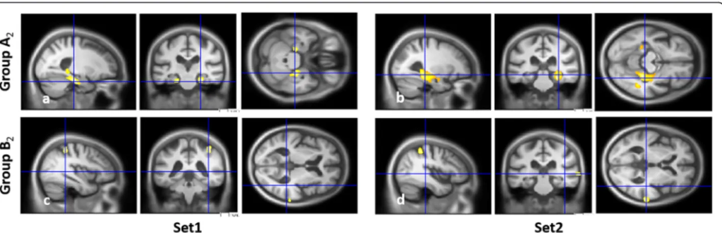
The DMS-48 is a visual forced-choice recognition mem-ory test. It is based on a delayed matching-to-sample paradigm, where the subjects are being asked to choose between a target and a distractor. During an implicit encoding phase, the subjects are asked to decide whether they see more or fewer than three colours on 48 consecu-tive target items. The stimuli belong to three different categories: abstract pictures; concrete objects that belong to the same semantic category (e.g. two cats); and concrete objects that do not belong to the same category (e.g. car-rot and cat). The two first categories allow the use of verbal strategies to be limited. After the encoding phase, an immediate and a 1-hour delayed recognition trial are proposed with two different sets of distractors, Set1 and Set2, respectively (see detailed procedure in [37]). We used the norms published by Barbeau et al. [37], which take age into account. In order to test our hypothesis, according to which only patients whose scores declined between Set1 and Set2 truly have a memory deficit related to MTL dysfunction (‘storage-like’ deficit), we defined three groups depending on the memory profile as follows: group A2 for patients with a ‘storage-like’ deficit (Set2 < Set1);
group B2for patients with ‘encoding’ deficit (Set2 ≥ Set1);
and group C2for patients with normal scores on both Set1
and Set2. The whole cohort of MCI patients will be re-ferred to as group ABC. See Table 2 for the performances of each group on the DMS-48. Note that the groups of pa-tients were different for the DMS-48 and the FCSRT. Note Table 1 Demographic and main clinical data of the whole cohort
Age 79.1 (5.6) Gender (male/female) 47/91 EL: number of patients depending
on the category
EL3: 11; EL4: 34; EL5: 16; EL6: 19; EL7: 58 MCI subtypes: number of patients
depending on the subtype
aMCI: 97 (sd: 20, md: 77) naMCI: 41 (sd: 35, md: 6) MMSE 27.1 (1.7)
CDR: number of patients depending on the score
Score 0: 28 Score 0.5: 110 IADL 13.1 (1.3)
Data presented as mean (standard deviation) unless stated otherwise aMCI amnestic mild cognitive impairment, naMCI non-amnestic mild cognitive impairment, MCI mild cognitive impairment, sd single-domain, md multi-domain, EL educational level, CDR Clinical Dementia Rating Scale, IADL Instrumental Activity of Daily Living, MMSE Mini-Mental State Examination
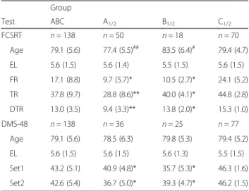
Table 2 Performances on the FCSRT and the DMS-48 according to the memory profile and demographic characteristics of the different groups Group Test ABC A1/2 B1/2 C1/2 FCSRT n = 138 n = 50 n = 18 n = 70 Age 79.1 (5.6) 77.4 (5.5)## 83.5 (6.4)# 79.4 (4.7) EL 5.6 (1.5) 5.6 (1.4) 5.5 (1.5) 5.6 (1.5) FR 17.1 (8.8) 9.7 (5.7)* 10.5 (2.7)* 24.1 (5.2) TR 37.8 (9.7) 28.8 (8.6)** 40.0 (4.1)* 44.8 (2.8) DTR 13.0 (3.5) 9.4 (3.3)** 13.8 (2.0)* 15.3 (1.0) DMS-48 n = 138 n = 36 n = 25 n = 77 Age 79.1 (5.6) 78.5 (6.3) 79.8 (5.3) 79.4 (5.2) EL 5.6 (1.5) 5.6 (1.5) 5.6 (1.3) 5.5 (1.5) Set1 43.2 (5.1) 40.9 (4.8)* 35.7 (5.3)* 46.3 (1.6) Set2 42.6 (5.4) 36.7 (5.0)* 39.3 (4.7)* 46.2 (1.5)
Data presented as mean (standard deviation)
#
Significant difference with P < 0.05 compared with group C1/2 ##
Significant difference with P < 0.05 compared with groups B1/2and C1/2
* Significant difference with P < 0.001 compared with group C1/2
** Significant difference with P < 0.001 compared with groups B1/2and C1/2
DMS-48 Delayed Matching-to-Sample—48 items, FCSRT Free and Cued Selective Reminding Test, EL educational level, FR free recall score, TR total recall score, DTR delayed total recall score, Set1 immediate score, Set2 1-hour delayed score
that no significant difference existed between the groups for age and EL.
Neuroimaging study
We used VBM to investigate the neuroanatomical corre-lates of anterograde memory performances in the MCI patients. To map the regions of atrophy related to the memory deficit, we tested correlation with the GM volume at a voxel level with the scores on both memory tests in the patients. Each participant underwent a high-resolution anatomical MRI scan at inclusion. T1-weighted three-dimensional anatomical images were obtained using 3 T MRI scanners in Strasbourg (Verio 32-channel Tim Siemens scanner; Siemens, Erlangen, Germany) and in CENIR, ICM, Paris (Verio and Trio 32-channel Tim Siemens scanner; Siemens) using a volumetric Magnetization Prepared Rapid Acquisition with Gradient Echo (MPRAGE) se-quence (FOV = 256 × 256 mm2, image matrix = 256 × 256, slice thickness = 1 mm; Strasbourg site: repetition time = 1900 ms, echo time = 2.52 ms, flip angle = 9°; Paris site: repetition time = 2300 ms, echo time = 4.18 ms, flip angle = 9°).
VBM analyses included image pre-processing and stat-istical analyses. These steps were carried out using the SPM12b software package (Wellcome Department of Im-aging Neuroscience, London; http://www.fil.ion.ucl.ac.uk/ spm) running on Matlab R2010a (MathWorks, Natick, MA, USA). Anatomical MRI images were spatially pre-processed using standard procedures [61]. All T1-weighted structural images were first segmented, bias corrected and spatially normalized to the Montreal Neurological Institute (MNI) space using an extension of the unified segmenta-tion procedure [62] that includes six classes of tissue. The DARTEL registration toolbox was then used to build a study-specific template and to bring into alignment all of the segmentation images. The VBM analysis was done on modulated GM images; that is, the GM value in each voxel was multiplied by the Jacobian determinant derived from the spatial normalization. This procedure preserves the total amount of GM from the original images. These mod-ulated GM images were smoothed with a Gaussian kernel (FWHM: 8 mm).
Statistical analysis Behavioural analyses
Intergroup differences between the demographic data and the memory scores on the two tests were compared using a Student’s t test.
VBM analyses
Statistical correlations between local GM volume and scores on both memory tests were then investigated using the General Linear Model (GLM). Raw scores on the FCSRT (FR, TR and DTR) and the DMS-48 (Set1 and Set2)
were tested successively for groups A1/2, B1/2 and C1/2,
pooled (group ABC) and independently, by entering each of them as a covariate of interest. The correlations were tested usingt contrasts (one-tailed test), assuming that de-creased memory performances would be associated with decreased GM volumes. Different nuisance covariates were considered in the model: the age of the subjects, EL, the total GM volume, and site of acquisition (because of two different MRI scans). We used a statistical thresh-old ofP < 0.05 with family-wise error (FWE) as the correc-tion for multiple analyses whenever possible. For each detected cluster, partial correlation analyses were conducted between the mean GM volume of the cluster and FCSRT or DMS-48 scores while taking into account the same set of nuisance covariates (results shown in Additional file 1: Figure S1). When no correlations were found using FWE, a less stringent statistical threshold ofP < 0.001, uncor-rected, was considered. A cluster spatial extent of 50 voxels was used in this case to avoid irrelevant and isolated detec-tions, unless no correlation over 50 voxels was found at all. The software Xjview (http://www.alivelearn.net/xjview8/) allowed us to identify the brain regions and to determine the number of voxels within each region included in each cluster. In the present work, we refer to the hippocampus according to the AAL atlas, namely as the hippocampus proper plus the dentate gyrus and uncus [63]. Reference to the‘MTL’ includes additionally the entorhinal (BA28 and BA34), perirhinal (BA35) and parahippocampal (BA36– BA37) cortices, which together constitute the parahippo-campal gyrus [64]. We also performed group analyses to compare the GM volume between groups A1/2vs. C1/2and
groups B1/2vs. C1/2, using at test, including the same
nuis-ance covariates, in order to check whether the MTL would be atrophied in the group A1/2, in which the MTL was
expected to be found in correlation analyses (results shown in Additional file 2: Figure S2).
Results
Free and Cued Selective Reminding Test
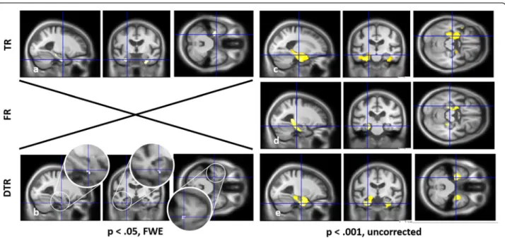
For group ABC (Fig. 1 and Table 3), VBM analyses on the whole cohort revealed correlations after FWE cor-rection for the TR and DTR scores. For TR, the cluster mainly involved the parahippocampal gyrus bilaterally, more particularly the perirhinal cortex (BA35), and to a lesser extent the anterior hippocampus (Fig. 1a and Additional file 1: Figure S1.a–c). For DTR, only the left perirhinal cortex (BA35) was found after FWE correc-tion (Fig. 1b and Addicorrec-tional file 1: Figure S1.d). With a less stringent threshold ofP < 0.001, uncorrected, a larger correlation with the MTL (hippocampus, entorhinal cor-tex (BA28 and BA34), perirhinal (BA35), parahippocampal cortex (BA36)) appeared bilaterally for the TR, DTR and FR scores, and with the lateral temporal cortex for TR and
DTR (Fig. 1c–e). In each case, the left clusters’ volumes were larger than those on the right.
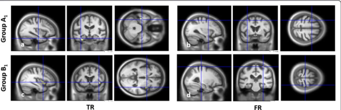
For groups A1and B1independently (Fig. 2 and Table 4),
no correlations were found when VBM analyses were per-formed independently for groups A1, B1and C1with FWE
correction; we therefore used a less stringent threshold of P < 0.001, uncorrected. VBM analyses then only revealed correlations with the MTL (perirhinal (BA35) and parahip-pocampal cortices (BA36)) and with the lateral temporal neocortex (fusiform gyrus) for TR for group A1(Fig. 2a).
The cluster was left-sided and mainly included parahippo-campal gyrus (BA35 and BA36) and, to a lesser extent, hippocampus (see Table 4). Conversely, TR scores were cor-related with insular cortex volume for group B1(Fig. 2c)
but not with the MTL. Finally, performances on FR were correlated with prefrontal aspects for both groups A1
(Fig. 2b) and B1(Fig. 2d). In the case of group A1, the
clus-ter size was smaller than 50 voxels, although we chose to take it into account since it was the only correlation found. Note that when comparing the cerebral volume of groups A1 and B1 vs. C1, the MTL was found to be atrophied
bilaterally in group A1 only (P < 0.001, uncorrected; see
Additional file 2: Figure S2).
Delayed Matching-to-Sample—48 items
For group ABC (Fig. 3 and Table 5), VBM analyses on the whole cohort revealed correlations after FWE correction only for the scores on Set2 of the DMS-48, which exclu-sively involved the right posterior hippocampus (Fig. 3a and Additional file 1: Figure S1.e). With a less stringent thresh-old ofP < 0.001, uncorrected, a larger correlation with the
MTL appeared for both Set1 and Set2 (Fig. 3b, c), bilaterally but more prominently on the right, including hippocampus as well as parahippocampal gyrus (parahippocampal cortex (BA36 and BA37), perirhinal cortex (BA35), posterior ento-rhinal cortex (BA28) and BA19). The precuneus was also involved for Set1.
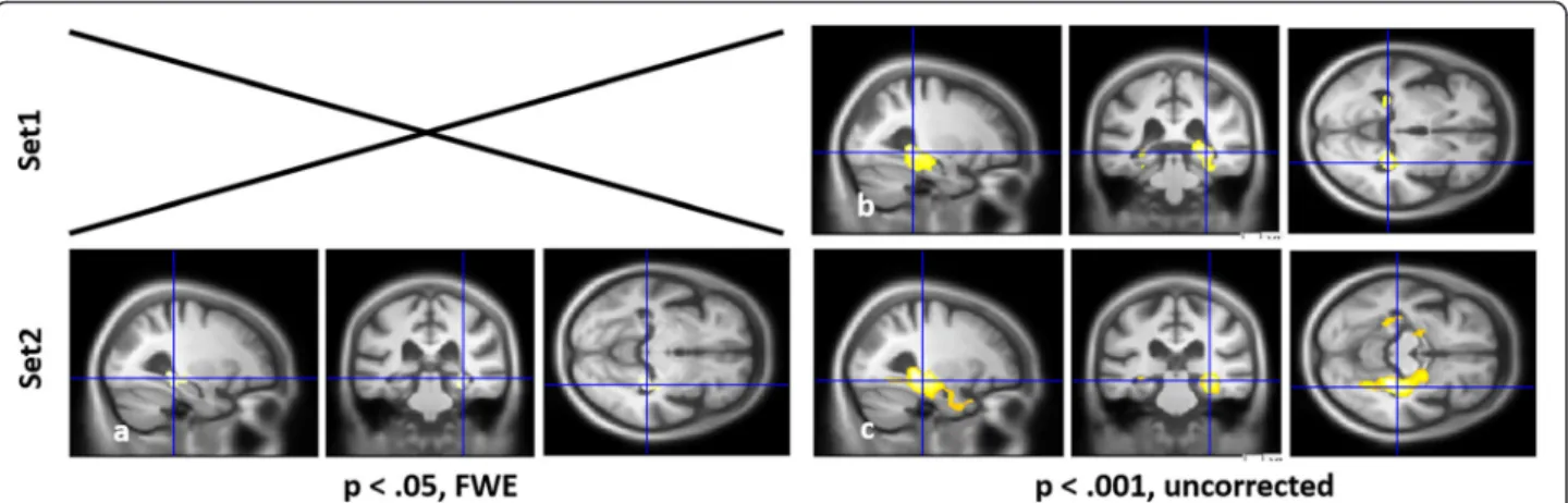
For groups A2and B2independently (Fig. 4 and Table 6),
when VBM analyses were performed using FWE correc-tion only one correlacorrec-tion with the right hippocampus was found for group A2with Set2 (see Table 6). We therefore
used a less stringent threshold of P < 0.001, uncorrected. VBM analyses then only revealed correlations with the MTL (hippocampus, parahippocampal cortex (BA36– BA37), perirhinal cortex (BA35) and posterior entorhi-nal cortex (BA28)) for group A2 both with Set1 and
Set2 (Fig. 4a, b). The clusters were bilateral but larger on the right, and prominently included parahippocampal gyrus for Set1 and hippocampus for Set2, when consider-ing both the volume and T value (Table 6). Conversely, scores on Set1 and Set2 were correlated with temporal and parietal volumes for group B2(Fig. 4c, d) but not with the
MTL. Note than when comparing the cerebral volume of groups A2and B2vs. C2, respectively, the MTL was found
to be atrophied in group A2only, specifically on the right
(P < 0.005, uncorrected; see Additional file 2: Figure S2).
Discussion
In the present study, we investigated the neuroanatom-ical correlates of two anterograde memory tests, the FCSRT and the DMS-48, in a cohort of MCI patients using VBM. Overall, we found that the scores on the
Fig. 1 VBM analyses for the FCSRT in the whole group of patients (group ABC). GM volume regions positively correlated with the TR a, c, FR d and DTR b, e, including age, gender, EL, total GM volume and centre as nuisance covariates, with a threshold of P = 0.05, FWE a, b or P = 0.001, uncorrected c–e. DTR delayed total recall, FR free recall, FWE family-wise error, TR total recall
two memory tests were correlated with the volume of the MTL, prominently on the left side for the FCSRT and on the right side for the DMS-48, concordant with the respective verbal and visual modality of the two tests. However, when analysing different subgroups of patients according to their memory profile, we showed that a correlation with the MTL existed only in patients with a deficient TR score on the FCSRT. Similarly, a correlation with the MTL existed only when considering the subgroup of patients with worsening performances be-tween Set1 and Set2 of the DMS-48. The present study confirms that both tests are reliable topographical markers, when properly interpreted, to indicate a profile of the MTL. Partially contradicting our hypothesis, the parahip-pocampal gyrus was prominently involved for both the visual recognition memory task and the verbal memory task supposed to assess ‘episodic’ memory, whereas the
hippocampus was prominently involved for the delayed recall of the visual recognition memory task. Our conclu-sions are based on correlational analysis rather than group analysis since we did not include a control group without memory complaints, which is one limitation of the study. This finally allowed us to unravel more precisely the differ-ent memory phases and the contribution of their cerebral substrates.
Neuroanatomical correlates of the FCSRT
Regarding the FCSRT, our study confirms in a larger co-hort the results of previous studies showing the implication of the MTL in MCI or Alzheimer’s disease [13, 30–32]. As expected [32], implication of the MTL was found for the TR scores, but was also the case for the FR scores when considering the whole group of patients (for similar results with other memory tests, see also [12, 65–67]). Within the Table 3 GM volume regions positively correlated with the scores on the FCSRT for the whole group of patients (group ABC) using VBM
Group (score) P Brain region Side BA k x y z T ABC (TR), n = 138 PFWE= 0.05 Parahippocampal g. L 35 212 –26 –15 –27 5.59
Hippocampus NA 33 –21 –27 –12 5.13 Parahippocampal g. R 35 154 26 –10 –32 5.48 P = 0.001 Hippocampus L NA 1227/4337 –28 –10 –24 5.02 Parahippocampal g. 28, 34–36 818/4337 –26 –15 –27 5.59 Temporal pole 38 281/4337 –31 6 –21 4.25 Temporal lobe 38 905/4337 –31 6 –21 4.25 Amygdala NA 372/4337 –28 –3 –21 4.23 Parahippocampal g. R 20, 28, 36 812/1700 27 –15 –33 4.56 Hippocampus NA 172/1700 –25 –9 –24 3.91 Temporal pole NA 20/53 37 13 –18 3.39 Hippocampus NA 99/1200 –25 –9 –24 3.91 ABC (FR), n = 138 P = 0.001 Hippocampus L NA 470/1188 –24 –34 –3 3.82 Parahippocampal g. 28, 35 295/1188 –19 –28 –13 3.97 Amygdala NA 25/1188 –21 –9 –12 3.26 Hippocampus R NA 264/572 25 –35 1 3.65 Parahipocampal g. 27 104/572 21 –34 –3 3.76 ABC (DTR), n = 138 PFWE= 0.05 Parahippocampal g. L 35 25 –26 –15 –27 5.06
P = 0.001 Parahippocampal g. L 28, 34–36 1646/2401 –26 –15 –27 5.06 Hippocampus NA 711/2401 –27 –10 –24 4.38 Temporal lobe 20 151/2401 –37 –12 –25 3.29 Amygdala NA 313/2401 –25 0 –18 3.65 Parahippocampal g. R 28, 35, 36 496/850 25 –12 –36 4.04 Hippocampus NA 36/850 27 –10 –25 3.61 Temporal lobe 20 68/850 37 13 –18 3.39
Statistical analyses were performed including age, gender, education level, total GM volume and centre as nuisance covariates, with a threshold of P = 0.05, FWE or P = 0.001, uncorrected, including a minimal k of 50 voxels
GM grey matter, FCSRT Free and Cued Selective Reminding Test, VBM voxel-based morphometry, L left, R right, BA Brodmann area, k cluster size in voxel (specific region’s volume/cluster’s global volume), x, y, z Talairach coordinates, T T value, g. gyrus, DTR delayed total recall, FR free recall, FWE family-wise error, TR total recall
MTL, we found a correlation with the parahippocampal gyrus, which was stronger for the perirhinal cortex (BA35), encompassing the entorhinal cortex [31, 32] (see also [67–69]), and the parahippocampal cortex [13, 32], as well as a correlation with the hippocampus [30–32] (see also [12, 65, 70, 71]). These results are consistent with the involvement of the MTL in the consolidation and storage of new information [10, 36]. This correl-ation existed prominently on the left, as reported previ-ously with the FCSRT [31, 32] and other tests [12, 66, 68], consistent with the verbal modality [66, 72, 73].
Interestingly, we were able to distinguish the neuro-anatomical correlates of the group of patients who had low performances on TR (group A1) from those who
had deficient FR scores only (group B1). Thus, we found
that MTL atrophy existed only in group A1 and
corre-lated with their deficient TR scores, reflecting the exist-ence of a storage deficit. We therefore confirmed that the memory profile with few cueing improvements could indeed be considered an ‘amnestic syndrome of the medial temporal type’ [5, 17]. The correlation obtained with the MTL for the whole group was probably driven by group A1, since no correlation was found with the
MTL in group B1. Regarding the MTL subregions found,
when considering the results obtained for group A1 or
after FWE correction, the parahippocampal gyrus, cluding the perirhinal cortex, was more prominently in-volved than the hippocampus for the TR scores. In other words, our results suggest that the parahippocampal gyrus rather than the hippocampus allows the storage of ‘episodic’ memory, which contradicts the prediction of the dual process model [35, 36]. In this model, the stor-age of contextualized information associated with recol-lection is supposed to involve the hippocampus. Even though the FCSRT is based on recollection rather than familiarity, it is questionable to consider that strong contextual information is associated with such labora-tory memory. Indeed, the retrieval of learned word lists is not comparable with real-life episodic memory such as autobiographical memory [74], which indeed in-volves the hippocampus (e.g. [75–77]). Moreover, cued recall performances on the FCSRT were also found to be associated with the parahippocampal region by Lekeu et al. [13], which was interpreted as reflecting the specific semantic process associated with cueing (e.g. [78, 79]).
Table 4 GM volume regions positively correlated with the scores on the FCSRT independently for groups A1and B1using VBM
Group (score) P Brain region Side BA k x y z T A1(TR), n = 50 0.001 Parahippocampal g. L 35/36 50/283 –28 –15 –28 3.62 Hippocampus NA 16/283 –34 –16 –21 3.47 Fusiform g. 20 135/283 –37 –18 –24 3.68 A1(FR), n = 50 0.001 Mid. Frontal g. L NA 16 –31 –1 49 3.41 B1(TR), n = 18 0.001 Insula R 13 88/170 42 3 0 4.38 B1(FR), n = 18 0.001 Precentral g. R 4 62/63 28 –30 60 5.60 Statistical analyses were performed including age, gender, education level, total GM volume and centre as nuisance covariates, with a threshold of P = 0.001, uncorrected, including a minimal k of 50 voxels except for the FR in group A1
GM grey matter, FCSRT Free and Cued Selective Reminding Test, VBM voxel-based morphometry, L left, R right, BA Brodmann area, k cluster size in voxel (specific region’s volume/cluster’s global volume), x, y, z Talairach coordinates, T T value, g. gyrus, FR free recall, TR total recall
Fig. 2 VBM analyses for the FCSRT depending on the memory profile. GM volume regions positively correlated with the scores on the FCSRT independently for group A1a, b and group B1c, d, for TR a, c (left) and FR b, d (right), including age, gender, EL, total GM volume and centre as
Finally, we found correlations involving extra-MTL re-gions, either for the TR scores in group B1or for the FR
scores in both groups A1and B1. TR scores in group B1
were associated with the volume of the right insula, the significance of this correlation being limited by the fact that the TR scores were not deficient and no focal atro-phy was found in group analyses. The FR scores in both groups A1and B1were associated with prefrontal aspects,
as was the case in Lekeu et al.’s study [13], concordant with the implication of search activity and strategic re-trieval of the information during FR [80]. Concordant with the pattern of regions associated with the two different memory profiles, in a longitudinal study involving MCI patients [34], those who had deficient scores on TR of the FCSRT developed GM atrophy within the left temporal lobe, whereas those who had deficient scores on FR only developed subcortical and frontal GM loss. Additionally, a correlation was also found for the whole group with the
lateral temporal cortex, probably reflecting semantic aspects associated with verbal memory [81].
Neuroanatomical correlates of the DMS-48
As expected, the MTL volume also proved to be corre-lated to the performances on the DMS-48, concordant with previous studies [44, 45, 47]. Namely, the implica-tion of the MTL encompasses both the parahippocampal gyrus (including the entorhinal, perirhinal and parahip-pocampal cortex) and the hippocampus—although when the analyses were performed independently for each group, the implication of the MTL remained for group A2 only. In this group, performances declined between
the immediate and delayed recall, which can therefore also be considered an ‘amnestic profile of the medial temporal type’, concordant with our hypothesis. Taken together with these previous imaging studies [44, 45, 47] and clinical longitudinal studies [48, 49], our results Table 5 GM volume regions positively correlated with the score on the DMS-48 for the whole group of patients (group ABC) using VBM
Group (score) P Brain region Side BA k x y z T ABC (Set1), n = 138 P = 0.001 Hippocampus R NA 606/1377 27 –34 –1 4.31
Parahippocampal g. 28, 35–37 557/1377 28 –39 –6 4.36 Hippocampus L NA 67/264 –28 –40 0 3.47 Parahippocampal g. 19, 31 38/264 –27 –45 –10 3.41 Precuneus 31 70/70 –2 –71 21 3.43 ABC (Set2), n = 138 PFWE= 0.05 Hippocampus R NA 370 28 –37 –3 5.42
Parahippocampal g. 36–37 370 27 –37 –9 5.33 P = 0.001 Hippocampus R NA 1303/3820 30 –25 –12 4.65 Parahippocampal g. 19, 28, 35–37 834/3820 22 –19 –22 4.02 Hippocampus L NA 94/279 –25 –39 –3 3.67
Statistical analyses were performed including age, gender, education level, total GM volume and centre as nuisance covariate, with a threshold of P = 0.05, FWE or P = 0.001, uncorrected, including a minimal k of 50 voxels
GM grey matter, DMS-48, Delayed Matching-to-Sample—48 items, VBM voxel-based morphometry, L left, R right, BA Brodmann area, k cluster size in voxel (specific region’s volume/cluster’s global volume), x, y, z Talairach coordinates, T T value, g. gyrus, FWE family-wise error, Set1 immediate score, Set2 1-hour delayed score
Fig. 3 VBM analyses for the DMS-48 in the whole group of patients (group ABC). GM volume regions positively correlated with Set1 b and Set2 a, c, including age, gender, EL, total GM volume and centre as nuisance covariates, with a threshold of P = 0.05, FWE a or P = 0.001, uncorrected b, c. FWE family-wise error, Set1 immediate score, Set2 1-hour delayed score
confirm that the DMS-48 constitutes an interesting clin-ical marker for potential Alzheimer’s disease pathology, with our additional contribution about the interpretation of the memory profile, which should decline over time to be suspected of MTL dysfunction. Additionally, the implication of the MTL was prominently right-sided (or exclusively right-sided for Set2 in group A2 after FWE
correction), as was the case in a previous study [44], which is concordant with the visual modality of the task [66, 72, 73]. This shows that the test was particularly well designed to avoid the use of verbal strategy.
We will further consider the results obtained with group A2, which had an‘amnestic profile of the medial
temporal type’, to analyse more precisely the MTL
subregions involved. Whereas the correlation with the MTL prominently involved the parahippocampal gyrus, namely the perirhinal cortex, over the hippocampus for immediate recall, the opposite pattern was found for delayed recall. In this case, the hippocampus alone was found after FWE correction. The involvement of the parahippocampal gyrus is widely demonstrated for rec-ognition memory [35], with the perirhinal cortex being more particularly associated with object recognition, and the parahippocampal cortex with spatial information, while the hippocampus would bind item and contextual informa-tion [36]. In human studies, the right hippocampus was found to be associated with spatial navigation [82, 83] or with the visuo-spatial component of autobiographical Table 6 GM volume regions positively correlated with the score on the DMS-48 independently for groups A2and B2using VBM
Group (score) P Brain region Side BA k x y z T A2(Set1), n = 36 P = 0.001 Parahippocampal g. R 28, 35–37 493/907 24 –27 –20 5.00
Hippocampus NA 190/907 28 –38 –1 4.26 Parahippocampal g. L 28, 35 194/211 –24 –27 –20 4.35 A2(Set2), n = 36 PFWE= 0.05 Hippocampus R NA 25 27 –34 –12 6.59
P = 0.001 Hippocampus R NA 1091/2669 30 –33 –7 5.05 Parahippocampal g. 28, 35, 36 776/2669 24 –36 –15 3.78 Parahippocampal g. L 28, 35 260/381 –18 –17 –22 5.04 Hippocampus NA 79 –21 –15 –20 3.49 B2(Set1), n = 25 P = 0.001 Inf. parietal lobule R 40 153 37 –43 45 4.28
Sup. Temporal g. 22 80 66 –35 1 4.61 Inf. Temporal g. L 20 101 –51 –15 –34 4.08 Sup. Temporal g. 22 54 –60 –48 –10 4.73 B2(Set2), n = 25 P = 0.001 Sup. Temporal g. R 22 179 66 –33 1 5.98
Inf. parietal lobule 40 161 37 –43 45 5.46
Statistical analyses were performed including age, gender, education level, total GM volume and centre as nuisance covariate, with a threshold of P = 0.05, FWE or P = 0.001, uncorrected, including a minimal k of 50 voxels
GM grey matter, DMS-48, Delayed Matching-to-Sample—48 items, VBM voxel-based morphometry, L left, R right, BA Brodmann area, k cluster size in voxel (specific region’s volume/cluster’s global volume), x, y, z Talairach coordinates, T T value, g. gyrus, FWE family-wise error, Set1 immediate score, Set2 1-hour delayed score
Fig. 4 VBM analyses for the DMS-48 depending on the memory profile. GM volume regions positively correlated with the scores on the DMS-48 independently for group A2a, b and group B2c, d, for Set1 a, c (left) and Set2 b, d (right), including age, gender, EL, total GM volume and centre
memory [84]. In our study, the switch from parahippocam-pus to hippocamparahippocam-pus during delayed recall was unexpected insofar as recognition memory was involved. This could re-flect the existence of a deeper encoding of the memory trace due to a double registration of items after the first trial [38]. In particular, we cannot exclude that it is due to the fact that the encoding is no longer implicit once the subject realized he/she was performing a memory task on the first trial and sees the target items for the second time. Moreover, one can hypothesize that the recognition of items could be associated with a sense of recollection dur-ing the second trial and therefore involve the hippocampus [36]. Although this could be the case during an activation study, the results obtained in a correlation study probably reflect the minimal memory ability of the patient, namely a sense of familiarity. An R/K paradigm [85] should ideally have been included during the assessment of recognition memory to control for this parameter. Additionally, our results could indicate that the hippocampus is involved in the retention of information after a 1-hour delay, even when recognition memory is concerned.
Finally, performances of group B2were correlated with
extra-MTL regions, namely the temporal and parietal cor-tices. The implication of the inferior temporal gyrus prob-ably reflects the process of visual identification through the visual ventral pathway [86, 87]. Moreover, visual atten-tional processes are probably supported by the inferior parietal lobule [88, 89], as well as by the superior temporal gyrus according to some authors [90, 91]. Similarly, the precuneus, which was also implicated for Set1 in the whole group, is thought to be involved in attention shift between object features [92, 93]. Thus, lower performances on the first trial probably reflect perceptual and attentional pro-cesses involved for encoding during the registration phase. Overall, our study suggests that recognition memory tests involving a unique trial might not be reliable enough to in-dicate MTL dysfunction because they do not allow a per-ceptual or attentional deficit to be distinguished from a memory deficit per se.
Factors for the involvement of MTL subregions in memory
Altogether, we found the prominent involvement of the parahippocampal gyrus for the FCSRT and for the im-mediate trial of the DMS-48, whereas the hippocampus was prominently involved for the 1-hour delayed recall of the DMS-48. Considering the classical view of a dual process model distinguishing recollection vs. familiarity-based memory, respectively involved in contextualized memory and recognition memory [35, 36], the implica-tion of the parahippocampus was expected for the DMS-48 but we would have expected the implication of the hippocampus for the FCSRT. Conversely, the para-hippocampus was prominently involved for ‘episodic’
memory as assessed by the FCSRT and the hippocampus for the delayed recall of the DMS-48, even though recogni-tion memory was involved. Therefore, our results cannot be explained by the different level of awareness between fa-miliarity and recollection. Our results also contradict the hypothesis that a deeper encoding process, such as seman-tic judgment, would involve the hippocampus because it triggers a stronger memory trace than a shallower encoding process, such as perceptual judgment [38, 94]. Finally, the fact that the task consists of explicit or implicit encoding neither explains our results, since the FCSRT involves the parahippocampus even though the instruction to memorize is explicit.
Most interestingly, the implication of the hippocam-pus for the delayed recall of the DMS-48 suggests that it might be triggered by the duration of the memory trace. To the best of our knowledge, recognition mem-ory tasks in humans do not usually involve such a delayed trial, making our results difficult to compare. However, this hypothesis is consistent with some ani-mal studies with hippocampal damage [95, 96], in which recognition memory was found to be impaired after long retention delays [38]. According to this hy-pothesis of a time dependency, the hippocampus could also be expected for the delayed recall of the FCSRT but the delay involved in this case is shorter. A 1-hour delayed trial should ideally have been included. However, in support of this hypothesis, another study involving cor-relational analysis with MCI patients [31] showed that the entorhinal cortex was correlated with the performances on the FCSRT during the learning phase, and the left hippocampus was additionally involved for delayed recall. Similarly, Schmidt-Wilcke et al. [67] found perfor-mances on the CERAD to be correlated with the hippo-campus for delayed recall and with the entorhinal/ perirhinal cortex for immediate recall, suggesting the implication of short-term memory during the encoding phase [97]. However, demonstrating the persistence of such a correlation during the learning phase in our study would favour its implication beyond the encoding phase. Hence, we suggest that there might be a sequential involvement of the parahippocampus and the hippocam-pus, independent of the‘episodic’ vs. recognition nature of memory, with the parahippocampus being preferen-tially involved during the encoding/learning phases and the hippocampus during storage after a 30-minute delay. This temporal sequence would be consistent with the anatomical organization of the MTL, with the dif-ferent parahippocampal subregions constituting entry into the hippocampus [98]. This is also consistent with the existence of an accelerated forgetting in patients with hippocampal sclerosis, whose performances are normal on immediate recall but decline after a 1-hour delay [99].
Conclusions
Our study confirms the interest in the FCSRT and the DMS-48 as topographical markers of the MTL, therefore constituting pertinent clinical markers for potential under-lying incipient Alzheimer’s disease pathology. However, we have confirmed in a large cohort of MCI patients that solely the existence of deficient scores after cueing on the FCSRT constitutes an amnestic profile of the MTL type, whereas FR is associated with prefrontal aspects due to strategic retrieval processes. In a similar vein, our study shows that solely declining scores on recognition memory implicate the MTL, whereas attentional and perceptual processes are involved when the scores improve, highlight-ing the contribution of a delayed trial to interpret the performances on a recognition task. These tasks also allow a reliable assessment of lateralization since the anatomical correlates are prominently left-sided for the FCSRT and right-sided for the DMS-48, concordant with the respective verbal and visual modality of the two tests. When ana-lysing the MTL subregions involved, our results suggest a sequential involvement of the parahippocampus and the hippocampus in time, with the hippocampus being prominently involved after a prolonged delay, independent of the existence of a sense of recollection or familiarity associated with the memory, and of the encoding strength. This study could be complemented by the inclusion of a region-of-interest analysis to directly compare the involve-ment of the hippocampus and the parahippocampus, as well as by the use of amyloid biomarkers to assess the diagnostic value for the underlying pathology.
Additional files
Additional file 1: Figure S1. Showing partial correlations between the normalized behavioural scores and the normalized volumes of the clusters: normalized scores were obtained taking into account nuisance covariates (i.e. age of the subjects, EL, total GM volume, site of acquisition) for the mean FCSRT (Fig. S1.a-c for TR and S1.d. for DTR) or DMS-48 scores (Fig. S1.e.) and the mean GM volume of the clusters found using FWE correction in the whole group of patients (group ABC). (TIF 278 kb)
Additional file 2: Figure S2. Showing group analyses: local GM loss in group A1S2.a and group B1S2.b as compared with group C1and in
group A2S2.c and group B2S2.d as compared with group C2including
age, gender, EL, total GM volume and centre as nuisance covariates, with a threshold of P = 0.001 S2.a, S2.b, S2.d or P = 0.005 S2.c, uncorrected. (TIF 709 kb)
Abbreviations
aMCI:amnestic mild cognitive impairment; DMS-48: Delayed Matching-to-Sample—48 items; DTR: delayed total recall; EL: educational level; FCSRT: Free and Cued Selective Reminding Test; FR: free recall; FWE: family-wise error; GM: grey matter; MCI: mild cognitive impairment; md: multi-domain; MTL: medial temporal lobe; naMCI: non-amnestic mild cognitive impairment; sd: single-domain; TR: total recall; VBM: voxel-based morphometry.
Acknowledgements
The authors are grateful to the patients and their families. They would also like to thank Barbara Jung, Clélie Phillipps, Nadine Longato, Catherine Kleitz, Mathias Bilger, Laetitia Monjoin, Emmanuelle Ehrhardt, Jenny Kemp, Laure Di
Bitonto, Natacha Vogt, Laetitia Berly, Timothée Albasser, Anne Botzung, Mélanie Stackfleth, Xavier De Petigny, Christine Delmaire and Nassim Jeremie for their participation in this study.
Authors’ contributions
NP, ED, BC, CB, MLS, CM-H, EC, CD, OH and FB were responsible for acquisition of clinical data. SK, SL, DG and JPA were responsible for acquisition of MRI data and imaging analysis. VN, NP and IW were responsible for imaging and statistical analysis. NP, VN, BC, IW and FB were responsible for manuscript preparation. ED, CB, MLS, CM-H, EC, CD, SK, SL, DG, JPA and OH were responsible for critical revision of the manuscript. NP, VN, OH and FB were responsible for study conception and design. OH and FB were responsible for study coordination. All authors read and approved the final manuscript.
Competing interests
The authors declare that they have no competing interests. Author details
1Department of Neurology, University Hospital of Strasbourg,
Neuropsychology Unit, Strasbourg, France.2University of Strasbourg, CNRS,
ICube laboratory, FMTS, Strasbourg, France.3University Hospital of
Strasbourg, Centre Mémoire Ressources et Recherche, Strasbourg, France.
4Department of Geriatrics, University Hospital of Strasbourg, Strasbourg,
France.5Department of Geriatrics, Broca Hospital, Assistance
Publique-Hôpitaux de Paris, Paris, France.6Department of Radiology,
University Hospital of Strasbourg, Strasbourg, France.7Department of Neuroradiology, Groupe Hospitalier Pitié-Salpêtrière, Assistance Publique-Hôpitaux de Paris, Paris, France.8UPMC Paris 6—Inserm U1127,
CNRS 7225, Institut du Cerveau et de la Moelle (ICM), Centre de NeuroImagerie de Recherche (CENIR), Paris, France.9Paris Descartes University, Sorbonne Paris Cité, EA4468 Paris, France.
Received: 29 January 2016 Accepted: 29 April 2016
References
1. Albert MS, DeKosky ST, Dickson D, Dubois B, Feldman HH, Fox NC, et al. The diagnosis of mild cognitive impairment due to Alzheimer’s disease: recommendations from the National Institute on Aging-Alzheimer’s Association workgroups on diagnostic guidelines for Alzheimer’s disease. Alzheimers Dement. 2011;7(3):270–9. doi:10.1016/j. jalz.2011.03.008.
2. Dubois B, Feldman HH, Jacova C, Hampel H, Molinuevo JL, Blennow K, et al. Advancing research diagnostic criteria for Alzheimer’s disease: the IWG-2 criteria. Lancet Neurol. 2014;13(6):614–29. doi:10.1016/S1474-4422(14)70090-0. 3. Petersen RC, Smith GE, Waring SC, Ivnik RJ, Tangalos EG, Kokmen E. Mild
cognitive impairment: clinical characterization and outcome. Arch Neurol. 1999;56(3):303–8.
4. Petersen RC. Mild cognitive impairment as a diagnostic entity. J Intern Med. 2004;256(3):183–94. doi:10.1111/j.1365-2796.2004.01388.x.
5. Dubois B, Albert ML. Amnestic MCI or prodromal Alzheimer’s disease? Lancet Neurology. 2004;3(4):246–8. doi:10.1016/S1474-4422(04)00710-0. 6. Delacourte A, David JP, Sergeant N, Buee L, Wattez A, Vermersch P, et al.
The biochemical pathway of neurofibrillary degeneration in aging and Alzheimer’s disease. Neurology. 1999;52(6):1158–65.
7. Scheltens P, Leys D, Barkhof F, Huglo D, Weinstein HC, Vermersch P, et al. Atrophy of medial temporal lobes on MRI in‘probable’ Alzheimer’s disease and normal ageing: diagnostic value and neuropsychological correlates. J Neurol Neurosurg Psychiatry. 1992;55(10):967–72.
8. Silbert LC, Quinn JF, Moore MM, Corbridge E, Ball MJ, Murdoch G, et al. Changes in premorbid brain volume predict Alzheimer’s disease pathology. Neurology. 2003;61(4):487–92.
9. Scoville WB, Milner B. Loss of recent memory after bilateral hippocampal lesions. J Neurol Neurosurg Psychiatry. 1957;20(1):11–21.
10. Squire LR, Zola-Morgan S. The medial temporal lobe memory system. Science. 1991;253(5026):1380–6.
11. Tulving E, Markowitsch HJ. Episodic and declarative memory: role of the hippocampus. Hippocampus. 1998;8(3):198–204. doi:10.1002/(SICI)1098-1063(1998)8:3<198::AID-HIPO2>3.0.CO;2-G.
12. Desgranges B, Baron JC, Eustache F. The functional neuroanatomy of episodic memory: the role of the frontal lobes, the hippocampal formation, and other areas. NeuroImage. 1998;8(2):198–213. doi:10.1006/nimg.1998.0359. 13. Lekeu F, Van der Linden M, Chicherio C, Collette F, Degueldre C, Franck G,
et al. Brain correlates of performance in a free/cued recall task with semantic encoding in Alzheimer disease. Alzheimer Dis Assoc Disord. 2003;17(1):35–45. 14. Dubois B, Feldman HH, Jacova C, Dekosky ST, Barberger-Gateau P,
Cummings J, et al. Research criteria for the diagnosis of Alzheimer’s disease: revising the NINCDS-ADRDA criteria. Lancet Neurol. 2007;6(8):734–46. doi:10. 1016/S1474-4422(07)70178-3.
15. Sarazin M, Berr C, De Rotrou J, Fabrigoule C, Pasquier F, Legrain S, et al. Amnestic syndrome of the medial temporal type identifies prodromal AD: a longitudinal study. Neurology. 2007;69(19):1859–67. doi:10.1212/01.wnl. 0000279336.36610.f7.
16. Grober E, Buschke H, Crystal H, Bang S, Dresner R. Screening for dementia by memory testing. Neurology. 1988;38(6):900–3.
17. Tounsi H, Deweer B, Ergis AM, Van der Linden M, Pillon B, Michon A, et al. Sensitivity to semantic cuing: an index of episodic memory dysfunction in early Alzheimer disease. Alzheimer Dis Assoc Disord. 1999;13(1):38–46. 18. Grober E, Lipton RB, Hall C, Crystal H. Memory impairment on free and cued
selective reminding predicts dementia. Neurology. 2000;54(4):827–32. 19. Grober E, Sanders AE, Hall C, Lipton RB. Free and cued selective reminding
identifies very mild dementia in primary care. Alzheimer Dis Assoc Disord. 2010;24(3):284–90. doi:10.1097/WAD.0b013e3181cfc78b.
20. Grober E, Kawas C. Learning and retention in preclinical and early Alzheimer’s disease. Psychol Aging. 1997;12(1):183–8.
21. Grober E, Hall C, Sanders AE, Lipton RB. Free and cued selective reminding distinguishes Alzheimer’s disease from vascular dementia. J Am Geriatr Soc. 2008;56(5):944–6. doi:10.1111/j.1532-5415.2008.01652.x.
22. Pillon B, Gouider-Khouja N, Deweer B, Vidailhet M, Malapani C, Dubois B, et al. Neuropsychological pattern of striatonigral degeneration: comparison with Parkinson’s disease and progressive supranuclear palsy. J Neurol Neurosurg Psychiatry. 1995;58(2):174–9.
23. Fossati P, Coyette F, Ergis AM, Allilaire JF. Influence of age and executive functioning on verbal memory of inpatients with depression. J Affect Disord. 2002;68(2–3):261–71.
24. Lemos R, Maroco J, Simoes MR, Santiago B, Tomas J, Santana I. The free and cued selective reminding test for predicting progression to Alzheimer’s disease in patients with mild cognitive impairment: a prospective longitudinal study. J Neuropsychol. 2015;1–16. doi:10.1111/jnp.12075. 25. Mura T, Proust-Lima C, Jacqmin-Gadda H, Akbaraly TN, Touchon J, Dubois B,
et al. Measuring cognitive change in subjects with prodromal Alzheimer’s disease. J Neurol Neurosurg Psychiatry. 2014;85(4):363–70. doi:10.1136/jnnp-2013-305078.
26. Derby CA, Burns LC, Wang C, Katz MJ, Zimmerman ME, L’Italien G, et al. Screening for predementia AD: time-dependent operating characteristics of episodic memory tests. Neurology. 2013;80(14):1307–14. doi:10.1212/WNL. 0b013e31828ab2c9.
27. Auriacombe S, Helmer C, Amieva H, Berr C, Dubois B, Dartigues JF. Validity of the free and cued selective reminding test in predicting dementia: the 3C study. Neurology. 2010;74(22):1760–7. doi:10.1212/WNL.0b013e3181df0959. 28. Wagner M, Wolf S, Reischies FM, Daerr M, Wolfsgruber S, Jessen F, et al.
Biomarker validation of a cued recall memory deficit in prodromal Alzheimer disease. Neurology. 2012;78(6):379–86. doi:10.1212/WNL.0b013e318245f447. 29. Grober E, Dickson D, Sliwinski MJ, Buschke H, Katz M, Crystal H, et al.
Memory and mental status correlates of modified Braak staging. Neurobiol Aging. 1999;20(6):573–9.
30. Deweer B, Lehericy S, Pillon B, Baulac M, Chiras J, Marsault C, et al. Memory disorders in probable Alzheimer’s disease: the role of hippocampal atrophy as shown with MRI. J Neurol Neurosurg Psychiatry. 1995;58(5):590–7. 31. Sanchez-Benavides G, Gomez-Anson B, Molinuevo JL, Blesa R, Monte GC,
Buschke H, et al. Medial temporal lobe correlates of memory screening measures in normal aging, MCI, and AD. J Geriatr Psychiatry Neurol. 2010; 23(2):100–8. doi:10.1177/0891988709355271.
32. Sarazin M, Chauvire V, Gerardin E, Colliot O, Kinkingnehun S, de Souza LC, et al. The amnestic syndrome of hippocampal type in Alzheimer’s disease: an MRI study. J Alzheimers Dis. 2010;22(1):285–94. doi:10.3233/JAD-2010-091150. 33. Zimmerman ME, Pan JW, Hetherington HP, Katz MJ, Verghese J, Buschke H,
et al. Hippocampal neurochemistry, neuromorphometry, and verbal memory in nondemented older adults. Neurology. 2008;70(18):1594–600. doi:10.1212/01.wnl.0000306314.77311.be.
34. Koric L, Ranjeva JP, Felician O, Guye M, de Anna F, Soulier E, et al. Cued recall measure predicts the progression of gray matter atrophy in patients with amnesic mild cognitive impairment. Dement Geriatr Cogn Disord. 2013;36(3-4):197–210. doi:10.1159/000351667.
35. Brown MW, Aggleton JP. Recognition memory: what are the roles of the perirhinal cortex and hippocampus? Nat Rev Neurosci. 2001;2(1):51–61. doi:10.1038/35049064.
36. Eichenbaum H, Yonelinas AP, Ranganath C. The medial temporal lobe and recognition memory. Annu Rev Neurosci. 2007;30:123–52. doi:10.1146/ annurev.neuro.30.051606.094328.
37. Barbeau E, Didic M, Tramoni E, Felician O, Joubert S, Sontheimer A, et al. Evaluation of visual recognition memory in MCI patients. Neurology. 2004; 62(8):1317–22.
38. Squire LR, Wixted JT, Clark RE. Recognition memory and the medial temporal lobe: a new perspective. Nat Rev Neurosci. 2007;8(11):872–83. doi:10.1038/nrn2154.
39. Meunier M, Bachevalier J, Mishkin M, Murray EA. Effects on visual recognition of combined and separate ablations of the entorhinal and perirhinal cortex in rhesus monkeys. J Neurosci. 1993;13(12):5418–32.
40. Bussey TJ, Muir JL, Aggleton JP. Functionally dissociating aspects of event memory: the effects of combined perirhinal and postrhinal cortex lesions on object and place memory in the rat. J Neurosci. 1999;19(1):495–502. 41. Vargha-Khadem F, Gadian DG, Watkins KE, Connelly A, Van Paesschen W, Mishkin M. Differential effects of early hippocampal pathology on episodic and semantic memory. Science. 1997;277(5324):376–80.
42. Mayes AR, Holdstock JS, Isaac CL, Hunkin NM, Roberts N. Relative sparing of item recognition memory in a patient with adult-onset damage limited to the hippocampus. Hippocampus. 2002;12(3):325–40. doi:10. 1002/hipo.1111.
43. Braak H, Braak E. Neuropathological stageing of Alzheimer-related changes. Acta Neuropathol. 1991;82(4):239–59.
44. Barbeau EJ, Ranjeva JP, Didic M, Confort-Gouny S, Felician O, Soulier E, et al. Profile of memory impairment and gray matter loss in amnestic mild cognitive impairment. Neuropsychologia. 2008;46(4):1009–19. doi:10.1016/j. neuropsychologia.2007.11.019.
45. Guedj E, Barbeau EJ, Didic M, Felician O, de Laforte C, Ceccaldi M, et al. Identification of subgroups in amnestic mild cognitive impairment. Neurology. 2006;67(2):356–8. doi:10.1212/01.wnl.0000225076.73312.d4. 46. Chetelat G, Desgranges B, de la Sayette V, Viader F, Eustache F, Baron JC.
Mild cognitive impairment: Can FDG-PET predict who is to rapidly convert to Alzheimer’s disease? Neurology. 2003;60(8):1374–7.
47. Didic M, Ranjeva JP, Barbeau E, Confort-Gouny S, Fur YL, Felician O, et al. Impaired visual recognition memory in amnestic mild cognitive impairment is associated with mesiotemporal metabolic changes on magnetic resonance spectroscopic imaging. J Alzheimers Dis. 2010;22(4): 1269–79. doi:10.3233/JAD-2010-101257.
48. Didic M, Felician O, Barbeau EJ, Mancini J, Latger-Florence C, Tramoni E, et al. Impaired visual recognition memory predicts Alzheimer’s disease in amnestic mild cognitive impairment. Dement Geriatr Cogn Disord. 2013; 35(5-6):291–9. doi:10.1159/000347203.
49. De Anna F, Felician O, Barbeau E, Mancini J, Didic M, Ceccaldi M. Cognitive changes in mild cognitive impairment patients with impaired visual recognition memory. Neuropsychology. 2014;28(1):98–105. doi:10. 1037/neu0000032.
50. Hughes CP, Berg L, Danziger WL, Coben LA, Martin RL. A new clinical scale for the staging of dementia. Br J Psychiatry. 1982;140:566–72.
51. Lawton MP, Brody EM. Assessment of older people: self-maintaining and instrumental activities of daily living. Gerontologist. 1969;9(3):179–86. 52. Katz S, Ford AB, Moskowitz RW, Jackson BA, Jaffe MW. Studies of illness in
the aged. The index of Adl: a standardized measure of biological and psychosocial function. JAMA. 1963;185:914–9.
53. Thurstone LL, Thurstone TG. Manuel d’application de la batterie factorielle P. M.A (primary mental activities). Adaptation française du Centre de Psychologie Appliquée. 2d ed. Paris: CPA; 1964.
54. Deloche G, Hannequin D. Test de dénomination orale d’images: DO 80. Paris: Les Editions du Centre de psychologie appliquée; 1997. 55. Goul WR, Brown M. Effects of age and intelligence on trail making test
performance and validity. Percept Mot Skills. 1970;30(1):319–26. doi:10. 2466/pms.1970.30.1.319.
56. Wechsler D. Wechsler adult intelligence scale. 3d ed. Paris: Les Editions du Centre de Psychologie Appliquée; 1997.