HAL Id: hal-01795643
https://hal.sorbonne-universite.fr/hal-01795643
Submitted on 18 May 2018
HAL is a multi-disciplinary open access
archive for the deposit and dissemination of sci-entific research documents, whether they are pub-lished or not. The documents may come from teaching and research institutions in France or abroad, or from public or private research centers.
L’archive ouverte pluridisciplinaire HAL, est destinée au dépôt et à la diffusion de documents scientifiques de niveau recherche, publiés ou non, émanant des établissements d’enseignement et de recherche français ou étrangers, des laboratoires publics ou privés.
disease associated with duplications and missense
mutations in APP gene, Down syndrome and sporadic
Alzheimer’s disease
David Mann, Yvonne Davidson, Andre Robinson, Nancy Allen, Tadafumi
Hashimoto, Anna Richardson, Matthew Jones, Julie Snowden, Neil Pendleton,
Marie-Claude Potier, et al.
To cite this version:
David Mann, Yvonne Davidson, Andre Robinson, Nancy Allen, Tadafumi Hashimoto, et al.. Patterns and severity of vascular amyloid in Alzheimer’s disease associated with duplications and missense mutations in APP gene, Down syndrome and sporadic Alzheimer’s disease. Acta Neuropathologica, Springer Verlag, 2018, Epub ahead of print]. �10.1007/s00401-018-1866-3�. �hal-01795643�
https://doi.org/10.1007/s00401-018-1866-3
ORIGINAL PAPER
Patterns and severity of vascular amyloid in Alzheimer’s disease
associated with duplications and missense mutations in APP gene,
Down syndrome and sporadic Alzheimer’s disease
David M. A. Mann1 · Yvonne S. Davidson1 · Andrew C. Robinson1 · Nancy Allen1 · Tadafumi Hashimoto2 ·
Anna Richardson3 · Matthew Jones3 · Julie S. Snowden1,3 · Neil Pendleton1 · Marie‑Claude Potier4 ·
Annie Laquerrière5,6 · Vee Prasher7 · Takeshi Iwatsubo2 · Andre Strydom8,9
Received: 15 February 2018 / Revised: 4 May 2018 / Accepted: 10 May 2018 © The Author(s) 2018
Abstract
In this study, we have compared the severity of amyloid plaque formation and cerebral amyloid angiopathy (CAA), and the subtype pattern of CAA pathology itself, between APP genetic causes of AD (APPdup, APP mutations), older individuals with Down syndrome (DS) showing the pathology of Alzheimer’s disease (AD) and individuals with sporadic (early and late onset) AD (sEOAD and sLOAD, respectively). The aim of this was to elucidate important group differences and to provide mechanistic insights related to clinical and neuropathological phenotypes. Since lipid and cholesterol metabolism is implicated in AD as well as vascular disease, we additionally aimed to explore the role of APOE genotype in CAA severity and subtypes. Plaque formation was greater in DS and missense APP mutations than in APPdup, sEOAD and sLOAD cases. Conversely, CAA was more severe in APPdup and missense APP mutations, and in DS, compared to sEOAD and sLOAD. When stratified by CAA subtype from 1 to 4, there were no differences in plaque scores between the groups, though in patients with APPdup, APP mutations and sEOAD, types 2 and 3 CAA were more common than type 1. Conversely, in DS, sLOAD and controls, type 1 CAA was more common than types 2 and 3. APOE ε4 allele frequency was greater in sEOAD and sLOAD compared to APPdup, missense APP mutations, DS and controls, and varied between each of the CAA phenotypes with APOE ε4 homozygosity being more commonly associated with type 3 CAA than types 1 and 2 CAA in sLOAD and sEOAD. The differing patterns in CAA within individuals of each group could be a reflection of variations in the efficiency of perivascular drainage, this being less effective in types 2 and 3 CAA leading to a greater burden of CAA in parenchymal arteries and capillaries. Alternatively, as suggested by immunostaining using carboxy-terminal specific antibodies, it may
relate to the relative tissue burdens of the two major forms of Aβ, with higher levels of Aβ40 promoting a more ‘aggressive’
form of CAA, and higher levels of Aβ42(3) favouring a greater plaque burden. Possession of APOE ε4 allele, especially ε4
homozygosity, favours development of CAA generally, and as type 3 particularly, in sEOAD and sLOAD.
Keywords Alzheimer’s disease · Down syndrome · APP mutations · Cerebral amyloid angiopathy · Amyloid plaques
Introduction
Alzheimer’s disease (AD) is a neurodegenerative disorder characterised clinically by a progressive loss of memory and cognition, accompanied by functional impairments of orien-tation and praxis. Pathologically, the major changes involve a deposition of amyloid β protein (Aβ) in brain parenchyma (as amyloid plaques) and hyperphosphorylated tau within neurones (as neurofibrillary tangles). Additionally, most cases display deposits of Aβ within blood vessel walls—a change known as cerebral amyloid angiopathy (CAA). While more than 90% cases of AD are without obvious genetic
Electronic supplementary material The online version of this article (https ://doi.org/10.1007/s0040 1-018-1866-3) contains supplementary material, which is available to authorized users. * David M. A. Mann
david.mann@manchester.ac.uk
cause, and termed ‘sporadic’, the remainder is associated with mutational events involving either the Amyloid Precur-sor Protein (APP) or presenilin (PSEN) genes.
With respect to the transmembrane protein APP, missense mutations changing the amino acid sequence at either the amino- or carboxy-terminal points of the Aβ sequence (e.g. APP670/671, APP717) result in increased catabolic break-down of APP by β- and/or γ-secretase into Aβ, and confer a pathological picture similar to that seen in sporadic AD. On the other hand, mutations lying in the juxtamembrane region, such as APP692 or APP693, are more associated with CAA than plaques, and often manifest clinically as
acute stroke. There are also rare French [6, 14, 37], Dutch
[41], Finnish [38], Japanese [19], Swedish [48] and British
[30] families where AD is linked to duplications at the APP
locus, resulting in APP overproduction. In most of these families, the duplication has been validated only in living patients and confirmed cases with brain donation are scarce. An APP duplication has also been reported in a Spanish
patient with apparently sporadic AD and severe CAA [21],
but other studies of sporadic AD with CAA have not
identi-fied such duplications [3, 11]. It has long been known that
most individuals with Down syndrome (DS), who live into middle age and beyond, show a pathological picture
indistin-guishable from that of AD [24, 25]. In most DS individuals,
there is a complete triplication of chromosome 21, including the APP locus. In both APPdup and DS individuals, it is presumed that the early deposition of Aβ plaques and CAA stems from an overexpression of APP and the consequent degradation of an excessive production of APP. In addition, recent work suggests that a mutation in the 3′untranslated region of APP also result in APP overexpression and might
act as a genetic determinant in some cases of CAA [33].
Although all cases of AD are defined pathologically by the presence of numerous plaques and tangles, and usually CAA, throughout the cerebral cortex and hippocampus, the morphological appearance of these changes, especially plaques and CAA, can vary according to the underlying genetic background. For example, a much more severe CAA is seen in patients with presenilin-1 (PSEN-1) muta-tions located beyond codon 200 compared to those where the
mutation lies before codon 200 [28]. Also, certain
PSEN-1 mutations are associated with an unusual morphological form of amyloid plaques, known as ‘cotton wool’ plaques,
and often present clinically with a spastic paresis [29].
The cardinal clinical presentation in APP duplica-tions (APPdup) is that of progressive dementia frequently accompanied by seizures and intracerebral haemorrhage
(ICH) [6, 14, 19, 37, 38, 41], and neuropathological
stud-ies have revealed severe CAA in association with abundant Aβ plaques and neurofibrillary tangles, and occasionally
Lewy bodies [6, 14]. CAA is also often prominent in
indi-viduals with DS [25] where there is also an additional APP
copy number. Nevertheless, CAA can present in sporadic and familial AD in various phenotypic histological forms, involving differing combinations of leptomeningeal and parenchymal arterial pathology, sometimes extending into
the capillary bed [2, 46].
We have recently reviewed epidemiological data on stroke (including haemorrhagic stroke) related to CAA to make
comparisons between DS and APPdup [5]. Although ICH,
the main clinical consequence of vascular amyloidosis, is a common clinical occurrence in APPdup, this is a more poorly defined feature of individuals with DS, suggesting the
presence of a mechanism(s) that acts protectively [5]. This
might seem somewhat paradoxical given that DS only differs from APPdup in the ~ 270 other genes located on chromo-some 21 that are also triplicated.
However, a direct comparison of vascular amyloidosis at pathological level between DS, APPdup, and other APP mutations has never been undertaken as far as we are aware, and consequently the degree and nature of tissue differences in CAA, ICH, and Aβ deposition between these disorders remain unclear. Therefore, in this analysis, we aim to com-pare the severity of amyloid plaque formation and CAA, and the subtype pattern of CAA pathology itself, and the relationship between CAA severity and CAA phenotype, in APP genetic causes of AD (APPdup, APP mutations), DS and sporadic (early and late onset) AD (sEOAD and sLOAD, respectively). The observations might help to elu-cidate important differences between the patient groups, and thus provide mechanistic insights related to clinical and neuropathological phenotypes. Since lipid and cholesterol metabolism is implicated in AD as well as vascular disease, we additionally aimed to explore the role of APOE genotype in CAA severity and subtypes.
Materials and methods
Patients
The study included 152 subjects in total, comprising six groups categorised according to the pathological and genetic basis of their condition. First, brains were obtained from four patients (three males, one female, patients #1–4) with genetically proven duplications in APP gene through Prof Annie Laquerrière at University of Rouen. These were from two separate families. Three patients (patients #1–3) were members of the same family (known as F037, and
identi-fied as II-4, II-5 and II-6, respectively, in [6]). The other
patient (patient #4) was a member of a second family
(iden-tified as II.1 in [14]). Second, brains were obtained from 34
individuals with DS (21 males, 13 females, patients #5–38) ranging (at death) from 36 to 69 years. Where karyotyp-ing had been performed, all were full trisomy 21. Eight of
these were drawn from the Manchester Brain Bank (MBB), 4 were obtained from the Neurodegenerative Diseases Brain Bank at Institute of Psychiatry, Psychology and Neurosci-ence (IOPPN) Brain Bank, London (courtesy of C Troakes), 8 from the Thomas Willis Brain Bank (TWBB), Oxford (courtesy of M Esiri), with the remaining 14 being obtained through Professor V P Prasher at University of Birming-ham. Third, brains were obtained from 16 patients (seven males, nine females, patients #39–54) with missense muta-tions in APP gene, 14 with point mutamuta-tions at codon 717 (10 Val717Ile, 3 Val717Gly and 1 Val717Ala) and 2 with a point mutation at codon 692 (Flemish mutation). Four of
these were obtained through MBB, eight from IOPPN Brain Bank (courtesy of C Troakes), and four from Queen Square Brain Bank (QSBB) (courtesy of L Parsons). Additionally, brains were obtained from 34 patients with clinical diagnosis of sEOAD (21 males, 13 females, patients #55–88), 34 with sLOAD (18 males, 16 females, patients #89–122) and 30 elderly controls (11 males, 19 females, subjects #123–152)
through MBB (see Table 1 for group, and Supplementary
Table 1 for individual, patient details). All brains had been obtained at autopsy through appropriate consenting proce-dures with Local Ethical Committee approval. Patients #1–4 and #39–122 all fulfilled relevant pathological diagnostic
Table 1 Mean ± SD age at onset, age at death and duration of illness for cases of APP duplication (APPdup), Down syndrome, missense APP mutations and sporadic early onset Alzheimer’s disease (sEOAD), sporadic late onset Alzheimer’s disease and controls, both overall and stratified according to each CAA phenotype
Age range is given in parentheses na not applicable
X Group age at onset and duration of illness data based on 14 individuals (2 CAA type 1, 7 CAA type 2 and 5 CAA type 3)
**Significantly different from CAA type 2, p < 0.01 ***Significantly different from sEOAD, p < 0.001 +++ Significantly different from sLOAD group, p < 0.001 $$ Significantly different from sLOAD group, p < 0.01
!,!!! Significantly different from missense APP group, p < 0.05, < 0.001, respectively
Age at onset (years) Age at death (years) Duration of illness (years) APPdup All (n = 4) 51.0 ± 5.0+++ (44–55) 61.0 ± 5.7+++ (55–68) 10.0 ± 3.2 (7–14) CAA type 2 (n = 1) 54.0 68.0 14.0 CAA type 3 (n = 3) 50.0 ± 5.6 (44–55) 58.7 ± 4.0 (55–63) 8.7 ± 2.1 (7–11) Down syndromex All (n = 34) 53.1 ± 3.7+++ (48–59) 58.7 ± 6.0+++*** (36–69) 5.9 ± 2.9!!! (2–14) CAA type 1 (n = 15) 52.0 ± 5.0 (50–54) 58.7 ± 8.0 (36–69) 5.0 ± 1.4 (4–6) CAA type 2 (n = 9) 52.6 ± 3.6 (49–57) 58.9 ± 3.2 (53–64) 6.0 ± 1.7 (2–9) CAA type 3 (n = 10) 54.4 ± 6.0 (48–59) 58.4 ± 4.8(47–62) 6.0 ± 4.7(2–14) Missense APP All (n = 16) 51.6 ± 7.4+++ (40–62) 61.8 ± 6.2+++ (51–70) 10.8 ± 4.6 (6–21) CAA type 1 (n = 7) 52.0 ± 8.3(42–62) 60.0 ± 6.4 (51–61) 7.8 ± 2.3 (6–12) CAA type 2 (n = 7) 52.3 ± 7.3 (40–59) 64.6 ± 5.6 (59–72) 12.3 ± 4.6 (6–21) CAA type 4 (n = 2) 45.0 58.0 ± 7.7 (53–63) 18.0 sEOAD All (n = 34) 55.1 ± 5.9+++ (35–64) 63.6 ± 5.5+++ (45–69) 8.6 ± 2.4 (5–14) CAA type 1 (n = 9) 52.1 ± 7.4(35–60) 62.3 ± 7.3 (45–69) 10.4 ± 1.9** (7–14) CAA type 2 (n = 20) 55.8 ± 4.9 (43–64) 63.7 ± 4.7(50–68) 7.7 ± 2.3 (5–10) CAA type 3 (n = 5) 58.8 ± 2.2 (56–61) 66.6 ± 1.7(64–67) 7.9 ± 1.9(5–10) sLOAD All (n = 34) 72.9 ± 7.1 (65–90) 80.8 ± 7.4 (70–95) 8.0 ± 2.7! (3–14) CAA type 1 (n = 17) 73.7 ± 6.8 (65–84) 82.1 ± 7.7 (70–95) 8.4 ± 2.7 (3–14) CAA type 2 (n = 7) 70.1 ± 7.4 (65–86) 77.6 ± 7.2 (71–92) 7.4 ± 3.1 (3–13) CAA type 3 (n = 6) 71.3 ± 6.0 (65–80) 78.8 ± 6.4 (70–87) 8.2 ± 2.6 (4–12) Controls All (n = 30) na 87.0 ± 6.2+++$$ (69–100) na CAA type 1 (n = 15) na 87.3 ± 6.0 (76–100) na CAA type 2 (n = 2) na 87.5 ± 7.1(87–88) na
criteria for AD [16, 31, 32]. The sEOAD patients acquired through MBB had been investigated longitudinally within specialist dementia clinics at Salford Royal Hospital using the Manchester Neuropsychological Profile (Man-NP)
[42, 47] to determine and characterise the nature of their
dementia. The sLOAD patients and elderly controls were drawn partly from the Manchester recruits for the Brains for Dementia Research (BDR) cohort, and partly from The Uni-versity of Manchester Longitudinal Study of Cognition in
Normal Healthy Old Age [36]. The 14 individuals with DS
who had been assessed clinically by Professor V P Prasher at University of Birmingham had all been diagnosed as suf-fering from non-familial ‘Dementia in Alzheimer’s disease’
according to DCR-10 criteria [51]. This kind of clinical
information was not, or no longer, available for the other 20 DS individuals whose brains had been acquired many years ago by MBB, TWBB or IOPPN Brain Banks.
None of the individuals in any of the pathological groups had been known to have suffered from stroke or ICH.
Histological methods
Paraffin sections were cut at 6 µm from formalin fixed blocks of frontal lobe (BA8/9), temporal lobe (BA21/22) with pos-terior hippocampus, occipital lobe (BA17/18) and cerebel-lar hemisphere. Following titration to determine optimal immunostaining, antibodies were identically employed in a
standard immunohistochemical protocol [10]. Sections were
immunostained for Aβ using 4G8 antibody (Cambridge Bio-science, clone 4G8, 1:3000) and for tau proteins phospho-rylated at Ser202 and Thr205 (P-tau) using AT8 antibody (Source Bioscience, clone AT8, 1:750). Sections immu-nostained with 4G8 antibody were subject to formic acid pretreatment before antigen unmasking by pressure cooking in citrate buffer (pH 6.0, 10 mM) for 30 min, reaching 120 degrees Celsius and > 15 kPa pressure. The formic acid pre-treatment step was omitted for AT8 (tau) immunostaining. Following immunostaining, all cases were staged as Thal
phase of Aβ deposition [45] and Braak stage of
neurofibril-lary degeneration [4]. Additional immunostaining, using
previously validated end-specific Aβ40 and Aβ42(3)
mono-clonal antibodies (known as BA27 and BC05, respectively) was performed on 30 cases, selected to be representative of each CAA phenotype within each pathological group, as
described previously [17, 18]. For this work, we chose three
APPdup cases (case #1–3), six with DS (#12, 17, 19–21 and 26), five with missense APP mutations (#49–52 and 54), six with sEOAD (#58, 62, 68, 75, 78 and 81), seven with sLOAD (#90, 93, 95–98 and 122) and three controls (#129, 140 and 143).
Sections were examined microscopically for the appearance, severity and topographical distribution of immunostaining
within brain parenchyma (as plaques) and cerebral ves-sels (as CAA). A five-point scoring system, similar to that
employed by Olichney et al. [34], was employed to
sepa-rately grade the severity of Aβ plaques and CAA as total Aβ
(4G8) and separately as Aβ40 (BA27) and Aβ42(3) (BC05)
specific changes. Plaques
Grade 0—no Aβ plaques in parenchyma.
Grade 1—A few Aβ plaques in parenchyma occupying each low power (× 10 microscope objective) field. Grade 2—A moderate number of Aβ plaques in paren-chyma occupying each low power (× 10 microscope objective) field.
Grade 3—Many dispersed Aβ plaques in parenchyma occupying each low power (× 10 microscope objective) field.
Grade 4—Very many densely packed Aβ plaques in parenchyma occupying each low power (× 10 microscope objective) field.
CAA
Grade 0—No CAA in blood vessel walls in leptomenin-ges or brain parenchyma.
Grade 1—Occasional blood vessels with CAA in lep-tomeninges and/or within brain parenchyma, usually not occupying the full thickness of the wall.
Grade 2—A moderate number of blood vessels with CAA in leptomeninges or brain parenchyma in leptomeninges or within brain parenchyma, some occupying the full thickness of the wall.
Grade 3—Many blood vessels with CAA in leptomenin-ges or brain parenchyma, most occupying the full thick-ness of the wall.
Grade 4—Most or all blood vessels with severe CAA in leptomeninges or within brain parenchyma, occupying the full thickness of the wall.
Representative images for each plaque and CAA grade are presented in Supplementary Fig. 1.
Plaque and CAA scores were separately summated across all four brain regions examined to provide a ‘global’ plaque and CAA score for each patient.
CAA subtype, based on examination of frontal, tem-poral and occipital cortex in sections immunostained for
Aβ, was assigned to all cases as previously described [2].
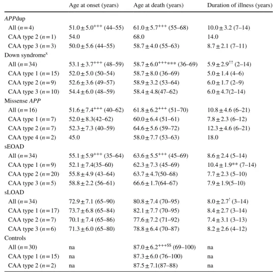
Type 1 describes cases predominantly with many diffuse and cored Aβ plaques, throughout the cerebral cortex, in which CAA is confined within leptomeningeal vessels. Type 2 describes cases where, along with many diffuse and cored Aβ plaques, CAA is present in both leptomeningeal and
deeper penetrating arteries, especially within occipital cor-tex. Type 3 describes cases where capillary CAA is present along with arterial CAA, especially within primary visual cortex, but with relatively few Aβ plaques. Type 4 describes a predominantly vascular phenotype, where Aβ deposition is much more prevalent in and around blood vessels throughout the brain and Aβ plaques are scarce or absent.
Representa-tive images for each CAA subtype are shown in Fig. 1. All
phenotype assessments were performed by a single, highly experienced neuropathologist (Mann), blinded to patient grouping, based on a ‘template’ derived from previous
work [2] in which the assessment methodology had been
validated.
APOE genotyping
DNA was extracted from frozen cerebellum or frontal cortex by routine methods from which APOE genotype was
deter-mined [50]. APOE genotyping was possible for all 4 APPdup
patients, 25/34 DS individuals, 13/16 missense APP muta-tion patients, all 34 sEOAD patients, 33/34 sLOAD patients, and all 30 controls. The lack of available frozen tissue pre-vented APOE genotyping for those remaining individuals.
Statistical analysis
Rating data were entered into an excel spreadsheet and analysed using Statistical Package for Social Sciences (SPSS) software (version 20.0). Patients were stratified according to genetic subtype for statistical analysis of the effect of each mutation on the underlying Aβ pathology. Comparisons of semi-quantitative scores for intensity of Aβ immunostaining were performed using Kruskal–Wallis test with post hoc Mann–Whitney test where Kruskal–Wal-lis yielded a significant difference between antibody stain-ing scores. Group comparisons of age at onset, age at death and duration of illness were made using ANOVA with post hoc Tukey test. Comparisons of frequency of APOE alleles and genotypes were performed using chi-square test. Cor-relations between rating data were made using Spearman rank correlation test. In all instances, significance levels were set at p < 0.05.
Fig. 1 The four different CAA phenotypes as seen on immunostaining for Aβ. Type 1 describes cases predominantly with many diffuse and cored Aβ plaques, throughout the cerebral cortex in which CAA is confined in leptomeningeal vessels. Type 2 describes cases where, along with many diffuse and cored Aβ plaques, CAA is present in both leptomeningeal and deeper penetrating arter-ies, especially within occipital cortex. Type 3 describes cases where capillary CAA is present along with arterial CAA, especially within primary visual cortex, but with relatively few Aβ plaques. Type 4 describes a predominantly vascular phenotype, where Aβ deposition is much more prevalent in and around blood vessels throughout the brain and Aβ plaques are scarce or absent. Immunoper-oxidase–haematoxylin
CAA type 1 CAA type 2
CAA type 3 CAA type 4
Results
Demographic observations
There were significant differences between APPdup, DS, missense APP mutations, sEOAD and sLOAD groups
with respect to mean age at onset of disease (F4,93 = 52.6,
p < 0.001). By definition, patients with sLOAD had a sig-nificantly later age at onset of illness (p < 0.001) compared to those with APPdup, DS, missense APP mutations and sEOAD, but there were no significant differences in age at onset between any of the early onset AD and DS groups
(Table 1). Similarly, there were significant differences
between APPdup, DS, missense APP mutations, sEOAD and sLOAD cases and control groups with respect to
mean age at death (F5,146 = 99.1, p < 0.001) with patients
with sLOAD, and controls, all (by definition) dying at a later age (p < 0.001) than patients with APPdup and mis-sense APP mutations, sEOAD and individuals with DS. Again, there were no significant differences in age at death between any of the early onset AD and DS groups
(Table 1). Patients with sEOAD were slightly older at
death than those individuals with DS (p = 0.017). The controls died at a later age than the patients with sLOAD
(p = 0.002) (Table 1). There were also significant
differ-ences in mean duration of illness (F4,93 = 5.1, p = 0.001)
between patients with APPdup, DS, missense APP muta-tions, sEOAD and sLOAD. Patients with missense APP mutations had a longer duration of illness than individuals with DS (p < 0.001) and patients with sLOAD (p = 0.042).
Consistent with general longevity, mean age at onset and mean age at death, were both significantly later in females compared to males in the sLOAD group alone (p = 0.004), but no significant gender difference was seen with respect to duration of illness. There were no gender differences in age at onset, age at death or duration of illness for each CAA subtype within each patient group.
Braak and Thal stageing
Based on AT8 immunostaining for tau proteins (Braak stage) and 4G8 immunostaining for Aβ (Thal phase), all 4 patients with APPdup and all 16 with missense APP muta-tions were at Braak tangle stage 6 and Thal amyloid phase 5. Of the 34 individuals with DS, 11 (32%) were at Braak stage 5 and 23 (68%) at Braak stage 6; all were at Thal stage 5. Of the 34 patients with sEOAD, 10 (29%) were at Braak stage 5 and 24 (71%) were at Braak stage 6, and 2 (6%) were at Thal stage 4 and 32 (94%) were at Thal stage 5. Of the 34 patients with sLOAD, 2 (6%) were at Braak stage 4, 6 (18%) were at Braak stage 5 and 26 (76%) were
at Braak stage 6, and 6 (18%) were at Thal stage 4 and 28 (82%) were at Thal stage 5. Of the 30 controls, 1 (3%) was Braak stage 0, 13 (43%) were at Braak stage 1 and 16 (54%) were at Braak stage 2, and 9 (30%) were Thal stage 0, 10 (33%) were at Thal stage 1 and 11 (37%) were at Thal stage 2 (see Supplementary Table 1).
Pathological changes and CAA phenotypes
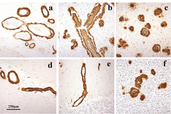
APPdup
As evidenced by 4G8 immunostaining, CAA was severe in all four patients, but did not present with a uniform appearance.
In patients #1 (Fig. 2a–c), #3 (Fig. 2g–i) and #4
(Fig. 2j–l), there was very severe involvement of all arteries
in occipital cortex, both leptomeningeal and intraparenchy-mal extending to variable degrees into the capillary bed, with some perivascular deposition of Aβ. There were rela-tively few amyloid plaques in comparison with the degree of vascular and perivascular involvement, most of these being
cored-type plaques (Fig. 2b, h, k). This pattern of CAA,
which most closely resembled type 3 (see [2]), was also
seen in frontal and temporal cortical regions. CAA was also severe in the cerebellum, but mostly confined to
leptome-ningeal arteries (Fig. 3a, c, e). There were variable
num-bers of amyloid plaques, mostly as diffuse deposits within
the molecular layer in patient #3 (Fig. 3f), but in patient
#1 there were coarser, irregular deposits in Purkinje and
granule cell layers (Fig. 3b). The cerebellum was not
avail-able for study in patient #4. In general, the severity of the changes in patient #3 was less than that in patients #1 and #4, though they essentially retained the same pathological characteristics.
In patient #2, severe CAA was seen across all three
corti-cal regions affecting leptomeningeal (Fig. 2d) and
paren-chymal (Fig. 2e) arteries but only very rarely extending
into the capillary bed (Fig. 2f). Dense perivascular deposits
were not seen, and Aβ plaques were present in all regions. In the cerebellum, moderate CAA was present confined to
leptomeningeal arteries (Fig. 3e) but many diffuse deposits
were seen in the molecular layer and some coarser deposits
in Purkinje cell layer (Fig. 3f). This pattern, in the main,
conformed to type 2 CAA [2] though the very limited
capil-lary involvement in occipital cortex (Fig. 2f) would suggest
a phenotype verging on type 3 CAA.
Down syndrome
As seen in 4G8 immunostaining, amyloid deposition in the form of plaques and CAA was widely present in all 34 indi-viduals with DS. CAA was present as type 1 in 15 (44%)
cases, type 2 in 9 (26%) cases and type 3 in 10 cases (29%)
(Table 1).
Missense APP mutations
By 4G8 immunostaining, CAA was present in all 16 patients with missense APP mutations, being type 1 in 7 of the 14 patients with codon 717 mutation and type 2 in the
other 7 patients (Table 1). The two patients with APP692
mutation displayed a unique phenotype (type 4) in which arteriolar and capillary CAA with perivascular deposition of amyloid (dyshoric angiopathy), but few/no plaques, was predominant within occipital cortex, in addition to severe
leptomeningeal CAA (Fig. 4a–c). In the cerebellum, CAA
was severe and sometimes affected parenchymal arteries in both the molecular and granule cell layer, again with perivascular deposition especially in granule cell layer
(Fig. 4d–f).
Fig. 2 Patterns of immunostaining for Aβ as CAA and plaques in occipital cortex in APPdup patients #1 (a–c), #2 (d–f), #3 (g–i) and #4 (j–l). Immunoperoxidase–haematoxylin
Sporadic EOAD and LOAD and control subjects
Using 4G8 immunostaining, CAA was present in all 34 patients with sEOAD (as type 1 in 9 patients (26%), type 2 in 20 patients (59%) and type 3 in 5 patients (15%)), in 30/34 patients with sLOAD (as type 1 in 17 patients (57%), type 2 in 7 patients (23%) and type 3 in 6 patients (20%), but only in 17 controls (57%) (as type 1 in 15 subjects (88%) and type
2 in the other 2 subjects (12%) (Table 1).
We carefully examined routine haematoxylin–eosin stained sections from the cortical and cerebellar regions assessed for CAA in all cases for evidence of overt ICH/ microbleeds, sections, but found none to be significantly, or at least consistently, present in any patient group or CAA subtype.
Comparisons of CAA phenotype
The proportion of patients showing different CAA
pheno-types varied between the pathological groups (Fig. 5). The
proportion of individuals with DS showing type 1 CAA (44%) was significantly greater (p = 0.003) than those with sEOAD (27%) but not those with sLOAD (57%). Also, the proportion of individuals with sLOAD (57%), and that of controls (88%), showing type 1 CAA was both significantly greater than that proportion of sEOAD patients with type 1 CAA (p = 0.014 and 0.0003, respectively). Conversely, the proportion of patients with sEOAD with type 2 CAA (59%) was significantly greater than that in those with DS (24%, p = 0.002) or sLOAD (23%, p = 0.004) or in controls (12%, p = 0.001). For type 3 CAA, the proportion of individuals with APPdup (75%) was significantly greater than those with DS (27%, p = 0.014), sEOAD (15%, p = 0.005) and sLOAD (20%, p = 0.019), and was also greater (p < 0.05 by Fisher exact test) than those with missense APP mutations and con-trols (0% in both).
When stratified according to each CAA phenotype, there were no significant differences with respect to mean age at onset of disease for patients with APPdup, missense APP mutations, sEOAD and sLOAD, or for mean age at death between any of the CAA phenotypes for APPdup, DS, mis-sense APP mutations, sEOAD, sLOAD and control cases
(Table 1). Only in sEOAD group did duration of illness vary
(F2,29 = 5.5, p = 0.01) between CAA phenotypes, with CAA
type 1 cases having a significantly longer duration of illness
than those with type 2 CAA (p = 0.01) (Table 1).
Comparisons of plaque and CAA scores
Both overall plaque (χ2 = 109.2, p < 0.001) (Fig. 6a) and
CAA (χ2 = 68.0, p < 0.001) scores differed significantly
across the six groups. The degree of plaque formation (over-all plaques scores) was significantly greater in both DS and missense APP mutations than in sEOAD and sLOAD cases (p < 0.001 in every instance). As would be expected, all 5 pathological (AD) groups had a significantly greater degree of plaque formation than controls (p < 0.001), but the degree of plaque formation was not significantly different between
sEOAD and sLOAD cases (Fig. 6a).
As would be expected, all five pathological (AD) groups had a significantly greater degree of CAA than controls (p < 0.001) but, in contrast to plaque scores, the degree of CAA was also significantly greater in sEOAD than sLOAD
(p = 0.014) (Fig. 6b). On the other hand, the severity of CAA
was significantly greater in both APPdup (p = 0.005 and p = 0.009, respectively), missense APP mutations (p = 0.016 and p = 0.001, respectively) and DS (p = 0.008 and p < 0.001, respectively) than in sEOAD and sLOAD, and the severity of CAA was greater in APPdup than that in DS (p = 0.014) but not so for missense APP mutations (p = 0.056), with there being no significance difference between the missense APP mutations and DS (Fig. 6b).
Fig. 3 Patterns of immunostaining for Aβ as CAA and plaques in cer-ebellum in APPdup patients #1 (a, b), #2 (c, d) and #3 (e, f). Immun-operoxidase–haematoxylin
There were no significant differences between males and females in either plaque or CAA scores for each pathological group, or for each CAA subtype within each pathological group.
Relationship between CAA subtypes and plaque or CAA severity
When stratified by CAA subtype, there were no significant
Fig. 4 Patterns of immunostaining for Aβ as CAA and plaques in occipital cortex (a, b) and cerebellum (c, d) in missense APP patient #61. Immunoperoxidase–haematoxylin
Fig. 5 Percentage frequency of the four CAA phenotypes within each of the six different pathological groups
APPdup DS missense APP sEOAD sLOAD control
Percentage of case
differences in overall plaque scores between each CAA subtype for any of the six groups. However, overall sever-ity of CAA did vary significantly across subtypes for DS
(χ2 = 13.0, p < 0.001), sEOAD (χ2 = 13.1, p = 0.001) and
sLOAD (χ2 = 14.4, p < 0.001), but not for patients with
APP-dup or missense APP mutation, or controls. In both mis-sense APP mutations and sEOAD there was a significantly greater level of CAA as both CAA type 2 (p = 0.001 and 0.004, respectively) and type 3 (p < 0.001 and 0.002, respec-tively) than as type 1, with no significant differences in CAA
severity between type 2 and type 3 cases. In DS, sLOAD and controls, there was a significantly greater level of CAA in type 1 than type 2 and type 3 (p < 0.006 and 0.002), with no significant differences in CAA severity between type 2 and type 3 cases.
There were significant correlations between CAA severity scores and CAA phenotype when all cases were considered
together (rs = 0.646, p < 0.001) or separately as individual
pathological groups (DS, rs = 0.604, p < 0.001; missense
APP mutations, rs = 0.612, p = 0.012; sEOAD, rs = 0.628,
Fig. 6 Box plots of scores for severity of plaques (a) and CAA (b) across the six different pathological groups. +++ Sig-nificantly different (p < 0.001) from controls. ***Significantly different (p < 0.001) from both sEOAD and sLOAD. ! Signifi-cantly different (p < 0.05) from DS. $Significantly different (p < 0.05) from sLOAD
APPdup DS missense APP sEOAD sLOAD control
APPdup DS missense APP sEOAD sLOAD control
Plaque scor e CAA scor e
a
b
p < 0.001; sLOAD, rs = 0.680, p < 0.001). For controls,
this correlation failed to reach significance (rs = 0.377,
p = 0.136). The number of cases with APPdup was too low to permit this particular analysis. For all groups, CAA severity scores progressively increased with ascending CAA
pheno-type class (Fig. 7).
Comparisons of patterns of immunostaining using end‑specific antibodies
The pattern of immunostaining for Aβ as plaques or CAA was compared for 4G8, BC05 and BA27 antibodies. Irre-spective of pathological/genetic grouping, all blood vessels stained for CAA by 4G8 also appeared to be detected by
BA27 but fewer were detected (and less strongly so) with BC05. On the other hand, all plaques detected by 4G8 also appeared to be detected by BC05, but only a subset (of cored
plaques) was detected by BA27 (Fig. 8).
Semi-quantitative analysis of the rating data for the level of immunostaining of plaques and CAA by 4G8, BC05 and BA27 antibodies was performed on the 30 cases collectively. The degree of immunostaining of plaques
dif-fered significantly between the three antibodies (χ2 = 37.9,
p < 0.001) with scores for rating of plaque density with 4G8 antibody being significantly greater than that for BC05 antibody (p = 0.019) and BA27 antibody (p < 0.001), and that for BC05 also being significantly greater than BA27 (p < 0.001). Similarly, the degree of immunostaining
Fig. 7 Box plots of scores for CAA severity scores across the four different CAA phenotypes for all cases collectively (a) and individually for APPdup (b), DS (c), missense APP muta-tions (d), sEOAD (e), sLOAD (f) and control (g) groups
a
b
c
d
e
f
g
1 2 3 4 CAA subtype 5 10 15 20 CAA scor e CAA scor e1 CAA subtype 2 1 CAA subtype2 3
CAA subtype 1 2 3 CAA subtype 1 2 3 1 2 3 1 2 CAA subtype CAA scor e CAA scor e 5 10 15 20 5 10 15 20 5 10 15 20 CAA scor e CAA scor e CAA scor e 26 10 26 10 CAA subtype 2 68 10
for CAA differed significantly between the three
antibod-ies (χ2 = 6.8, p = 0.033) with scores for rating of CAA
density with 4G8 and BA27 antibodies being significantly greater than that for BC05 antibody (p = 0.035 and 0.019, respectively). No significant difference between scores for 4G8 and BA27 antibodies was found (p = 0.582).
Despite there being significant differences in the levels of immunostaining for plaques and CAA between the three antibodies, regression analysis showed highly significant correlations between plaque scores for each of the three antibodies (p < 0.001 in every instance), and likewise for CAA scores (p < 0.001 in every instance).
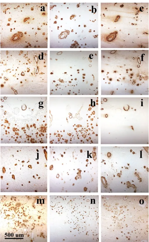
Fig. 8 Adjacent sections of occipital cortex from APPdup case #1 (a–c), DS case #17 (d–f), missense APP mutation cases #49 (g–i) and #54 (j–l) and sEOAD case #58 (m–o) immunostained for Aβ using 4G8 antibody to detect total Aβ (a, d, g, j, m), BC05 antibody to detect Aβ42(3) (b, e, h, k, n) and BA27 antibody to detect Aβ40 (c, e, i, l, o). All blood vessels stained for CAA by 4G8, irrespective of genetic or pathological group, or CAA phenotype, also appeared to be strongly immunoreactive for Aβ40 but less strongly for Aβ42(3). On the other hand, all plaques detected by 4G8 were strongly immunoreactive for Aβ42(3) but only a subset (of cored plaques) appeared to con-tain Aβ40. Immunoperoxidase (with haematoxylin counterstain in a, d, g, j, m)
Effect of APOE genotype
Overall, APOE ε4 allele frequency in sEOAD patients was significantly higher than that in DS individuals (p < 0.0001), missense APP patients (p < 0.001) and controls (p < 0.001), as was this in sLOAD patients compared to DS (p < 0.001), missense APP mutations (p < 0.001) and controls (p < 0.001)
(Table 2). There were no significant differences in ε4 allele
frequency between DS and missense APP patients, between DS and controls, or between sEOAD and sLOAD patients
(Table 2). None of the APPdup patients bore APOE ε4 allele
and, therefore, could not be included in the comparisons. There were no significant differences in APOE ε2 allele frequency between any of the groups, except that this was significantly higher in controls (p = 0.038) compared to
sLOAD patients (Table 2). None of the APPdup or missense
APP mutation patients bore APOE ε2 allele (Table 2) and so could not be included in these comparisons.
There was no significant effect of possession of at least one copy of APOE ε4 allele, or possession of none, one or two ε4 alleles, on overall plaque severity in DS, sEOAD or sLOAD. Likewise, there was no significant effect of pos-session of at least one copy of APOE ε4 allele on overall CAA severity in DS and sEOAD, but in sLOAD severity of CAA was significantly greater in APOE ε4 allele bearers compared to non-bearers (p = 0.040). Moreover, when strati-fied according to possession of none, one or two ε4 alleles, the severity of CAA in sLOAD was significantly greater in patients with two APOE ε4 alleles than both those with one or no APOE ε4 alleles (p = 0.013 in both instances) with no significant difference between those with one or no APOE ε4 alleles. No such effect was seen in sEOAD. It was not possible to test for the effects of homozygosity for APOE ε4 allele in DS as there were no such individuals represented in the cohort. Likewise, the effects of possession of APOE
ε4 allele could not be tested in APPdup or missense APP mutation due to lack of patient representation.
Due to the low frequency of APOE ε4 allele, it was not possible to ascertain whether APOE genotype influenced CAA phenotype in patients with APPdup and missense APP
mutations (Tables 2, 3). Comparisons of APOE ε4 allele
frequency between each of the CAA phenotypes within DS, sEOAD, sLOAD and control groups showed no sig-nificant differences, except that APOE ε4 allele frequency was significantly greater in type 1 CAA and type 3 CAA in comparisons between sEOAD and sLOAD, and sEOAD
and controls (Table 3). By contrast, the proportion of ε4ε4
homozygotes in sEOAD and sLOAD groups combined was numerically greater in type 2 (32%) and type 3 (67%) CAA compared to type 1 (11%), and the proportion of these was significantly greater in type 3 CAA (p = 0.004), and tending
Table 2 APOE allele and genotype numbers (percentage frequency in parentheses) in APP duplication (APPdup), Down syndrome, missense APP mutations, sporadic early onset Alzheimer’s disease (sEOAD), late onset Alzheimer’s disease (sLOAD) and controls
***Significantly different from sEOAD, p < 0.001
+,+++ Significantly different from sLOAD, p < 0.05, < 0.001 respectively
APPdup (n = 4) Down syndrome (n = 25) Missense APP (n = 13) sEOAD (n = 34) sLOAD (n = 33) Controls (n = 30)
ε2/ε2 0 (0) 0 (0) 0 (0) 1 (2.1) 0 (0) 0 (0) ε2/ε3 0 (0) 5 (17.8) 0 (0) 2 (4.2) 1 (3.0) 5 (16.7) ε2/ε4 0 (0) 1 (3.6) 0 (0) 0 (0) 0 (0) 1 (3.3) ε3/ε3 4 (100) 16 (57.1) 11 (84.6) 14 (41.2) 7 (21.2) 20 (66.7) ε3/ε4 0 (0) 6 (21.4) 2 (15.4) 9 (26.5) 16 (48.4) 4 (13.3) ε4/ε4 0 (0) 0 (0) 0 (0) 8 (23.5) 9 (27.3) 0 (0) ε2 0 (0) 6 (10.7) 0 (0) 4 (6.0) 1 (1.5) 6 (10.0)+ ε3 8 (100) 44 (78.6) 24 (92.3) 39 (57.3) 31 (46.9) 49 (81.2) ε4 0 (0) 6 (10.7)***,+++ 2 (7.7)***,+++ 25 (36.7) 34 (51.5) 5 (8.8)***,+++
Table 3 Number (percentage in parentheses) of cases in APP
duplica-tion (APPdup), Down syndrome, missense APP mutaduplica-tions, sporadic early onset Alzheimer’s disease (sEOAD), sporadic late onset Alz-heimer’s disease (sLOAD) and controls with each CAA phenotype (upper half)
Also shown (lower half) are APOE ε4 allele frequencies associated with each CAA phenotype
**,***Significantly different from controls, p < 0.01, < 0.001, respec-tively
APOE ε4 allele frequencies in each CAA phenotype 1 2 3 4 APPdup (n = 4) 0.000 0.000 0.000 0.000 Downs syndrome (n = 28) 0.100 0.188 0.100 0.000 Missense APP (n = 13) 0.000 0.071 0.000 0.250 sEOAD (n = 34) 0.272** 0.388 0.500 0.000 sLOAD (n = 30) 0.438*** 0.643 0.833 0.000 Controls (n = 17) 0.067 0.25 0.000 0.000
to be greater in type 2 CAA (p = 0.065), compared to type 1 CAA, with no significance between type 2 and type 3 CAA.
Discussion
In the present study, we have compared the extent and pattern of Aβ deposition, both as plaques and as CAA, in patients with duplications in APP, others with missense mutations in APP, patients with sEOAD and sLOAD, older individuals with DS and elderly controls. The main findings to emerge were:
1. The degree of plaque formation was greater in both DS and missense APP mutations than in sEOAD and sLOAD cases, while the degree of plaque formation was not significantly different between sEOAD and sLOAD. 2. Conversely, the severity of CAA was significantly
greater in both APPdup and missense APP mutations, and DS, compared to sEOAD and sLOAD. Again, all five pathological (AD) groups had a significantly greater degree of CAA than controls, and the degree of CAA was also significantly greater in sEOAD than sLOAD, and in APPdup compared to DS.
3. When stratified by CAA subtype, no significant differ-ences in overall plaque scores were seen between each CAA subtype for any of the six groups. However, in both APP mutations and sEOAD there was a significantly greater level of CAA as types 2 and 3 CAA compared to type 1. Conversely, in DS, sLOAD and controls there was a significantly greater level of CAA in type 1 CAA than types 2 and 3. In APPdup type 3 was the predomi-nant CAA phenotype.
4. CAA severity scores progressively increased across CAA types 1–4 for all cases combined and for each pathological group individually.
5. APOE ε4 allele frequency was overall significantly higher in sEOAD than in DS, missense APP and con-trols, as was ε4 allele frequency in sLOAD compared to DS, missense APP mutations and controls. There were no significant differences in APOE ε4 allele frequency between DS, missense APP and controls. None of the APPdup patients bore APOE ε4 allele.
6. All blood vessels stained for CAA by 4G8 appeared to be detected by BA27 but fewer were detected with BC05. Conversely, all plaques detected by 4G8 appeared to be detected by BC05, but only a subset was detected by BA27.
7. APOE ε4 allele frequency varied numerically between each of the CAA phenotypes in APPdup, missense APP mutations, sEOAD, sLOAD, DS and controls, but not significantly so. Nonetheless, APOE ε4 homozygosity was more commonly associated with type 3 CAA than
types 1 and 2 CAA, and was associated with a greater severe of CAA overall in sLOAD but not in sEOAD or DS.
In a recent study, Head and colleagues [15] compared
the overall extent of CAA, atherosclerosis and arteriolo-sclerosis in 32 individuals with DS, ranging in age from 43 to 70 years, and in 80 individuals mostly with late onset sporadic AD (sLOAD) and 37 controls. Younger patients with sEOAD and APP mutations were not specifically inves-tigated in this latter study. Nonetheless, like ourselves, these authors found that CAA occurred at significantly higher fre-quencies in the brains of individuals with DS compared to sLOAD cases and controls, with the DS cohort being 1.2 times more likely to have CAA relative to sLOAD cases, and 4.6 times more likely to have CAA compared to con-trol cases. On the other hand, atherosclerosis and arteriolo-sclerosis were rare in cases with DS. Such observations of significantly more frequent CAA, and a greater severity of CAA when present, in people with DS relative to sLOAD and control cases are consistent with the hypothesis that such changes are driven, at least partially, by an overexpression of APP. The lack of AD neuropathology and CAA, even at greater than 70 years of age, in rare cases of DS with partial
trisomy 21 where APP is not overexpressed [12, 35] would
be consistent with this argument.
Nonetheless, how an overexpression of APP might be translated into enhanced CAA remains unclear, but could involve deficiencies in clearance mechanisms when faced with such an overload of Aβ. In this latter context, it has been postulated that the strong association between age, CAA and AD pathology in the general population is driven, at least partially, by an impaired efficiency of cerebral ves-sels in later life in expelling extracellular fluid containing soluble forms of Aβ as a consequence of atherosclerosis/
arteriosclerosis within such vessels [49]. Nonetheless,
indi-viduals with DS appear to be protected against
hyperten-sion [1] and atherosclerosis, and show less cerebrovascular
pathology typically associated with cardiovascular risk fac-tors, including atherosclerotic lesions and
arteriolosclero-sis [15, 22]. Paradoxically, this ought to result in a better
preservation of perivascular drainage channels in DS, and consequently a less severe, rather than more severe, CAA compared to sLOAD. Potential inefficiencies in perivascu-lar drainage might not appertain to individuals with APP mutations or DS since these would only be anticipated to occur beyond an age at which most would generally survive to. However, Aβ can also be cleared from the brain through several other routes, involving endocytosis by microglia and astrocytes, or enzymatic degradation. It is, therefore, pos-sible that failures in these latter pathways, in conjunction with the overexpression of APP, might foster an inability to expel Aβ and result in severe CAA.
Despite the commonality of sharing duplication at the APP locus, and an overexpression of APP protein, there are clear differences in clinical presentation between APPdup and DS individuals. Stroke and ICH are the main clinical consequences of vascular amyloidosis in APPdup, occurring in at least one-third of all cases, but both are uncommon in individuals with DS, with only a handful
of case reports of this in the literature (see [5]). Indeed,
it has been estimated that haemorrhagic stroke occurs in
only about 3–4% of older people with DS [43], some ten
times less frequent than in patients with APPdup. Why these clinical differences should occur is unclear, given that DS differs from APPdup only in the number of other genes located on chromosome 21 that are also triplicated. In most instances of DS, a full triplication of chromosome 21 is present, whereas in APPdup triplication of APP locus
is variable, generally ranging from 0.5 to 6.5 Mb [48],
with the region of triplication in some instances being
limited to APP gene alone [41], while in others it may
extend to include up to 12 other genes [37]. This raises
the question as to whether the possession of other tripli-cated genes in some way confer some degree of protec-tion against the likelihood of stroke or ICH in DS. These genes may be involved in the production of Aβ, of which there are two main pathways—the secretory pathway or the endo-lysosomal pathway. In the latter, Aβ cleavage occurs in the endosomal compartment where pH is optimal for β-secretase activity. Enlargement of the early endoso-mal compartment is considered one of the earliest mor-phological alterations detectable in postmortem tissues in
sporadic AD, in most APP mutations and in DS [8]. APP
overexpression is implicated in the formation of enlarged endosomes, but the mechanism is uncertain. Interestingly, it was recently shown that increase of β-CTF, the C-termi-nal fragments of APP generated after β-secretase cleavage, can produce enlarged endosomes in fibroblasts from DS
individuals [20] while in lymphoblastoid cell lines from
individuals with APPdup endosomes appeared to be of
normal size [9].
However, with regards to clinical phenotype, it is
nota-ble that in the Dutch family [41] where triplication of APP
locus was restricted to APP gene alone, and in the Swedish family the region of triplication extended to cover only those two genes either side of APP, no instances of ICH
were reported [48]. Conversely, ICH commonly occurred
in a Finnish family with 0.55 Mb duplication covering APP and four other neighbouring genes [38]. By contrast, in the French families where there is a greater and more
variable extension of triplication of APP locus [6, 37],
ICH occurred in about 30% of patients though, notably, dementia but not ICH defined the clinical course of the four APPdup patients studied here in whom the region of
triplication was 0.78 Mb [6]. Hence, factors other than
size of APP triplication per se may account for the low prevalence of ICH in DS compared to APPdup.
Alternatively, it may be differences in the actual CAA phenotype present that are responsible. Type 1 CAA tended to be more common in DS and sLOAD, and type 2 CAA was more common in missense APP mutations and sEOAD than in the other groups. Type 3 CAA was more common in individuals with APPdup than those with DS as well as others with sEOAD or missense APP mutations. Type 4 was only seen in patients with APP692 mutation, though 2 of the APPdup patients (patients #1 and 3), although designated as CAA type 3, showed a particularly severe phenotype that was reminiscent of type 4 CAA. Consequently, patients with APPdup in the present study had a more severe CAA phe-notype than most individuals with DS, despite the similar ages, with only 30% DS individuals showing this type 3 phenotype, and then not to the same degree of severity as in patients with APPdup. It is possible, therefore, that it is the lesser extent of CAA in DS (compared to APPdup) that low-ers the risk of CAA-related stroke and haemorrhage in such individuals. Correlations between CAA severity scores and CAA phenotype suggest that the different CAA phenotypes exist on a continuum with type 1 being the least ‘aggres-sive’ form, and types 2–4 following progressively as Aβ becomes deposited further along the arterial tree reaching into parenchymal arteries and arterioles (type 2) and finally into capillaries (types 3 and 4). Certainly, this would accord
with the proposal put forward by Weller et al. [49] based
on a progressive slowing of perivascular drainage leading to build up of deposits in vessel walls. However, if so, this would not explain the relative absence of amyloid plaques in types 3 and 4 CAA, and the highest levels of plaques in type 1 CAA.
APP is processed by proteolytic enzymes known as secretases. Cleavage within the APP domain containing the Aβ sequence by α-secretase precludes its formation, whereas sequential cleavage at the amino- and carboxyl- termini of the Aβ sequence by β- and γ-secretases, respectively, releases Aβ into the extracellular fluid of the brain. Most of
this occurs as a more slowly aggregating form, Aβ40,
com-pared to the longer, more rapidly aggregating form, Aβ42(3).
It has been shown that in both AD and DS the Aβ in CAA
is composed mostly of Aβ40, whereas in plaques it is Aβ42(3)
that predominates [17, 18, 40, 44]. The differential pattern
of composition of Aβ within brain parenchyma and blood vessel walls can be explained by the relative aggregation
properties of Aβ40 and Aβ42(3), with the less aggregation
prone Aβ40 travelling further along perivascular drainage
channels and ultimately reaching blood vessel walls, com-pared to the less abundant though more rapidly
aggregat-ing Aβ42(3) which coalesces into plaques within the brain
parenchyma. In familial AD due to certain missense muta-tions in APP (for example those at or around codon 717)
proteolytic processing of APP elevates levels of Aβ42(3)
rela-tive to Aβ40 [39] with excessive numbers of Aβ42(3)
contain-ing plaques becontain-ing formed as a consequence [26]. Other
mis-sense mutations (for example, those around codons 692 and
693) enhance the aggregation properties of both Aβ40 and
Aβ42(3) without affecting levels of production. As mentioned earlier triplication at APP locus will increase production of
both Aβ40 and Aβ42(3).
Hence, the different CAA phenotypes seen here in the different forms of AD might in some way reflect the relative
proportions of Aβ40 and Aβ42(3) being generated. In APPdup
and DS, where excess amounts of Aβ40 (and Aβ42(3)) are
pro-duced, this could lead to failure to expel this from the extra-cellular fluid leading to a massive build up in smaller arteries and capillaries evidenced as the more extensive type 3 CAA. In missense APP mutations such as those at codon 692,
the mutated, more highly aggregation prone, form of Aβ40
would promote its excessive deposition in vessel walls, and
again result in the severe type 4 CAA (see [26] for APP693
mutations), whereas no excess of Aβ40 is generated in
mis-sense mutations in APP occurring around codon 717, and in these cases the less extensive type 1 and 2 CAA predomi-nate. In sEOAD and sLOAD, where normal levels of both
Aβ40 and Aβ42(3) are produced, the different CAA
pheno-types present might reflect the relative efficiencies in which
Aβ40 is cleared through the perivascular drainage channels.
Present observations that all blood vessels stained for CAA by 4G8, irrespective of genetic or pathological group, or CAA phenotype, also appeared to be strongly
immunoreac-tive for Aβ40 but less strongly for Aβ42(3). On the other hand,
all plaques detected by 4G8 were strongly immunoreactive
for Aβ42(3) but only a subset (of cored plaques) appeared to
contain Aβ40. Such findings are consistent with our previous
studies in familial AD and DS [17, 18]. Consequently, in
addition to potential differences in Aβ production, differ-ences in CAA phenotypes between DS and APPdup might also involve amyloid clearance, but alternative mechanisms could involve a unique oxidative stress profile or immune
response in DS [52].
Interestingly, as others have shown [34, 40, 46],
posses-sion of two copies of APOE ε4 allele was associated with a greater severity of CAA in sLOAD patients alone. However, APOE genotype per se did not greatly influence the actual CAA phenotype in any pathological group. Although there was a numerical increase in ε4 allele frequency from type 1 to type 3 CAA in sEOAD and sLOAD, these differences were not substantiated statistically. Nonetheless, it is known that an increase in ε4 allele copy from 0 to 2 is associated
with higher levels of Aβ40 deposition (as plaques) in sLOAD
[13, 27], possession of ε4 allele/ApoE E4 isoform decreases
brain clearance of Aβ [7], and ApoE E4 isoform promotes
fibrillogenesis [23]. In these respects, possession of APOE
ε4 allele/E4 isoform could potentiate the development of
CAA, as type 3 CAA, in those patients with sLOAD bearing APOE ε4ε4 genotype, a suggestion in keeping with previous
studies [46]. The absence of type 3 CAA in elderly controls
and missense APP mutations involving codon 717, would also accord with the relative infrequency of APOE ε4 allele and ε4ε4 homozygosity in such individuals. However, hav-ing said that, overall severity of CAA and prevalence of type 3 CAA were equivalent in DS individuals as in patients with sEOAD and sLOAD, despite there being in DS a relative lack of APOE ε4 alleles, and a complete absence of ε4ε4 homozygotes.
While there were no significant differences in overall plaque scores between sEOAD and sLOAD cases, overall CAA scores were lower in sLOAD than sEOAD. Further-more, age at death did not vary significantly between CAA phenotypes for these two groups though the proportion of cases with the less severe type 1 CAA was greater in sLOAD than sEOAD, suggesting that advancing age per se may, if anything, lessen the severity of CAA and phenotype present, at least in AD. This observation was not due simply to a shorter duration of disease in sLOAD compared to sEOAD which might potentially have terminated disease progression at an early stage in the later onset cases.
Hence, the factors that determine CAA phenotype are complex and remain unclear, possibly involving differential
levels of production or clearance of Aβ40 or shorter sized
peptides, or factors which promote its aggregation such as ApoE E4 isoform, or some combination of all of these. Moreover, why there should be a distinction in CAA phe-notype profiles between DS and APPdup is puzzling and it is curious why sEOAD should be more strongly associ-ated with type 2 CAA compared to sLOAD (and vice versa for type 1 CAA), but differential possession of APOE ε4 allele does not appear to determine this. Interestingly,
previ-ous studies [28] have shown type 2 CAA to be particularly
common in early onset familial AD associated with PSEN-1 mutations, especially in those where the mutation is located after codon 200, in the absence of any APOE ε4 allele modifying effect. Possibly sEOAD shares some genetic or mechanistic risk affinity with such PSEN-1 mutations, which ultimately translate into a similar CAA phenotype. The neu-ropathological differences between the different forms of AD highlighted in this study require further study to elu-cidate the underlying mechanisms. The scientific value in knowing what CAA phenotype is present will help to reduce variability of findings, and provide greater consistency of results, when factors relating to promotion of CAA are being investigated.
Acknowledgements We acknowledge the support of the Manchester Brain Bank by Alzheimer’s Research UK and Alzheimer’s Society through their funding of the Brains for Dementia Research (BDR) Pro-gramme. Manchester Brain Bank also receives Service Support costs from Medical Research Council. This work was supported by Medical
Research Council of UK, Grant Number G0701441 and was also par-tially funded by a Wellcome Trust Strategic Award (Grant Number: 098330/Z/12/Z) conferred upon The London Down Syndrome (Lon-DownS) Consortium.
Open Access This article is distributed under the terms of the
Crea-tive Commons Attribution 4.0 International License (http://creat iveco mmons .org/licen ses/by/4.0/), which permits unrestricted use, distribu-tion, and reproduction in any medium, provided you give appropriate credit to the original author(s) and the source, provide a link to the Creative Commons license, and indicate if changes were made.
References
1. Alexander M, Petri H, Ding Y, Wandel C, Khwaja O, Foskett N (2016) Morbidity and medication in a large population of indi-viduals with Down syndrome compared to the general population. Dev Med Child Neurol 58:246–254
2. Allen N, Robinson AC, Snowden S, Davidson YS, Mann DMA (2014) Patterns of cerebral amyloid angiopathy define histopatho-logical phenotypes in Alzheimer’s disease. Neuropathol Appl Neurobiol 40:136–148
3. Blom ES, Viswanathan J, Kilander L, Helisalmi S, Soininen H, Lannfelt L, Ingelsson M, Glaser A, Hiltunen M (2008) Low preva-lence of APP duplications in Swedish and Finnish patients with early-onset Alzheimer’s disease. Eur J Hum Genet 16:171–175 4. Braak H, Braak E (1991) Neuropathological stageing of
Alzhei-mer-related changes. Acta Neuropathol 82:239–259
5. Buss L, Fisher E, Hardy J, Nizetic D, Groet J, Pulford L, Strydom A (2016) Intracerebral haemorrhage in Down syndrome: protected or predisposed [version 1; referees: 2 approved]. F1000Research 5(F1000 Faculty Rev):876. https ://doi.org/10.12688 /f1000 resea rch.7819.1
6. Cabrejo L, Guyant-Maréchal L, Laquerrière A, Vercelletto M, De la Fournière F, Thomas-Antérion C, Verny C, Letournel F, Pasquier F, Vital A, Checler F, Frebourg T, Campion D, Hanne-quin D (2006) Phenotype associated with APP duplication in five families. Brain 129:2966–2976
7. Castellano JM, Kim J, Stewart FR, Jiang H, DeMattos RB, Pat-terson BW, Fagan AM, Morris JC, Mawuenyega KG, Cruchaga C, Goate AM, Bales KR, Paul SM, Bateman RJ, Holtzman DM (2011) Human apoE isoforms differentially regulate brain amyloid-β peptide clearance. Sci Transl Med 3:89ra57
8. Colacurcio DJ, Pensalfini A, Jiang Y, Nixon RA (2018) Dysfunc-tion of autophagy and endosomal-lysosomal pathways: roles in pathogenesis of Down syndrome and Alzheimer’s Disease. Free Radic Biol Med 114:40–51
9. Cossec JC, Lavaur J, Berman DE, Rivals I, Hoischen A, Stora S, Ripoll C, Mircher C, Grattau Y, OlivoMarin JC, de Chaumont F (2012) Trisomy for synaptojanin1 in Down syndrome is func-tionally linked to the enlargement of early endosomes. Hum Mol Genet 21:3156–3172
10. Davidson Y, Barker H, Robinson AC, Troakes C, Smith B, Al-Saraj S, Shaw C, Rollinson S, Masuda-Suzukake M, Hasegawa M, Pickering-Brown S, Snowden JS, Mann DMA (2014) Brain distribution of dipeptide repeat proteins in frontotemporal lobar degeneration and motor neurone disease associated with expan-sions in C9ORF72. Acta Neuropathol Commun 2:70
11. Domingues-Montanari S, Parés M, Hernández-Guillamon M, Fernández-Cadenas I, Mendioroz M, Ortega G, Boada M, Mas-juan J, Huertas N, Alvarez-Sabín J, Delgado P, Montaner J, Stroke Project of Cerebrovascular Diseases Study Group, Spanish
Society of Neurology (2011) No evidence of APP point mutation and locus duplication in individuals with cerebral amyloid angi-opathy. Eur J Neurol 18:1279–1281
12. Doran E, Keator D, Head E, Phelan MJ, Kim R, Totoiu M, Barrio JR, Small GW, Potkin SG, Lott IT (2017) Down syndrome, partial Trisomy 21, and absence of Alzheimer’s disease: the role of APP. J Alzheimers Dis 56:459–470
13. Gearing M, Mori H, Mirra SS (1996) Ab peptide length and apolipoprotein E genotype in Alzheimer’s disease. Ann Neurol 39:395–399
14. Guyant-Marechal I, Berger E, Laquerrière A, Rovelet-Lecrux A, Viennet G, Frebourg T, Rumbach L, Campion D, Hannequin D (2008) Intrafamilial diversity of phenotype associated with APP duplication. Neurology 71:1925–1926
15. Head E, Phelan MJ, Doran E, Kim RC, Poon WW, Schmidt FA, Lott IT (2017) Cerebrovascular pathology in Down syndrome and Alzheimer disease. Acta Neuropathol Commun 5:93
16. Hyman BT, Phelps CH, Beach TG, Bigio EH, Cairns NJ, Carrillo MC, Dickson DW, Duyckaerts C, Frosch MP, Masliah E, Mirra SS, Nelson PT, Schneider JA, Thal DR, Thies B, Trojanowski JQ, Vinters HV, Montine TJ (2012) National Institute on Aging–Alz-heimer’s Association guidelines for the neuropathologic assess-ment of Alzheimer’s disease. Alzheimers Deassess-ment 8:1–13 17. Iwatsubo T, Odaka A, Suzuki N, Mizusawa H, Nukina N, Ihara
Y (1994) Visualization of A beta 42(43) and A beta 40 in senile plaques with end-specific A beta monoclonals: evidence that an initially deposited species is A beta 42(43). Neuron 13:45–53 18. Iwatsubo T, Mann DMA, Odaka A, Suzuki N, Ihara Y (1995)
Amyloid β-protein (Aβ) deposition: Aβ42(43) precedes Aβ(40) in Down’s syndrome. Ann Neurol 37:294–299
19. Kasuga K, Shimohata T, Nishimura A, Shiga A, Mizuguchi T, Tokunaga J, Ohno T, Miyashita A, Kuwano R, Matsumoto N, Onodera O, Nishizawa M, Ikeuchi T (2009) Identification of independent APP locus duplication in Japanese patients with early-onset Alzheimer’s disease. J Neurol Neurosurg Psychiatry 80:1050–1052
20. Kim S, Sato Y, Mohan PS, Peterhoff C, Pensalfini A, Rigoglioso A, Jiang Y, Nixon RA (2016) Evidence that the rab5 effector APPL1 mediates APP-βCTF-induced dysfunction of endosomes in Down syndrome and Alzheimer’s disease. Mol Psychiatry 21:707
21. Lladó A, Grau-Rivera O, Sánchez-Valle R, Balasa M, Obach V, Amaro S, Rey MJ, Molinuevo JL, Gelpi E, Antonell A (2014) Large APP locus duplication in a sporadic case of cerebral haem-orrhage. Neurogenetics 15:145–149
22. Lott IT, Head E (2005) Alzheimer disease and Down syndrome: factors in pathogenesis. Neurobiol Aging 26:383–389
23. Ma J, Yee A, Brewer HB Jr, Das S, Potter H (1994) Amyloid-associated proteins alpha 1-antichymotrypsin and apolipoprotein E promote assembly of Alzheimer beta-protein into filaments. Nature 372:92–94
24. Mann DMA, Esiri MM (1988) The site of the earliest lesions of Alzheimer’s disease. N Engl J Med 318:789–790
25. Mann DMA, Younis N, Jones D, Stoddart RW (1992) The time course of pathological events concerned with plaque formation in Down’s syndrome with particular reference to the involvement of microglial cells. Neurodegeneration 1:201–215
26. Mann DMA, Iwatsubo T, Ihara Y, Cairns NJ, Lantos PL, Bogda-novic N, Lannfelt L, Winblad B, Maat-Schieman MLC, Rossor MN (1996) Predominant deposition of amyloid β42(43) in plaques in cases of Alzheimer’s disease and Hereditary cerebral haemor-rhage associated with mutations in the amyloid precursor protein gene. Am J Pathol 148:1257–1266
27. Mann DMA, Iwatsubo T, Pickering-Brown SM, Owen F, Saido TC, Perry RH (1997) Preferential deposition of amyloid β protein (Aβ) in the form Aβ40 in Alzheimer’s disease is associated with