HAL Id: inserm-00408027
https://www.hal.inserm.fr/inserm-00408027
Submitted on 28 Jul 2009
HAL is a multi-disciplinary open access
archive for the deposit and dissemination of
sci-entific research documents, whether they are
pub-lished or not. The documents may come from
teaching and research institutions in France or
abroad, or from public or private research centers.
L’archive ouverte pluridisciplinaire HAL, est
destinée au dépôt et à la diffusion de documents
scientifiques de niveau recherche, publiés ou non,
émanant des établissements d’enseignement et de
recherche français ou étrangers, des laboratoires
publics ou privés.
Neuronal activity of histaminergic tuberomammillary
neurons during wake-sleep states in the mouse.
Kazumi Takahashi, Jian-Sheng Lin, Kazuya Sakai
To cite this version:
Kazumi Takahashi, Jian-Sheng Lin, Kazuya Sakai. Neuronal activity of histaminergic
tuberomammil-lary neurons during wake-sleep states in the mouse.. Journal of Neuroscience, Society for Neuroscience,
2006, 26 (40), pp.10292-8. �10.1523/JNEUROSCI.2341-06.2006�. �inserm-00408027�
Behavioral/Systems/Cognitive
Neuronal Activity of Histaminergic Tuberomammillary
Neurons During Wake–Sleep States in the Mouse
Kazumi Takahashi, Jian-Sheng Lin, and Kazuya Sakai
Institut National de la Sante´ et de la Recherche Me´dicale U628, Universite´ Claude-Bernard-Lyon I, 69373 Lyon Cedex 08, France
Using extracellular single-unit recordings alone and in combination with neurobiotin juxtacellular labeling and histamine
immunohis-tochemistry, we have identified, for the first time in nonanesthetized, head-restrained mice, histamine neurons in the tuberomammillary
nuclei of the posterior hypothalamus. They are all characterized by triphasic broad action potentials. They are active only during
wakefulness, and their activity is related to a high level of vigilance. During waking states, they display a slow (
⬍10 Hz) tonic, repetitive,
irregular firing pattern. Their activity varies in the different waking states, being lowest during quiet waking, moderate during active
waking, and highest during attentive waking. They cease firing during quiet waking before the onset of EEG synchronization, the EEG sign
of sleep (drowsy state), and remain silent during slow-wave sleep and paradoxical (or rapid eye movement) sleep. They exhibit a
pronounced delay in firing during transitions from sleep to wakefulness or remain quiescent during the same transitions if the animals
are not fully alert. They either respond with a long delay, or do not respond, to an arousing stimulus if the stimulus does not elicit an overt
alert state. These data support the view that the activity of histaminergic tuberomammillary neurons plays an important role, not in the
induction of wakefulness per se, but in the maintenance of the high level of vigilance necessary for cognitive processes. Conversely,
cessation of their activity may play an important role in both the initiation and maintenance of sleep.
Key words: mouse; histamine neuron; neurobiotin; vigilance; extracellular single-unit recording; hypothalamus
Introduction
Since von Economo’s anatomopathological observations on the
viral encephalitic epidemic of 1918, several lines of experimental
evidence have pointed to a crucial role of the posterior
hypothal-amus in the maintenance of wakefulness (W) (Moruzzi, 1972).
Experimental destruction or inactivation of posterior
hypotha-lamic neurons results in somnolence and hypersomnia. In
addi-tion, inactivation of these neurons restores sleep in various
mod-els of insomnia, produced either by destruction of the preoptic
anterior hypothalamic area or by administration of drugs, such as
para-chlorophenylalanine, a 5-hydroxytryptamine (serotonin or
5-HT) synthesis inhibitor, amphetamine, a psychostimulant, or
modafinil, a newly discovered wake-promoting drug (Lin, 2000).
Of the posterior hypothalamic neurons, the histaminergic
neu-rons in the tuberomammillary nuclei (TM) are the sole source of
neuronal histamine (HA) in the mammalian brain and are
con-tained in widely branching neuronal pathways influencing large
target fields, such as the noradrenergic neurons in the nucleus
locus ceruleus and serotonergic neurons in the dorsal raphe
nu-cleus (DRN) (Schwartz et al., 1991; Wada et al., 1991; Haas and
Panula, 2003). The HA system is therefore thought to play an
important role in a wide variety of physiological functions,
in-cluding sleep/waking regulation, learning, memory, attention,
and control of affective states (Schwartz et al., 1991; Wada et al.,
1991; Haas and Panula, 2003). HA neurons play an important
role in the forebrain waking systems (Haas and Panula, 2003; Lin,
2000). Although much is known about the membrane properties
of HA neurons in the rat posterior hypothalamus (Haas and
Reiner, 1988; Greene et al., 1990; Haas, 1992; Llinas and Alonso,
1992; Stevens and Haas, 1996; Yang and Hatton, 1997), no
infor-mation is available about the unit activity of identified HA
neu-rons in vivo in general and across wake–sleep states in rodents,
including the mouse in particular, despite its increasing use in
experimental models. Determination of the activity of HA
neu-rons across sleep–waking cycles is a prerequisite for an
under-standing of their precise roles in behavioral state control and of
their physiological functions. Here we report for the first time
that the unit activity of identified mouse HA neurons is waking
specific and related to the high level of vigilance necessary for
cognitive processes.
Materials and Methods
Animals and surgery. All experimental procedures followed European Economic Community Guidelines (86/609/EEC) and the Policy on Eth-ics approved by the Society for Neuroscience (1993). Every effort was made to minimize the number of animals used as well as any pain and discomfort.
Male adult C57BL/6 mice (28 –35 g at the time of surgery; Harlan, Gannat, France) were used. Under pentobarbital anesthesia (50 mg/kg
Received June 2, 2006; revised Aug. 15, 2006; accepted Aug. 25, 2006.
This study was supported by Institut National de la Sante´ et de la Recherche Me´dicale U628, Claude Bernard University, and European Contract QLRT-2001-00826 (5th Programme cadre de recherche technologique). K.T. was supported by grants from the Fyssen Foundation. We thank Ge´rard Guidon for his expert technical assistance.
Correspondence should be addressed to Kazuya Sakai, Institut National de la Sante´ et de la Recherche Me´dicale U628, Universite´ Claude-Bernard-Lyon 1, 8 Avenue Rockefeller, 69373 Lyon Cedex 08, France. E-mail: sakai@univ-lyon1.fr.
K. Takahashi’s present address: Department of Physiology, Fukushima Medical University, Fukushima 960-1295, Japan.
DOI:10.1523/JNEUROSCI.2341-06.2006
i.p.), electrodes were implanted to record neocortical and hippocampal electroencephalograms (EEGs), neck muscle activity [electromyogram (EMG)], and heart rate [electrocardiogram (EKG)]. For EEG recordings, screw electrodes were fixed in the frontal, parietal, and occipital cortices, and a stainless steel wire (200m diameter, bare except for 0.5 mm at the tip) was implanted in the dorsal hippocampus. For EMG and EKG re-cordings, two stainless steel wires (250m diameter) were inserted in the neck muscles and one in the lateral chest. In addition, a U-shaped plastic plate (18 mm wide, 16 mm long, 5 mm thick) was fixed stereotaxically to the skull using dental acrylic cement so that the cranium could be pain-lessly returned to the same stereotaxic position using a semichronic head holder (SA-8; Narishige, Tokyo, Japan). A small hole was drilled in the skull above the posterior hypothalamus and covered by antibiotic cream for the subsequent insertion of microelectrodes. After a recovery period of 1 week, the animals were habituated to the head-restrained position by being placed on a cotton sheet inside a plastic box, painlessly restraining the head with a semichronic head holder and preventing large body movements with a cotton-coated plastic covering. During experiments, the head was covered to reduce visual stimuli. Under these conditions, the mice displayed complete sleep–waking cycles, consisting of W, slow– wave sleep (SWS), and paradoxical [or rapid eye movement (REM)] sleep (PS) during all experiments lasting 6 – 8 h. During experiments, sugared water was regularly given through a fine tube attached to a syringe.
Extracellular single-unit and polygraphic recordings. Single neuronal activity was recorded extracellularly using a glass pipette microelectrode filled with either a 0.5Msodium acetate solution containing 2% pon-tamine sky blue or 0.5MNaCl containing 1.5% Neurobiotin (Nb; Vector Laboratories, Burlingame, CA). The electrode was vertically inserted in increments of 1–3m using a pulse motor microdrive manipulator (MO-81; Narishige, Tokyo, Japan). The neuronal activity was amplified and filtered (NeuroLog; Digitimer, Hertfordshire, UK) with a cutoff fre-quency of 100 Hz and then digitized at a sampling rate of 16.7–20.0 kHz using a CED 1410 data processor (Cambridge Electronic Design, Cam-bridge, UK). The polygraphic signals were also digitized at a sampling rate of either 504 or 512 Hz and stored on a personal computer.
At the end of each experiment, pontamine sky blue was injected from the recording electrode by passing a negative current (5A for 5 min) so as to mark one or two recording sites in each electrode track.
Juxtacellular labeling with neurobiotin. Nb was dissolved at a concen-tration of 1.5% in 0.5MNaCl. When a single neuron was recorded across the sleep–waking cycle, Nb was delivered through the recording elec-trode with a 50% duty cycle of 200 ms anodal current pulses of increasing intensity (⬍10 nA) under continuous electrophysiological monitoring (Pinault, 1996). The pulse intensity was adjusted so as to modulate spon-taneous firing without damaging the recorded neuron. The position of the microelectrode was also adjusted by moving the electrode with the hydraulic manipulator to prevent obvious cellular damage. Within 8 h after the end of Nb labeling, the mice received a lethal dose of pentobar-bital and were processed for histochemistry.
Histochemistry. At the end of experiments, the animals were deeply anesthetized with pentobarbital and then perfused through the ascend-ing aorta with 50 ml of Rascend-inger’s solution, followed by 150 ml of fixative consisting of 4% N-(3-dimethylaminopropyl)-carbodiimide (CD; Sigma-Aldrich, Saint Quentin, France) in 0.1Mphosphate buffer (PB), pH 7.4, followed by 100 ml of 4% paraformaldehyde (PF). The brain was removed and postfixed for 48 h at 4°C in PB containing 4% CD and 1% PF and placed in PB containing 30% sucrose for 48 h at 4°C. Twenty micrometer sections were then cut on a cryostat and stored in 0.1MPBS containing 0.3% Triton X-100 (PBST) and 0.1% sodium azide until stained.
For juxtacellular Nb application experiments, the sections were first incubated overnight at 4°C with ABC (diluted 1:2000; Vector Laborato-ries) and then, after three rinses, were processed for visualization using DAB (Vector Laboratories) as chromogen. The sections were then incu-bated for 7– 8 d at 4°C with rabbit anti-histamine antibodies (Millipore, Billerica, MA), diluted 1:80,000 in PBST. After several washes, the sec-tions were incubated overnight at 4°C with biotinylated anti-rabbit IgG antibodies (Vector Laboratories), diluted 1:1000 in PBST. After several
rinses, they were incubated for 90 min at room temperature with ABC (diluted 1:2000; Vector Laboratories) and then processed for visualiza-tion using DAB–nickel (Vector Laboratories) as chromogen.
Data analysis. Sleep–waking stages were defined using the EEG and neck EMG signals. W was defined as low-voltage, desynchronized EEG or waves accompanying sustained EMG activity. The drowsy state (D) was defined as the first 3 s period from the onset of EEG synchronization during the transition from W to SWS. SWS was defined by sustained high-voltage slow waves in the EEG and lowered EMG activity. PS was defined by sustained theta waves and decreased␦ waves in the EEG and the absence of EMG activity. Power spectra were computed for 1 s epochs using a Fast Fourier Transform routine and the Spike2 analysis program (Cambridge Electronic Design). The lead from dorsal hippocampus– occipital cortex was usually used for the analysis of the␦ (0.5–4.0 Hz) and (6.0–12.0 Hz) frequency bands. Analysis of unitary activity was per-formed using Spike2 software (Cambridge Electronic Design). Mean dis-charge rates were calculated from a total analysis time of⬎10–30 s using 2–10 s bins for each of the following states: (1) attentive waking (AtW), elicited by touching the tail with a soft brush or removing the cover placed over the head of the mouse (5 s bin). This state is characterized by a low-amplitude (desynchronized) EEG with a sustained EMG activity; (2) active waking (AW), characterized by overt body, limb, or oral-buccal movements, including grooming (2–10 s bin); (3) quiet waking (QW), characterized by the absence of gross movements (3–10 s bin); (4) drowsy state (3 s bin); (5) SWS (10 s bin); and (6) PS (10 s bin). The coefficient of variation for the firing interval (SD/mean discharge interval) was calcu-lated during the waking states. Statistical analysis was performed using one-way ANOVA or a two-tailed t test, a p value of⬍0.05 or 0.01 being considered, respectively, as significant or highly significant.
Results
Extracellular recordings were made from a total of 181 neurons in
the TM region of the posterior hypothalamus of 17 mice during
the complete sleep–wake cycle, including at least one episode of
PS. The present report is mainly based on a homogenous group of
42 neurons recorded in the TM (Figs. 1, right side, 2a). In the rat
brain, the TM has been subdivided into either three (medial,
ventral, and diffuse) (Ericson et al., 1987) or five (E1–E5)
(Ina-gaki et al., 1988) subgroups. As shown in Figure 1 (left side), the
HA neurons were also found in these regions in the mouse
pos-terior hypothalamus. In the mouse TM, we found a strikingly
homogeneous group of neurons that were clearly distinguished
from other posterior hypothalamic neurons in terms of spike
shape, firing rate, and firing pattern across the sleep–waking cycle
and in response to an arousing sound stimulus. They were judged
to be HA neurons on the basis of their characteristic action
po-tential waveform, their spontaneous discharge pattern across the
sleep–waking cycle, and their double immunostaining with
jux-tacellularly applied Nb and HA, as described below.
General characteristic of HA TM neurons
All HA TM neurons displayed a broad, triphasic action potential
(Fig. 3A). The HA TM neurons could be clearly distinguished in
terms of wave form from other posterior hypothalamic neurons,
which exhibited either narrow (Fig. 3B1) or broad (Fig. 3B2)
biphasic action potentials, such as those of “waking-active”
neu-rons (see below).
All HA TM neurons discharged tonically during W and fell
completely silent during both SWS and PS (Fig. 4) and are
here-after referred to as “waking-specific” neurons. During waking
states, characterized by desynchronized EEG or theta waves, they
displayed a slow (
⬍10 Hz) tonic, repetitive, irregular firing
pat-tern, as shown by the large (⬎0.5) coefficients of variation for the
spike interval (supplemental Table 1, available at www.jneurosci.
org as supplemental material). As shown in Figure 5 and
rized in supplemental Table 1 (available at www.jneurosci.org as
supplemental material), the activity of these neurons varied in the
different waking states, being lowest during QW without motor
behavior (Mean
⫾ SD, 3.00 ⫾ 1.21 spikes/s), moderate during
AW with motor activities (4.12
⫾ 1.42 spikes/s), and highest
during AtW (5.15
⫾ 1.99 spikes/s) elicited by touching the tail
with a soft brush or removal of the cover placed over the mouse.
The difference in firing rate for the three states was statistically
highly significant (F
⫽ 16.21, p ⬍ 0.001). A significant difference
in discharge rate was also seen between AtW and AW and
be-tween AW and QW ( p
⬍ 0.05, two-tailed t test).
In addition to the 42 waking-specific HA TM neurons, we
found 12 waking-active TM neurons, which discharged at a
higher rate during waking, at a lower rate during the drowsy state
and SWS, and at the lowest rate during PS (Fig. 1, indicated by
circles on the right side, and Fig. 6 A, B). The discharge rate and
firing pattern of these waking-active neurons were variable, and
none displayed complete cessation of discharge during sleep, thus
differing from the HA TM waking-specific neurons. Their mean
discharge rates
⫾ SD during AW, QW, SWS, and PS were 5.97 ⫾
5.09, 2.76
⫾ 1.57, 0.86 ⫾ 1.04, and 0.30 ⫾ 0.25 Hz, respectively.
Change in unit activity during state transitions
We then examined changes in unit activity of the waking-specific
neurons during behavioral state transitions. During the
transi-tion from W to SWS (Fig. 7A, t2), they stopped discharging
be-fore the onset of EEG synchronization, the first sign of EEG sleep
(drowsy state), with a mean (⫾ SEM) latency of 1078.3 ⫾ 72.7 ms
(range, 450.2–3016.0). During the transition from SWS to W
(Fig. 7A, t1), they displayed firing not before, but after, the onset
of EEG desynchronization, with a mean latency of 800.18
⫾ 62.7
ms (range, 318.6 –2099.6). It should be mentioned that, when the
animals entered a steady sleep cycle consisting of multiple SWS
and PS episodes, brief interruptions of sleep caused by phasic
body movements were not accompanied by any spike discharge
(Fig. 4, arrowheads on the hypnogram). During the transition
from PS to W (Fig. 7A, t3), they exhibited unit discharges soon
after the end of the PS episode, defined by the interruption of
sustained theta waves and the onset of EEG desynchronization
(see also Fig. 4). The mean latency to onset of the spike discharge
from the end of PS was 3849.2
⫾ 833.8 ms (range,
223.9 –24893.0).
Response to an arousing sound stimulus
When applied during SWS, an arousing sound stimulus (hand
clapping) elicited a change in the EEG pattern from
synchroniza-tion to desynchronizasynchroniza-tion (Fig. 7B). HA TM neurons either
re-sponded to the stimulus with a pronounced delay (mean minimal
latency
⫾ SEM, 384.5 ⫾ 81.1 ms; range, 57.0–1941.5) (Fig. 7B, 1)
or did not respond at all (latency,
⬎2 s), especially when the
stimulus did not elicit a steady state of EEG desynchronization or
waves, the sign of alertness (Fig. 4, indicated by arrows with “s,”
and Fig. 7B, 2). The mean minimal latency (
⫾ SEM) for
waking-active neurons was 105.3
⫾ 26.6 ms (range, 14.1–313.1),
signifi-cantly shorter ( p
⬍ 0.01, two-tailed t test) than that of
waking-specific neurons.
Juxtacellular labeling by Nb and double immunostaining
Successful recordings across all sleep–wake states were made
from 28 neurons, which were then labeled with Nb and found to
be located in the TM region of the posterior hypothalamus (Fig.
1, right side). Eight were HA immunopositive (Fig. 2b), and all
eight were waking-specific neurons, characterized by a triphasic
broad action potential. The remaining 20 neurons were HA
im-munonegative (Fig. 2c), and all were characterized by a biphasic
action potential. Of these, 2 were W-active, 6 were W/PS-active, 5
were SWS/PS-active, and 7 were state indifferent (Fig. 1).
Discussion
The present study is the first to investigate single-unit discharge
of HA TM neurons in nonanesthetized, head-restrained mice
across wake–sleep states. We have identified, for the first time in
vivo, HA neurons in the TM nuclei of the posterior hypothalamus
and found that they are active only during W and that their
Figure 1. Camera lucida drawings of frontal sections (four different planes at 0.2 mminter-vals rostral to caudal) showing the distribution of HA-immunoreactive neurons (dots on the left side) and that of specific neurons (white and black triangles on the right side), waking-active neurons (white and black circles on the right side), and other Nb-labeled non-HA neurons (dots on the right side). The white triangles and circles indicate cells from which recordings were made but which were not tested for Nb labeling and subsequent identification of neurotrans-mitter content. The black triangles indicate waking-specific neurons labeled with both Nb and HA. The black circles indicate waking-active neurons labeled with Nb but immunonegative for HA. 3V, Third ventricle; Arc, arcuate nucleus; Br, bregma; cp, cerebral peduncle; DM, dorsome-dial hypothalamic nucleus; DTM, diffuse tuberomammillary nucleus; MTM, medorsome-dial tuberomam-millary nucleus; VTM, ventral tuberomamtuberomam-millary nucleus; f, fornix; MM medial mamtuberomam-millary nucleus; LM, lateral mammillary nucleus; mt, mammillothalamic tract; MTu, medial tuberal nucleus; PH, posterior hypothalamic area; pm, principal mammillary tract; PMV, ventral pre-mammillary nucleus; STh, subthalamic nucleus; SuMM, medial suprapre-mammillary nucleus.
activity is related to a high level of vigilance. They cease firing
during the drowsy state characterized by a low vigilance level and
remain quiescent during the transition from sleep to W when the
animals are not fully alert and respond with a long delay, or do
not respond at all, to an arousing stimulus if it does not elicit an
overt alert state. Several lines of evidence support the view that
these waking-specific neurons are all HA. They were recorded in
all subgroups of the TM in which HA
neu-rons are found, but not in other regions of
the posterior hypothalamus. These mouse
TM neurons exhibited a characteristic
spike shape and a remarkably
homoge-neous pattern of unit activity in different
behavioral states and in response to an
arousing stimulus, as discussed below.
Double immunostaining with Nb and HA
showed that the waking-specific neurons
characterized by a triphasic broad action
potential were indeed HA positive,
whereas other types of neurons with either
a narrow or broad biphasic action
poten-tial were HA negative.
Characteristics of HA TM neurons in vitro and in vivo
All mouse TM waking-specific neurons had a broad triphasic
action potential. This finding is consistent with those previously
obtained by in vitro intracellular recording from identified HA
TM neurons in the rat brain (Haas and Reiner, 1988; Greene et
al., 1990; Kamondi and Reiner, 1991; Llinas and Alonso, 1992;
Reiner and Kamondi, 1994; Stevens et al., 2001).
In brain slice preparations, most HA TM neurons are reported
to fire spontaneously at a slow-regular rate (0.5–10 Hz) at the
resting membrane potential (Stevens et al., 2001). In
urethane-anesthetized rats, HA TM neurons are reported to discharge
slowly, with a relatively regular pattern (Reiner and McGeer,
1987). In the present in vivo experiments in nonanesthetized,
head-restrained mice, HA TM neurons were completely
quies-cent during both the drowsy state and sleep, and, during waking
states, displayed a tonic (
⬍ 10 Hz) but irregular firing pattern, as
shown by the large coefficient of variation for the spike interval
(CV
⬎ 0.5), suggesting that mouse HA TM neurons are very
sensitive to changes in vigilance level and various synaptic inputs.
In this regard, mouse HA TM neurons differ from those in the cat
and dog, in which presumed HA TM neurons are reported to fall
silent only during deep SWS and PS (Vanni-Mercier et al., 1985;
Sakai et al., 1990; John et al., 2004). Steininger et al. (1999)
re-cently reported the presence of PS (REM)-off neurons in the rat
posterior hypothalamus, including the ventrolateral subgroup of
the TM. These neurons appear to be similar to the waking-active
neurons described in the present study but differ from HA TM
waking-specific neurons in terms of their location, biphasic spike
shape, and reduced but persistent discharge during SWS and PS.
In the present study, two waking-active neurons were labeled
with Nb, and both were found to be HA negative.
Role of HA neurons in the control of sleep–wake regulation
Three HA receptors, H
1, H
2, and H
3, have been identified in the
brain. H
1and H
2receptors mainly mediate excitatory
postsynap-tic actions, whereas H
3receptors, located on histaminergic and
other cell somata, dendrites, and axons, cause autoinhibition of
HA TM neurons and inhibition of the release of other
neuro-transmitters (Haas and Panula, 2003). H
1receptors mediate
ex-citatory actions on whole brain activity, and systemic
administra-tion of H
1receptor antagonists, commonly known as
antihistamines, evokes sedation in humans (Nicholson, 1983;
Schwartz et al., 1991). Our previous study showed that local
in-jection of muscimol in the TM causes sleep (Lin et al., 1989),
suggesting a role of HA TM neurons in the sedation and sleep.
Neuropeptides, named orexins (Orx) [also called hypocretins
(Hcrt)], have been discovered in the posterior hypothalamus and
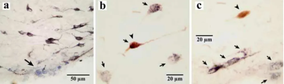
Figure 2. HA and non-HA neurons marked with pontamine sky blue or labeled for HA in the TM. a, Photomicrographs showingthe recording site of a waking-specific neuron marked with pontamine sky blue in the ventral TM. b, A waking-specific neuron stained with both Nb and HA (arrows and arrowhead). c, A non-waking-specific neuron labeled only with Nb (arrowhead). b, c, Arrows indicate neurons stained only with HA. The arrow in a shows the recording site of a waking-specific neuron in the ventral tuberomammillary nucleus marked with pontamine sky blue.
Figure 3. A, B, Spike shape of waking-specific (A) and waking-active (B) neurons. Note that waking-specific neurons are characterized by a broad triphasic action potential, whereas waking-active and other TM neurons are characterized by a biphasic (either narrow or broad) action potential. The arrowheads in A1 and A2 indicate the positive deflection. In A3 and B3, D1–D4 indicate the duration of the averaged action potential measured from onset (t0) to the peak (D1), the first zero crossing (D2), the peak of the afterhyperpolarization (D3), and the second zero crossing (D4 ), shown in A1.
are reported to play a role in the mechanisms of wake–sleep
reg-ulation and in the pathophysiology of narcolepsy/cataplexy
(Chemelli et al., 1999; Lin et al., 1999; Peyron et al., 2000). Orx/
Hcrt directly excites HA TM neurons in vitro in rat brain slice
preparations (Bayer et al., 2001; Eriksson et al., 2001; Yamanaka
et al., 2002). Recent in vivo studies in mice showed that
intrace-rebroventricular infusion of orexin A selectively activates the HA
system (Hong et al., 2005) and causes a significant increase in W
in wild-type mice but not in H
1receptor gene knock-out mice
(Huang et al., 2001), suggesting that the arousal effect of orexin A
is mainly mediated by activation of the HA system.
In the present experiments in mice, all HA TM neurons fell
silent during quiet waking before the onset of EEG
synchroniza-tion observed during the drowsy state, suggesting that the
cessa-tion of activity of HA TM neurons may play a role in the
induc-tion of SWS. The mechanisms underlying this cessainduc-tion of
activity during sleep are not known, but it is currently believed
that it is caused by GABAergic inputs from sleep-active neurons
in the preoptic and basal forebrain areas (Saper et al., 2001).
During the transition from sleep to W or in response to an
arous-ing stimulus durarous-ing sleep, there was a marked delay in the onset of
firing of HA TM neurons. It has been shown in vitro (Haas and
Reiner, 1988; Greene et al., 1990; Llinas and Alonso, 1992;
Stevens et al., 2001) that, when depolarizing pulses are applied
from a hyperpolarized membrane potential level, there is a long
delay before activation of cells because of a pronounced
voltage-gated transient outward current (A-type current) (Connor and
Stevens, 1971). It is possible that mouse HA TM neurons are
hyperpolarized during sleep and that the transient outward
cur-rent ensures that the membrane potential remains below
thresh-Figure 4. Activity of a representative waking-specific neuron. Note that activity is confinedto waking periods, with the cell being completely silent during sleep. Note also the absence of discharge during short awakening periods (arrowheads) during sleep. The large arrow indicates the end of PS, as shown by the interruption of theta waves and the onset of EEG desyn-chronization. The small arrows with an “s” indicate short awakenings elicited by the arousing sound stimulus.
Figure 5. Activity of a representative waking-specific neuron during the different wak-ing states.
Figure 6. A, B, Activity of a representative waking-active neuron (A) and discharge profile of waking-active (B1) and waking-specific (B2) neurons across the sleep–waking cycle.
old during sleep. Cessation of activity of HA TM neurons may
therefore play a role in the maintenance of sleep.
As shown in the present study, HA TM neurons display
arousal-specific neuronal activity. Although the activity of
human HA TM neurons remains unknown, presumed HA TM
neurons in cats and dogs are reported to display a highly
arousal-specific neuronal activity similar to that seen in mice
(Vanni-Mercier et al., 1985; Sakai et al., 1990; John et al., 2004). An
increase in histaminergic transmission enhances W, whereas its
blockade causes somnolence (for review, see Lin, 2000). HA
in-duces a switch in neuronal firing mode from rhythmic burst to
single spike activity in thalamic relay neurons (McCormick and
Williamson, 1991) and an increase in spiking by reducing
spike-frequency adaptation (McCormick and Williamson, 1989). In
addition, HA stimulates cholinergic neurons of the brainstem
and basal forebrain (Khateb et al., 1995; Lin et al., 1996; Crochet
and Sakai, 1999; Koyama and Sakai, 2000) and serotonergic
neu-rons of the DRN (Sakai and Crochet, 2000; Brown et al., 2002),
both implicated in waking and attention. Collectively, these data
indicate an important role of HA in the control of arousal.
In the present study, HA TM neurons ceased firing during the
drowsy state, characterized by a low vigilance level, remained
quiescent during the transition from sleep to W when the animals
were not fully alert, and responded with a long delay, or dis not
respond at all, to an arousing stimulus if it did not elicit an overt
alert state. These findings suggest that the activity of HA TM
neurons plays an important role not in the induction of W per se,
but in the maintenance of a high level of vigilance, and that lack of
HA neuronal activity results in somnolence. These data are in
good agreement with those of our recent study showing that KO
mice for histidine decarboxylase, the HA-synthesizing enzyme,
display a deficit in W and signs of somnolence when faced with a
novel environment (Parmentier et al., 2002). Additional detailed
studies in freely behaving animals are required to determine
whether or not a particular waking state drives the neuronal
ac-tivity of HA TM neurons or vice versa. The alternation between
waking and sleep is determined by multiple arousal- and
sleep-promoting systems, which are widely distributed in the brain
(Jones, 2005). Precise knowledge of the unit activity profiles of
these systems during the state transition should be helpful to
understand how each system contributes to the generation
and/or maintenance of the behavioral states.
In conclusion, the present study shows, for the first time in
mice, that HA neuronal activity is specific to the waking state with
a high vigilance level and that these neurons may play a role not in
the initiation of W per se, but in the maintenance of the high level
of vigilance necessary for cognitive processes. Conversely,
cessa-tion of their activity may play an important role in both the
initiation and maintenance of sleep.
References
Bayer L, Eggermann E, Serafin M, Saint-Mleux B, Machard D, Jones B, Mu¨-hlethaler M (2001) Orexins (hypocretins) directly excite tuberomam-millary neurons. Eur J Neurosci 14:1571–1575.
Brown RE, Sergeeva OA, Eriksson KS, Haas HL (2002) Convergent excita-tion of dorsal raphe serotonin neurons by multiple arousal systems (orexin/hypocretin, histamine, and noradrenaline). J Neurosci 22:8850 – 8859.
Chemelli RM, Willie JT, Sinton CM, Elmquist JK, Scammell T, Lee C, Rich-ardson JA, Williams SC, Xiong Y, Kisanuki Y, Fitch TE, Nakazato M, Hammer RE, Saper CB, Yanagisawa M (1999) Narcolepsy in orexin knockout mice: molecular genetics of sleep regulation. Cell 98:437– 451. Connor JA, Stevens CF (1971) Voltage clamp studies of a transient outward
membrane current in gastropod neural somata. J Physiol (Lond) 213:21–30.
Crochet S, Sakai K (1999) Effects of microdialysis application of mono-amines on the EEG and behavioural states in the cat mesopontine teg-mentum. Eur J Neurosci 11:3738 –3752.
Ericson H, Watanabe J, Ko¨hler CH (1987) Morphological analysis of the tuberomammillary nucleus in the rat brain: delineation of subgroups with antibody againstL-histidine decarboxylase as a marker. J Comp Neurol 263:1–24.
Eriksson KS, Sergeeva O, Brown RE, Haas HL (2001) Orexin/hypocretin excites the histaminergic neurons of the tuberomammillary nucleus. J Neurosci 21:9273–9279.
Greene RW, Haas HH, Reiner PB (1990) Two transient outward currents in histamine neurons of the rat hypothalamus in vitro. J Physiol (Lond) 420:149 –163.
Haas H, Panula P (2003) The role of histamine and the tuberomamillary nucleus in the nervous system. Nat Rev 4:121–130.
Haas HL (1992) Electrophysiology of histamine receptors. In: The hista-mine receptor (Schwartz JC, Haas HL, eds), pp161–177. New York: Wiley. Haas HL, Reiner P (1988) Membrane properties of histaminergic tube-romammillary neurons of the rat hypothalamus in vitro. J Physiol (Lond) 399:633– 646.
Hong ZY, Huang ZL, Qu WM, Eguchi N (2005) Orexin A promotes hista-mine, but not norepinephrine or serotonin, release in frontal cortex of mice. Acta Pharmacol Sin 26:155–159.
Huang ZL, Qu WM, Li WD, Mochizuki T, Eguchi N, Watanabe T, Urade Y, Hayaishi O (2001) Arousal effect of orexin A depends on activation of the histaminergic system. Proc Natl Acad Sci USA 98:9965–9970. Inagaki N, Yamatodani A, Ando-Yamamoto M, Tohyama M, Watanabe T,
Wada H (1988) Organization of histaminergic fibers in the rat brain. J Comp Neurol 273:283–300.
Figure 7. A, B, Discharge pattern of a waking-specific neuron during behavioral state tran-sitions (A) and response to an arousing sound stimulus (B). In A, the transition from SWS to W is shown as t1, that from W to drowsy state is shown as t2, and that from PS to W is shown as t3. Note that the unit fires with a long delay after the onset of EEG desynchronization of W, whereas it stops firing during quiet waking before the onset of the drowsy state, defined by EEG synchro-nization. Note also that there is no, or a long-delayed, response to a sound stimulus given during sleep.
John J, Wu MF, Boehmer LN, Siegel JM (2004) Cataplexy-active neurons in the hypothalamus: implications for the role of histamine in sleep and waking behavior. Neuron 27:619 – 634.
Jones BE (2005) Basic mechanisms of sleep-wake states. In: Principles and practice of sleep medicine (Kryger MH, Roth T, Dement WC, eds), pp 136 –153. Philadelphia: Elsevier.
Kamondi A, Reiner PB (1991) Hyperpolarization-activated inward current in histaminergic tuberomammillary neurons of the rat hypothalamus. J Neurophysiol 66:1902–1911.
Khateb A, Fort P, Pegna A, Jones BE, Muhlethaler M (1995) Cholinergic nucleus basalis neurons are excited by histamine in vitro. Neuroscience 69:495–506.
Koyama Y, Sakai K (2000) Modulation of presumed cholinergic mesopon-tine tegmental neurons by acetylcholine and monoamines applied ionto-phoretically in unanesthetized cats. Neuroscience 96:723–733. Lin JS (2000) Brain structures and mechanisms involved in the control of
cortical activation and wakefulness, with emphasis on the posterior hy-pothalamus and histaminergic neurons. Sleep Med Rev 4:471–503. Lin JS, Sakai K, Vanni-Mercier G, Jouvet M (1989) A critical role of the
posterior hypothalamus in the mechanisms of wakefulness determined by microinjection of muscimol in freely moving cats. Brain Res 479:225–240. Lin JS, Hou Y, Sakai K, Jouvet M (1996) Histaminergic descending inputs to the mesopontine tegmentum and their role in the control of cortical activation and wakefulness in the cat. J Neurosci 16:1523–1537. Lin L, Faraco J, Li R, Kadotani H, Rogers W, Lin X, Qiu X, de Jong PJ, Nishino
S, Mignot; E (1999) The sleep disorder canine narcolepsy is caused by a mutation in the hypocretin (orexin) receptor 2 gene. Cell 98:365–376. Llinas RR, Alonso A (1992) Electrophysiology of the mammillary complex
in vitro. I. Tuberomammillary and lateral mammillary neurons. J Neuro-physiol 68:1307–1320.
McCormick DA, Williamson A (1989) Convergence and divergence of neu-rotransmitter action in human cerebral cortex. Proc Natl Acad Sci USA 86:8098 – 8102.
McCormick DA, Williamson A (1991) Modulation of neuronal firing mode in cat and guinea pig LGNd by histamine: possible cellular mechanisms of histaminergic control of arousal. J Neurosci 11:3188 –3199.
Moruzzi G (1972) The sleep-waking cycle. Ergeb Physiol 64:1–165. Nicholson AN (1983) Antihistamines and sedation. Lancet 2:211–212. Parmentier R, Ohtsu H, Djebbara-Hannas Z, Valatx JL, Watanabe T, Lin JS
(2002) Anatomical, physiological, and pharmacological characteristics of histidine decarboxylase knock-out mice: evidence for the role of brain histamine in behavioral and sleep-wake control. J Neurosci 22:7695–7711.
Peyron C, Faraco J, Rogers W, Ripley B, Overeem S, Charnay Y, Nevsimalova S, Aldrich M, Reynolds D, Albin R, Li R, Hungs M, Pedrazzoli M,
Padi-garu M, Kucherlapati M, Fan J, Maki R, Lammers GJ, Bouras C, Kucher-lapati R, et al. (2000) A mutation in a case of early onset narcolepsy and a generalized absence of hypocretin peptides in human narcoleptic brains. Nat Med 6:991–997.
Pinault D (1996) A novel single-cell staining procedure performed in vivo under electrophysiological control: morpho-functional features of juxta-cellular labeled thalamic cells and other central neurons with biocytin or Neurobiotin. J Neurosci Meth 65:113–136.
Reiner PB, Kamondi A (1994) Mechanisms of antihistamine-induced seda-tion in the human brain: H1 receptor activaseda-tion reduces a background leakage potassium current. Neuroscience 59:579 –588.
Reiner PB, McGeer EG (1987) Electrophysiological properties of cortically projecting histamine neurons of the rat hypothalamus. Neurosci Lett 73:43– 47.
Sakai K, Crochet S (2000) Serotonergic dorsal raphe neurons cease firing by disfacilitation during paradoxical sleep. NeuroReport 11:3237–3241. Sakai K, El Mansari M, Lin JS, Zhang JG, Vanni-Mercier G (1990) The
posterior hypothalamus in the regulation of wakefulness, and paradoxical sleep. In: The diencephalon and sleep (Mancia M, Marini M, eds), pp 171–198. New York: Raven.
Saper CB, Chou TC, Scammell TE (2001) The sleep switch: hypothalamic control of sleep and wakefulness. Trends Neurosci 24:726 –731. Schwartz JC, Arrang JM, Garbarg M, Pollard H, Ruat M (1991)
Histamin-ergic transmission in the mammalian brain. Physiol Rev 71:1–51. Steininger TL, Alam MN, Gong H, Szymusiak R, McGinty D (1999)
Sleep-waking discharge of neurons in the posterior lateral hypothalamus of the albino rat. Brain Res 840:138 –147.
Stevens DR, Haas HL (1996) Calcium-dependent prepotentials contribute to spontaneous activity in rat tuberomammillary neurons. J Physiol (Lond) 493:747–754.
Stevens DR, Eriksson KS, Brown RE, Haas HL (2001) The mechanism of spontaneous firing in histamine neurons. Behav Brain Res 124:105–112. Vanni-Mercier G, Sakai K, Jouvet M (1985) Waking-statespecific neurons
in the posterior hypothalamus of the cat. Sleep 84:238 –240.
Wada H, Inagaki N, Yamatodani A, Watanabe T (1991) Is the histaminergic neuron system a regulatory center for whole-brain activity? Trends Neu-rosci 14:415– 418.
Yamanaka A, Tsujino N, Funabashi H, Honda K, Guan JL, Wang OP, Tomi-naga M, Goto K, Shioda S, Sakurai T (2002) Orexins activate histamin-ergic neurons via the orexin 2 receptor. Biochem Biophys Res Commun 2002:1237–1245.
Yang QZ, Hatton GI (1997) Electrophysiology of excitatory and inhibitory afferents to rat histaminergic tuberomammillary nucleus neurons from hypothalamus and forebrain sites. Brain Res 773:162–172.