HAL Id: inserm-02265304
https://www.hal.inserm.fr/inserm-02265304
Submitted on 9 Aug 2019
HAL is a multi-disciplinary open access
archive for the deposit and dissemination of sci-entific research documents, whether they are pub-lished or not. The documents may come from teaching and research institutions in France or abroad, or from public or private research centers.
L’archive ouverte pluridisciplinaire HAL, est destinée au dépôt et à la diffusion de documents scientifiques de niveau recherche, publiés ou non, émanant des établissements d’enseignement et de recherche français ou étrangers, des laboratoires publics ou privés.
Desiccation-induced viable but nonculturable state in
Pseudomonas putida KT2440, a survival strategy
Laura Pazos-Rojas, Ligia Catalina Muñoz-Arenas, Osvaldo
Rodríguez-Andrade, Lesther Emanuel López-Cruz, Orestes López-Ortega,
Fábio Lopes-Olivares, Silvia Luna-Suarez, Antonino Baez, Yolanda Elizabeth
Morales-García, Verónica Quintero-Hernández, et al.
To cite this version:
Laura Pazos-Rojas, Ligia Catalina Muñoz-Arenas, Osvaldo Rodríguez-Andrade, Lesther Emanuel López-Cruz, Orestes López-Ortega, et al.. Desiccation-induced viable but nonculturable state in Pseudomonas putida KT2440, a survival strategy. PLoS ONE, Public Library of Science, 2019, 14 (7), pp.e0219554. �10.1371/journal.pone.0219554�. �inserm-02265304�
Desiccation-induced viable but nonculturable
state in Pseudomonas putida KT2440, a
survival strategy
Laura Abisaı´ Pazos-Rojas1,2,3, Ligia Catalina Muñoz-ArenasID4, Osvaldo
Rodrı´guez-Andrade1, Lesther Emanuel Lo´ pez-Cruz1
, Orestes Lo´ pez-Ortega5
, Fa´bio Lopes-Olivares6, Silvia Luna-Suarez2, Antonino Baez1, Yolanda Elizabeth Morales-Garcı´aID1,3,
Vero´ nica Quintero-Herna´ndez1,7
, Miguel Angel Villalobos-Lo´ pezID2, Jesu´ s De la TorreID8,
Jesu´ s Muñoz-RojasID1*
1 Ecology and Survival of Microorganisms Research Group (ESMRG), Laboratorio de Ecologı´a Molecular Microbiana (LEMM), Centro de Investigaciones en Ciencias Microbiolo´ gicas (CICM), Instituto de Ciencias (IC), Beneme´ rita Universidad Auto´noma de Puebla (BUAP), Puebla, Mexico, 2 Centro de Investigacio´n en Biotecnologı´a Aplicada, Instituto Polite´cnico Nacional, Tepetitla, Tlaxcala, Mexico, 3 Licenciatura en Biotecnologı´a, Facultad de Ciencias Biolo´gicas, BUAP, Puebla, Mexico, 4 Facultad de Ingenierı´a Ambiental, Universidad Popular Auto´noma de Puebla, Puebla, Mexico, 5 Inserm U932, Institute Curie, Paris, France, 6 Nu´cleo de Desenvolvimento de Insumos Biolo´gicos para a Agricultura (NUDIBA), Universidade Estadual do Norte Fluminense Darcy Ribeiro (UENF), Rio de Janeiro, Brazil, 7 CONACYT, ESMRG, LEMM, CICM, IC, BUAP, Puebla, Me´xico, 8 Department of Environmental Protection, CSIC-Estacio´n Experimental del Zaidı´n, Granada, Spain
*joymerre@yahoo.com.mx
Abstract
The potential of Pseudomonas putida KT2440 to act as a plant-growth promoter or as a bior-emediator of toxic compounds can be affected by desiccation. In the present work, the bac-terial survival ratio (BSR) in response to air desiccation was evaluated for P. putida KT2440 in the presence of different protectors. The BSR in the presence of nonreducing disaccha-rides, such as trehalose, was high after 15 days of desiccation stress (occurring at 30˚C and 50% relative humidity), whereas in the absence of a protector the bacterial counts dimin-ished to nondetectable numbers (ca 2.8 log CFU/mL). The LIVE/DEAD staining method showed that bacteria protected with trehalose maintained increased numbers of green cells after desiccation while cells without protection were all observed to be red. This indicated that nonprotected bacteria had compromised membrane integrity. However, when nonpro-tected bacteria subjected to 18 days of desiccation stress were rehydrated for a short time with maize root exudates or for 48 h with water (prolonged rehydration), the bacterial counts were as high as that observed for those not subjected to desiccation stress, suggesting that the cells entered the viable but nonculturable (VBNC) state under desiccation and that they returned to a culturable state after those means of rehydration. Interestingly an increase in the green color intensity of cells that returned to a culturable state was observed using LIVE/ DEAD staining method, indicating an improvement in their membrane integrity. Cellular activity in the VBNC state was determined. A GFP-tagged P. putida strain expressing GFP constitutively was subjected to desiccation. After 12 days of desiccation, the GFP-tagged strain lost culturability, but it exhibited active GFP expression, which in turn made the cells green. Furthermore, the expression of 16S rRNA, rpoN (housekeeping), mutL, mutS a1111111111 a1111111111 a1111111111 a1111111111 a1111111111 OPEN ACCESS
Citation: Pazos-Rojas LA, Muñoz-Arenas LC, Rodrı´guez-Andrade O, Cruz LE, Lo´pez-Ortega O, Lopes-Olivares F, et al. (2019) Desiccation-induced viable but nonculturable state in Pseudomonas putida KT2440, a survival strategy. PLoS ONE 14(7): e0219554.https://doi. org/10.1371/journal.pone.0219554
Editor: Seon-Woo Lee, Dong-A University,
REPUBLIC OF KOREA
Received: January 4, 2019 Accepted: June 26, 2019 Published: July 19, 2019
Copyright:© 2019 Pazos-Rojas et al. This is an open access article distributed under the terms of theCreative Commons Attribution License, which permits unrestricted use, distribution, and reproduction in any medium, provided the original author and source are credited.
Data Availability Statement: All relevant data are
within the manuscript and its Supporting Information files.
Funding: This work is supported by: JMR: Apoyo
Redes PRODEP 2015-2016 (CA-262 and CA-244); JMR: DITCo2016-3, JMR: DITCo2016-4; VIEP-BUAP-2018 (VQH: COL525704-VIEP2018; ABR: 100524554-VIEP2018, JMR: 100425788-VIEP2018). The funders had no role in study
(encoding proteins from the mismatch repair complex), and oprH (encoding an outer mem-brane protein) were examined by RT-PCR. All evaluated genes were expressed by both types of cells, culturable and nonculturable, indicating active molecular processes during the VBNC state.
Introduction
Pseudomonas putida KT2440 is a nonpathogenic gram-negative bacterium that is able to colo-nize the rhizosphere of several plants [1], degrade aromatic compounds [2,3], and promote the growth and health of plants [4–6].P. putida KT2440 has been widely used as a model in bio-degradation and environmental adaptation studies [7,8], showing complex chemosensory sys-tems, signal transduction, genetic regulation, and environmental stress responses that explain its high metabolic and adaptive versatility [7,9–11]. Despite this versatility, the survival ofP. putida KT2440 decreases drastically after the loss of water, as documented in studies of freeze-drying and freeze-drying, both under vacuum conditions [12,13]. The potential ofP. putida KT2440 for use in the bioremediation of soils and plant growth promotion could be affected by drought, temperature and pH fluctuations, high salinity, low nutrient availability, and desicca-tion. These conditions are limiting factors that determine the survival of all bacteria [14–17]. In particular, desiccation is a highly restrictive factor in regard to the development of any organism, including bacteria [17–19]. Some bacteria are highly tolerant to desiccation, such as Enterobacter sp. UAPS03001, Klebsiella variicola T29A and Paraburkholderia unamae MTl-641, but others are very sensitive to desiccation stress, such asBradyrhizobium japonicum USDA 110 andBurkholderia sacchari LMG 19450 [6,20,21]. Under water-limited conditions, tolerance to desiccation is fundamental for any bacterial species associated with seeds to main-tain their plant-growth promoting features, which normally recover after rehydration [20]. The bacteria that are best adapted to desiccation-rehydration processes will be the most com-petitive in environments with low water availability; for example, tolerant bacteria that were adhered to seeds and desiccated for 18 days were rehydrated and showed good root coloniza-tion during plant development [6]. In addition, bacteria tolerant to desiccation have the capa-bility to rapidly resume activity and show increased transcription levels when water becomes available again [22].
The presence of stressors such as UV radiation, heavy metals, nutrient limitation, low tem-peratures, salinity, desiccation and oxidation can lead to a viable but nonculturable (VBNC) state [23–26], in which bacteria remain viable and metabolically active but fail to grow on stan-dard culture media [27,28]. Cells suffer metabolic changes, such as a reduction in nutrient transport, respiration rates, and macromolecular synthesis, during the VBNC state [29]. Fur-thermore, a continuous gene expression occurs in cells in VBNC state [30], which has been proposed to be definitive proof that cells remain metabolically active and are not dead [23]. In some cases, the removal of the inducing stressors and/or the provision of suitable conditions for VBNC cells can restore their ability to grow and therefore their culturability [31,32]. At least eighty-five bacterial species have been reported to enter the VBNC state, most of them pathogenic species [32], but the behavior of beneficial plant-associated microorganisms remains to be fully investigated.
Bacterial desiccation under vacuum conditions is completely different from that occurring in natural environments because desiccation in the environment occurs without negative
pres-Viable but nonculturable state in P. putida KT2440 as a survival strategy
design, data collection and analysis, decision to publish, or preparation of the manuscript.
Competing interests: The authors have declared
and their beneficial potential could be affected by water limitation. Therefore, in the present work, the capability ofP. putida KT2440 to tolerate air desiccation was studied, and its entrance into a VBNC state as a survival strategy is discussed.
Materials and methods
Bacterial growth and desiccation assays
P. putida KT2440 cells were grown until the stationary phase in LB (Luria-Bertani) liquid medium supplemented with 100μg/mL chloramphenicol (LB-Cm100) [12]. Desiccation assays were conducted as previously described [33]. Three hundred and sixty milliliters of bacterial growth suspension was subdivided into aliquots of 15 mL. Each aliquot was centrifuged at 5000 rpm for 10 min, the supernatant was removed and the pellet was resuspended in the same volume of sterile distilled water. This process was repeated twice but in the second round the bacterial pellet in each tube was resuspended with 200 mM of different protectors. A con-trol using only water was included. Each suspension was aliquoted in microtubes of 1.5 mL capacity with 500μL of bacterial suspension and covered with sterile cotton. Five samples from each treatment were used to determine the bacterial density (Colony Forming Units (CFU)/mL) contained in the suspensions before desiccation using the Massive Stamping Drop Plate (MSDP) method [34–36]. Desiccation was carried out at 30˚C and 50% relative humidity (RH). The bacterial density (CFU/mL) was monitored every 3 days after the beginning of des-iccation (DABD) by taking 5 samples and rehydrating them with water (500μl) for 20 min. The medium used for bacterial quantification was LB agar (1.5% W/V)-Cm100. The BSR to air desiccation was calculated as the ratio of the log of the number of bacterial cells present in the suspension at any time post desiccation (PD) plus one to the log number of viable cells before desiccation (BD), all multiplied by 100; BSR = [(logPD + 1)/logBD]× 100 [6,12,20]. A BSR value of 100 indicates that all bacteria survived after desiccation stress, while a BSR value of 0 indicates that no bacteria survived. Samples were weighed both before desiccation and after desiccation to calculate the water lost from each sample. All samples attained complete desic-cation at 5 DABD.
Adherence to maize sprouts and colonization of
P. putida KT2440 after
desiccation
Cells ofP. putida KT2440 were desiccated with or without a protector (trehalose 200 mM) fol-lowing the methodology described above. At 18 DABD, a total of 25 dried samples with and without the protector were rehydrated with water (500μL) for 20 min and transferred to a Fal-con 15 mL centrifuge tube. Axenic maize sprouts were obtained according to Morales-Garcı´a et al. (2011) [37] and then inoculated by submerging the germinate in the bacterial suspension for 1 h. Two control treatments were included, the first consisting of 20 sprouting seeds sub-merged in water for 1 h and the second consisting of 20 sprouting seeds subsub-merged in 200 mM trehalose. Five sprouts from each treatment were used to determine the number of bacte-ria adhered to the maize germinates using the MSDP method [6,20,37]. The remaining sprouts were transferred to 50 mL Falcon centrifuge tubes containing 6.4 g of sterile vermiculite amended with 25 mL of sterile water, and the tubes were placed in a plant growth chamber for 15 days at 25˚C and 80% RH with a photoperiod of 16 h light/8 h darkness. Every 3 days, the plants were irrigated to maintain substrate moisture. Rhizosphere samples were collected to evaluate colonization as previously described using five replicate plants from each treatment [20,36,38]. For this, the vermiculite adhered to the roots and considered as the rhizosphere was resuspended in water at a ratio of 1:10 (W/V). This suspension was vortexed for 3 min,
and the resulting suspension was serially diluted. The bacterial abundance was determined according to the MSDP method using LB-agar (1.5% W/V)-Cm100. In addition to coloniza-tion, the bacterial membrane integrity was evaluated for each treatment (with and without tre-halose) at each stage of bacterial establishment. Thus, suspensions of the bacteria before desiccation, at 18 DABD and rehydrated for 20 min, bacterial suspensions from sprouting seeds and from the rhizosphere were assayed. For all the evaluated samples, the MFI (mean of fluorescence intensity) values were calculated. Both the membrane integrity assays and MFI measurements are described in the section “Membrane damage toP. putida KT2440 during desiccation”.
Rehydration of
P. putida KT2440 with maize root exudates
Two types of root exudates were obtained: 1) Root exudates from the early stages of growth. For this treatment, five mL of water was used to wash an agar-water plate in which 10 axenic seeds had previously germinated [37]. The collected exudates were stored at -20˚C until they were used. 2) Root exudates from plants grown for 12 days. This type of root exudate was obtained from hydroponic axenic systems. Each system consisted of a 300 mL flask containing 100 mL of liquid MS-J medium (Morales-Garcı´aet al., 2011). A metallic ring was placed inside each flask in contact with the medium, and the systems were covered with cotton. The hydro-ponic systems were sterilized before use. A previously germinated axenic maize sprout was placed in each ring such that the root remained in contact with the MS-J medium, and the root exudates were allowed to accumulate within the medium. The plant growth conditions were 25˚C and 80% RH with a photoperiod of 16 h light/8 h darkness. The root exudates were collected after 12 days of plant growth and were stored at -20˚C until they were used.
Both types of root exudates were used to rehydrate desiccated cells ofP. putida KT2440. Desiccated bacterial cells were obtained as described before, and 5 samples were prepared for each treatment. The bacterial abundances in the suspensions before desiccation and at 18 DABD were determined using the MSDP method with five independent samples for each treatment. The desiccated cells were rehydrated for 20 min and 3, 6, 9, 12, 24, 27, 30 and 48 h with the plant root exudates. Cells rehydrated only with water were used as controls.
Ability of
P. putida KT2440 to grow in the presence of maize root exudates
under static conditions
To test whetherP. putida KT2440 can use maize root exudates as a carbon source and grow under static conditions, similar to the procedure used in the rehydration experiments, bacterial cells were grown until the stationary phase in liquid LB-Cm100medium (two 50 mL flasks con-taining 15 mL of culture). The bacterial suspensions were washed and resuspended with the same volume of maize root exudates and water as a control. The suspensions were serially diluted (1:10) to quantify the number of bacteria. For the exudate treatments, the bacterial dilutions were performed using the exudates. All dilution tubes were kept at room temperature under static conditions for 24 and 48 h. The bacterial abundance in each dilution was deter-mined at those experimental times.
Membrane damage to
P. putida KT2440 during desiccation
Bacterial membrane damage was evaluated using the L7007 LIVE/DEAD BacLight Bacterial Viability Kit for microscopy (Molecular Probes Invitrogen Detection Technologies) and the observation of stained cells using fluorescence microscopy. This kit uses SYTO 9 and propi-dium iodide to discriminate between live cells with intact membranes (green fluorescence)
were grown until the stationary phase, and samples of washed bacterial suspension were desic-cated for 18 days following the methodology described previously. Membrane integrity was tested in 5 samples before desiccation, and in five samples from 3, 6, 9, 12, 15 and 18 DABD rehydrated for 20 min, and the bacterial abundances were determined to calculate the BSR val-ues. The cells at 18 DABD were also rehydrated for 24 and 48 h to determine the bacterial abundance and membrane integrity. The samples were observed with the VE-146YT fluores-cence microscope at 100× using G (excitation 500–550 nm, red bacteria) and B filters (excita-tion 420–490 nm, green bacteria) following the supplier’s instruc(excita-tions. The bacterial images were examined by means of fluorescence intensity (MFI) graphs to evaluate the distribution of propidium iodide and SYTO 9 alone or in combination (MERGE); the analysis was performed along a line randomly traced through the cells. Pixel intensity information per fluorescence channel was extracted with ImageJ (v1.43u, NIH, USA, public domain). Each graph was gener-ated from a minimum of fifteen randomly selected bacteria for each condition per experiment using Microsoft Excel (v14.3.9, Microsoft Corporation, Redmond, WA, USA). The MFI data were examined sequentially by ImageJ and graphed using Prism software.
TEM analysis of
P. putida KT2440 under desiccation
TEM analysis was performed with the BD samples and those from 6, 12 and 18 DABD. The methodology used to prepare the samples was carried out in four stages: 1) fixation of the sam-ple with a mixture of 2.5% glutaraldehyde, 4% paraformaldehyde and 0.05 M phosphate buffer; 2) sample washing with phosphate buffer; 3) sample dehydration with alcohol at different centrations (15, 30, 50, 70, 90 and 100%), starting the wash from the lowest to the highest con-centration of alcohol; and 4) inclusion of the sample in LR White (Sigma) starting with a dilution of 2:1 (alcohol:resin) for 6 h at 4˚C. Subsequently, the solution was discarded, and a dilution of 1:1 (alcohol:resin) was added under the same conditions. Finally, the solution was discarded, pure resin was added for 24 h at 4˚C (two times) and the samples were allowed to polymerize at 60˚C for 24 h. After inclusion, the samples were microtomized for the analysis of contrast, and the samples were observed with TEM.
Desiccation of
P. putida KT2440 tagged with GFP
GFP-taggedP. putida KT2440, generated by the site-specific insertion of miniTn7-gfp at an extragenic location nearglmS, which constitutively expresses the gfp gene [39], was used in this work. The GFP-tagged bacterial strain was subjected to desiccation for 18 days following the methodology described above. The bacterial abundance was determined at 3, 6, 9, 12, 15 and 18 DABD after rehydration, and BSR values were calculated. The microscopic examination of GFP fluorescence using a Zeiss Axioplan ZE155 microscope (Germany) (filters BP546/ FT580/ LP590) was performed with the same samples used to quantify the bacterial abundance, and the fluorescence intensity was measured with a HIDEX Chameleon Multilabel Detection Plat-form multiwell plate reader at an excitation wavelength of 395 nm and emission wavelength of 509 nm. The samples were observed at 100×.
Expression of some constitutive genes of
P. putida KT2440 under
desiccation
Total RNA was extracted fromP. putida KT2440 cells before desiccation, and from 20 min-rehydrated cells (from samples at 6, 12 and 18 DABD). RNA extraction was performed using the hot acidic phenol-chloroform method (3 replicates of 1.5 mL) [40]. Cells were lysed using 0.5% SDS, 20 mM sodium acetate and 10 mM EDTA, and RNA was extracted twice with hot acid phenol:chloroform followed by two extractions with phenol:chloroform isoamyl alcohol.
Total RNA was precipitated with absolute ethanol and then washed in 70% ethanol (molecular grade). Finally, the RNA sample was resuspended in DEPC water and stored at -80˚C until use. To reduce genomic DNA contamination, the RNA isolated from each sample was treated with the Invitrogen TURBO DNA-free Kit. RNA integrity was evaluated in 2% agarose gel, and its concentration was measured using a Thermo Scientific NanoDrop 2000/2000c spectro-photometer. The cDNA of each gene for which expression was evaluated was obtained using the High-Capacity cDNA Reverse Transcription Kit from Applied Biosystems, with 2μg RNA as the template and 10μM of specific DNA primers (antisense primers,Table 1). Retrotran-scription reactions were performed at 25˚C per 10 min, 37˚C per 120 min, 85˚C per 5 min, and 4˚C per 10 min. Total cDNA was quantified using a Thermo Scientific NanoDrop 2000/ 2000c spectrophotometer. To amplify each gene of interest from the cDNA obtained, PCRs were performed. The PCR conditions were 1 cycle for 5 min at 95˚C, 25 cycles at 95˚C for 30 seconds, 63˚C for 30 seconds, and 72˚C for 20 seconds, 1 cycle at 72˚C for 8 min, and a final step of 10 min at 4˚C. The amplified products were visualized by electrophoresis in 1% agarose gel (30 min, 100 volts) using GelRed Nucleic Acid Gel Stain (Biotium).
Real-time qPCR was performed using an Applied Biosystems 7500 Fast Real Time PCR Sys-tem. Reactions were performed by using SYBR Green PCR Master Mix as a signal reporter and 30 amplification cycles. Each reaction was composed of 10 ng of cDNA and 6μM of sense and antisense primers in a total volume of 20μL. RT-qPCR was performed in 96-well microti-ter PCR plates using the following amplification conditions: 1 cycle of 5 min at 95˚C and 30 two-step cycles at 95˚C for 30 seconds and 63˚C for 30 seconds. Each reaction was performed in triplicate. Data were analyzed using the 2-ΔΔCTmethod [41]. In our work,ΔΔCT= [(CTgene
of interest–CTinternal control) AD–(CTgene of interest–CTinternal control) BD], where AD
means after desiccation and BD means before desiccation. The expression of therpoN gene was used as an endogenous control to normalize the amount of mRNA obtained from a target gene. The expression data obtained for each time point were normalized to the expression of each gene obtained before desiccation.
Results
Survival of
P. putida KT2440 under air desiccation (30˚C and 50% RH) was
increased by nonreducing disaccharides
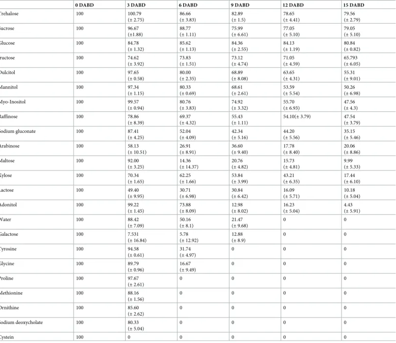
The tolerance ofP. putida KT2440 to air desiccation was explored in the presence or absence of 200 mM of diverse substances used as possible protectors (Table 2).P. putida KT2440 was sensitive to desiccation, and bacteria were not detected at 12 DABD after rehydration when no protector was added. Some of the explored compounds protected this bacterium from desicca-tion stress (Table 2). The best protectors were the nonreducing disaccharides (trehalose and sucrose) and the monosaccharide glucose, followed by fructose and some polyalcohols (dulci-tol, mannitol and myo-inositol). Other compounds were less successful in protecting this
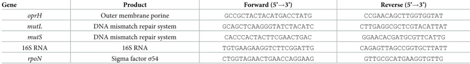
bac-Table 1. Oligonucleotides used for the analysis by RT-PCR and RT-qPCR.
Gene Product Forward (5’!3’) Reverse (5’!3’)
oprH Outer membrane porine GCCGCTACTACATGACCTATG CCGAACAGCTTGGTGGTAT
mutL DNA mismatch repair system GCAGCTCAAGGGTATCTACATC CTTGAGGCGCTCGTACATTAT
mutS DNA mismatch repair system CACCCACTACTTCGAACTGAC GGAACACGATGCGTTCATTG
16S RNA 16S RNA TGTGAAGAAGGTCTTCGGATTG CAGAGTTAGCCGGTGCTTATT
rpoN Sigma factorσ54 CTGGTAGAACTGAACCAGGAAG GTTGCGCATGAAGGTGTTG
https://doi.org/10.1371/journal.pone.0219554.t001
KT2440 from desiccation, and the BSR was similar to that observed without protector addi-tion. The presence of amino acids had a negative effect on bacterial survival under the evalu-ated desiccation conditions in comparison with that in the absence of a protector.
Desiccation of
P. putida KT2440, its adherence to maize seeds after
rehydration, and its capability to colonize the rhizosphere
The BSR ofP. putida KT2440 decreased to an undetectable level after 9 DABD without a pro-tector, but in the presence of trehalose as a propro-tector, the bacterial abundance was approxi-mately 108CFU/mL for all experimented time points (Fig 1). In this experiment, bacterial
Table 2. BSR ofP. putida KT2440 cells subjected to air desiccation in the presence or absence of protectors (200 mM).
0 DABD 3 DABD 6 DABD 9 DABD 12 DABD 15 DABD
Trehalose 100 100.79 (± 2.75) 86.66 (± 3.83) 82.89 (± 1.5) 78.65 (± 4.41) 79.56 (± 2.79) Sucrose 100 96.67 (±1.88) 88.77 (± 1.11) 75.99 (± 6.61) 77.05 (± 5.10) 79.05 (± 5.10) Glucose 100 84.78 (± 1.32) 85.62 (± 1.13) 84.36 (± 2.55) 84.13 (± 1.19) 80.84 (± 0.82) fructose 100 74.62 (± 3.92) 73.83 (± 1.51) 73.12 (± 4.74) 71.05 (± 4.59) 65.793 (± 6.05) Dulcitol 100 97.65 (± 0.58) 80.00 (± 2.35) 68.89 (± 8.08) 63.65 (± 4.31) 55.31 (± 9.01) Mannitol 100 97.34 (± 1.15) 80.33 (± 0.69) 68.61 (± 2.61) 53.59 (± 5.54) 50.26 (± 6.98) Myo-Inositol 100 99.57 (± 0.94) 80.76 (± 3.83) 74.92 (± 3.32) 55.70 (± 6.93) 47.56 (± 4.3) Raffinose 100 78.86 (± 8.39) 69.37 (± 4.32) 55.43 (± 1.11) 54.10(± 3.79) 47.54 (± 3.79) Sodium gluconate 100 87.41 (± 4.25) 52.04 (± 4.09) 42.34 (± 5.16) 44.20 (± 5.56) 35.15 (± 5.46) Arabinose 100 58.13 (± 10.51) 26.91 (± 8.91) 36.60 (± 9.40) 17.78 (± 8.40) 20.06 (± 8.86) Maltose 100 92.00 (± 3.25) 14.36 (± 14.37) 20.76 (± 4.82) 15.73 (± 4.81) 9.99 (± 5.33) Xylose 100 70.34 (± 1.65) 62.25 (± 1.66) 53.84 (± 3.99) 43.21 (± 6.35) 17.44 (± 6.10) Lactose 100 49.40 (± 9.95) 30.71 (± 6.98) 30.84 (± 6.42) 16.09 (± 5.71) 10.18 (± 5.04) Adonitol 100 99.22 (± 1.45) 73.88 (± 8.09) 12.98 (± 8.02) 16.23 (± 5.04) 4.43 (± 5.91) Water 100 88.42 (± 7.09) 50.16 (± 8.1) 21.47 (± 9.68) 0 0 Galactose 100 7.531 (± 16.84) 5.78 (± 12.92) 12.88 (± 8.9) 0 0 Tyrosine 100 94.58 (± 0.61) 31.74 (± 4.97) 0 0 0 Glycine 100 89.79 (± 0.96) 16.67 (± 9.49) 0 0 0 Proline 100 97.67 (± 2.61) 0 0 0 0 Methionine 100 88.16 (± 1.56) 0 0 0 0 Ornithine 100 85.60 (± 2.62) 0 0 0 0 Sodium deoxycholate 100 80.33 (± 5.04) 0 0 0 0 Cystein 100 0 0 0 0 0 https://doi.org/10.1371/journal.pone.0219554.t002
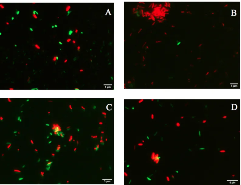
viability was evaluated in a suspension before desiccation and at 18 DABD using the LIVE/ DEAD BacLight Bacterial Viability Kit. Before desiccation, the observed cells were green in both conditions, with and without trehalose (Fig 2A and 2B). However, at 18 DABD, the pro-tected suspension contained green bacterial cells, and the nonpropro-tected suspension contained only red bacterial cells (Fig 2C and 2D). Red cells lack an intact cell membrane and are conven-tionally scored as dead. At 18 DABD, the rehydrated samples were used to inoculate maize sprouts for 1 h. The number of bacteria in the suspensions did not change after interaction with sprouts (high numbers of bacteria in the presence of the protector and no detected bacte-ria in the absence of the protector). The sprouts were transferred to Falcon tubes containing 6.4 g of vermiculite, and 25 mL of water was added. The adherence of the bacteria was tested 2 h after sowing, and the bacterial abundance was 9.1×10⁶CFU/sprouted seed in the treatment with trehalose, while bacteria were not detected in the treatment without a protector. Mem-brane integrity was tested with the LIVE/DEAD Kit, and several bacterial cells were observed to be green in the trehalose treatments (Fig 2E), while the nonprotected cells were all red (Fig 2F). Rhizosphere colonization byP. putida KT2440 was tested at 15 days post inoculation (dpi). Surprisingly,P. putida KT2440 colonized the rhizosphere of the plants in high numbers in both treatments: 4×10⁸CFU/gV for the treatment with trehalose and 9×10⁸
CFU/gV for the treatment without a protector (Fig 3A). The recuperation of bacterial cells in the rhizosphere of plants derived from seeds on which bacteria were not detected after desiccation could mean that the bacteria entered a viable but nonculturable state during the desiccation process (treat-ment without the use of protectors) and had returned to a culturable state when the bacteria interacted with the plants. The membrane integrity of the bacteria colonizing the plant rhizo-sphere was evaluated, and all observed bacteria were green in both treatments, suggesting that the membranes of the cells, from the treatment without protection were recovered in terms of their integrity and that the crossing of propidium iodide was prevented during their interac-tion with the plants (Fig 3B and 3C). The MFI analysis corroborated the observations of mem-brane integrity restoration after bacterial colonization because the MFI of SYTO 9 increased with respect to that of propidium iodide both in the presence and absence of the protector (Figs4and5).
Desiccated nonculturable
P. putida KT2440 returns to a culturable state
after rehydration with plant exudates or under prolonged rehydration
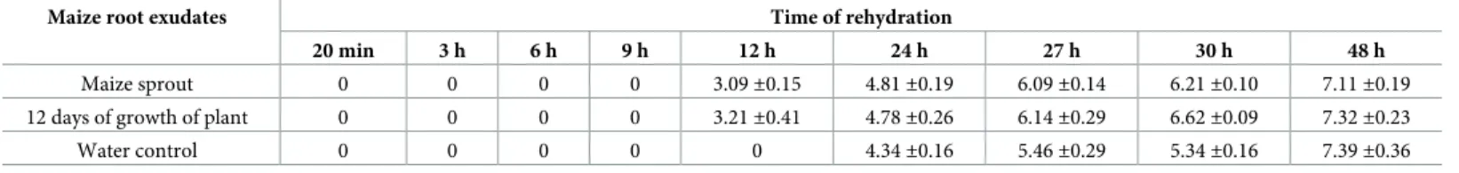
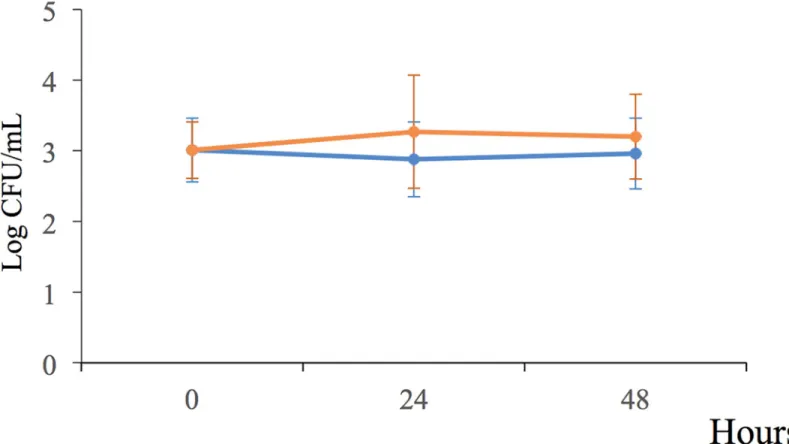
To explore whether the exudates of plants returnP. putida KT2440 to a culturable state, cells in the stationary phase were desiccated until 18 DABD and rehydrated three different suspen-sions: 1) maize sprout exudates, 2) maize root exudates following 12 days of growth, and 3) water only as a control. Rehydration was carried out for 20 min and 1, 3, 6, 9, 12, 24, 27, 30 and 48 h, and the bacterial abundance was determined. As expected, the bacterial abundance had decreased to nondetectable levels at 18 DABD, but the bacteria returned to a culturable state after rehydration with plant root exudates or with only water; interestingly, this return was faster in the presence of exudates; in contrast, in water, the bacterial abundance was simi-lar to that of the samples rehydrated with the exudates for up to 48 h (Table 3). It is noteworthy that the plant root exudates obtained from the early stages were able to accelerate the return to a culturable state of theP. putida KT2440 cells (Table 3). During rehydration, the bacterial sus-pension in the microtubes remained static, and independent experiments showed that the bac-terial cells were unable to duplicate under this condition in the presence of both exudates and
Fig 1. Bacterial survival ratio (A) and log CFU/mL (B) ofP. putida KT2440 under desiccation stress (30˚C and 50% RH).
DABD means days after the beginning of desiccation. https://doi.org/10.1371/journal.pone.0219554.g001
only water (Fig 6). Therefore, the increase inP. putida KT2440 observed after the rehydration of desiccated cells corresponds to cells returning to a culturable state but not to active growth under static rehydration. Because the bacteria returning to a culturable state reached levels of cultivability similar to that observed for the bacteria rehydrated with the plant root exudates, later experiments were conducted under prolonged water rehydration.
Evaluating the membrane integrity of
P. putida KT2440 after desiccation
and rehydration
Bacterial membrane damage caused during desiccation and rehydration was related to bacte-rial viability and cultivability.P. putida KT2440 was desiccated at 30˚C and 50% RH until 18 DABD, and every 3 days, the number of culturable bacteria and membrane integrity were eval-uated. Before desiccation, the bacterial abundance was approximately 4.5×10⁸CFU/mL, with a BSR of 100 (Fig 7H), and the majority of the observed cells were green when the LIVE/DEAD BacLight Bacterial Viability Kit was used, indicating the presence of healthy membranes (Fig 7A,S5AandS6AFigs). The BSR decreased to 0 at 12 DABD (Fig 7H), and the number of green bacteria decreased with a concomitant increase in red bacteria (Fig 7), in accordance with the MFI analysis (Fig 8). This result indicates that the bacterial membrane is damaged during desiccation stress, which is associated with the inability of the bacteria to grow in the culture media after a short rehydration period. At 18 DABD, the samples were rehydrated for 24 and 48 h, and an increase in the number of culturable bacterial cells to 2.3×10⁴and 3.2×10⁷
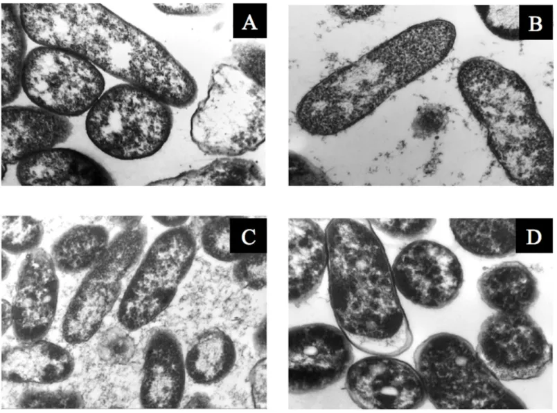
CFU/mL, respectively, was observed, with a similar increase in the number of green bacterial cells, which indicated that membrane reparation had occurred (Fig 9,S7andS8Figs). The increase in green intensity was supported by the MFI values (Fig 10A); interestingly, the MFI values associated with propidium iodide were similar to those found under rehydration (Fig 10B). Bacterial staining using toluidine blue showed a decrease in bacterial size among the des-iccated bacteria (S9 Fig). TEM observation of the bacterial cells during desiccation showed that the bacteria suffered a retraction of the cell cytoplasm with a concomitant incremease of periplasmic space (Fig 11).
Desiccation of
P. putida KT2440::gfp (green fluorescent protein)
A miniTn7-GFP mutant strain ofP. putida KT2440, which constitutively expresses the green fluorescent protein [39], was used to explore whether this protein is expressed by bacterial cells after desiccation. The desiccation tolerance ofP. putida KT2440::gfp was similar to that of the wild-type strain; this strain was nonculturable at 12 DABD (Fig 12H), but under fluorescence microscopy, cells ofP. putida tagged with GFP were green during different stages of desicca-tion (Fig 12), indicating constitutive expression and active synthesis of this protein. However, the fluorescence intensity of the desiccated bacterial cells partially decreased until 18 DABD (Fig 12H).
Fig 2. Fluorescence micrographs ofP. putida KT2440 cells treated with the LIVE/DEAD BacLight Bacterial Viability Kit. (A) Bacterial cells
before desiccation with trehalose (200 mM). (B) Bacterial cells before desiccation without a protector. (C) Bacterial cells protected with trehalose at 18 DABD. (D) Bacterial cells without a protector at 18 DABD. (E) Bacterial cells protected with trehalose adhered to germinated seeds after rehydration. (F) Bacterial cells without protection adhered to germinated seeds after rehydration. The samples were observed at 100×. Each image represents the MERGE of two captured images (green and red cells). The generation of MERGE images is shown inS1andS3Figs, and examples of MERGE cell analysis are shown inS2andS4Figs.
Fig 3. Rhizosphere colonization byP. putida KT2440. (A) Cell abundance from plants inoculated with rehydrated 18 DABD cells without protection in comparison to
cell abundance from plants inoculated with rehydrated DABD cells in the presence of trehalose (200 mM). (B and C) Fluorescence micrographs ofP. putida KT2440
using the LIVE/DEAD BacLight Bacterial Viability Kit. The samples were observed at 100×. (B) Cells obtained from the rhizosphere of plants inoculated with rehydrated 18 DABD cells without protection. (C) Bacterial cells obtained from the rhizosphere of plants inoculated with rehydrated 18 DABD cells in the presence of trehalose (200 mM). Each image represents the MERGE of two captured images (green and red cells). The generation of MERGE images is shown inS1DandS3DFigs, and examples of the analysis of cells in MERGE images are shown inS2DandS4DFigs.
Bacterial gene expression of some constitutive genes before and after
desiccation and rehydration
RNA was extracted from nondesiccated and desiccated-rehydrated cells to evaluate the active expression of genes from culturable and nonculturable cells. Desiccated cells from 18 DABD and, in some cases, 40 DABD were rehydrated for 20 min or 24 h. The explored genes were 16S rRNA,rpoN (housekeeping), mutL, mutS (codifying proteins from the mismatch repair complex), and a gene codifying an outer membrane protein widely distributed inP. putida KT2440 (oprH). All evaluated genes were expressed by cells both before desiccation and after desiccation-rehydration independent of the time of desiccation or rehydration (S10andS11
Figs), which means that the desiccated-rehydrated cells were alive at all evaluation times, even though they had membrane damage or were nonculturable.
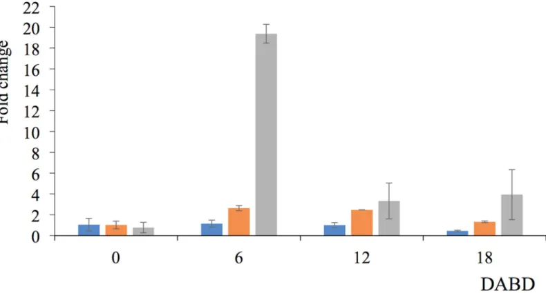
The level of expression of themutL, mutS y oprH genes from P. putida KT2440 was quanti-fied before desiccation and at 6, 12 and 18 DABD with the RT-qPCR method. Interestingly, oprH gene expression was markedly increased at 6 DABD (19.37 times), and the expression declined at 12 or 18 DABD (Fig 13), but it was higher than the levels observed before desicca-tion. The level of expression of themutS gene increased more than 2 times at 6 and 12 DABD and returned to levels similar to those of nondesiccated cells at 18 DABD. The level of expres-sion of themutL gene was constant, but it decreased at 18 DABD.
Discussion
P. putida KT2440 has high agro-biotechnological potential [4,6,42]. According to our results, this bacterium is very sensitive to air desiccation, and the benefits of a sensitive bacterial spe-cies could be lost after desiccation occurs in the environment [20,21]. Bacterial tolerance to desiccation is a key factor in designing stable inoculants because tolerant bacteria can adhere to seeds, tolerate desiccation in field soils, and colonize plant roots after rehydration when environmental conditions are favorable, maintaining their ability to improve plant growth [6,20]. Therefore, the high tolerance ofP. putida KT2440 and studies about how this bacterium tolerates desiccation are very important to exploit its capabilities in fields. Disaccharides have been reported to be good protectors ofP. putida KT2440 under freeze-drying conditions [12,13]. However, in our work, only nonreducing disaccharides were able to effectively protect P. putida KT2440 cells from the effects of air desiccation. The best protector was trehalose, resulting in high cultivability in this bacterium, and this disaccharide could be used to formu-late bacterial powder inoculants. Although trehalose produced by genetically modified cells of P. putida KT2440 was unable to protect the cells from the effects of freeze-drying [13], this disaccharide was shown to be able to protectP. putida KT2440 from the effects of the freeze-drying process when this sugar was added at 200 mM [12], as was also observed in terms of protection against air desiccation in our work.
Trehalose facilitated high bacterial culturability even at 18 DABD; however, in the treat-ments without protection, the number of culturable cells decreased to a nondetectable value after 12 DABD. Therefore, we hypothesize that only rehydrated protected cells can adhere to roots and colonize the rhizosphere of maize plants. As expected, only protected cells were detected in the adhesion assays. Surprisingly, the number of bacteria colonizing the
rhizo-Fig 4. Mean fluorescence intensity (MFI) ofP. putida KT2440 cells with protection (trehalose 200 mM). (A)
Analysis of SYTO 9 and (B) analysis of propidium iodide. (1) Samples obtained from the bacterial suspension before desiccation, (2) cells at 18 DABD rehydrated for 20 min, (3) cells adhered to maize sprouts, and (4) cells from rhizosphere colonization.
https://doi.org/10.1371/journal.pone.0219554.g004
We propose that during desiccation,P. putida KT2440 enters a nonculturable state as a strat-egy to cope with stress. However, the bacterial cells return to a culturable state after interacting with plant roots, likely because the exudates of plants activate mechanisms related to this return. To determine whether root exudates allowP. putida KT2440 to return to cultivability, the bacterial cells were desiccated until 18 DABD, and they were rehydrated with only water or with maize root exudates;P. putida KT2440 cells returned to a culturable state after rehydra-tion with plant root exudates faster than they did with only water, suggesting that some com-pounds or molecules from exudates favor the return to a culturable state. Studies have shown that whenBacillus atrophaeus UCMB-5137 grows in the presence of corn exudates, the expres-sion of genes related to stress and detoxification is stimulated [43]; it is probable that the maize root exudates favor the expression of genes necessary for the return to a culturable state forP. putida KT2440 after rehydration. Among the compounds present in the exudates of corn plants are nitrogen compounds, fatty acids, organic acids, sugars, volatile compounds, steroids, terpenoids and other substances [44,45]. It will be interesting to carry out future trials to clarify which compounds favor the quickest return to a culturable state inP. putida KT2440 following the rehydration of desiccated cells. The return to a culturable state occurs when bacteria find favorable conditions and when the stress is completely withdrawn [25], and this could explain why in our work bacterial cells were also able to return to a culturable state after prolonged rehydration.
Bacteria in the VBNC state can be identified using techniques that determine whether they are metabolically active even if they are not culturable. These methodologies include assays with the LIVE/DEAD BacLight Bacterial Viability Kit, which identifies damage to the plas-matic membrane [25], studies of the green fluorescent protein (GFP), which is constitutively expressed by nonculturable bacteria [30,46], and gene expression studies using molecular tools, such as RT-PCR and RT-qPCR [23,47]. In the present work, we used these techniques to evaluate whetherP. putida KT2440 remain active in the VBNC state during desiccation. By using the LIVE/DEAD BacLight Bacterial Viability Kit, it was observed that there was an increase in the number of red bacteria and that the propidium iodide MFI values were greater than the MFI values of SYTO 9, indicating that the injury to membranes ofP. putida KT2440 increases as the duration of desiccation progresses. During desiccation, bacterial membrane changes in the composition of fatty acids and experience alterations in its fluidity [48], and destabilization could allow propidium iodide to cross the membrane and the red staining of cells. According to the kit and other works, red cells are considered dead [49]. However, our results showed that the red bacterial cells from 18 DABD (after rapid rehydration) were non-culturable, but they returned to a culturable state after prolonged rehydration or rehydration in the presence of exudates, indicating that the red cells never died.
Fig 5. Mean fluorescence intensity (MFI) ofP. putida KT2440 cells without protection. (A) Analysis of SYTO 9 and (B)
analysis of propidium iodide. (1) Samples obtained from the bacterial suspension before desiccation, (2) cells at 18 DABD rehydrated for 20 min, (3) cells adhered to maize sprouts, and (4) cells from rhizosphere colonization.
https://doi.org/10.1371/journal.pone.0219554.g005
Table 3. Number of cells ofP. putida KT2440 (log CFU/mL) 18 DABD and rehydrated with root exudates of maize.
Maize root exudates Time of rehydration
20 min 3 h 6 h 9 h 12 h 24 h 27 h 30 h 48 h
Maize sprout 0 0 0 0 3.09±0.15 4.81±0.19 6.09±0.14 6.21±0.10 7.11±0.19
12 days of growth of plant 0 0 0 0 3.21±0.41 4.78±0.26 6.14±0.29 6.62±0.09 7.32±0.23
Water control 0 0 0 0 0 4.34±0.16 5.46±0.29 5.34±0.16 7.39±0.36
Interestingly, under prolonged rehydration (24 and 48 h), the bacteria decreased in terms of the intensity of their red coloration, as indicated by the microscopic analysis and MFI val-ues, and increased in terms of the intensity of their green coloration. This result indicates that the penetration of the bacterial cells by propidium iodide decreased, likely due to the recovery of membrane integrity, and again, that the crossing of propidium iodide was prevented. In fact, in association with plants, the red intensity declined, and only green cells were observed, indicating the total recovery of the integrity of the membranes. However, the red staining by propidium iodide do not necessarily mean “dead cells”. It was reported that viableP. putida (ATCC 12633) cells were stained by propidium iodide suggesting that the use of LIVE/DEAD BacLight Bacterial Viability kit may give a confusing result in determining live cells [50]. We propose that additional studies like TEM, changes in membrane fluidity, changes in mem-brane potential and changes in memmem-brane phospholipid composition will be required to clarify the level of membrane damage in our viable but non-culturable bacterial cells subjected to des-iccation-rehydration process.
The first assay to verify the metabolic activity of nonculturable cells involved the desiccation ofP. putida KT2440::gfp, a strain that constitutively expresses the green fluorescent protein (GFP) [39].P. putida KT2440::gfp tolerated air desiccation at a level similar to that of wild-type and the presence of nonculturable cells were observed after 12 DABD. However, in all the experiments, fluorescent cells were observed without an apparent decrease in intensity at 15 DABD, with a slight decrease at 18 DABD. This result suggests the occurrence of active meta-bolic activity in rehydrated nonculturable cells. GFP studies have also been carried out with other models to evaluate the VBNC state of cells [46,51]. In our work, the fluorescence
Fig 6. Bacterial behavior ofP. putida KT2440 cells rehydrated with water (orange line) or in the presence of root exudates (blue line) under static conditions for
48 h.
intensity and microscopic observation of green cells confirmed the viability and cellular activ-ity of nonculturable rehydratedP. putida KT2440::gfp cells.
Molecular tools such as RT-PCR and RT-qPCR have been widely used to determine cellular viability because the average lifetime of mRNA is approximately 3 to 5 min. Thus, amplified RNA molecules arise from active and recent transcription and are an excellent indicator of metabolic activity in VBNC cells [52,53]. To increase our knowledge of the cellular activity of nonculturable rehydratedP. putida KT2440 cells, we monitored the expression of housekeep-ing genes (RpoN and 16S rRNA), genes encoding proteins from the mismatch repair complex (mutL and mutS), and a gene encoding an outer membrane protein (oprH) both before desic-cation and at 18 DABD. All explored genes in the present work were amplified from RNA samples after a short period of rehydration of nonculturableP. putida KT2440 cells, indicating active expression of the genes in that state and consequently active metabolic activity. 16S rRNA has been widely used to explore the VBNC state of bacteria [54,55], and in our work, it was the first gene selected to evaluate cell activity, as ribosomal genes were observed to be active after rehydration. The generpoN encodes the σ54factor from RNA polymerase and reg-ulates the expression of several genes [56–58]. This housekeeping gene (rpoN) was used as a reference gene for the RT-qPCR in this study. After amplification with RT-qPCR, it was observed that theoprH, mutL and mutS genes in nonculturable rehydrated P. putida KT2440 cells were expresed. TheoprH gene encodes one of the most abundant membrane proteins of the genusPseudomonas, which functions mainly as an aquaporin and a drug transporter efflux protein and is important for the maintenance and support of the membrane [59]. Expression of theoprH gene has been observed to be highly induced by high oxygen pressure conditions, likely as a consequence of plasmatic membrane destabilization caused by this type of stress [59]. In our work, the expression of theoprH gene highly increased at 6 DABD, suggesting that under desiccation conditions, the destabilization of cell membranes occurs, which is supported by the increase in red bacterial cells observed under fluorescence microscopy. AlthoughoprH gene expression decreased at 12 and 18 DABD, this expression was higher than that observed for themutL and mutS genes; therefore, we propose this gene as an indicator of viability in P. putida KT2440 under desiccation stress.
ThemutL and mutS genes codify proteins from the mismatch repair complex, which acts in DNA replication and is also involved in the signaling process to prevent chemical damage that could occur under air desiccation, such as oxidation, Maillard reactions, and DNA damage [18,60]. Therefore,mutL and mutS could be fundamental genes involved in DNA reparation, and the study of the rehydration of desiccatedP. putida KT2440 cells was carried out. Our results show a decrease in the expression of themutL gene in nonculturable cells after rehydra-tion, but this gene was expressed over time. For themutS gene, the expression was similar in culturable and nonculturable cells. Therefore, these genes could be involved in DNA repara-tion in cells damaged by desiccarepara-tion. In our work, RT-PCR of thegfp gene from nonculturable cells was not performed, and it will be interesting to carry out such assays in the future.
Conclusions
Pseudomonas putida KT2440 has very low tolerance to air desiccation in comparison to other bacteria; however, trehalose protects it from stress and could be used to design powder-stable
Fig 7. Fluorescence ofP. putida KT2440 cells desiccated for 18 days and stained with the LIVE/DEAD BacLight
Bacterial Viability Kit. (A) Cells before desiccation and (B) 3, (C) 6, (D) 9, (E) 12, (F) 15, and (G) 18 DABD. Each
image represents the MERGE of two captured images (green and red cells). The samples were observed at 100×. The generation of MERGE images is shown inS5 Fig, and examples of the analysis of MERGE images are shown inS6 Fig. (H) The BSR ofP. putida KT2440 during desiccation.
inoculants with the capability to maintain the viability of the strain without the use of a vac-uum, decreasing the cost of production. Furthermore, this bacterium enters the VBNC state during desiccation without protector addition, which could be a strategy to mitigate the stress. P. putida KT2440 cells in a desiccated state have a decreased cell size and retracted cytoplasm. Desiccation also causes damage to the membranes ofP. putida KT2440, and these membranes can apparently be repaired after prolonged rehydration or rehydration in the presence of plant root exudates. After a short rehydration period, the bacterial cells are not capable of growth in culture media (VBNC state); however, the transcription of several genes remains active under
Fig 8. Mean fluorescence intensity (MFI) of the same images as inFig 7. (A) Analysis of SYTO 9. (B) Analysis of
propidium iodide.
https://doi.org/10.1371/journal.pone.0219554.g008
Fig 9. Fluorescence ofP. putida KT2440 cells desiccated for 18 days and stained with the LIVE/DEAD BacLight Bacterial Viability Kit. (A) Cells before
desiccation, (B) cells at 18 DABD rehydrated for 20 min, (C) cells at 18 DABD rehydrated for 24 h, (D) cells at 18 DABD rehydrated for 48 h. The samples were observed at 100×. Each image represents the MERGE of two captured images (green and red cells). Generation of MERGE images is shown inS7 Fig, and examples of analysis of MERGE images are shown inS8 Fig.
these conditions in the wild-type strain, and GFP activity was also detected in cells targeted with thegfp gene, showing active metabolic activity after the rehydration of desiccated cells. Data from the present work support that during desiccation, this bacterial strain suffers dam-age to its membranes and enters the VBNC state, and the bacterial cells return to a culturable state after their interaction with plant roots or prolonged rehydration when the membranes are repaired. Therefore, this work will contribute to the development of bacterial inoculants containing live bacteria in a VBNC state that could return to a culturable state after interaction with root plants when water conditions are completely favorable to germination, leading to successful colonization and the maintenance of their beneficial effects.
This work opens new avenues of research in the field of bacterial survival under desiccation stress and breaks the paradigm of the bacterial membrane integrity concept. Today, people
Fig 10. Mean fluorescence intensity (MFI) of the same images as inFig 9. (A) Analysis of SYTO 9. (B) Analysis of
propidium iodide.
https://doi.org/10.1371/journal.pone.0219554.g010
Fig 11. Transmission electron microscopy ofP. putida KT2440. (A) Before desiccation, 50,000×, (B) at 6 DABD, 30,000×, (C) at 12 DABD, 30,000× and (D) at 18
DABD, 50,000×.
think that if the membrane is affected, then the bacteria are dead, but we showed that even though theP. putida KT2440 membrane was affected, this bacterium was able to return to a culturable state if environmental conditions were favorable for its growth. The results of the present work could be taken into account to design bacterial inoculants for application in agri-culture, by using live bacteria in a VBNC state that can return to a culturable state after the interacting with plant roots when the conditions are favorable for germination and coloniza-tion. The inoculant industry adheres to some norms regarding the number of bacteria in inoc-ulant formulations that indicate that when the number decreases below a specified threshold, the inoculant should be discarded. However, whether the bacteria are truly dead or remain only nonculturable should be considered because the bacteria could return to a culturable state after their interaction with plants.
Supporting information
S1 Fig. Fluorescence microscopy ofP. putida KT2440 cells treated with the kit “Live/Dead
BacLight Bacterial Viability” from treatments desiccated in presence of trehalose (200 mM). A) Bacterial cells before desiccation. B) Bacterial cells at 18 DABD. C) Bacterial cell
adhered to germinated seeds. D) Rhizosphere colonization ofP. putida KT2440 from plants inoculated with rehydrated cells of 18 DABD. Each row shows two captured imagens; the images of column SYTO 9 were taken at filter with excitation 420–490 nm, images of column
Fig 12. Fluorescence ofP. putida KT2440 tagged with GFP desiccated for 18 days. (A) Cells before desiccation and (B) 3, (C) 6, (D) 9, (E)
12, (F) 15, and (G) 18 DABD. The samples were observed at 100×. (H) The BSR of P. putida tagged with GFP during desiccation (blue line) and fluorescence intensity ofP. putida tagged with GFP during desiccation (orange line).
https://doi.org/10.1371/journal.pone.0219554.g012
Fig 13. Gene expression levels ofmutL (blue bar), mutS (orange bar) and oprH (gray bar) obtained by RT-qPCR. Before desiccation (0) and 6, 12 and 18 DABD.
propidium iodide were taken at filter excitation 500-550nm. MERGE correspond to combina-tion of both images (SYTO 9 and propidium iodide).
(PDF)
S2 Fig. MERGE imagens and histograms that represent distribution of SYTO 9 and propi-dium iodide from cells random selected. Treatments desiccated with trehalose. A) Bacterial
cells before desiccation. B) Bacterial cells at 18 DABD. C) Bacterial cell adhered to germinated seeds. D) Rhizosphere colonization ofP. putida KT2440 from plants inoculated with rehy-drated cells of 18 DABD.
(PDF)
S3 Fig. Fluorescence microscopy ofP. putida KT2440 cells treated with the kit “Live/Dead
BacLight Bacterial Viability” from treatments desiccated without protectors. A) Bacterial
cells before desiccation. B) Bacterial cells at 18 DABD. C) Bacterial cell adhered to germinated seeds. D) Rhizosphere colonization ofP. putida KT2440 from plants inoculated with rehy-drated cells of 18 DABD. Each row shows two captured imagens; the images of column SYTO 9 were taken at filter with excitation 420–490 nm, images of column propidium iodide were taken at filter excitation 500-550nm. MERGE correspond to combination of both images (SYTO 9 and propidium iodide).
(PDF)
S4 Fig. MERGE Imagens and histograms that represent distribution of SYTO 9 and propi-dium iodide from cells random selected. Treatments desiccated without protectors. A)
Bacte-rial cells before desiccation. B) BacteBacte-rial cells at 18 DABD. C) BacteBacte-rial cell adhered to germinated seeds. D) Rhizosphere colonization ofP. putida KT2440 from plants inoculated with rehydrated cells of 18 DABD.
(PDF)
S5 Fig. Fluorescence ofP. putida KT2440 cells desiccated during 18 days, and stained with
LIVE/DEAD BacLight Bacterial Viability kit. A) Before desiccation, B) 3, C) 6, D) 9, E) 12,
F) 15, and G) 18 DABD. Each row shows two captured imagens; the images of column SYTO 9 were taken at filter with excitation 420–490 nm, images of column propidium iodide were taken at filter excitation 500-550nm. MERGE correspond to combination of both images (SYTO 9 and propidium iodide).
(TIFF)
S6 Fig. MERGE imagens and histograms that represent distribution of SYTO 9 and propi-dium iodide from cells random selected. A) Before desiccation, B) 3, C) 6, D) 9, E) 12, F) 15,
and G) 18 DABD (PDF)
S7 Fig. Fluorescence ofP. putida KT2440 (A) before desiccation (B) after desiccation by 18 days and rehydrated by 20 minutes (C) 24 h (D) 48 h stained with LIVE/DEAD BacLight Bac-terial Viability kit. The images of column SYTO 9 were taken at filter with excitation 420–490 nm, images of column propidium iodide were taken at filter excitation 500-550nm and images MERGE corresponding to combination of both images (SYTO 9 and propidium iodide). (PDF)
S8 Fig. MERGE imagens and histograms that represent distribution of SYTO 9 and propi-dium iodide from cells random selected. A) Before desiccation. B) Twenty-min rehydrated
bacterial cells of 18 DABD. C) Twenty four-hour rehydrated bacterial cells of 18 DABD. D)
Forty eight-hour rehydrated bacterial cells of 18 DABD. (PDF)
S9 Fig. Optical microscopy ofP. putida KT2440 stained with toluidine blue along
desicca-tion assay. A) Before desiccadesicca-tion, B) 6 DABD, C) 9 DABD, D) 12 DABD, E) 15 DABD, and F)
18 DABD. (PDF)
S10 Fig. Amplification of the16S rRNA gene fromP. putida KT2440 by using the RT-PCR
method. 1) Marker 1 kb DNA Leader Jena Bioscience, 2) cells before desiccation, 3)
twenty-min rehydrated cells of 18 DABD. 4) Twenty four-hours rehydrated cells of 18 DABD, 5) twenty-min rehydrated cells of 40 DABD, 6) negative control; reaction without retrotranscrip-tase, and 7) negative control; reaction without template.
(PDF)
S11 Fig. Amplification of the genesmutL, rpoN and oprH from P. putida KT2440 by using
the RT-PCR method. 1) Marker 1 kb DNA Leader Jena Bioscience, 2)mutL before desicca-tion, 3)mutL from twenty-min rehydrated cells of 18 DABD, 4) mutL from Twenty four-hours rehydrated cells of 18 DABD, 5)mutL from twenty-min rehydrated cells of 40 DABD, 6) rpoN before desiccation, 7) rpoN from twenty-min rehydrated cells of 18 DABD, 8) rpoN from twenty four-hours rehydrated cells of 18 DABD, 9)rpoN from twenty-min rehydrated cells of 40 DABD, 10)oprH before desiccation, 11) oprH from twenty-min rehydrated cells of 18 DABD, 12)oprH from twenty four-hours rehydrated cells of 18 DABD, 13) oprH from twenty-min rehydrated cells of 40 DABD.
(PDF)
Acknowledgments
We are grateful to CONACYT for the fellowship awarded to Laura Abisaı´ Pazos-Rojas, Ligia Catalina Muñoz-Arenas, Osvaldo Rodrı´guez-Andrade and Lesther Emanuel Lo´pez-Cruz.
Author Contributions
Conceptualization: Jesu´s Muñoz-Rojas.
Data curation: Laura Abisaı´ Pazos-Rojas, Ligia Catalina Muñoz-Arenas, Osvaldo Rodrı´guez-Andrade, Orestes Lo´pez-Ortega, Silvia Luna-Suarez.
Formal analysis: Laura Abisaı´ Pazos-Rojas, Ligia Catalina Muñoz-Arenas, Osvaldo Rodrı´-guez-Andrade, Orestes Lo´pez-Ortega, Fa´bio Lopes-Olivares, Jesu´s Muñoz-Rojas.
Funding acquisition: Jesu´s Muñoz-Rojas.
Investigation: Laura Abisaı´ Pazos-Rojas, Ligia Catalina Muñoz-Arenas, Osvaldo Rodrı´guez-Andrade, Lesther Emanuel Lo´pez-Cruz, Silvia Luna-Suarez, Jesu´s De la Torre.
Methodology: Laura Abisaı´ Pazos-Rojas, Lesther Emanuel Lo´pez-Cruz, Yolanda Elizabeth Morales-Garcı´a.
Project administration: Yolanda Elizabeth Morales-Garcı´a, Jesu´s Muñoz-Rojas.
Supervision: Fa´bio Lopes-Olivares, Yolanda Elizabeth Morales-Garcı´a, Miguel Angel
Villalo-bos-Lo´pez, Jesu´s De la Torre, Jesu´s Muñoz-Rojas.
Validation: Fa´bio Lopes-Olivares.
Writing – original draft: Laura Abisaı´ Pazos-Rojas, Antonino Baez, Vero´nica
Quintero-Her-na´ndez, Miguel Angel Villalobos-Lo´pez, Jesu´s Muñoz-Rojas.
Writing – review & editing: Laura Abisaı´ Pazos-Rojas, Antonino Baez, Vero´nica
Quintero-Herna´ndez, Jesu´s Muñoz-Rojas.
References
1. Espinosa-Urgel M, Kolter R, Ramos J-L. Root colonization by Pseudomonas putida: love at first sight. Microbiology. 2002; 148: 341–344.https://doi.org/10.1099/00221287-148-2-341PMID:11832496 2. Ramos-Gonzalez MI, Ramos-Diaz MA, Ramos JL. Chromosomal gene capture mediated by the
Pseu-domonas putida TOL catabolic plasmid. J Bacteriol. 1994; 176: 4635–4641.https://doi.org/10.1128/jb. 176.15.4635-4641.1994PMID:8045894
3. Timmis KN. Pseudomonas putida: a cosmopolitan opportunist par excellence. Environ Microbiol. 2002; 4: 779–781.https://doi.org/10.1046/j.1462-2920.2002.00365.xPMID:12534460
4. Matilla MA, Ramos JL, Bakker PAHM, Doornbos R, Badri D V., Vivanco JM, et al. Pseudomonas putida KT2440 causes induced systemic resistance and changes in Arabidopsis root exudation. Environ Microbiol Rep. 2009; 2: 381–388.https://doi.org/10.1111/j.1758-2229.2009.00091.xPMID:23766110 5. Molina-Romero D, Morales-Garcı´a YE, Herna´ndez-Tenorio A-L, Castañeda-Lucio M,
Netzahuatl-Muñoz AR, Muñoz-Rojas J. Pseudomonas putida estimula el crecimiento de maı´z en funcio´n de la tem-peratura. Rev Iberoam Ciencias. 2017; 4: 80–88.
6. Molina-Romero D, Baez A, Quintero-Herna´ndez V, Castañeda-Lucio M, Fuentes-Ramı´rez LE, Bustil-los-Cristales M del R, et al. Compatible bacterial mixture, tolerant to desiccation, improves maize plant growth. PLoS One. Public Library of Science; 2017; 12: e0187913. Available:https://doi.org/10.1371/ journal.pone.0187913PMID:29117218
7. Martins Dos Santos VAP, Heim S, Moore ERB, Stra¨tz M, Timmis KN. Insights into the genomic basis of niche specificity of Pseudomonas putida KT2440. Environ Microbiol. 2004; 6: 1264–1286.https://doi. org/10.1111/j.1462-2920.2004.00734.xPMID:15560824
8. Wackett LP. Pseudomonas putida—a versatile biocatalyst. Nat Biotechnol. Nature Publishing Group; 2003; 21: 136–138. Available:http://dx.doi.org/10.1038/nbt0203-136PMID:12560839
9. Pineda-Molina E, Reyes-Darias JA, Lacal J, Ramos JL, Garcı´a-Ruiz JM, Gavira JA, et al. Evidence for chemoreceptors with bimodular ligand-binding regions harboring two signal-binding sites. Proc Natl Acad Sci U S A. 2012; 109: 18926–18931.https://doi.org/10.1073/pnas.1201400109PMID:23112148 10. Krell T, Lacal J, Reyes-Darias JA, Jimenez-Sanchez C, Sungthong R, Ortega-Calvo JJ. Bioavailability
of pollutants and chemotaxis. Curr Opin Biotechnol. 2013; 24: 451–456.https://doi.org/10.1016/j. copbio.2012.08.011PMID:22981870
11. Wu X, Monchy S, Taghavi S, Zhu W, Ramos J, van der Lelie D. Comparative genomics and functional analysis of niche-specific adaptation in Pseudomonas putida. FEMS Microbiol Rev. 2011; 35: 299–323. https://doi.org/10.1111/j.1574-6976.2010.00249.xPMID:20796030
12. Muñoz-Rojas J, Bernal P, Duque E, Godoy P, Segura A, Ramos JL. Involvement of cyclopropane fatty acids in the response of Pseudomonas putida KT2440 to freeze-drying. Appl Environ Microbiol. 2006; 72: 472–477.https://doi.org/10.1128/AEM.72.1.472-477.2006PMID:16391080
13. Manzanera M, Castro AG De, Tøndervik A, Strøm AR, Tunnacliffe A. Hydroxyectoine is superior to tre-halose for anhydrobiotic engineering of Pseudomonas putida KT2440. Appl Environ Microbiol. 2002; 68: 4328–4333.https://doi.org/10.1128/AEM.68.9.4328-4333.2002PMID:12200283
14. Roszak DB, Colwell RR. Survival strategies of bacteria in the natural environment. Microbiol Rev. 1987; 51: 365–379. doi: 0146-0749/87/030365-15 PMID:3312987
15. Chookietwattana K, Maneewan K. Selection of efficient salt-tolerant bacteria containing ACC deami-nase for promotion of tomato growth under salinity stress. Soil Environ. Faisalabad: Soil Science Soci-ety of Pakistan; 2012; 31: 30–36.
16. He H, Chen Y, Li X, Cheng Y, Yang C, Zeng G. Influence of salinity on microorganisms in activated sludge processes: A review. Int Biodeterior Biodegradation. 2017; 119: 520–527.https://doi.org/10. 1016/j.ibiod.2016.10.007
17. Vilchez S, Manzanera M. Biotechnological uses of desiccation-tolerant microorganisms for the rhizore-mediation of soils subjected to seasonal drought. Appl Microbiol Biotechnol. 2011; 91: 1297.https://doi. org/10.1007/s00253-011-3461-6PMID:21769483
19. Potts M, Slaughter SM, Hunneke F-U, Garst JF, Helm RF. Desiccation tolerance of Prokaryotes: appli-cation of principles to human cells. Integr Comp Biol. 2005; 45: 800–809.https://doi.org/10.1093/icb/45. 5.800PMID:21676831
20. Pazos-Rojas LA, Rodrı´guez-Andrade O, Muñoz-Arenas LC, Morales-Garcı´a YE, Corral-Lugo A, Quin-tero-Herna´ ndez V, et al. Desiccation-tolerant rhizobacteria maintain their plant growth- promoting capa-bility after experiencing extreme water stress. SciFed J Appl Microbiol. 2018; 2: 1–13.
21. Streeter JG. Effect of trehalose on survival of Bradyrhizobium japonicum during desiccation. J Appl Microbiol. 2003; 95: 484–491.https://doi.org/10.1046/j.1365-2672.2003.02017.xPMID:12911696 22. Barnard RL, Osborne CA, Firestone MK. Responses of soil bacterial and fungal communities to
extreme desiccation and rewetting. ISME J. International Society for Microbial Ecology; 2013; 7: 2229– 2241. Available:https://doi.org/10.1038/ismej.2013.104
23. Oliver JD. Recent findings on the viable but nonculturable state in pathogenic bacteria. FEMS Microbiol Rev. 2010; 34: 415–425.https://doi.org/10.1111/j.1574-6976.2009.00200.xPMID:20059548 24. Orruño M, Kaberdin VR, Arana I. Survival strategies of Escherichia coli and Vibrio spp.: contribution of
the viable but nonculturable phenotype to their stress-resistance and persistence in adverse environ-ments. World J Microbiol Biotechnol. Springer Netherlands; 2017; 33: 45.https://doi.org/10.1007/ s11274-017-2218-5PMID:28161849
25. Stokell JR, Steck TR. Viable but nonculturable bacteria. eLS. 2012. pp. 1–8.https://doi.org/10.1002/ 9780470015902.a0000407.pub2
26. Vriezen JAC, de Bruijn FJ, Nu¨sslein KR. Desiccation induces viable but non-culturable cells in Sinorhi-zobium meliloti 1021. AMB Express. 2012; 2: 1–9.https://doi.org/10.1186/2191-0855-2-1
27. Oliver JD. The public health significance of viable but nonculturable bacteria. In: Colwell RR, Grimes DJ, editors. Nonculturable Microorganisms in the Environment. Boston, MA: Springer US; 2000. pp. 277–300.https://doi.org/10.1007/978-1-4757-0271-2_16
28. Dopp E, Richard J, Dwidjosiswojo Z, Simon A, Wingender J. Influence of the copper-induced viable but non-culturable state on the toxicity of Pseudomonas aeruginosa towards human bronchial epithelial cells in vitro. Int J Hyg Environ Health. Elsevier; 2017; 220: 1363–1369.https://doi.org/10.1016/j.ijheh. 2017.09.007PMID:28941772
29. Oliver JD. The viable but nonculturable state in bacteria. J Microbiol. Department of Biology, University of North Carolina at Charlotte, Charlotte, NC 28223–0001, USA.jdoliver@uncc.edu; 2005; 43: 93–100. Available:http://europepmc.org/abstract/MED/15765062PMID:15765062
30. Trevors JT. Viable but non-culturable (VBNC) bacteria: Gene expression in planktonic and biofilm cells. J Microbiol Methods. Elsevier B.V.; 2011; 86: 266–273.https://doi.org/10.1016/j.mimet.2011.04.018 PMID:21616099
31. Robben C, Fister S, Witte AK, Schoder D, Rossmanith P, Mester P. Induction of the viable but non-cul-turable state in bacterial pathogens by household cleaners and inorganic salts. Sci Rep. Springer US; 2018; 8: 15132.https://doi.org/10.1038/s41598-018-33595-5PMID:30310128
32. Zhao X, Zhong J, Wei C, Lin CW, Ding T. Current perspectives on viable but non-culturable state in foodborne pathogens. Front Microbiol. 2017; 8: 1–16.https://doi.org/10.3389/fmicb.2017.00001 33. Molina-Romero D, Baez A, Quintero-Herna´ndez V, Castañeda-Lucio M, Fuentes-Ramı´rez LE,
Bustil-los-Cristales M del R, et al. Selection assay to identify desiccation tolerant bacteria [Internet]. Protocols. io PLOS one. 2017. pp. 1–2.https://doi.org/10.17504/protocols.io.j4icque
34. Corral-Lugo A, Morales-Garcı´a YE, Pazos-Rojas LA, Ramı´rez-Valverde A, Martı´nez-Contreras RD, Muñoz-Rojas J. Cuantificacio´n de bacterias cultivables mediante el me´todo de “goteo en placa por sell-ado (o estampsell-ado) masivo.” Rev Colomb Biotecnol. 2012; 14: 147–156.
35. Reyes-Darias JA, Garcı´a V, Rico-Jime´ nez M, Corral-Lugo A, Lesouhaitier O, Jua´rez-Herna´ndez D, et al. Specific gamma-aminobutyrate chemotaxis in pseudomonads with different lifestyle. Mol Micro-biol. 2015; 97: 488–501.https://doi.org/10.1111/mmi.13045PMID:25921834
36. Rodrı´guez-Andrade O, Fuentes-Ramı´rez LE, Morales-Garcı´a YE, Molina-Romero D, Bustillos-Cristales MR, Martı´nez-Contreras RD, et al. The decrease in the population of Gluconacetobacter diazotrophicus in sugarcane after nitrogen fertilization is related to plant physiology in split root experiments. Rev Argentina Microbiol. 2015; 47: 335–43.https://doi.org/10.1016/j.ram.2015.09.004PMID:26652262 37. Morales-Garcı´a YE, Jua´rez-Herna´ndez D, Arago´n-Herna´ ndez C, Mascarua-Esparza MA, Bustillos-Cristales MR, Fuentes-Ramı´rez LE, et al. Growth response of maize plantlets inoculated with Entero-bacter spp., as a model for alternative agriculture. Rev Argent Microbiol. 2011; 43: 287–293.https://doi. org/10.1590/S0325-75412011000400009PMID:22274827
38. Muñoz-Rojas J, Caballero-Mellado J. Population dynamics of Gluconacetobacter diazotrophicus in sug-arcane cultivars and its effect on plant growth. Microb Ecol. 2003; 46: 454–464.https://doi.org/10.1007/ s00248-003-0110-3PMID:14722690