Modi
fied Blalock Taussig shunt: a not-so-simple palliative procedure
Verena Dirks
a, René Prêtre
b, Walter Knirsch
c,d, Emanuela R. Valsangiacomo Buechel
c,d, Burkhardt Seifert
e,
Martin Schweiger
a,d, Michael Hübler
a,dand Hitendu Dave
a,d,*
a Division of Congenital Cardiovascular Surgery, University Children’s Hospital Zurich, Zurich, Switzerland b Department of Cardiovascular Surgery, University Hospital Lausanne, Lausanne, Switzerland
c Department of Paediatric Cardiology, University Children’s Hospital Zurich, Zurich, Switzerland d Children’s Research Centre, University of Zurich, Zurich, Switzerland
e
Division of Biostatistics, Institute for Social and Preventive Medicine, University of Zurich, Zurich, Switzerland
* Corresponding author. Division of Congenital Cardiovascular Surgery, University Children’s Hospital Zurich, Zurich, Switzerland. Fax: +41-44-2668021; e-mail: [email protected]; [email protected] (H. Dave).
Received 7 January 2013; received in revised form 26 February 2013; accepted 27 February 2013
Abstract
OBJECTIVES: Thirty-two consecutive isolated modified Blalock Taussig (BT) shunts performed in infancy since 2004 were reviewed and analysed to identify the risk factors for shunt intervention and mortality.
METHODS: Sternotomy was the only approach used. Median age and weight were 10.5 (range 1–74) days and 2.9 (1.9–4.4) kg, respect-ively. Shunt palliation was performed for biventricular hearts (Tetralogy of Fallot/double outlet right ventricle/transposition of great arteries_ventricular septal defect_pulmonary stenosis/pulmonary atresia_ventricular septal defect/others) in 21, and univentricular hearts in 11, patients. Hypoplastic left heart syndrome patients were excluded. Two procedures required cardiopulmonary bypass. Median shunt size was 3.5 (3–4) mm and median shunt size/kg body weight was 1.2 (0.9–1.7) mm/kg. Reduction in shunt size was necessary in 5 of 32 (16%) patients.
RESULTS: Three of 32 (9%) patients died after 3 (1–15) days due to cardiorespiratory decompensation. Lower body weight (P = 0.04)
and bigger shunt size/kg of body weight (P = 0.004) were significant risk factors for mortality. Acute shunt thrombosis was observed in
3 of 32 (9%), none leading to death. Need for cardiac decongestive therapy was associated with univentricular hearts (P < 0.001), bigger
shunt size (P = 0.054) and longer hospital stay (P = 0.005). Twenty-eight patients have undergone a successful shunt takedown at a
median age of 5.5 (0.5–11.9) months, without late mortality.
CONCLUSIONS: Palliation with a modified BT shunt continues to be indicated despite increased thrust on primary corrective surgery.
Though seemingly simple, it is associated with significant morbidity and mortality. Effective over-shunting and acute shunt thrombosis are the lingering problems of shunt therapy.
Keywords:Modified Blalock Taussig shunt • Palliation • Mortality • Cyanotic heart disease
INTRODUCTION
While the classic Blalock Taussig shunt was a breakthrough in
treating cyanotic heart diseases [1], it involves sacrificing
ante-gradeflow to the subclavian artery with its attendant risks [2,3].
In 1975, de Leval modified the technique, using a
polytetra-fluoroethylene interposition graft popularly known as the
modi-fied Blalock Taussig (BT) shunt (MBTS) [4]. While MBTS has
become an established palliative procedure with progressive
improvements in the outcome [5], growing experience has led to
increasing thrust on primary corrective procedures. Palliative strategy has obvious disadvantages, such as the need for two operations, potentially two scars, possibility of distortion of the branch pulmonary artery, volume loading of the ventricles, lower diastolic pressures, etc. In spite of these, primary shunt palliation continues to be indicated in neonates with physiological pul-monary hypertension, which makes a bidirectional Glenn shunt
untenable. Many centres also consider a primary neonatal cor-rection of Tetralogy of Fallot to be riddled with risks and hence, still prefer to palliate neonates in blue spells.
While acknowledging its role even in the modern era, mortal-ity of the MBTS procedure is relatively high, tending to be
around 10% [6]. This report is based on 32 consecutive ‘first
time’ MBTS palliations performed at our institution since 2004, with a view to analysing the risk factors for mortality, shunt thrombosis and need for decongestive therapy.
PATIENTS AND METHODS
Patients
Thirty-two MBTS procedures performed in neonates and infants at our institution since 2004 were analysed. MBTS performed in
© The Author 2013. Published by Oxford University Press on behalf of the European Association for Cardio-Thoracic Surgery. All rights reserved. doi:10.1093/ejcts/ezt172 Advance Access publication 28 March 2013
patients with HLHS or as a part of other complex procedures, such as unifocalization, were excluded. Twenty-six patients were
neonates and 6 were infants. Twenty-five patients were male.
The demographic data are detailed in Table 1. Diagnosis was
established using trans-thoracic echocardiography.
End points
Primary end points were mortality and shunt thrombosis. Secondary end points such as need for excessive inotropic support and cardiac decongestive therapy were also analysed. A
brief analysis comparing Era 1 (2004–07) and 2 (2008–11) was
performed to see if the results had changed over time and to identify the factors that may have accounted for that change.
Surgical technique
All MBTS procedures were performed through a sternotomy. Two procedures required cardiopulmonary bypass. After full sternotomy, the right lobe of the thymus was excised; the course of the brachiocephalic trunk up to its bifurcation was dissected. The right pulmonary artery up to the hilum was dissected. A bolus of 100 IU/kg crystalline heparin was administered. The brachiocephalic trunk was clamped with a Cooley clamp, and the distal right subclavian artery, temporarily with a ligaclip.
A longitudinal arteriotomy was performed at the ‘premarked’
undersurface of the truncus to subclavian artery continuity. An obliquely fashioned end of the thin-wall Gore-Tex stretch vascu-lar graft (W. L. Gore & Associates, Inc., AZ, USA) was sutured end-to-side to the arteriotomy. Clamps were released and good
shunt flow through the anastomosis ascertained. The Cooley
clamp was placed again to exclude the proximal anastomosis from the circulation. The shunt length was trimmed so as to
avoid it being too long. The shunt lumen wasflushed to remove
microthrombi. The right pulmonary artery was excluded from circulation using two vascular clamps. The transversely fashioned distal end of the graft was anastomosed to the right pulmonary
artery. The target arterial saturation was 75–85% (QP:Qs≈ 1).
Patent duct: to ligate or not?
Our preference was to ligate the persistent ductus arteriosus
(PDA) in all patients having forwardflow through their main
pul-monary artery. In shunt-dependent circulations, the patent duct was often circumvented and almost obliterated using a silastic sling and ligaclips. The aim of this manoeuvre was to allow a
quick rescue by re-establishing ductal flow in case of a shunt
thrombosis emergency.
Selection of shunt size
As a rule of thumb, a 3-mm graft was used for children around 3 kg or lower in body weight, whereas a 3.5-mm graft was used for children around 3.5 kg. The indication, whether palliating for
a univentricular or a biventricular heart, influenced the size
se-lection in borderline weight-class children. Fine regulation of flow and pressure was influenced by displacing the proximal
inflow anastomosis either to the proximal subclavian artery or to
the brachiocephalic trunk, as well as by slightly titrating the length of the shunt. The details of shunt size and positioning are
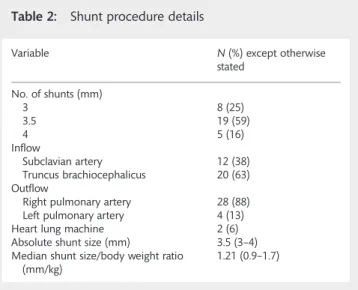
summarized in Table2.
Postoperative left open sternum
The primary goal was to close the chest at the shunt procedure. However, if there were any fears about the fate of the shunt (especially in totally shunt-dependent pulmonary circulation) or about the shunt getting squeezed behind the aorta, etc. the sternum was left open. It was believed that an open sternum lends itself to a quick response in case of an emergency when compared with a closed chest.
Anticoagulation
Anticoagulation regimen was decided on a case-by-case basis. Therapeutic heparinization was performed in high-risk shunt scenarios, such as shunt-dependent pulmonary perfusion, in
Table 1: Preoperative clinical characteristics
Variable Median (range)
N 32
Age (days) 10.5 (1–74)
Weight at operation (kg) 2.9 (1.9–4.4)
Size (cm) 48 (39–52)
Body surface area (m²) 0.2 (0.14–0.24)
SpO2(%) 85 (50–95)
Diagnosis n (%)
TOF/DORV/TGA_VSD_PS, pulmonary
atresia_VSD, Ebstein’s anomaly 21 (66) Single ventricle inclusive of pulmonary
atresia_intact septum
11 (34)
TOF: Tetralogy of Fallot; DORV: double outlet right ventricle; TGA: transposition of great arteries; VSD: ventricular septal defect; PS: pulmonary stenosis.
Table 2: Shunt procedure details
Variable N (%) except otherwise stated No. of shunts (mm) 3 8 (25) 3.5 19 (59) 4 5 (16) Inflow Subclavian artery 12 (38) Truncus brachiocephalicus 20 (63) Outflow
Right pulmonary artery 28 (88) Left pulmonary artery 4 (13) Heart lung machine 2 (6) Absolute shunt size (mm) 3.5 (3–4) Median shunt size/body weight ratio
(mm/kg) 1.21 (0.9–1.7) CO N G E N IT A L
cases with shunt clipping (shunt size reduction) or technical pro-blems encountered during shunt construction. Heparin infusion
starting with 5–10 IU/kg/h, followed by therapeutic dose as early
as 2 h postoperatively, was planned if surgical bleeding was not an issue. In effect, however, the therapeutic anticoagulation was often achieved later than 2 h. Shunts without complications and considered normal risk received aspirin in the long term.
Three patients did not have long-term anticoagulation/platelet inhibitor medication: 2 who died early and 1 who survived with
early shunt thrombosis in a neonatal Ebstein’s anomaly with
antegrade pulmonaryflow.
All patients were evaluated postoperatively with trans-thoracic echocardiography.
Statistical analysis
Statistical analysis was performed using IBM SPSS Statistics 19 (SPSS, Inc., Chicago, IL, USA). Categorical variables are presented
as numbers with percents and are compared using Fisher’s exact
test. Continuous variables are presented as median with range
and are compared between groups using the Mann–Whitney
test.P-values <0.05 were considered significant.
RESULTS
Mortality
Three of the 32 (9%) patients died at a median of 3(1–15) days.
The details of patients who died are as presented in Table 3.
Lower body weight (P = 0.04) and bigger shunt size/kg of body
weight (P = 0.004) were significant risk factors for mortality
(Table4).
Shunt thrombosis
Acute shunt thrombosis was observed in 3 of the 32 (9%)
patients, none of whom died. These 3 patients had Ebstein’s
anomaly with antegrade pulmonary flow (2.2 kg 3 mm right
MBTS), double outlet right ventricle-hypoplastic left ventricle (LV)_pulmonary stenosis (2.9 kg 3 mm right MBTS) and
unba-lanced atrio-ventricular septal defect hypoplastic LV
d-transposition of great arteries_pulmonary stenosis (3 kg 3.5
mm left MBTS). All shunt thrombosis occurred within the first
24 h and were diagnosed by echocardiography. Two patients had shunt revision with evacuation of the thrombus. One patient
with stable saturation due to normal antegrade pulmonaryflow
was not subjected to shunt revision. Shunt thrombosis could not
be statistically related to shunt size (P = 0.1) or shunt size/kg
body weight (P = 0.92). Early anticoagulation regimen (P = 0.33),
competitive blood flow (P = 1.0), diagnosis (P = 0.27), need for
cardiopulmonary bypass (P = 0.18), increased inotropic support
(P = 0.71) and shunt size reduction (P = 1.0) were not associated
with acute shunt thrombosis.
Inotropic support
Need for adrenalin ≤0.05 and/or noradrenaline ≤0.05 and/or
milrinone ≤0.75 μg/kg/min was defined as normal inotropic
support. Accordingly, 6 of the 32 (19%) patients needed normal inotropic support and 23 of the 32 (72%) needed higher ino-tropic support. Three patients were without any ionoino-tropic support.
Decongestive therapy
Need for cardiac decongestive therapy (over and above that of diuretics) was necessary in 10 of 29 survivors to discharge. Decongestive therapy was required in 2 of 19 (20%) biventricular hearts compared with 8 of 10 (80%) univentricular hearts
(P < 0.001), obviously resulting in a significantly longer hospital
stay (P = 0.005). Bigger shunts were associated with the need for
decongestive therapy (P = 0.054).
Open thorax
The sternum was left open postoperatively in 11 (34%) patients.
Other complications
Postoperative complications included chylothorax, phrenic nerve palsy, necrotizing enterocolitis and abdominal bleeding with unclear focus in 1 patient each.
Corrective surgery
Of the 29 survivors, 28 (97%) have undergone corrective surgery with takedown of the BT shunt at the time of this study. Ten patients were subjected to a bidirectional Glenn anastomosis, while 18 underwent a biventricular repair. None of the shunt survivors died during or after the corrective surgery.
Fate of branch pulmonary artery
Seven of 28 patients (25%) had reconstruction of the branch pul-monary artery at the distal shunt insertion site. Residual stenosis after BT shunt takedown occurred in 6 patients. In 3 of the patients, the stenosis occurred despite the pulmonary artery being reconstructed, while in 3, the stenosis occurred without the artery being reconstructed.
Era
A brief comparative analysis between 12 shunts in Era I and 20
shunts in Era II is depicted in Table5.
DISCUSSION
An ideal shunt helps promote uniform growth of the pulmonary arteries, without causing distortion. An excessive shunt results in
significant diastolic run-off in the short term and elevated
pul-monary vascular resistance or impaired ventricular and atrioven-tricular valve performance in the long term. Although various
has become established as the procedure of choice [4,12]. MBTS continues to be a subject of academic interest, because of per-sistent risks associated with this simple-looking procedure.
Sternotomy or thoracotomy
MBTS was classically performed through a thoracotomy. However, recent trends have shown increasing preference for a
sternotomy approach [6]. A sternotomy saves the child from a
second scar, avoids morbid damage to the thorax with pro-spects of late scoliosis, but more importantly, the target pul-monary artery being intrapericardial, it is more accessible for eventual reconstruction after takedown. Avoiding a thoracotomy in the prospective Fontan patients has an added advantage of reducing build-up of lung adhesions to the thoracic wall and the consequent development of systemic-to-pulmonary artery
collaterals. Other disadvantages of a thoracotomy approach
enumerated in the literature include Horner’s syndrome,
distor-tion of lobar branch pulmonary arteries and preferentialflow to
one lung with unbalanced growth [6]. Depending on the side of
the thoracotomy, it may not be always possible to perform PDA ligation, but with a sternotomy, it is always possible. In the end, whether or not to close the duct remains a strategic
decision [13].
The sternotomy approach does confront the surgeon with the challenges of a central run-off from the systemic artery leading to greater steal, low diastolic pressures, coronary malperfusion and pulmonary hyperperfusion. In addition, the often-used truncus brachiocephalicus to the right pulmonary artery shunt may be at danger of being squashed between the dominant aorta and the superior vena cava, for which the parietal
pericar-dial reflection over the superior vena cava to the trachea should
be divided to create space for the shunt.
Table 3: Mortality details
Diagnosis Age (days) Weight (kg) Shunt size (mm)
Cause of death Died (Postop day)
DORV_TGA_PS 19 2.5 3.5 Cardiorespiratory
decompensation
15 Pulmonary atresia VSD, hepatopulmonary syndrome, catheter
perforation and emergency shunt
4 2.2 3.5 Cardiorespiratory decompensation
1 Pulmonary atresia intact ventricular septum 6 2.4 4 Myocardial ischaemia 3 (ECMO) DORV: double outlet right ventricle; TGA: transposition of great arteries; PS:pulmonary stenosis; ECMO: extra corporeal membrane oxygenation.
Table 4: Risk factors for mortality
Variable Alive n (%) Deadn (%) P-value N 29 3 Weight (kg) 2.94 (1.9–4.4) 2.37 (2.2–2.49) 0.04 Diagnosis Biventricular hearts 19/29 (66) 2/3 (67) 1.00 Univentricular hearts 10/29 (34) 1/3 (33)
Competitive pulmonary flow 21/29 (72) 1/3 (33) 0.22
Use of heart lung machine 2/29 (7) 0/3 (0) 1.00
Size of shunt (mm):
3 8/29 (28) 0/3 (0) 0.47
3.5 17/29 (59) 2/3 (67)
4 4/29 (14) 1/3 (33)
Shunt size/kg body weight 1.19 (0.88–1.58) 1.59 (1.41–1.69) 0.004
Anticoagulation regimen:
1 LDH 7/28a(25) 0/2c(0) 0.38
2 ETH 4/28a(14) 1/2c(50)
3 LTH 17/28a(61) 1/2c(50)
Long-term anticoagulation 7/28a(25) 0/1b(0) 1.00
Postoperative high ionotropes 20/29 (69) 3/3 (100) 0.52
Shunt size reduction 3/29 (10) 2/3 (67) 0.056
Shunt thrombosis 3/29 (10) 0/3 (0) 1.00
SaO2postoperative (day of operation) 83 (77–94) 85 (80–90) 0.97
Hospital stay 23 (5–95) 3 (1–15) 0.02
LDH: low dose (10 IU/kg/h) heparin; ETH: early therapeutic heparin; LTH: late therapeutic heparin.
aOne patient data missing.
bOne patient on immediate ECMO was not analysed for acute anticoagulation regimen. cTwo patients who died early were not analysed for long-term anticoagulation.
CO N G E N IT A L
While the proposed alternative shunts such as Potts or
Waterston/Cooley shunts were difficult to regulate, the MBTS
flow is restricted by the size of the graft as also by the size of the
inflow vessel.
While the Boston group [6] reported four times higher risk of
shunt failures through a thoracotomy when compared with a
sternotomy, Shauqet al. [13] have reported significantly longer
ventilation time, inotropic support, intensive care unit (ICU) stay
and hospital stay in the sternotomy group. Thesefindings reflect
the learning curve involved with shunts created through a sternotomy.
Competitive
flow and PDA strategy
A completely left open duct may be difficult to regulate in the
presence of a MBTS. With our technique of duct obliteration using a silastic sling, one retains the possibility of quickly restor-ing duct patency in the case of a shunt thrombosis. While a
patent duct imparts significant safety in the case of a shunt
failure, some reports have associated patent duct with shunt
thrombosis [14]. Petrucci et al. (Society of Thoracic Surgeons
[STS] database) have shown no association between closed duct
and the risk of composite morbidity [15]. Closing or keeping the
duct open during the MBTS procedure has advantages and dis-advantages and, hence remains in the end, an individual decision.
Mortality
In spite of overall improvement in results [5], mortality reported
ranges from 2.3 to 16% [15]. Our overall postoperative mortality
was 9.4%. Low body weight (P = 0.041) and bigger shunt size/kg
body weight (P = 0.011) were factors associated with
post-operative mortality. There was a trend towards significance
between the need for postoperative shunt size reduction and
mortality (P = 0.056). These findings point towards over-shunting
as a possible indicator of mortality in our series. The Boston group has reported a mortality of 9 of 102 (8.7%) patients, with
indications that excessive pulmonary bloodflow could have
con-tributed to mortality in the sternotomy group. Multivariate risk factors for mortality in their analysis included small graft size, left
MBTS and male sex [6]. The same group also suggested the use
Table 5: Comparison between eras
Variable Era I (2004–07) n (%) Era II (2008–11)n (%) P-value N 12 (38) 20 (62) Diagnosis Biventricular hearts 6/12 (50) 15/20 (75) 0.25 Univentricular hearts 6/12 (50) 5/20 (25) Weight (kg) 2.89 (2.2–4.4) 2.93 (1.9–3.8) 0.99 Size of shunt (mm) 3.5 (3.5–4) 3.5 (3–3.5) 0.002 Shunt size/weight 1.24 (0.91–1.69) 1.16 (0.88–1.58) 0.13
Presence of competitive blood flow 7/12 (58) 15/20 (75) 0.44
Use of HLM 0/12 (0) 3/20 (10) 0.52
Shunt distribution, mm (in %)
3 0/12 (0) 8/20 (40) 0.001
3.5 7/12 (58) 12/20 (60)
4 5/12 (42) 0/20 (0)
Right vs left 11/20 (92) right 17/20 (85) right 1.00
1/12 (8) left 3/20 (15) left
Site of take-off (truncus brachiocephalicus vs subclavian artery) 10/12 (83) vs 2/12 (17) 10/20 (50) vs 10/20 (50) 0.08 Early anticoagulation strategy
Low dose 3/10 (30) 4/20 (20) 0.71
Early therapeutic 1/10 (10) 4/20 (20)
Late therapeutic (as defined inTable 4) 6/10 (60) 12/20 (60) Long-term anticoagulation
Aspirin 8/10 (80) 14/19 (74) 1.00
Therapeutic 2/10 (20) 5/19 (26)
Postoperative ionotropic support
None 1/12 (8) 2/20 (10) 0.48
Normal 1/12 (8) 5/20 (25)
Higha 10/12 (83) 13/20 (65)
Need for shunt reduction 3/12 (25) 2/20 (10) 0.34
Decongestive therapy (more than diuretics) 6/9 (67) 4/20 (20) 0.03
Duration of ventilation 2 (1–15) 1.5 (0–9) 0.53
ICU stay 4 (1–15) 5 (1–13) 0.39
Duration of hospital stay 18.5 (1–95) 22.5 (5–84) 0.69
Mortality 3/12 (25) 0/20 (0) 0.04
Shunt thrombosis 0/12 (0) 3/20 (15) 0.27
SaO2before takedown 82 (72–93) 81.5 (73–94) 0.66
Residual branch PA stenosis at shunt insertion site 4/9 (44) 1/15 (7) 0.05
of smaller (3.5 mm) shunts through a sternotomy approach instead of the 4-mm shunts for the thoracotomy approach. An
STS database harvest study [15] has identified preoperative
venti-lation, pulmonary atresia_intact ventricular septum, univentricu-lar hearts and weight <3 kg as risk factors for mortality.
Pulmonary atresia with intact ventricular septum, when speci
fic-ally analysed, did not come out as a significant risk factor for
mortality in our cohort, probably because of small numbers.
While Alkhulaifi et al. [16] identified weight <2 kg and
preopera-tive ventilation, Rao et al. [17] identified restrictive atrial septal
defect, univentricular physiology and postoperative intervention as risk factors for mortality.
Shunt thrombosis
Shunt thrombosis is a grave complication of the MBTS proced-ure. Our overall acute shunt thrombosis of (3 of 32) 9.4% corre-sponds with those of (9 of 76) 11.8% reported from Bristol and
(14 of 102) 13.7% reported from Boston [6]. We could not show
an association between smaller shunt size and occurrence of
thrombosis, probably because of the small numbers. Tsai et al.
[18] and Tamisieret al. [12] have suggested that young age and
smaller size are significantly related to shunt thrombosis. Other
reports have also linked weight <2 kg [16] and weight <3.6 kg [19]
to shunt thrombosis. Gedickeet al. [14] have found weight <3 kg,
high preoperative haemoglobin (>18 g/dl) and a postoperative
patent duct as significant factors for shunt thrombosis.
Anticoagulation-coagulopathy
Although an association between an anticoagulation regimen and shunt thrombosis could not be established in our study, it does not belittle the role of postoperative anticoagulation,
par-ticularly in high-risk patients. Al Jubair et al. [20] have shown,
less-shunt failure occurs if heparin is given before clamping. An early postoperative phase with a fresh anastomosis, coupled with phases of low systemic pressures, pulmonary hypertension, ex-ternal compression and resulting stasis, can initiate thrombus
formation. It is these uncertainties that can be positively in
flu-enced by early anticoagulation. Liet al. [21] have demonstrated a
beneficial effect of acetylsalicylic acid in infants palliated with a
shunt, with reduced incidence of shunt thrombosis and death.
Another prospective study has shown the beneficial effect of
haemodilution with a significantly higher shunt patency rate [22].
Rare coagulopathies, such as protein C deficiency [23], and
primary antiphospholipid syndrome [24] have also been
reported to cause shunt thrombosis.
Late shunt obstruction
We have not observed any late shunt thrombosis in this series of patients. This has been reported as a cause in up to 15% of
out-of-hospital mortalities [14,15]. Wellset al. [25] have observed
>50% obstruction of the MBTS in 21% of their patients and have
identified a shunt size of <4 mm to be a risk factor for
high-grade stenosis (>50%).
Era
While the cohort did not change over time in terms of most
demographic and procedural variables, shunt size was signi
fi-cantly lower in the latter half of the series when compared with the former half. Shunt thrombosis was higher in the later era,
but did not reach statistical significance. These findings may
indi-cate that small shunts are prone to shunt thrombosis. Mortality
as well as need for cardiac decongestive therapy was significantly
higher in the previous era. With time, while mortality was avoided, shunt thrombosis remained worrisome. While shunt thrombosis morbidity could be partially attributed to the tech-nique, the importance of optimal intensive postoperative man-agement cannot be overemphasized. Interestingly, in spite of smaller shunt size selection in the later era, transcutaneous satur-ation before shunt takedown was 82% in both the eras. This
implies that the shunt flow was adequately regulated by the
artery from which the shunt was sourced.
LIMITATIONS
This is a retrospective study with a small patient cohort, which may not be powered enough to identify all risk factors contribut-ing to the various end points. The series becontribut-ing spread over a time frame of 8 years, even generalized improvements in opera-tive technique and perioperaopera-tive care may alone account for the improvements in outcome.
CONCLUSION
In spite of increasing confidence with primary neonatal intracardiac
repairs, the MBTS continues to be indicated for malformations of the univentricular pathway. Although seemingly innocuous,
the MBTS procedure is associated with significant morbidity and
mortality. While small shunts may have a tendency to shunt throm-bosis, large shunts may lead to pulmonary over-circulation and
volume loading of the heart. Various studies have identified
low body weight, small shunt size, over-shunting, univentricular
hearts—specifically pulmonary atresia with intact ventricular
septum, to be risk factors associated with postoperative morbidity
and mortality. It appears that timely and efficient early
anticoagula-tion as well as long-term antiplatelet therapy may help reduce the risk of early and late shunt dysfunction.
Conflict of interest: none declared.
REFERENCES
[1] Blalock A, Taussig HB. Landmark article May 19, 1945: the surgical treat-ment of malformations of the heart in which there is pulmonary stenosis or pulmonary atresia. By Alfred Blalock and Helen B Taussig. JAMA 1984; 251:2123–38.
[2] Mavroudis C, Backer CL. Palliative operations. In: Mavroudis C, Backer CL (eds). Pediatric Cardiac Surgery. 3rd edn. Philadelphia: Mosby, 2003, 160–70.
[3] Geiss D, Williams WG, Lindsay WK, Rowe RD. Upper extremity gangrene: a complication of subclavian artery division. Ann Thorac Surg 1980;30:487–9. [4] de Leval MR, McKay R, Jones M, Stark J, Macartney FJ. Modified Blalock-Taussig shunt. Use of subclavian artery orifice as flow regulator
CO N G E N IT A L
in prosthetic systemic-pulmonary artery shunts. J Thorac Cardiovasc Surg 1981;81:112–9.
[5] Williams JA, Bansal AK, Kim BJ, Nwakanma LU, Patel ND, Seth AKet al. Two thousand Blalock-Taussig shunts: a six-decade experience. Ann Thorac Surg 2007;84:2070–5; discussion 2070–5.
[6] Odim J, Portzky M, Zurakowski D, Wernovsky G, Burke RP, Mayer JE Jr et al. Sternotomy approach for the modified Blalock-Taussig shunt. Circulation 1995;92:II256–61.
[7] Potts WJ, Smith S, Gibson S. Anastomosis of the aorta to a pulmonary artery: certain types in congenital heart disease. J Am Med Assoc 1946; 132:627–31.
[8] Waterston DJ. Treatment of Fallot’s tetralogy in children under 1 year of age. Rozhl Chir 1962;41:181–3.
[9] Cooley DA, Hallman GL. Intrapericardial aortic-right pulmonary arterial anastomosis. Surg Gynecol Obstet 1966;122:1084–6.
[10] Gazzaniga AB, Elliott MP, Sperling DR, Dietrick WR, Eiseman JT, McRae DM et al. Microporous expanded polytetrafluoroethylene arterial prosthesis for construction of aortopulmonary shunts: experi-mental and clinical results. Ann Thorac Surg 1976;21:322–7.
[11] Amato JJ, Marbey ML, Bush C, Galdieri RJ, Cotroneo JV, Bushong J. Systemic-pulmonary polytetrafluoroethylene shunts in palliative opera-tions for congenital heart disease. Revival of the central shunt. J Thorac Cardiovasc Surg 1988;95:62–9.
[12] Tamisier D, Vouhe PR, Vernant F, Leca F, Massot C, Neveux JY. Modified Blalock-Taussig shunts: results in infants less than 3 months of age. Ann Thorac Surg 1990;49:797–801.
[13] Shauq A, Agarwal V, Karunaratne A, Gladman G, Pozzi M, Kaarne M et al. Surgical approaches to the Blalock shunt: does the approach matter? Heart Lung Circ 2010;19:460–4.
[14] Gedicke M, Morgan G, Parry A, Martin R, Tulloh R. Risk factors for acute shunt blockage in children after modified Blalock-Taussig shunt opera-tions. Heart Vessels 2010;25:405–9.
[15] Petrucci O, O’Brien SM, Jacobs ML, Jacobs JP, Manning PB, Eghtesady P. Risk factors for mortality and morbidity after the neonatal
Blalock-Taussig shunt procedure. Ann Thorac Surg 2011;92:642–51; discussion 641–51.
[16] Alkhulaifi AM, Lacour-Gayet F, Serraf A, Belli E, Planche C. Systemic pulmonary shunts in neonates: early clinical outcome and choice of surgical approach. Ann Thorac Surg 2000;69:1499–504.
[17] Rao MS, Bhan A, Talwar S, Sharma R, Choudhary SK, Airan B et al. Modified Blalock-Taussing shunt in neonates: determinants of immediate outcome. Asian Cardiovasc Thorac Ann 2000;8:339–43.
[18] Tsai KT, Chang CH, Lin PJ. Modified Blalock-Taussig shunt: statistical analysis of potential factors influencing shunt outcome. J Cardiovasc Surg (Torino) 1996;37:149–52.
[19] Bove EL, Kohman L, Sereika S, Byrum CJ, Kavey RE, Blackman MSet al. The modified Blalock-Taussig shunt: analysis of adequacy and duration of palliation. Circulation 1987;76:III19–23.
[20] Al Jubair KA, Al Fagih MR, Al Jarallah AS, Al Yousef S, Ali Khan MA, Ashmeg Aet al. Results of 546 Blalock-Taussig shunts performed in 478 patients. Cardiol Young 1998;8:486–90.
[21] Li JS, Yow E, Berezny KY, Rhodes JF, Bokesch PM, Charpie JR et al. Clinical outcomes of palliative surgery including a systemic-to-pulmonary artery shunt in infants with cyanotic congenital heart disease: does aspirin make a difference? Circulation 2007;116:293–7.
[22] Sahoo TK, Chauhan S, Sahu M, Bisoi A, Kiran U. Effects of hemodilution on outcome after modified Blalock-Taussig shunt operation in children with cyanotic congenital heart disease. J Cardiothorac Vasc Anesth 2007; 21:179–83.
[23] Watanabe M, Aoki M, Fujiwara T. Thrombotic occlusion of Blalock-Taussig shunt in a patient with unnoticed protein C deficiency. Gen Thorac Cardiovasc Surg 2008;56:544–6.
[24] Deally C, Hancock BJ, Giddins N, Hawkins L, Odim J. Primary antipho-spholipid syndrome: a cause of catastrophic shunt thrombosis in the newborn. J Cardiovasc Surg (Torino) 1999;40:261–4.
[25] Wells WJ, Yu RJ, Batra AS, Monforte H, Sintek C, Starnes VA. Obstruction in modified Blalock shunts: a quantitative analysis with clinical correl-ation. Ann Thorac Surg 2005;79:2072–6.