Primary follicular and marginal-zone lymphoma of the
breast: clinical features, prognostic factors and
outcome: a study by the International Extranodal
Lymphoma Study Group
G. Martinelli
1*, G. Ryan
2, J. F. Seymour
3, L. Nassi
1, S. Steffanoni
1, A. Alietti
1, L. Calabrese
1,
G. Pruneri
4, L. Santoro
5, M. Kuper-Hommel
6, R. Tsang
7, P. L. Zinzani
8, A. Taghian
9, E. Zucca
10&
F. Cavalli
101
Division of Haematology, European Institute of Oncology, Milan, Italy;2Division of Radiation Oncology, Peter MacCallum Cancer Centre;3Department of Haematology, Peter MacCallum Cancer Centre and University of Melbourne, Melbourne, Australia; Divisions of4Pathology and Laboratory Medicine and;5Statistical Analysis, European Institute of Oncology, Milan, Italy;6Department of Internal Medicine, Bernhoven Hospital, Oss, The Netherlands;7Department of Radiation Oncology, Princess Margaret Hospital, University Health Network, Toronto, Canada;8
Institute of Haematology and Medical Oncology, Policlinico Sant’Orsola, Bologna, Italy;
9
Department of Radiation Oncology, Massachusetts General Hospital, Boston, MA, USA and10
Department of Medical Oncology, Oncology Institute of Southern Switzerland, Bellinzona, Switzerland
Received 20 March 2009; accepted 23 March 2009
Background:Primary breast lymphoma (PBL) of low-grade histology is a rare disease. This multicentric retrospective study was carried out to determine clinical features, prognosis and relapse.
Patients and methods:Patients with histologically proven, previously untreated follicular or marginal-zone PBL (MZL PBL) diagnosed from 1980 to 2003 were included in the study. Major end points were progression-free survival (PFS), overall survival (OS) and potential prognostic factors.
Results:We collected data on 60 cases of PBL [36 follicular and 24 marginal-zone lymphoma (MZL)]. Stage was IEor
IIEin 57 patients and IVE in three patients due to bilateral breast involvement. Surgery, chemotherapy and radiotherapy
(RT), alone or in combination, were used as first-line treatments in 67%, 42% and 52% of patients, respectively. Overall response rate was 98%, with a 93% complete response rate. Five-year PFS were 56% for MZL and 49% for follicular PBL (P= 0.62). Relapses were mostly in distant sites (18 of 23 cases); no patients relapsed within RT fields.
Conclusions:Our data showed an indolent behaviour of MZL PBL, comparable to other primary extranodal MZL. Conversely, patients with follicular PBL had inferior PFS and OS when compared with limited-stage nodal follicular non-Hodgkin’s lymphomas, suggesting an adverse prognostic role of primary breast localisation in this histological subgroup.
Key words:breast malignancies, follicular lymphomas, MZLs, primary extranodal lymphomas
introduction
Primary breast lymphoma (PBL) is a clinicopathological entity that represents 1% of all non-Hodgkin’s lymphomas (NHL) [1] and <0.5% of all breast malignancies [2]. PBL typically affects an elderly population, but may rarely occur in younger women, at times associated with pregnancy or lactation; men are very rarely affected. The large majority of cases of PBL are represented by diffuse large B-cell lymphoma (DLBCL), with the remainder comprising follicular lymphoma and marginal-zone lymphoma (MZL) subtypes [3]. Primary breast
localisation represents a small proportion of extranodal
lymphomas (<5%) in both follicular lymphoma and MZL subgroups [4]. Although it has been hypothesised that DLBCL PBL could originate from germinal centre-related B-cells [5], there is even less known about the pathogenesis of histologically ‘low-grade’ PBL. Surgery, radiotherapy (RT), chemotherapy (CT) and more recently immunotherapy have been reported as treatment modalities for PBL either as monotherapy or in combination. Due to the small number of publications investigating PBL, there is limited information about this disease. Moreover, almost all papers are focused on DLBCL, thus information regarding follicular lymphoma and MZL subtypes are scarce [6–13]. A pathophysiologic role of chronic infections has been identified in some extranodal NHL such as gastric (Helicobacter pylori), ocular adnexal (Clamydia psittaci) and skin (Borrelia burgdorferi) MZL [14], but such correlations have not been reported in the literature regarding PBL.
original
article
*Correspondence to: Dr. G. Martinelli, Division of Haematology, European Institute of Oncology, Via Ripamonti 435, I-20141 Milan, Italy. Tel:+39-02-57489538; Fax:+39-02-94399219; E-mail: giovanni.martinelli@ieo.it
The International Extranodal Lymphoma Study Group (IELSG) has conducted a large multicentre retrospective study of PBL of all histological subtypes in order to review its clinical characteristics, natural history and prognosis. We present here the results of the analysis of patients with histologically proven diagnosis of follicular or MZL PBL.
materials and methods
trial overview and data collection
Data on all cases of PBL diagnosed at each participating institute from January 1980 to December 2003 were retrospectively collected. Inclusion criteria for the study were as follows: histologically proven NHL with primary localisation of one or both breasts, with or without regional lymph node involvement. Patients with disseminated lymphoma with breast involvement or breast presentation of recurrent/progressive lymphoma were excluded. All histological subtypes were eligible. Patients were staged according to the Ann Arbor classification [15]; the staging of extranodal NHL involving bilateral paired organs remains contentious, but for this study patients with bilateral breast disease were considered stage IV. Protocol and case report forms (CRFs) were approved by the local institutional review boards or ethics committees of each participating institution. CRFs were designed to collect data on patient and tumour characteristics, diagnostic test results, potential prognostic factors, treatment approaches, response and survival. We also evaluated the International Prognostic Index (IPI) in all patients [16] and the Follicular Lymphoma International Prognostic Index (FLIPI) in patients with follicular PBL [17]. Data for each patient were collected by local investigators and CRFs were then sent to IELSG headquarters and analysed centrally.
definition of study outcomes
Tumour response was assessed after the completion of planned treatment according to the published response criteria [18]. Progression-free survival (PFS) was calculated as the interval between start of treatment and progression of disease, death or last known follow-up; overall survival (OS) was defined as the period between the start of treatment and death from any cause or last known follow-up; cause-specific survival (CSS) was defined as the period between the start of treatment and death from disease or treatment complications.
pathology review
Various histological classifications were in use throughout the study period, and patients classified according to Kiel, Working Formulation, Revised European-American Lymphoma or World Health Organisation (WHO) were all eligible. All cases were locally reviewed, and this report is limited to the 60 patients with their histology reclassified as either follicular lymphoma or MZL according to WHO criteria [19].
statistical analysis
Baseline characteristics were summarised as a table of frequencies for categorical variables and as mean and standard deviation, median and range for continuous variables. The distribution of these characteristics among patients with histologically different lymphomas (follicular lymphoma versus MZL) was compared by the Pearson’s chi-square test for categorical variables and either t-test or Mann–Whitney–Wilcoxon test (depending on whether the variable was normally distributed or not) for continuous variables. Unadjusted time-to-event data distributions (PFS and OS) were estimated by the Kaplan–Meier method [20]; the comparison between histologically different lymphomas was carried out by the log-rank test [21]. The univariate Cox models [22] were used to detect and quantify
prognostic factors with potentially different roles between the histological subtypes. Due to the small number of events within the subpopulations delineated by the subtypes, a multivariate analysis was not statistically valid. The analyses were carried out using SAS statistical software (SAS Institute, Cary, NC).
results
patients
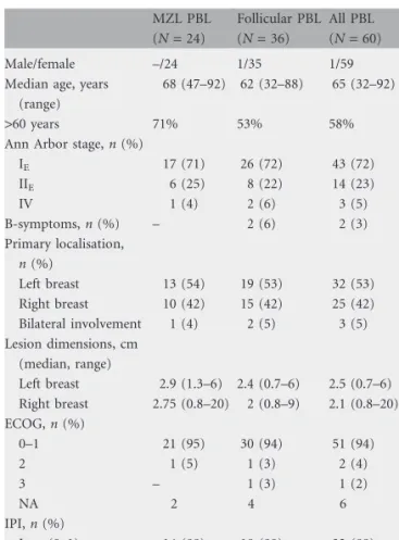
This IELSG study enrolled a total of 278 patients with PBL: 204 of them had DLBCL (73%) and were separately analysed and have been previously reported [23]. A total of 60 patients with PBL, 36 with follicular lymphoma and 24 with MZL were considered and analysed further for this report (Table 1).
Fifty-nine patients were female and only one was male. The median age was 65 years (range 32–92), 51 patients were postmenopausal (85%) and 36 patients aged ‡60 years (60%). No patient was pregnant or lactating at diagnosis. Eastern Cooperative Oncology Group performance status at diagnosis was zero to one in 51 patients (85%). Only two patients (3%)
Table 1. Patients’ characteristics
MZL PBL (N = 24) Follicular PBL (N = 36) All PBL (N = 60) Pa Male/female –/24 1/35 1/59 0.41 Median age, years
(range)
68 (47–92) 62 (32–88) 65 (32–92) 0.16
>60 years 71% 53% 58% Ann Arbor stage, n (%)
IE 17 (71) 26 (72) 43 (72) 0.95 IIE 6 (25) 8 (22) 14 (23) IV 1 (4) 2 (6) 3 (5) B-symptoms, n (%) – 2 (6) 2 (3) 0.24 Primary localisation, n (%) Left breast 13 (54) 19 (53) 32 (53) 0.97 Right breast 10 (42) 15 (42) 25 (42) Bilateral involvement 1 (4) 2 (5) 3 (5) Lesion dimensions, cm (median, range) Left breast 2.9 (1.3–6) 2.4 (0.7–6) 2.5 (0.7–6) 0.9b Right breast 2.75 (0.8–20) 2 (0.8–9) 2.1 (0.8–20) 0.8b ECOG, n (%) 0–1 21 (95) 30 (94) 51 (94) 0.91 2 1 (5) 1 (3) 2 (4) 3 – 1 (3) 1 (2) NA 2 4 6 IPI, n (%) Low (0–1) 14 (88) 18 (90) 32 (89) 0.67 Low–intermediate (2) 2 (12) 2 (10) 4 (11) NA 8 16 24
aPearson’s chi-square test. bWilcoxon’s test.
MZL PBL, marginal-zone PBL; PBL, primary breast lymphoma; ECOG, Eastern Cooperative Oncology Group; IPI, International Prognostic Index; NA, not available.
presented with B-symptoms. Forty-three patients were in stage IE(72%), 14 patients (23%) were in stage IIE, 11 due to axillary
and three due to supraclavicular nodal involvement, and three patients were in stage IVE(5%) due to bilateral breast disease.
IPI was evaluated in 36 patients, and it was low (0–1) in 32 patients (89%). FLIPI score was evaluated in 27 of 36 patients with follicular lymphoma: 24 presented with a low score and three an intermediate score. There were no statistically significant differences between baseline characteristics in patients with MZL and follicular PBL.
diagnosis
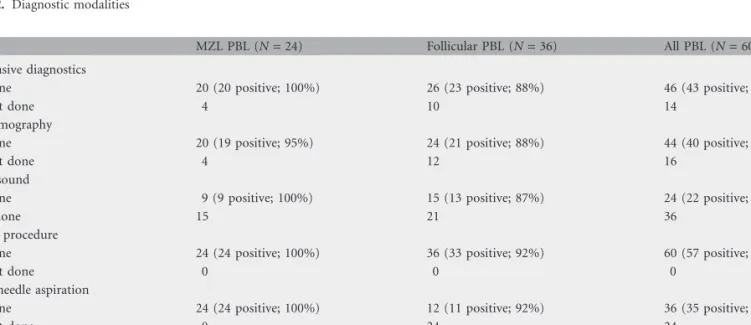
Both invasive and noninvasive procedures were carried out as a part of the diagnostic work-up of the patients (Table 2). Forty-six patients underwent a noninvasive diagnostic procedure, such as mammography and/or ultrasound, which confirmed the presence of pathological lesion in 43 patients (93%). An invasive procedure was carried out in all 60 patients, aimed at obtaining a histological diagnosis. Thirty-six patients underwent a fine-needle aspiration (FNA) for cytology, which was positive in 35 (97%). A surgical biopsy was carried out in 59 patients, and it was diagnostic in 53 (90%). Overall, FNA and/or surgical biopsy led to a histological diagnosis in 57 of 60 patients (95%). The remaining three patients required more invasive diagnostic surgery: mastectomy and lymph nodal dissection in one patient and lumpectomy in the other two patients.
treatment
As patients were identified retrospectively from various institutions, there was no uniform treatment policy, and management was determined by the individual managing clinicians. RT, CT and surgery were used alone or in combination (Table 3).
Forty patients (67%) were managed with initial surgery: a breast-conserving resection was carried out in 31 patients; the remaining nine underwent a mastectomy. Surgery was used
alone in 11 patients, while the other 29 patients received an additional treatment modality: six with CT, 15 with RT and eight patients received both CT and RT. Twenty patients (34%) did not undergo a surgical procedure: seven of them received CT alone, nine patients underwent RT alone and four patients received both CT and RT. Overall, CT was administered in 25 patients with different schedules. Fifteen patients received an anthracycline- or anthracenedione-based regimen, such as combination chemotherapy with cyclophosphamide, doxorubicin, vincristine and prednisone (CHOP) or
cyclophosphamide, mitoxantrone, vincristine, prednisone, the others alkylating agent-based CT (e.g. chlorambucil or cyclophosphamide, vincristine, prednisone). No patients received immunotherapy with rituximab or intrathecal prophylaxis.
RT was administered to breast fields in 36 patients (dose range 25–50 Gy, median 38 Gy) and nodal fields (axilla and supraclavicular) were irradiated in 18 patients (dose range 30– 46 Gy, median 36 Gy). While RT was used alone in nine patients (15%), it was used in association with other therapies in 27 cases (45%).
Local treatments (e.g. surgery and/or RT) were similarly distributed between MZL and follicular PBL, as well as the extent of surgery (i.e. mastectomy). Conversely, systemic CT was administered in 19 of 36 patients (53%) with follicular PBL and in 6 of 24 (25%) with MZL PBL (P = 0.03).
response and survival
Considering all first-line treatment combinations, the overall response rate (ORR) was 98% (100% for MZL and 97% for follicular PBL): 56 patients (93%) obtained a complete remission (CR) and three patients (5%) a partial response. Only one patient with a follicular PBL did not respond to treatment and had initially stable disease; after 8 years he died from progressive lymphoma.
Table 2. Diagnostic modalities
MZL PBL (N = 24) Follicular PBL (N = 36) All PBL (N = 60) Noninvasive diagnostics
Done 20 (20 positive; 100%) 26 (23 positive; 88%) 46 (43 positive; 93%)
Not done 4 10 14
Mammography
Done 20 (19 positive; 95%) 24 (21 positive; 88%) 44 (40 positive; 91%)
Not done 4 12 16
Ultrasound
Done 9 (9 positive; 100%) 15 (13 positive; 87%) 24 (22 positive; 92%)
Not done 15 21 36
Invasive procedure
Done 24 (24 positive; 100%) 36 (33 positive; 92%) 60 (57 positive; 95%)
Not done 0 0 0
Fine-needle aspiration
Done 24 (24 positive; 100%) 12 (11 positive; 92%) 36 (35 positive; 97%)
Not done 0 24 24
Surgical biopsy
Done 24 (22 positive; 92%) 35 (31 positive; 89%) 59 (53 positive; 90%)
Not done 0 1 1
Fourteen patients with follicular PBL (39%) relapsed after a median interval of 26 months (range 1.2–85 months). Nine patients with a MZL PBL (37%) relapsed after a median interval of 12 months (range 4–102 months). No patients who received RT in first-line treatment (eight with follicular and eight with MZL PBL) relapsed in the irradiated fields. Of the 23 relapses, three were locoregional, while the remaining were in contralateral breast (two patients) and in distant sites (18 patients). Considering the three local relapses, two of them had been treated with systemic CT, whereas the third patients received combined surgery and chemoradiotherapy: this patient relapsed outside the irradiated field (ipsilateral axillary nodes). Relapse rate was also evaluated in order to investigate the role
of an additional treatment (CT and/or RT) after surgery; results are reported in Table 4.
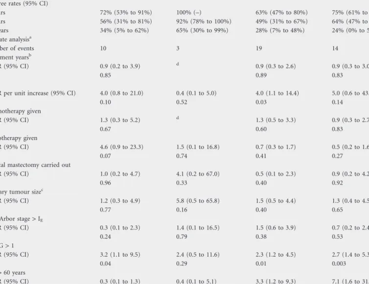
PFS for MZL and follicular PBL was 72% and 63% at 3 years, 56% and 49% at 5 years and 34% and 28% at 10 years, respectively (log-rank test: P = 0.55). The median PFS was 7.3 years in MZL and 3.9 years in follicular PBL (Figure 1). No difference in terms of PFS was detected between the patients who received a CHOP-like and those treated with alkylating agent-based CT.
After a median follow-up time of 44 months (range 5–156 months), 17 patients died; 14 with follicular lymphoma and three with MZL. OS for MZL and follicular PBL was 100% and 75% at 3 years, 92% and 64% at 5 years and 64% and 47% at 10 years, respectively (log-rank test: P = 0.04). The median OS was 8.1 years in follicular PBL and was not reached in MZL PBL (Figure 2). Univariate analyses of potential prognostic factors for PFS and OS were carried out, and the results are
summarised in Table 5. Only impaired PS was shown to have an impact on OS in follicular PBL, but the analysis was greatly limited by the small sample size.
Nine patients died from disease: eight of them (89%) had follicular lymphomas. The CSS for MZL and follicular PBL was 100% and 84% at 3 years, 100% and 79% at 5 years and 80% and 66% at 10 years, respectively (log-rank test: P = 0.05; Figure 3).
Three patients developed a second malignancy, but only a case of acute myeloid leukaemia developing 27 months after CHOP CT could be considered as potentially treatment related. The other two patients developed a lung carcinoma and
Table 3. Treatment approach
MZL PBL (N = 24), n (%) Follicular PBL (N = 36), n (%) Total, n (%) Surgery alone 5 (21) 6 (17) 11 (18) CT alone 1 (4) 6 (17) 7 (12) RT alone 5 (21) 4 (11) 9 (15) Surgery and CT 2 (8) 4 (11) 6 (10) Surgery and RT 8 (34) 7 (19) 15 (25) Surgery + CT + RT 2 (8) 6 (17) 8 (13) CT + RT 1 (4) 3 (8) 4 (7)
Surgery (alone or not) 17 (71) 23 (64) 40 (67) CT (alone or not)a 6 (25) 19 (53) 25 (42) RT (alone or not) 16 (67) 20 (56) 36 (52)
aP = 0.03, Pearson’s chi-square test.
MZL, marginal-zone lymphoma; PBL, primary breast lymphoma; CT, chemotherapy; RT, radiotherapy.
Table 4. Disease progression or death during follow-up according to stage and surgery
MZL PBL (N = 24), n (%) P (log rank)a Follicular PBL (N = 36), n (%) P (log rank)a Total events 10 (41.7) 19 (52.8) Surgery Alone 1/5 (20.0) 0.16 4/6 (66.7) 0.50 With adjuvant therapy 6/12 (50.0) 8/17 (47.1) Surgery not performed 3/7 (42.8) 7/13 (53.8) Ann Arbor stage IE 9/17 (52.9) 12/26 (46.1)
Surgery
Alone 1/3 (33.3) 0.37 4/5 (80.0) 0.07 With adjuvant therapy 5/10 (50.0) 5/14 (35.7) Surgery not performed 3/4 (75.0) 3/7 (42.8) Ann Arbor stage IIE+ IV 1/7 (14.3) 7/10 (70)
Surgery
Alone 0/2 (–) 0/1 (–)
With adjuvant therapy 1/2 (50.0) 3/3 (100) Surgery not performed 0/3 (–) 4/6 (66.7)
aComparing surgery versus surgery + adjuvant therapy.
MZL, marginal-zone lymphoma; PBL, primary breast lymphoma.
Figure 1. Fifteen-year progression-free survival in follicular and marginal-zone primary breast lymphomas.
Figure 2. Fifteen-year overall survival in follicular and marginal-zone primary breast lymphomas.
a cholangiocarcinoma: these patients were treated for PBL with surgery alone and a combination of surgery and breast RT, respectively.
discussion
We present the results of the largest study ever conducted on histologically low-grade PBL. We collected information on many aspects of this disease, including clinical characteristics, treatment and outcomes. Despite the large number of patients, this study still has some weaknesses typical of multicentric retrospective analysis, including different national regulations concerning shipment of histological specimens, which prevented the performance of central histology review.
As shown by a large investigation recently published by IELSG [23], DLBCL is the most frequent histology among patients with PBL. Low-grade histological subtypes are a minority, comprising 14% for follicular lymphomas and 9% for MZL. This is a lower proportion than suggested by prior literature on PBL [6–13] and considering the more frequent presentation of MZL as primary extranodal lymphomas. Clinical characteristics were similar in the two histological groups, except that patients with follicular PBL had a lower median age than those with MZL lymphoma (62 versus 68 years, respectively), and the median lesion diameter was smaller in follicular lymphoma than in MZL (2.0 versus 2.8 cm, respectively), but these differences were not statistically significant. Bilateral synchronous PBL was uncommon,
Table 5. Summary table of time-to-event analyses
Analysis MZL PBL (N = 24) Follicular PBL (N = 36)
PFS OS PFS OS
Event-free rates (95% CI)
3 Years 72% (53% to 91%) 100% (–) 63% (47% to 80%) 75% (61% to 90%) 5 Years 56% (31% to 81%) 92% (78% to 100%) 49% (31% to 67%) 64% (47% to 81%) 10 Years 34% (5% to 62%) 65% (30% to 99%) 28% (7% to 48%) 24% (0% to 59%) Univariate analysisa Number of events 10 3 19 14 Treatment yearsb HR (95% CI) 0.9 (0.2 to 3.9) d 0.9 (0.3 to 2.6) 0.9 (0.3 to 3.0) P 0.85 0.89 0.83 IPIb
HR per unit increase (95% CI) 4.0 (0.8 to 21.0) 0.4 (0.1 to 5.0) 4.0 (1.1 to 14.4) 5.0 (0.6 to 43.1)
P 0.10 0.52 0.03 0.14 Chemotherapy given HR (95% CI) 1.3 (0.3 to 5.2) d 1.3 (0.5 to 3.3) 0.9 (0.3 to 2.7) P 0.67 0.60 0.83 Radiotherapy given HR (95% CI) 4.6 (0.9 to 23.3) 1.5 (0.1 to 16.8) 0.7 (0.3 to 1.7) 0.5 (0.2 to 1.6) P 0.07 0.74 0.41 0.27
Radical mastectomy carried out
HR (95% CI) 1.0 (0.2 to 4.7) 4.1 (0.2 to 67.0) 0.5 (0.1 to 2.3) 0.9 (0.2 to 4.2)
P 0.96 0.33 0.40 0.92
Primary tumour sizec
HR (95% CI) 1.2 (0.3 to 4.9) 5.8 (0.5 to 65.8) 1.5 (0.5 to 4.4) 1.3 (0.4 to 4.5)
P 0.77 0.16 0.40 0.65
Ann Arbor stage > IE
HR (95% CI) 0.3 (0.1 to 2.3) 1.4 (0.1 to 16.5) 1.5 (0.6 to 3.9) 0.7 (0.2 to 2.4) P 0.24 0.79 0.38 0.53 ECOG > 1 HR (95% CI) 3.2 (1.1 to 9.5) 2.4 (0.5 to 11.6) 2.3 (1.2 to 4.5) 2.7 (1.4 to 5.3) P 0.04 0.29 0.01 0.003 Age > 60 years HR (95% CI) 0.3 (0.1 to 1.3) 0.4 (0.1 to 5.1) 3.3 (1.2 to 9.3) 7.1 (1.6 to 31.9) P 0.10 0.52 0.02 0.01
aAny hazard ratio and pertinent P value derive form unadjusted univariate analysis. bTreatment years categories: £1995 versus >1995.
cPrimary tumour size categories: £5 versus >5 cm.
dNot estimable due to lack of events within the reference category.
MZL, marginal-zone lymphoma; PBL, primary breast lymphoma; PFS, progression-free survival; OS, overall survival; HR, hazard ratio; CI, confidence interval.
occurring in only 5% of patients, less than previously reported in literature [24]. Our data showed a similar sensitivity of radiological imaging, such as mammography or ultrasound, as reported in literature [24].
Being a retrospective study, there was a large variability in the treatment. Surgery, RT and CT were used alone or in
combination, leading to a 98% of ORR with a 93% of CR. About two-thirds of the patients underwent further surgery after the diagnostic procedure; this wide use of surgery may be explained with the need to confirm the diagnosis of PBL after FNA cytology and the insensitivity of radiological imaging in obtaining a histological definition, not only among NHL subtypes but also with breast cancer. However, a total mastectomy was carried out in only nine cases, and the majority of patients underwent less extensive surgery such as lumpectomy or quadrantectomy. We did not observe any advantage in terms of PFS and OS in those patients who underwent mastectomy, shown in a large study conducted on both low- and high-grade PBL [25]. We also observed a higher risk of relapse in those patients who were treated with surgery only.
The majority of patients received RT as part of the initial treatment, which was usually delivered after surgery or biopsy. The majority of the relapses occurred in distant sites, with only 21% of the recurrences arising in the primary disease sites (13%) or in the contralateral breast (8%).
Because of the relatively small number of patients and the many possible combinations, it is impossible to evaluate the independent contribution of CT. The majority of patients who received CT had follicular PBL. This is an atypical treatment strategy for localised follicular NHL, considering that they were mostly in stage I and were already treated with surgery or RT or both, although there are some data supporting such
a combined chemoradiation approach in limited stage nodal follicular NHL [26]. In patients with follicular PBL, the addition of CT to surgery seemed to reduce the relapse rate, although not statistically significant, probably because of the small sample size. The retrospective nature of this study could not resolve the question of whether a systemic treatment provided additional benefit after surgery. Indeed, our data seem to indicate a possible benefit in those patients with follicular PBL who received an adjuvant treatment. However, the real utility of CT following definitive local therapy with RT in
early-stage follicular lymphoma and marginal-zone NHL remains unproven.
No patients who received RT presented a relapse within the irradiated fields, confirming the role of this modality
treatment to prevent local recurrence. Based on that, we recommend the use of RT to the involved sites to prevent locoregional recurrence. Concerning the possible role of RT to the contralateral breast to reduce the risk of relapse, we do not recommend its routine use because of the relative low incidence of relapse observed in the contralateral breast (8%).
As may have been expected, OS and PFS were better in marginal-zone PBL than in follicular PBL, although the differences were not statistically significant. In particular, 10-year OS and PFS for follicular PBL were, respectively, 47% and 28%, lower than observed in limited-stage nodal follicular lymphomas, that are characterised by a far better prognosis, even after a ‘watch-and-wait’ approach [27], suggesting that breast localisation of follicular NHL may be an adverse prognostic factor. The longer CSS observed in follicular PBL could be related to the advanced age of the patients and the associated risks of unrelated deaths.
Different clinical behaviour of lymphomas originating in different sites is accepted, even where histological appearances are similar, such as cutaneous and central nervous system DLBCL compared with the nodal counterpart. Patients with marginal-zone PBL have a similar outcome compared with patients with other primary extranodal MZL, such as gastric MZL after eradication therapy. Similarly, up to 50% of relapses occur within the first 5 years of follow-up; however, most relapses are again responsive to treatments, not immediately affecting OS.
In our study, no patients received immunotherapy with rituximab as part of their treatment. We could reasonably hypothesise that rituximab could be effective and could improve the outcome in these CD20-positive lymphomas, as already demonstrated in nodal follicular lymphomas [28] and in extranodal MZL [29].
acknowledgements
The following collaborators also contributed cases to this study: S. Crabb, Royal South Hants Hospital, The Wessex Medical Oncology Unit, Southampton, UK; V. Ballova, National Cancer Institute, Bratislava, Slovakia; L. Devizzi, National Cancer Institute of Milan, Milan, Italy; M. Federico, Policlinico Cattedra di Oncologia Medica Oncology Department, University of Modena, Modena, Italy; H. Gomez Moreno, Instituto de Enfermedadas Neoplasicas, Lima, Peru; J. Vose and M. Bast, Department of Internal Medicine, University of Nebraska Medical Centre Omaha, Omaha, NE, USA; A. Grigg, Royal Melbourne Hospital, Melbourne, Australia; A. Lennard, Department of Haematology, Royal Victoria Infirmary, Newcastle-Upon-Tyne, UK; I. Poddubnaja, Cancer Research Centre of RAMS, NN Blokhin Cancer Research Center, Russian Academy of Medical Sciences, Moscow, Russia; M. Musso, Department of Haematoncology and Bone Marrow Transplant, Hospital ‘‘La Maddalena’’, Palermo, Italy; G. Pinotti, Unita` Operativa di Oncologia Medica, Ospedale di Circolo
Figure 3. Fifteen-year cause-specific survival in follicular and marginal-zone primary breast lymphomas.
Fondazione Macchi, Varese, Italy; D. Christie, East Coast Cancer Center, Tugun, Australia.
references
1. Ha CS, Dubey P, Goyal LK et al. Localized primary non-Hodgkin’s lymphoma of the breast. Am J Clin Oncol 1998; 21: 376–380.
2. Giardini R, Piccolo C, Rilke F. Primary non-Hodgkin’s lymphoma of the female breast. Cancer 1992; 69: 725–735.
3. Bobrow LG, Richards MA, Happerfield LC et al. Breast lymphomas: a clinicopathologic review. Hum Pathol 1993; 24: 274–278.
4. Zucca E, Conconi A, Pedrinis E et al. Nongastric marginal zone B-cell lymphoma of mucosa-associated lymphoid tissue. Blood 2003; 101: 2489–2495. 5. Rossi D, Berra E, Marino M et al. Histogenesis and pathogenesis of primary
breast lymphoma. Blood 2004; 104: (Abstr. 1371).
6. DeBlasio D, McCormick B, Straus D et al. Definitive irradiation for localized non-Hodgkin’s lymphoma of breast. Int J Radiat Oncol Biol Phys 1989; 17: 843–846. 7. Au WY, Chan AC, Chow LW, Liang R. Lymphoma of the breast in Hong Kong
Chinese. Hematol Oncol 1997; 15: 33–38.
8. Domchek SM, Hecht JL, Fleming MD et al. Lymphomas of the breast: primary and secondary involvement. Cancer 2002; 94: 6–13.
9. Wong WW, Schild SE, Halyard MY, Schomberg PJ. Primary non-Hodgkin lymphoma of the breast: the Mayo clinic experience. J Surg Oncol 2002; 80: 19–25.
10. Aviles A, Delgado S, Nambo MJ et al. Primary breast lymphoma: results of a controlled clinical trial. Oncology 2005; 69: 256–260.
11. Ryan GF, Roos DR, Seymour JF. Primary non-Hodgkin’s lymphoma of the breast: retrospective analysis of prognosis and patterns of failure in two Australian centers. Clin Lymphoma Myeloma 2006; 6: 337–341.
12. Choo SP, Lim ST, Wong EH, Tao M. Breast lymphoma: favorable prognosis after treatment with standard combination chemotherapy. Onkologie 2006; 29: 14–18.
13. Ganjoo K, Advani R, Mariappan MR et al. Non-Hodgkin’s lymphoma of the breast. Cancer 2007; 110: 25–30.
14. Suarez F, Lortholary O, Hermine O, Lecuit M. Infection-associated lymphomas derived from marginal zone B cells: a model of antigen-driven
lymphoproliferation. Blood 2006; 107: 3034–3044.
15. Carbone PP, Kaplan HS, Musshoff K et al. Report of the committee on Hodgkin’s disease staging classification. Cancer Res 1971; 31: 1860–1861.
16. Shipp MA, Harrington DP, Anderson JR et al. The International Non-Hodgkin’s Lymphoma Prognostic Factors Project. A predictive model for aggressive non-Hodgkin’s lymphoma. N Engl J Med 1993; 329: 987–994.
17. Solal-Ce´ligny P, Roy P, Colombat P et al. Follicular lymphoma international prognostic index. Blood 2004; 104: 1258–1265.
18. Miller AB, Hoogstraten B, Staquet M et al. Reporting results of cancer treatment. Cancer 1981; 47: 207–214.
19. Jaffe E, Harris NL, Stein H et al. World Health Organization Classification of Tumours: Pathology and Genetics of Tumours of the Haematopoietic and Lymphoid Tissues. Lyon, France: IARC Press 2001.
20. Kaplan EL, Meier P. Nonparametric estimation from incomplete observations. J Am Stat Assoc 1958; 53: 457–481.
21. Mantel N. Evaluation of survival data and two new rank order statistics arising in its consideration. Cancer Chemother Rep 1966; 50: 163–170.
22. Cox DR. Regression models and life-tables. J R Stat Soc 1972; 34: 187–220. 23. Ryan G, Martinelli G, Kuper-Hommel M et al. Primary diffuse large B-cell
lymphoma of the breast: prognostic factors and outcomes of a study by the International Extranodal Lymphoma Study Group. Ann Oncol 2008; 19: 233–241.
24. Yang WT, Lane DL, Le-Petross HT et al. Breast lymphoma: imaging findings of 32 tumors in 27 patients. Radiology 2007; 245: 692–702.
25. Jennings WC, Baker RS, Murray SS et al. Primary breast lymphoma. The role of mastectomy and the importance of lymph node status. Ann Surg 2007; 245: 784–789.
26. Seymour JF, Pro B, Fuller LM et al. Long-term follow up of a prospective study of combined modality therapy for stage I-I indolent non-Hodgkin’s lymphoma. J Clin Oncol 2003; 21: 2115–2122.
27. Advani R, Rosenberg SA, Horning SJ. Stage I and II follicular non-Hodgkin’s lymphoma: long-term follow-up of no initial therapy. J Clin Oncol 2004; 22: 1454–1459.
28. Marcus R, Imrie K, Belch A et al. CVP chemotherapy plus rituximab compared with CVP as first-line treatment for advanced follicular lymphoma. Blood 2005; 105: 1417–1423.
29. Conconi A, Martinelli G, Thie´blemont C et al. Clinical activity of rituximab in extranodal marginal zone B-cell lymphoma of MALT type. Blood 2003; 102: 2741–2745.