Vancomycin-induced deletion of the methicillin resistance gene
mecA in Staphylococcus aureus
Rajan P. Adhikari
1, Georgina C. Scales
1, Kere Kobayashi
1, John M. B. Smith
1,
Brigitte Berger-Ba¨chi
2and Gregory M. Cook
1*
1
Department of Microbiology, Otago School of Medical Sciences, University of Otago, P.O. Box 56, Dunedin,
New Zealand;
2Department of Medical Microbiology, University of Zu¨rich, CH-8028, Zu¨rich, Switzerland
Received 23 March 2004; returned 15 April 2004; revised 2 June 2004; accepted 3 June 2004
Objective: To elucidate factors that contribute to the development of vancomycin resistance in methi-cillin-resistant Staphylococcus aureus (MRSA).
Methods: Forty-nine MRSA isolates were subjected to passage selection with vancomycin to isolate mutants with reduced susceptibility to vancomycin. One mutant was chosen for detailed molecular and biochemical characterization.
Results: Five vancomycin-resistant mutants (vancomycin MICs, 6 – 12 mg/L) were obtained in vitro from five MRSA parent isolates. Upon acquisition of vancomycin resistance, all mutants showed a concomi-tant decrease in oxacillin resistance. In one particular MRSA strain, selection for vancomycin resist-ance repeatedly produced deletions and rearrangements, including loss of the mecA gene. Pleiotropic phenotypical changes, such as yellow pigment formation, loss of haemolysis, thickened cell wall, increased resistance to lysostaphin and reduced cell wall turnover were observed in this mutant. Conclusion: Acquisition of vancomycin resistance in one MRSA strain triggered mecA deletion suggesting that this deletion, coupled to other rearrangements and/or mutations, may be responsible for the increased vancomycin resistance phenotype.
Keywords: MRSA, staphylococci, vancomycin resistance, S. aureus
Introduction
In Staphylococcus aureus, resistance to methicillin and related b-lactam antibiotics is encoded by the mecA gene, which is carr-ied on a mobile genetic element termed the staphylococcal chromosome cassette mec (SCCmec). The transfer of this element is mediated by two site-specific recombinases, CcrA and CcrB, which catalyse precise excision of SCCmec and its orientation-specific integration into the chromosome of recipient cells.
The stability of SCCmec in S. aureus is influenced by environmental factors. For example, spontaneous mecA deletion has been observed during long-term storage in drug-free med-ium,1and in cultures that have been aged/starved, grown at high temperatures, or given small doses of UV radiation.2,3 Spon-taneous loss of SCCmec has also been reported to occur in some lineages of S. aureus in vivo.4,5
Several reports have implicated a role for mecA in the devel-opment of MRSA isolates with decreased susceptibility to vancomycin. These studies have demonstrated a correlation
between decreased oxacillin resistance with a concomitant increase in resistance to vancomycin either through the deletion of mecA6or mecA mutation.7
This study examined 49 clinical MRSA isolates for mutants with decreased susceptibility to vancomycin after passage selec-tion in vancomycin-containing medium. We report here that vancomycin stress repeatedly triggered deletion of mecA in one MRSA strain. This event coincided with a decrease in suscepti-bility to vancomycin and pleiotropic phenotypic changes.
Materials and methods
Strains and culture conditionsThe 49 MRSA isolates used in this study were a diverse collection of clinical isolates from the culture collections of this laboratory and the Environmental Science and Research, Communicable Disease Centre, Porirua, New Zealand. S. aureus were defined as MRSA with oxacillin MICs >_ 4 mg/L, and all were vancomycin-susceptible
...
*Corresponding author. Tel: +64-3-479-7722; Fax: +64-3-479-8540; E-mail: [email protected]
...
Journal of Antimicrobial Chemotherapy (2004) 54, 360–363 DOI: 10.1093/jac/dkh350
Advance Access publication 8 July 2004
JAC
360
(MIC <_ 2 mg/L). All MICs of vancomycin and oxacillin were deter-mined using Etests (AB Biodisk, Solna, Sweden) on Mueller – Hinton agar (MHA-Difco, Detroit, MI, USA) containing 2% NaCl. Disc dif-fusion sensitivity tests were carried out as described by the National Committee for Clinical Laboratory Standards (NCCLS). Haemolytic activity was sought by spotting 10 mL of a 0.5 McFarland Standard suspension on sheep blood agar plates with incubation for 24 h at 378C. The resulting growth was observed for their surrounding lytic zones. Presence of b-lactamase was shown with nitrocefin discs (Becton Dickinson, MD, USA).
Selection of mutants with decreased susceptibility to vancomycin
For each of the 49 vancomycin-susceptible MRSA isolates, a single colony was inoculated into 10 mL of tryptic soy broth (TSB) con-taining 6 mg/L vancomycin and incubated at 378C until the optical density (OD600, 1 cm light path) was >1.0. The resulting suspension
was then diluted to achieve a cell concentration of approximately 100 colonies per 100 mL. Diluted cell suspensions (100 mL) were spread on to tryptic soy agar (TSA) plates containing the same con-centration of vancomycin as the broth culture. Plates were then incubated at 378C until colonies became evident, which took 1 – 7 days for positive isolates. Colonies were picked from plates that exhibited good growth, excluding pinpoint colonies, and the cycle of broth and agar growth was repeated three times with medium containing the same concentration of vancomycin. With each repeat passage, the time taken for colonies to appear on plates was reduced to approximately 1 – 2 days for all isolates.
Pulsed-field gel electrophoresis
Pulsed-field gel electrophoresis (PFGE) of Sma I-digested DNA was carried out to confirm that clonal identity was maintained between each parent and the respective passaged mutant. PFGE was carried out by contour-clamped homogeneous electric field (CHEF) electro-phoresis using the CHEF-DRIII system (Bio-Rad Laboratories, Richmond, CA, USA) as previously described.8Gels were routinely run at 6 V/cm, 148C, at an included angle of 1208, on a 1.2% aga-rose gel (USB Corporation, Cleveland, OH, USA) with pulse times of 5 – 35 s for 22 h. The Low Range PFG Marker (New England Bio-labs, Inc., Beverly, MA, USA), containing lambda concatemers and HindIII-digested lambda fragments, was used as a size standard.
All strains were tested for the presence of mecA by PCR and Southern blot hybridization using standard protocols and primers as previously described.8Primer pair agr S3 (50 -GATTTAAGTCGCA-GTATTGGT-30) and agr S4 (50 -ACGCGTCATATTTAATTTTGT-30) were used to amplify a 1.2 kb amplicon containing agrC and agrA.
Cell wall turnover, autolysis assays, lysostaphin susceptibilities and transmission electron microscopy
Cell wall turnover was determined as described by Hanaki et al.9
using tritium-labelled N-acetylglucosamine ([3H]GlcNAc) and measuring the release of radioactive cell wall components. Autolysis activity of whole cells was determined by Triton X-100 treatment as described by Mani et al.10 The cells were incubated at 308C with shaking and the OD620 was measured at 60 min intervals for 4 h.
Lysostaphin (1 mg/L)-mediated cell lysis was carried out as described by Peschel et al.11The cells were incubated at 308C with shaking and the OD620was measured at 10 min intervals for 30 min.
The results shown for all experiments are the mean values of two to four independent experiments that were carried out in triplicate. The
standard error of the mean did not differ by more than 15%. For electron microscopy, strains were grown in 10 mL of TSB to the late-log phase of growth to an OD600around 1.0. Cells were pelleted
by centrifugation and fixed in 3% glutaraldehyde in 0.1 M phosphate buffer (pH 7.0), with 3 mg/mL Ruthenium Red for 2 h at room temp-erature. The cells were then washed in 0.1 M sodium cacodylate and mixed for 5 min. This cycle was repeated a further two times. Cells were then post-fixed in 2% osmium tetroxide with 3 mg/mL Ruthe-nium Red in 0.1 M sodium cacodylate buffer (pH 7.0) for 2 h at room temperature. Three more wash steps were then carried out, and the cells stored at 48C overnight. Samples were then dehydrated through an ethanol series and propylene-oxide, and embedded in Agar 100 resin (Agar Scientific, Stansted, Essex, UK). The embedded samples were sectioned (80 nm thick) and stained with uranyl acetate and lead citrate. Grids were viewed with a trans-mission electron microscope Akashi EM-002A at 100 kV accelerat-ing voltage. Mean cell wall measurements were calculated from an average of 30 cells.
Results
Acquisition of reduced susceptibility to vancomycin is concomitant with deletion of mecA in MRSA strain 1126 To elucidate the factors that contribute to the development of vancomycin resistance in MRSA, we subjected 49 clinical MRSA isolates (all vancomycin-susceptible) to passage selection with vancomycin. These 49 isolates consisted of: 15 isolates (oxacillin MICs, >_ 128 mg/L) with identical Sma I macrorestric-tion DNA profiles (group I); 24 isolates (oxacillin MICs, 4 to >_ 128 mg/L) with PFGE patterns that differed by only one DNA band (group II); and 10 isolates with distinct (>_ 6 band differ-ences) PFGE patterns (oxacillin MICs, 8 to >_ 128 mg/L) (group III). Based on these results, the 49 MRSA isolates selected for this study represented a relatively diverse genetic group.
Upon passage in liquid medium containing 6 mg/L vanco-mycin, five of 49 MRSA isolates yielded colonies after 1 – 7 days of incubation (Table 1). Two of the vancomycin-resistant mutants, Vr6-1126a and Vr8-1128a, were derived from group I MRSA isolates, and three (Vr6-1130a, Vr6-1132a and Vr12-1134a) from group II MRSA isolates. The vancomycin MICs for these mutants ranged from 6 to 12 mg/L (Table 1). These mutants were termed with the prefix Vr followed by the number indicating their vancomycin MIC. All corresponding parent iso-lates were mecA-positive, with oxacillin MIC values >_ 128 mg/L, and produced b-lactamase. The five Vr mutants exhibited decreased resistance to oxacillin upon acquisition of vancomycin resistance, but remained b-lactamase-positive.
Four of the five vancomycin-resistant mutants were positive for mecA, but one mutant, designated Vr6-1126a, had lost mecA (Table 1). In three independent experiments its parent MRSA, isolate 1126, yielded mutants that had completely lost oxacillin resistance. In this particular isolate, loss of oxacillin resistance correlated with vancomycin MICs above 4 mg/L. All mutants selected from strain 1126 were non-haemolytic and exhibited a pale yellow pigmentation in contrast with the parent, which was haemolytic and produced white colonies. Their vancomycin res-istance remained stable after 10 passages in non-selective medium.
PFGE analysis of mutants derived from strain 1126, and prob-ing with mecA, revealed that those with vancomycin MICs <_ 4 mg/L had the same PFGE pattern, termed 1a, as the parent
Vancomycin-induced mecA deletion in S. aureus
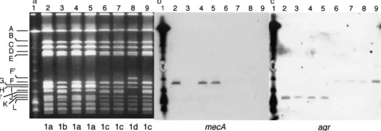
strain (Figure 1, lanes 2, 4 and 5). In contrast, mutants with vancomycin MICs >4 mg/L had lost mecA. This loss was associ-ated with large deletions in the SCCmec-carrying Sma I – G band (Figure 1, lanes 3, and 6 – 9), resulting either in a smaller additional band I0(Figure 1, lanes 3, 6, 7, and 9), or in a larger band F0, presumably due to a deletion fusing Sma I–G to SmaI–I in strain Vr10-1126a (Figure 1, lane 8).
Further analysis using the accessory gene regulator (agr) locus as a DNA probe revealed additional rearrangements in the Sma I – K band, which carried the agr operon (Figure 1, compare panel a and c). According to the published sequence of S. aureus N315 genome, agr maps in a region distant from the SCCmec integration site, suggesting that deletion of SCCmec may have induced loss of another genomic element, close to agr. This SmaI – K band became either slightly larger (Figure 1, lane 3), or became fused presumably to SmaI – J band to form a larger band co-migrating with Sma I – F (Figure 1, lanes 6 – 9). These mutants could be divided, based on the size of the deletion and their restriction patterns, into three groups: pattern 1b (21 kb deletion), represented by mutant Vr6-1126a (Figure 1, lane 3); pattern 1c
(93 kb deletion), represented by three mutants (Vr8-1126a, Vr8-1126b and Vr8-1126c; Figure 1, lanes 6, 7 and 9), which is similar to 1b, but with loss of Sma I – K; and pattern 1d (134 kb deletion), represented by Vr10-1126a (Figure 1, lane 8).
To rule out the possibility that the deletion of mecA from strain 1126 was due to a spontaneous event in the absence of vancomycin, 30 individual cultures of 1126 were incubated for 10 days or more in TSB without daily passage at 378C. Approxi-mately 200 colonies per culture were tested for oxacillin suscep-tibility. Of 2000 colonies tested, only three mutants (0.15%) with increased susceptibility to oxacillin were isolated. Two of three mutants were negative for mecA, but their susceptibility to vancomycin remained unchanged. The third mutant was still positive for mecA and susceptible to vancomycin. In contrast, the frequency of mecA deletion was 100% in vancomycin-induced, 1126-derived mutants with vancomycin MICs >4 mg/L. Hence, vancomycin triggered or selected for loss of mecA at a much higher frequency.
A detailed comparative phenotypic analysis was carried out between Vr6-1126a and its partially-isogenic parent 1126.
Figure 1.PFGE Sma I patterns of vancomycin-resistant mutants derived from parent strain 1126. (a) Lane 1, low range PFG marker (New England Biolabs); lane 2, parent strain 1126; lane 3, Vr6-1126a; lane 4, Vr4-1126a; lane 5, Vr4-1126b; lane 6, Vr8-1126a; lane 7 Vr8-1126b; lane 8, Vr10-1126a; lane 9, Vr8-1126c. The different banding patterns are indicated below each lane. (b) Southern hybridization of panel (a) with mecA; and (c) with agrCA.
Table 1. Vancomycin and oxacillin MICs for parent S. aureus isolates and vancomycin-resistant mutants
Vancomycin-resistant mutants MIC (mg/L)
Parent isolatea Oxacillin MIC (mg/L) Strain Vancomycin Oxacillin mecA
1126 >_ 128 Vr4-1126a 4 48 + Vr4-1126b 4 1.5 + Vr6-1126a 6 0.05 – Vr8-1126a 8 0.2 – Vr8-1126b 8 0.2 – Vr8-1126c 8 0.1 – Vr10-1126a 10 0.1 – 1128 >_ 128 Vr8-1128a 8 32 + 1130 >_ 128 Vr6-1130a 6 32 + 1132 >_ 128 Vr6-1132a 6 48 + 1134 >_ 128 Vr12-1134a 12 16 + a
All parent MRSA were vancomycin-susceptible (MIC <2 mg/L) and positive for mecA.
R. P. Adhikari et al.
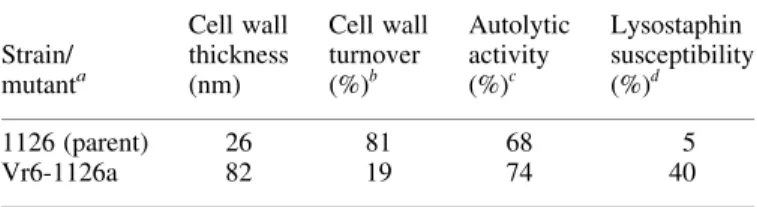
Mutant Vr6-1126a was more resistant to lysostaphin, and cell wall turnover was markedly reduced in the mutant compared with the parent (Table 2). The cell wall of Vr6-1126a was also three-fold thicker than the parent strain (Table 2), but Triton X-100-induced cell autolysis did not differ significantly.
Discussion
This study shows that vancomycin-induced stress can lead to the deletion of mecA in some strains of MRSA. In the strain reported here, this deletion correlated with increased resistance to vancomycin, chromosomal rearrangements and pleiotropic phenotypic changes. Subculturing of MRSA 1126 in non-selective liquid medium gave rise to spontaneous mutants with reduced oxacillin resistance at very low frequency (0.15%). However, subculturing of strain 1126 in vancomycin-containing medium triggered loss of mecA at a much higher frequency (100%). Reipert et al.6 identified a spontaneous mutant of methicillin-resistant vancomycin-intermediately-resistant S. aur-eus, where an increase in vancomycin MIC correlated with loss of a b-lactamase-encoding plasmid and a 32.5 kb deletion of SCCmec, which extended 65.4 kb into the chromosomal DNA. Hiramatsu et al.1 have hypothesized that penicillinase plasmids may play a role in the stability and phenotypic expression of mecA. However, it should be noted that the mecA deletion mutants generated in our study remained b-lactamase-positive and therefore appear to be novel.
The size of the mecA deletion in 1126 varied depending on the mutant, and rearrangements in other parts of the chromo-some (e.g. agr locus) were also observed. Some studies have reported that clinical glycopeptide-intermediate S. aureus (GISA) isolates are defective in the accessory gene regulator (agr) locus.12Whilst compromised agr function is not an absol-ute requirement for the development of GISA, it appears to be advantageous for the development of vancomycin hetero-resistance.
The phenotypic properties of Vr6-1126a are consistent with abnormalities in cell wall metabolism, as have been reported for other vancomycin-resistant mutants. The results of this study
contribute further to our knowledge on the development of vancomycin resistance in MRSA and we have shown that for one MRSA strain, this results in the deletion of mecA at the expense of vancomycin susceptibility. Future work will focus on the apparent incompatibility between oxacillin and vancomycin resistance in this strain.
Acknowledgements
We thank Richard Easingwood for expert technical assistance with electron microscopy, and Stefanie Keis for assistance with PFGE. This work was funded by an Otago Medical Research Foundation and Otago Research Grants awarded to GMC and JMBS. RPA was the recipient of a New Zealand Official Development Assistance (NZODA) study award and a grant from the Paul Ehrlich Gesellschaft (Germany).
References
1. Hiramatsu, K., Suzuki, E., Takayama, H. et al. (1990). Role of penicillinase plasmids in the stability of the mecA gene in methicillin-resistant Staphylococcus aureus. Antimicrobial Agents and Chemo-therapy 34, 600 – 4.
2. Inglis, B., Matthews, P. R. & Stewart, P. R. (1990). Induced deletions within a cluster of resistance genes in the mec region of the chromosome of Staphylococcus aureus. Journal of General Micro-biology 136, 2231 – 9.
3. Annear, D. I. & Grubb, W. B. (1976). Methicillin-sensitive variants in ageing broth cultures of methicillin-resistant Staphylococcus aureus. Pathology 8, 69 – 72.
4. Deplano, A., Tassios, P. T., Glupczynski, Y. et al. (2000). In vivo deletion of the methicillin resistance mec region from the chromosome of Staphylococcus aureus strains. Journal of Antimicrobial Chemother-apy 46, 617 – 20.
5. Donnio, P. Y., Louvet, L., Preney, L. et al. (2002). Nine-year surveillance of methicillin-resistant Staphylococcus aureus in a hospital suggests instability of mecA DNA region in an epidemic strain. Journal of Clinical Microbiology 40, 1048 – 52.
6. Reipert, A., Ehlert, K., Kast, T. et al. (2003). Morphological and genetic differences in two isogenic Staphylococcus aureus strains with decreased susceptibilities to vancomycin. Antimicrobial Agents and Chemotherapy 47, 568 – 76.
7. Sieradzki, K., Wu, S. W. & Tomasz, A. (1999). Inactivation of the methicillin resistance gene mecA in vancomycin-resistant Staphylo-coccus aureus. Microbial Drug Resistance 5, 253 – 7.
8. Adhikari, R. P., Cook, G. M., Lamont, I. et al. (2002). Phenotypic and molecular characterization of community occurring, Western Samoan phage pattern methicillin-resistant Staphylococcus aureus. Journal of Antimicrobial Chemotherapy 50, 825 – 31.
9. Hanaki, H., Kuwahara-Arai, K., Boyle-Vavra, S. et al. (1998). Activated cell-wall synthesis is associated with vancomycin resistance in methicillin-resistant Staphylococcus aureus clinical strains Mu3 and Mu50. Journal of Antimicrobial Chemotherapy 42, 199 – 209.
10. Mani, N., Tobin, P. & Jayaswal, R. K. (1993). Isolation and characterization of autolysis-defective mutants of Staphylococcus aureus created by Tn917-lacZ mutagenesis. Journal of Bacteriology 175, 1493 – 9.
11. Peschel, A., Vuong, C., Otto, M. et al. (2000). The D-alanine residues of Staphylococcus aureus teichoic acids alter the suscepti-bility to vancomycin and the activity of autolytic enzymes. Antimicrobial Agents and Chemotherapy 44, 2845 – 7.
12. Sakoulas, G., Eliopoulos, G. M., Moellering, R. C., Jr et al. (2002). Accessory gene regulator (agr) locus in geographically diverse Staphylococcus aureus isolates with reduced susceptibility to vanco-mycin. Antimicrobial Agents and Chemotherapy 46, 1492– 502.
Table 2. Summary of phenotypic properties of MRSA 1126 and Vr6-1126a Strain/ mutanta Cell wall thickness (nm) Cell wall turnover (%)b Autolytic activity (%)c Lysostaphin susceptibility (%)d 1126 (parent) 26 81 68 5 Vr6-1126a 82 19 74 40
The results shown are the mean values of 2 – 4 independent experiments. The standard error of the mean did not differ by more than 15%.
a1126 and Vr6-1126a were grown in the absence of vancomycin. b
Expressed as the percent radioactive cell wall components (CPM) released into the culture supernatant after 90 min resuspension in isotope-free medium containing 100 mg/L of cold N-acetylglucosamine.
cExpressed as percent remaining OD
620at 4 h after resuspension of culture in autolysis assay buffer.
dExpressed as percent remaining OD
620at 30 min after resuspension of cul-ture in assay buffer with lysostaphin (1 mg/mL). Control tubes incubated without lysostaphin showed less than a 10% decrease in OD620during the time course of the assay.
Vancomycin-induced mecA deletion in S. aureus