Highly efficient peptide binding and T cell
activation by MHC class II molecules of
CIITA-transfected cells
Eduardo Martinez-Soria, Claire-Anne Siegrist and Bernard Mach
L. Jeantet Laboratory of Molecular Genetics, Department of Genetics and Microbiology, University of Geneva Medical School, CMU, 9 Avenue de Champel, 1211 Geneva 4, Switzerland
Keywords: HLA restriction, peptide immunization, peptide loading, T cell recognition
Abstract
Expression of MHC class II, DM and li genes is controlled by the transactlvator CIITA, a mediator of the activation of these genes by IFN-y. Surprisingly, MHC class II molecules expressed on CIITA transfectants behave very differently from those expressed at the same level on IFN-y-induced cells in terms of peptide binding and peptide-specific T cell activation. MHC class ll-posltlve CIITA transfectants exhibit an unusually high capacity for binding exogenous peptldes, with a higher percentage of DR molecules occupied by a given peptide and are much more efficient at peptide-specific, HLA-DR-restricted activation of T lymphocytes. This unexpected phenotype reflects the antigen processing defect observed in CIITA transfectants. It suggests novel strategies for the use of CIITA-transformed cells in peptide-based Immunization.
Introduction
Activation of CD4+ antigen-specific T cells depends on the recognition by the TCR of specific complexes of peptides and MHC class II molecules displayed at the surface of antigen-presenting cells (APC). The formation of these com-plexes is a complicated process that favors presentation of peptide antigens derived from exogenous proteins. On their way to the cell surface, newly synthesized MHC class II invariant chain (li) complexes egress from the endoplasmic reticulum to a specialized endosomal compartment where, following removal of the li chain, they are loaded with peptides generated by the denaturation and proteolysis of endocytosed protein antigens (1,2) This complex peptide loading process requires the participation of HLA-DM molecules (3-5).
This essential role of MHC class II molecules for T cell activation implies that the very tight regulation of their expres-sion in various cell types represents an important level of control It is now established that both constitutive expression of MHC class II genes in specialized cells such as B lympho-cytes and their inducible expression in certain other cell types are under the control of the MHC class II transactivator CIITA (6,7). In MHC class ll-negative cells, CIITA is necessary for the induction of class II genes by IFN-y and is sufficient, in the absence of IFN-y, to turn on MHC class II expression (7). CIITA also controls the expression of the li and HLA-DM
genes (8,9). Surprisingly, however, when cells rendered MHC class ll-positive by either IFN-yor by transfection with CIITA, and expressing the same level of class II molecules, were compared for their ability to present exogenous protein anti-gens to specific CD4+ T cell clones, it was observed that IFN-7-treated cells could indeed activate T cells, whereas CIITA transfectants were unable to do so (10). This defect occurs at the level of antigen processing, through a yet undefined mechanism that can be corrected by treating the CIITA transfectants with IFN-y.
We have now compared the properties of MHC class II molecules expressed respectively on processing-defective CIITA-transfected cells and on IFN--y-induced cells for their ability to activate peptide-specific HLA-DR-restricted T cell lines. A drastic difference was observed in these two MHC class ll-positive cells: CIITA transfectants are much more efficient at peptide binding and a higher percentage of their surface MHC class II molecules can be loaded with a given specific peptide. Importantly, this results in a much more efficient peptide-specific, HLA-DR-restricted T cell activation than observed with IFN-y-treated cells. The data suggests that MHC class li-positive CIITA transfectants, once pulsed with selected peptide antigens, could be of interest for novel peptide-based immunization strategies.
Correspondence to: B. Mach
Methods
Cells and culture conditions
Human melanoma cells Me67 and Me208 and Epstein-Barr virus (EBV)-transformed lines OMW and BOLETH (11) were grown in RPMI 1640 medium complemented with glutamine, 10% heat-inactivated (56°C) FCS and antibiotics. Cells were incubated at 37°C in 5% CO2 and maintained in logarithmic growth phase with a viability >98% at all steps. For MHC class II induction, melanoma cells were incubated with human recombinant IFN-y (sp. act. 1.4X10⁷ U/mg; a gift of Biogen, Cambridge, MA) at 500 U/ml for a 48 h period.
Transfections
Melanoma cell lines were transfected by calcium phosphate precipitation followed 4 h later by a glycerol shock with either the expression vector EBO-Sfi alone or a full-length CIITA cDNA cloned into EBO-Sfi under control of the SV40 promoter (6,7). Stable transfectants were generated by selection with hygromycin B (Calbiochem, La Jolla, CA) and maintained in culture with hygromycin throughout the study, including during IFN-y induction. Identical results were obtained with untrans-fected cells and cells transuntrans-fected with the vector alone All studies were performed with bulk cultures rather than clones, thereby excluding the possible aberrant behavior of a single transfectant clone.
Surface MHC class II expression
Duplicate samples of 2 x 10⁵ cells were washed, pre-adsorbed with 10% normal rabbit serum (NRS), incubated with NRS or polymorphic HLA-DR mAb 2.06 (12) followed by fluorescein-conjugated rabbit anti-mouse IgG (Serotec), washed and analyzed by flow cytometry on a FACSan analyzer (Becton Dickinson, Mountain View, CA). Viable cells were gated based on propidium iodide uptake. A total of 10,000 cells were analyzed for each determination. The data were analyzed by the Lysys software package.
T cell lines
T61, T50, T54 and T87 are T cell lines specific for the p4 peptide of tetanus toxin (tt 1273-1284) which are restricted by the DRB3*0101 allele (data not shown). T19 is a T cell line specific for the p2 peptide of tetanus toxin (tt 830-843) which is restricted by DRB1*11 and DRB1*8 alleles (13). RPMI 1640 supplemented with 15% human AB+ serum from male volunteer donors was used as culture medium. T cell lines were re-stimulated for expansion with autologous irradiated peripheral blood mononuclear cells, preincubated with p4 or p2 peptides, in IL-2-supplemented culture medium. Expanded T cell lines were frozen in culture medium-DMSO 10% and stored in liquid nitrogen (13).
Antigen presentation to T cells
Melanoma cells (106) were fixed with 0.2% paraformaldehyde, washed and preincubated with various concentrations of tetanus synthetic peptides prior to washing and use as APC (3x10⁴ cells/well) in co-culture with tetanus-specific T cell lines (2X10⁴ cells/well) as described (10). The prohferative response of tetanus-specific T cell lines was measured after 48 h by [3H]thymidine incorporation.
Peptide binding
Binding assays were performed as described (14,15). Briefly, 3X10⁵ melanoma, either CIITA transfected or IFN-Y-treated, and EBV-transformed cell lines were incubated at 37°C for 4 h with various concentrations of a biotmylated peptide (HA307-319) of the influenza hemagglutmin (14) or medium alone. In competition experiments, cells were first incubated with a large excess of non-biotinylated HA peptide After washing, cells were incubated with FITC-streptavidin (4.22 ng/ml; Calbiochem) at 4°C for 30 mm. Stained cells were washed again and analyzed by flow cytometry as described above. To determine the relative amount of HLA-DR expressed on the surface of the cell lines, cells were incubated with a FITC-labeled munne mAb specific for DR (L243, Becton Dickinson) The fluorescein:protein ratios indicated by manufacturers were used to estimate the amount of cell surface DR. The effects of variations in DR expression between different cell lines were eliminated by dividing the fluorescence obtained with biotmylated peptide and fluoresceinated streptavidm by the level of staining with directly conjugated L243 (14). Assuming similar fluoresceinprotein ratios for both labeled reagents and stoichiometric binding, this ratio was interpreted as the estimate of the fraction of DR molecules occupied by peptides.
Results
Peptide binding on surface MHC class II molecules
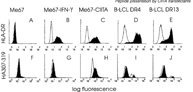
Indirect immunofluorescence analysis was first used to con-firm that IFN-Y- a n c J CIITA-transfected melanoma cells (Me67) express identical levels of surface HLA-DR molecules (Fig. 1B and C). It was also shown elsewhere (10) that the rate of newly made HLA-DR chains is identical under both conditions, and that both li and HLA-DM are induced in CIITA transfec-tants. When these two types of cells were incubated with a biotinylated influenza peptide (HA307-319) known to bind to HLA-DR4 (14), prior to addition of FITC-streptavidin, a drastic difference in peptide binding was observed (Fig 1, lower panel). Little staining above auto-fluorescence was detected on IFN-y-treated Me67 cells (Fig. 1G), which is consistent with peptide binding by <2% of surface HLA-DR molecules. In contrast, peptide binding on CIITA-transfected Me67 cells expressing similar levels of MHC class II molecules increased several fold as compared with IFN-Y-treated cells (Fig. 1H).
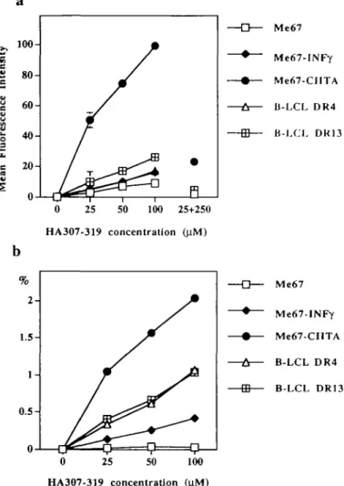
CIITA transfectants were then compared with homozygous EBV-transformed B cell lines, known to express unusually high densities of surface HLA-DR. HLA-DR4 and -DR13 homozygous cell lines (11) were chosen to match the two HLA-DR haplotypes expressed by Me67. On these EBV cell lines, the fluorescent signal resulting from binding of the HA307-319 peptide (Fig. 11 and J) was similar to that previ-ously reported by others (14,15). Surprisingly, HLA-DR molec-ules on CIITA transfectants appeared capable of binding higher amounts of peptide than EBV-transformed B cells (Fig. 1H). The greater total peptide binding capacity of MHC class ll-positive CIITA transfectants as compared to both IFN-Y-treated cells and B cell lines was confirmed in dose-response experiments (Fig. 2a). Competition assays in which cells were first incubated with a 10-fold excess of non-biotinylated
Me67
Me67-IFN-Y Me67-CIITA B-LCL DR4 B-LCLDR13
B i C D E
0 1 2 3 4 0 1 2 3 4 0 1 2 3 4 0 1 2 3 4 0 1 2 3 4 10 10 10 10 10 10 10 10 10 10 10 10 10 10 10 10 10 10 10 10 10 10 10 10 10 10° I01 102 103 I 04 10° 101 102 103 104 10° 101 102 103 104 10° 10' 10 2 103 104 10° 101 102 1 03 104log fluorescence
Fig 1. Peptide binding to MHC class II molecules at the surface of APC (Upper panel) Surface expression of HLA-DR on untransfected Me67
cells (A), Me67 cells incubated with IFN-y(500 U/ml) during 48 h (B), CIITA-transfected Me67 cells (C), EBV-transformed homozygous B cell lines (B-LCL) expressing HLA-DR4 (D) or DR13 (E) Cells were analyzed by flow cytofluorometry for the expression of HLA-DR (mAb 2 06) Blank profiles on the left represent cells incubated with NRS and the second reagent alone (Lower panel) Fluorescence of the same cells incubated with biotinylated HA307-319 peptide (100 (iM) prior to staining with FITC-streptavidm and analysis by flow cytofluorometry Blank profiles on the left indicate cells incubated with NRS and the second reagent alone Two peaks were observed in the case of the EBV B cell lines (I and J) that may reflect the presence of two populations of cells
peptide (Fig. 2a) as well as blocking experiments with MHC class ll-specific antibodies (data not shown) respectively confirmed peptide and HLA-DR specificity of the cell surface fluorescence. No binding of HA307-319 peptide was observed on a panel of MHC class ll-negative cell lines and on several cell lines expressing HLA-DR alleles that do not bind this particular peptide (14,15) (data not shown) Extent of HLA-DR occupancy by a specific peptide
It is known that occupation of MHC class II surface molecules by multiple peptides strongly limits their capacity to bind a given specific peptide provided exogenously. Although T cells have evolved to respond to very small amounts of peptide antigens (16,17), the heterogeneity of MHC class II occupancy limits the capacity of APC to be efficiently loaded with specific peptides. It was therefore of interest to explore if the higher peptide binding capacity displayed by CIITA transfectants as compared with IFN-y-treated cells resulted in a higher percentage of surface HLA-DR molecules capable of binding a given peptide. This was analyzed according to the approach previously described by Bush et al. (14). The relative amount of HLA-DR expressed on the surface of the cell lines was measured by incubating Me67 cells with FITC-labeled HLA-DR-specific mAb (L243-FITC; Becton Dickinson) at saturating concentrations (data not shown). Peptide-specific fluores-cence was then correlated to surface DR expression and corrected for respective fluorescein:protein ratios of both FITC-labeled reagents as described in Methods. The obtained ratio (Fig. 2b) thus represents an estimate of the fraction of surface DR molecules occupied by the biotinylated peptide (14). This estimation indicates that no more than 1% of surface MHC class II molecules at the surface of EBV-transformed B
cell lines are found to bind a given peptide at a concentration as high as 100 |iM, in agreement with figures obtained by others (14,15). This ppDR ratio is at least 50% lower in the case of MHC class II molecules of the same haplotypes induced upon IFN-y treatment of Me67 human melanoma cells. In contrast, the extent of HLA-DR occupancy by the HA307-319 peptide at the surface of CIITA transfectants was at least 7-fold higher than that of class II molecules on IFN-•y-treated cells, at each concentration of peptide.
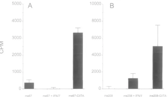
T cell activation by peptide-pulsed MHC class II molecules It was obviously of interest to explore the possible functional implications of the unusually high peptide binding capacity displayed by DR molecules on CIITA transfectants in terms of T cell activation CIITA-transfected and IFN-y-treated MHC class ll-positive cells were compared for their capacity to present tetanus peptides to tetanus-specific, HLA-DR-restricted T cell lines. Cells were fixed, preincubated with tetanus synthetic peptides, washed extensively and used as APC in co-culture with tetanus-specific T cell lines. The proliferative response of tetanus-specific T cell lines was measured after 48 h by [3H]thymidine incorporation. A dramatic difference in peptide-specific T cell activation was observed between IFN-y-induced and CIITA-transfected cells (Fig. 3). The capacity of peptide-pulsed IFN-y-treated Me67 cells to present a tetanus epitope (peptide p4, tt 1273-1284) to a DRB3*0101-restricted T cell line was either negligible or low. In contrast, when CIITA-transfected cells expressing the same levels of MHC class II molecules (Fig, 1) were incubated with the p4 tetanus peptide, a strong peptide-specific, DR-restricted activation was observed (Fig. 3A). This increased capacity of CIITA transfectants, as compared with
IFN-y-a 3— Me67 >> Intensi t •ne e uoresc t i . c V 1008 0 6 0 4 0 2 0 -U-1 /
J<
1—LJ™ 0 / ^^r—25 / *& — i — 50P
/
—i u— ' 100 25+250 Me67-INFy Me67-CIITA Ji-LCL DR4 B-LCL DR13 HA307-319 concentration (41M) Me67 Me67-INFy Me67-CIITA B-LCL DR4 B-LCL DR13 25 50 100 HA307-319 concentration jFig. 2. Relative peptide binding to cell surface HLA-DR molecules
(a) Dose-response analysis Me67 cells (control, IFN-y induced or CIITA transfected), DR4 and DR13 B-LCLs were incubated with increasing concentrations of biotmylated HA307-319 peptide prior to staining with FITC-streptavidin and analysis by flow cytofluorometry In competition experiments (25+250), cells were first incubated with an excess of non-biotinylated HA307-319 (250 nM) prior to addition of biotinylated peptide (25 (iM). Data are expressed as mean fluorescence intensity after subtraction of background auto-fluorescence given by cells incubated with NRS and the second reagent alone (b) Extent of HLA-DR occupancy by a specific peptide Percentage of HLA-DR molecules estimated to bind biotinylated HA307-319 at various peptide concentrations Ratios (%) represent mean fluorescence intensity due to binding of biotinylated peptide divided by mean anti-HLA-DR fluorescence intensity, after correction for fluorescence protein ratios of second reagents (14)
treated cells, to activate DR-restricted T cells after incubation with synthetic peptides was also observed with a different melanoma cell line (Me208, DRB1*1101/04) presenting a different tetanus peptide (p2, tt 830-843) to a different T cell line restricted bytheDRB1*11 and DRB1*08 alleles (Fig. 3B)
Peptide-specific T cell activation was studied further with four different DRB3*0101-restncted T cell lines and with various peptide concentrations (Fig. 4). In each case, a sharp dose-dependent peptide-specific, DR-restricted activation was observed. The possibility of an antiproliferative effect of IFN-y on T cells was excluded both by extensive washing of
Me67 cells prior to incubation with T cells and by the capacity of IFN-y-treated Me67 pulsed with native tetanus protein antigen to successfully activate the same T cell lines (10)
Discussion
The existence of an antigen processing-defective phenotype in MHC class ll-positive CIITA transfectants told us that, in addition to the genes controlled by CIITA, i.e. MHC class II, li and DM, another activity or protein is required for efficient presentation of protein antigens (10). Here we report that MHC class II molecules expressed on the surface of these antigen presentation-defective CIITA transfectants exhibit an unusually high affinity for exogenous peptides and that these peptide loaded non-professional APC become very effective T cell activators. CIITA transfection of MHC class ll-negative human melanoma cells induces the expression of surface MHC class II molecules able to bind and present exogenous peptides to antigen-specific, DR-restricted T cells to much higher levels than class II molecules expressed at the surface of IFN-y-treated cells This is not the consequence of an additional CIITA-dependant activity, since CIITA expression is itself induced by IFN-y (6,7).
The essential feature of antigen processing that is missing in CIITA transfectants, and that can be restored by IFN-y activation, could concern appropriate trafficking of MHC class ll-li chain complexes through specific cellular compartments or the activation of specific proteolytic enzymes required for adequate peptide loading onto MHC class II molecules within the endosomal compartment. Whatever the mechanism responsible for this antigen processing defect, it must lead to the expression of surface MHC class II molecules that are not occupied with a normal set of high-affinity peptides This results in the observed higher peptide binding capacity of surface MHC class II molecules and explains why CIITA transfectants exhibit such a high efficacy in T cell activation, when compared to IFN-y-treated cells.
Expression of the CIITA gene in a non-professional APC thus converts MHC class ll-negative cells into cells expressing high levels of autologous MHC class II molecules with an increased capacity for binding selected exogenous peptides. In normal cells, the enormous heterogeneity of peptide occu-pancy of MHC class II binding sites limits their capacity to bind and present a given peptide to T cells. It is shown here that, in the case of CIITA transfectants, a significantly higher proportion of MHC class II molecules can be loaded with a given specific peptide (Fig. 2b). Since occupation of only 0.1% of HLA-DR molecules by a given peptide has been shown to be sufficient to trigger T cell activation (16), it is not surprising that increasing this fraction by several fold results in a significant increase in peptide-dependant T cell activation. Peptide loading onto CIITA transfectants can be achieved with lower concentrations of exogenously provided synthetic peptides (down to <5 \iM, data not shown). This higher binding capacity of MHC class II molecules of CIITA transfec-tants could result either from occupancy of MHC class II molecules by low-affinity peptides or from the generation of 'empty' class II molecules. Since instability of empty molecules in SDS-PAGE is only observed with certain specific haplotypes (18), the normal SDS-PAGE pattern of HLA-DR molecules of
Q_
O
5000, 4000 3000-2000-
1000-10000-,
8000- 6000- 4000-2000-me67 2000-me67 + IFN-Y 2000-me67-CIITA me208 me208 me208-CIITA
Fig. 3. Peptide-specific T cell activation by two peptide-pulsed IFN-y-treated and CIITA-transfected melanoma cell lines Me67 and Me208
transfected with CIITA or incubated with IFN-y (500 U/ml) during 48 h were fixed and preincubated overnight with 10 ng/ml p4 (Me67) or p2 (Me208) tetanus toxin peptides Melanoma cells were extensively washed and tested as APC Me67 was tested with p4-specific T cell line T87 and Me208 was tested with p2-specific, DR11/8-restncted T cell line T19 Results are expressed as c p.m. obtained with peptide-pulsed melanoma cells after subtraction of c p m obtained with unpulsed melanoma cells
10-, 10-, 10-.
T54
0 0 0 1 1 0 1 0 o o o o T T o i o o o o 0 1 1 0 1 0 0 o o 0 1 1 0 1 0 0 p 4 p e p t i d e ( ( i g / m l )
Fig. 4. Peptide dose-response curve of T cell activation by IFN-y
and CIITA transfected melanoma cells Me67 cells either transfected with CIITA (solid circles) or incubated with (solid diamonds) or without (open squares) IFN-y (500 U/ml) during 48 h were fixed and preincubated overnight with various concentrations of p4 synthetic tetanus peptide prior to extensive washing and co-culture with T87, T61, T50 and T54 T cell lines. Results of peptide-specific T cell proliferation are expressed as the ratio of stimulation obtained with peptide-pulsed versus non-pulsed Me67 APC
Me67 Cl ITA transfectants (10) does not allow us to discriminate between these two possibilities.
IFN-Y allows correction of the protein processing defect seen in CIITA transfectants and thus restores presentation of a specific protein antigen to T cells (10) Treatment of CIITA transfectants with IFN-y, however, does not significantly decrease their high capacity for peptide binding (data not
shown). This observation was indeed expected. The peptide binding assay measures the overall occupancy of MHC class II molecules, of which only a fraction will be affected by IFN-Y treatment Whereas such a fraction of MHC class II molecules is sufficient to restore presentation of protein antigens (10), it is not expected to modify the overall capacity of the cell to bind exogenous peptides.
An interesting analogy can be made between the high binding capacity of MHC class II molecules observed here on CIITA transfectants and recent studies of MHC class I molecules that can be expressed under conditions where they also exhibit a very high affinity for exogenously provided peptides (19-21). The antigen processing-defective mutant cell line RMA-S expresses HLA class I molecules that are devoid of peptides and that exhibit a high affinity for exogen-ous peptides (19). It was also observed that, unexpectedly, transfection of RMA cells with the IL-10 gene lead to an RMA-S-like phenotype (20). In another system, it was possible to 'strip' experimentally MHC class I molecules on intact cells and to reload these molecules with exogenous peptides (21). These peptide-reloaded cells behaved as efficient inducers of a CD8 cytotoxic T lymphocyte (CTL) response (21).
In the field of tumor immunotherapy, the recent understand-ing of the importance of an MHC class ll-mediated component (22-25), together with the discovery of the role of CIITA as a general regulator of MHC class II expression (7,26), raised hopes that forcing the expression of CIITA, and consequently of MHC class II molecules, in tumor cells could lead to novel tumor immunization strategies. This approach has the obvious advantage of inducing expression of autologous MHC class II molecules on patients cells, as opposed to the need to transfect tumor cells with the relevant MHC class II genes specific of the particular HLA alleles of each different individual
(27-30). Our recent description of an antigen processing defect in MHC class ll-positive, CIITA-transfected melanoma cells (10) suggests, however, that these cells might not be that efficient at immunizing against exogenous protein antigens. Clearly, in vivo vaccination trials will be required to reach a definite conclusion on this issue.
Since the unusual property of surface MHC class II molec-ules on CIITA transfectants allows a strong peptide-specific activation of CD4 T lymphocytes, it might represent a practical advantage for peptide-based MHC class ll-mediated immun-ization. This applies to situations where a role of MHC class ll-mediated CD4 T cell activation and of MHC class ll-specific tumor antigens in the generation of tumor immunity is demon-strated or suspected (22,25,27). Two strategies can be envis-aged to exploit the efficient capacity of CIITA transfectants to activate T cells. Both have the advantage of dealing with autologous MHC class II molecules expressed on patient's cells; both involve CIITA expression, obtained either following transfection or by the delivery of the CIITA gene by an appropriate vector system; and both could be combined with the use of additional genes, such as B7 or granulocyte macrophage colony stimulating factor (25,30) The first approach would consist in loading CIITA-transfected, MHC class ll-positive, autologous tumor cells with specific peptides derived from proteins that are known to function as MHC class ll-restricted tumor antigens, such as tyrosinase in the case of melanoma (22). This would trigger a strong MHC class ll-specific CD4 T cell response, with the potential for eliciting CD4 antitumor CTL specific for the tumor antigen (31,32).
The second strategy relies on the advantage of providing locally an efficient, peptide-specific, CD4-mediated help signal that, in turn, could activate anti-tumor CD8 T lympho-cytes. CIITA-expressing MHC class ll-positive autologous tumor cells could thus be pulsed with a common promiscuous peptide antigen, such as a tetanus toxin peptide, and then used to immunize the same patient. Peptide-specific, HLA-restricted autologous, CD4 T cells would be activated and would deliver locally, within the micro-environment of the tumor cell, the appropriate help signals to bystander genuine tumor-specific CD8 CTL. It is known that efficient activation of tumor cell-specific CD8 CTL is facilitated by the simultaneous activation of CD4 T lymphocytes by the same cell (22). In addition to cancer immunotherapy, the capacity of inducing high levels of specific peptide binding and Tcell presentation by MHC class II molecules could also be relevant to immuniza-tion programs in various other fields, such as peptide-based tolerance induction in CD4+ T cell-mediated allergic responses (33-35).
Acknowledgements
We thank V Steimle (or providing CIITA expressing plasmids and for helpful discussion, and are grateful to S Carrell, G P Corradin, J Lamb and C. Irle for providing melanoma cell lines, tetanus toxin, biotmylated HA peptide and mAb This work was funded by grants from the Swiss National Foundation and the L Jeantet Foundation
Abbreviations
APC antigen-presenting cells
CTL cytotoxic T lymphocyte EBV Epstein-Barr virus h invariant chain NRS normal rabbit serum
References
1 Germain, R N and Margulies, D H 1993 The biochemistry and cell biology of antigen processing and presentation Annu Rev Immunol 11.403
2 Cresswell, P 1994 Assembly, transport, and function of MHC class II molecules Annu Rev Immunol 12259
3 Denzm, L K and Cresswell, P. 1995 HLA-DM induces CLIP dissociation from MHC class II afi dimers and facilitates peptide loading. Cell 82 155
4 Sloan, V S , Cameron, P, Porter, G , Gammon, M , Amaya, M , Mellins, E and Zaller, D M 1995 Mediation by HLA-DM of dissociation of peptides from HLA-DR Nature 375.802
5 Sherman, M A., Weber, D A and Jensen, P E 1995 DM enhances peptide binding to class II MHC by release of invariant chain-derived peptide Immunity 3'\97
6 Steimle, V, Often, L A , Zufferey, M and Mach, B 1993 Complementation cloning of an MHC class II transactivator mutated in hereditary MHC class II deficiency (or bare lymphocyte syndrome) Cell 75 135
7 Steimle, V., Siegnst, C A , Mottet, A , Lisowska Grospierre, B and Mach, B. 1994 Regulation of MHC class II expression by mterferon-gamma mediated by the transactivator gene CIITA Science 265 106
8 Chang, C H. and Flavell, R A. 1995 Class II transactivator regulates the expression of multiple genes involved in antigen presentation. J. Exp Med 181 765
9 Kern, I , Steimle, V, Siegnst, C A and Mach, B 1995 The two novel MHC class II transactivators RFX5 and CIITA both control expression of HLA-DM genes. Int. Immunol. 7 1295
10 Siegnst, C A , Martmez-Soria, E , Kern, I and Mach, B 1995 A novel antigen processing defective phenotype in MHC class II positive CIITA transfectants is corrected by mterferon-gamma J. Exp Med 1821793
11 Yang, S , Milford, E, Hammerlmg, U. and Dupont, B 1988 Description of the reference panel of B-lymphoblastoid cell lines for factors of the HLA system- the B-cell line panel designed for the Tenth International Histocompatibility Workshop In Immunobiology of HLA Histocompatibility Testing 1987. p 11 Sprmger-verlag, New-York
12 Charron, D. J and McDevitt, H O 1979 Analysis of HLA-D region-associated molecules with monoclonal antibody Proc Natl Acad. Sci USA 766567
13 Martinez Soria, E , Steimle, V, Burkhardt, C , Beffy, P, Tiercy, J M , Epplen, J T, Mach, B and Irle, C 1994 An HLA-DRB alpha-helix motif shared by DR11 and DR8 alleles is implicated in the plunallelic restriction of peptide-specific T-cell lines Hum Immunol 40 279
14 Busch, R , Strang, G., Howland, K and Rothbard, J B 1990 Degenerate binding of immunogemc peptides to HLA-DR proteins on B cell surfaces. Int Immunol 2443.
15 Rothbard, J B., Busch, R , Bal, V, Trowsdale, J , Lechler, R I and Lamb, J R 1989 Reversal of HLA restriction by a point mutation in an antigenic peptide Int Immunol 1 487
16 Demotz, S , Grey, H. M and Sette, A 1990 The minimal number of class II MHC-antigen complexes needed for T-cell activation Science 249-1028
17 Harding, C V. and Unanue, E. R 1990. Quantitation of antigen-presenting cell MHC class ll/peptide complexes necessary for T-cell stimulation Nature 346 574
18 Stebbins, C C , Loss, G E , Elias, C G., Chervonsky, A. and Sant A. J 1995. The requirement for DM in class ll-restricted antigen presentation and SDS-stable dimer formation is allele and species dependent J Exp Med 181 223
19 De Bruijn, M L , Schumacher, T N , Nieland, J D , Ploegh, H L , Kast, W M and Melief, C J 1991 Peptide loading of empty mapr histocompatibility complex molecules on RMA-S cells allows
the induction of primary cytotoxic T lymphocyte responses Eur J. Immunol 21:2963
20 Salazar Onfray, F, Petersson, M , Franksson, L, era/ 1995 IL-10 converts mouse lyrnphoma cells to a CTL-resistant, NK-sensitive phenotype with low but peptide-inducible MHC class I expression J Immunol 154 6291
21 Langlade Demoyen, P, Levraud, J P, Kounlsky, P. and Abastado, J. P 1994 Primary cytotoxic T lymphocyte induction using peptide-stripped autologous cells Int. Immunol. 6 1759 22 Topahan, S L 1994 MHC class II restricted tumor antigens and
the role of CD4+ T cells in cancer immunotherapy Curr Opm Immunol 6 741
23 Topalian, S L , Rivoltini, L , Mancini, M , Markus, N R , Robbms, P F, Kawakami, Y and Rosenberg, S A 1994 Human CD4+ T cells specifically recognize a shared melanoma-associated antigen encoded by the tyrosmase gene Proc Natl Acad Sci USA 91 9461
24 Ostrand Rosenberg, S 1994 Tumor immunotherapy the tumor cell as an antigen-presenting cell Curr Opm. Immunol 6 722 25 Pardoll, D M 1995 Paracrme cytokme adjuvants in cancer
immunotherapy Annu Rev Immunol 13 399
26 Mach, B, Steimle, V, Martmez-Sona, E and Reith, W 1995 Regulation of MHC class II genes lessons from a disease Annu Rev Immunol, in press
27 Ostrand Rosenberg, S, Thakur, A and Clements, V 1990 Rejection of mouse sarcoma cells after transfection of MHC class II genes J Immunol 144 4068
28 Ostrand Rosenberg, S , Roby, C , Clements, V K and Cole, G A 1991 Tumor-specific immunity can be enhanced by transfection of
tumor cells with syngeneic MHC-class-ll genes or allogeneic MHC-class-l genes Int J Cancer Suppl 6:61.
29 Dranoff, G , Jafiee, E , Lazenby, A., Golumbek, P., Levitsky, H , Brose, K , Jackson, V., Hamada, H., Pardoll, D. and Mulligan, R C 1993 Vaccination with irradiated tumor cells engineered to secrete murine granulocyte-macrophage colony-stimulating factor stimulates potent, specific, and long-lasting anti-tumor immunity Proc Natl Acad Sci. USA 90 3539.
30 Baskar, S , Ghmcher, L, Nabavi, N , Jones, R. T. and Ostrand-Rosenberg, S. 1995 Major histocompatibility complex class ll + B7-1+ tumor cells are potent vaccines for stimulating tumor rejection in tumor-bearing mice. J Exp Med 181:619
31 Frey, A B 1995 Rat mammary adenocarcinoma 13762 expressing IFN-gamma elicits antitumor CD4+ MHC class ll-restricted T cells that are cytolytic in vitro and tumoncidal in vivo. J Immunol 1544613
32 Takahashi, T, Chapman, P B , Yang, S. Y, Hara, I , Vljayasaradhi, S and Houghton, A N 1995 Reactivity of autologous CD4+ T lymphocytes against human melanoma J Immunol. 154 772 33 Gaur, A , Wiers, B , Liu, A , Rothbard, J. and Fathman, C. G
1992 Amelioration of autoimmune encephalomyehtis by myelm basic protein synthetic peptide-mduced anergy Science 258 1491
34 Hetzel, C and Lamb, J R 1994 CD4+ T cell-targeted immunomodulation and the therapy of allergic disease Clm Immunol Immunopathol 73.1
35 Larche, M , Hoyne, G , Lake, R. and Lamb, J R 1994 Immunological events underlying the induction of T cell non-responsiveness Int Arch Allergy Immunol 104 211