1999 Oxford University Press Nucleic Acids Research, 1999, Vol. 27, No. 1 289–291
The SWISS-2DPAGE database: what has changed
during the last year
Christine Hoogland*
,
Jean-Charles Sanchez
1,
Luisa Tonella
1,
Amos Bairoch
2,
Denis F. Hochstrasser
1and Ron D. Appel
Swiss Institute of Bioinformatics, c/o Medical Informatics Division, Geneva University Hospital, 24 rue Micheli-du-Crest, 1211 Geneva 14, Switzerland, 1Central Clinical Chemistry Laboratory, Geneva University
Hospital, Geneva, Switzerland and 2Department of Medical Biochemistry, University of Geneva, Geneva, Switzerland
Received October 1, 1998; Revised October 7, 1998; Accepted October 14, 1998
ABSTRACT
SWISS-2DPAGE (http://www.expasy.ch/ch2d/ ) is an annotated two-dimensional polyacrylamide gel elec-trophoresis (2-D PAGE) database established in 1993. The current release contains 21 reference maps from human and mouse biological samples, as well as from Saccharomyces cerevisiae, Escherichia coli and Dicty-ostelium discoideum origin. These reference maps now have 2480 identified spots, corresponding to 528 separate protein entries in the database, in addition to virtual entries for each SWISS-PROT sequence. During the last year, the SWISS-2DPAGE has undergone major changes. Six new maps have been added, and new functions to access the data have been provided through the ExPASy server. Finally, an important change concerns the database funding source.
INTRODUCTION
The SWISS-2DPAGE database collects data on proteins identi-fied on various two-dimensional polyacrylamide gel electro-phoresis (2-D PAGE) maps (1). Protein identification on 2-D PAGE maps is done by various techniques (2), including gel comparison, microsequencing, immunoblotting, amino acid composition analysis, peptide mass fingerprinting using mass spectrometry, or a combination of these techniques. The core of the SWISS-2DPAGE database consists of the description of the proteins identified, including mapping procedures, physiological and pathological information, experimental data (isoelectric point, molecular weight, amino acid composition, peptide mass) and bibliographical references, in addition to the 2-D PAGE images showing the protein locations. Cross-references are provided to Medline, to other 2-D PAGE databases [ECO2DBASE (3), HSC-2DPAGE (4), YEPD (5)], and to SWISS-PROT (6), which provides many links to other molecular databases [EMBL (7), GenBank (8), PROSITE (9), MIM (10), etc.]. The SWISS-2DPAGE database is maintained by the Swiss
Institute of Bioinformatics, in collaboration with the Central Clinical Chemistry Laboratory of the Geneva University Hospital.
FORMAT
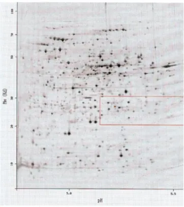
The protein entries in SWISS-2DPAGE are text files structured to be readable by human as well as by computer programs. Each entry is composed of defined lines, used to record various kinds of data (Fig. 1). For standardization purposes, the format of SWISS-2DPAGE entries is similar to the SWISS-PROT database (6), in addition to specific lines dedicated to 2-D PAGE data: (i) the master line (MT) lists the reference maps where the entry has been identified; (ii) the image line (IM) lists the 2-D PAGE images available for the entry; (iii) the 2D lines group different topics including the mapping procedure, spot coordinates, protein amino acid composition, peptide masses from mass spectrometry, protein expression levels and modifications. On the ExPASy Web server (see availability section) the data image associated with a protein entry displays the experimental location of the protein on the chosen map, in addition to a theoretical region computed from the protein sequence (Fig. 2).
CURRENT CONTENT
Release 8 of SWISS-2DPAGE (October 1998) contains 21 reference maps from human: cells or tissues (kidney, liver, lymphoma, platelet cells, red blood cells, colorectal epithelia cells), body fluids (cerebrospinal fluid, plasma), culture cells (erythroleukemia cell line, hepatoblastoma carcinoma derived cells, hepatoblastoma carcinoma derived cell line secreted proteins, macrophage like cell line, promyelocytic leukemia derived cells), from mouse cells or tissues (liver, gastrocnemius muscle, Pancreatic islet cells, epididymal fat pad), and from
Saccharomyces cerevisiae, Escherichia coli (for two pI ranges:
one from 3.5 to 10 and one from 4.5 to 5.5) and Dictyostelium
discoideum origin. Table 1 gives detailed descriptions for each of these maps, including creation and release dates, number of detected spots, number of identified spots, and number of distinct protein entries. There is a total of 528 protein entries. In addition,
Nucleic Acids Research, 1999, Vol. 27, No. 1
290
Figure 1. Example of a protein entry from SWISS-2DPAGE.
Figure 2. The protein from Figure 1 shown on the ECOLI4.5–5.5 reference map.
there are as many virtual entries as protein sequences in the SWISS-PROT database.
Table 1. Content of SWISS-2DPAGE
Abbreviations as follows: CEC, colorectal epithelia cells; CSF, cerebrospinal fluid; ELC, erythroleukemia cell line; HEPG2, hepatoblastoma carcinoma derived cells; HEPG2SP, hepatoblastoma carcinoma derived cell line se-creted proteins; HL60, promyelocytic leukemia derived cells; RBC, red blood cells; U937, macrophage like cell line; DICTYSLUG, D.discoideum; ECOLI, E.coli; ECOLI4.5–5.5, E.coli with pI range from 4.5 to 5.5; YEAST, S.cerevisiae; EPF, epididymal fat pad; ISLETS, pancreatic islet cells; MUSCLE, gastrocnemius muscle.
AVAILABILITY
The most efficient and user-friendly way to interactively browse in SWISS-2DPAGE is through the ExPASy World Wide Web molecular biology server at URL: http://www.expasy.ch/ . The SWISS-2DPAGE home page at URL: http://www.expasy.ch/ch2d/ provides several textual and graphical queries, and displays results with active links to other databases. A SWISS-2DPAGE entry may be reachable by keyword search (protein name, accession number, description or authors) or by selecting a spot on one of the 2-D PAGE maps. A full text search is now also available, allowing boolean operators (and, or, not) to restrict queries. Two new features have also been added to facilitate visualisation and differentiation of spots: (i) when clicking on a spot in one of the 2-D PAGE maps, the 2D line describing this spot in the corresponding protein entry will be highlighted in green; (ii) when displaying a protein entry, links are provided from each described spot to the corresponding 2-D PAGE map, in which the spot will be highlighted in green (in contrast to the other spots for that protein displayed in red).
291
Nucleic Acids Research, 1994, Vol. 22, No. 1Nucleic Acids Research, 1999, Vol. 27, No. 1 291
It is also possible to get a local copy of SWISS-2DPAGE using anonymous ftp (file transfer protocol) from the ExPASy FTP server (ftp://ftp.expasy.ch/databases/swiss-2dpage/ ).
Since September 1998, a system of an annual subscription fee for commercial users of the SWISS-2DPAGE database has been implemented. Increased data flow and especially automation of proteome analysis have created a requirement for resources, which cannot be addressed in full by public funding. It has therefore been decided to adopt the solution chosen for the SWISS-PROT database (6), namely to ask non-academic users to financially contribute to the maintenance of the database (for details, see URL http://www.expasy.ch/announce/ ).
ACKNOWLEDGEMENTS
The authors thank S. Frutiger, G. Hughes, M. Wilkins, A. Gooley, K. Williams, B. Walsh, K. Ou and P.-A. Binz for their assistance in protein identification, M. Raymond for providing colorectal data, J. Yan for Dicty data, C. Sarto for kidney data, R. Joubert-Carron for HL60 data, C. Pasquali for E.coli data, M.
Cawthorne for mouse data, and F. Ravier, S. Jaccoud, I. Demalte and V. Rouge for their technical assistance.
REFERENCES
1 Appel,R.D, Sanchez,J-C., Bairoch,A., Golaz,O., Miu,M., Vargas,R. and Hochstrasser,D. (1993) Electrophoresis, 14, 1232–1238.
2 Wilkins,M.R. and Gooley,A.A. (1997) In Wilkins,M.R., Williams,K.L., Appel,R.D. and Hochstrasser,D.F. (Eds), Proteome Research: New Frontiers in Functional Genomics. Springer Verlag, Berlin, pp. 35–64. 3 VanBogelen,R.A., Abshire,K.Z., Moldover,B., Olson,E.R. and
Neidhardt,F.C. (1997) Electrophoresis, 18, 1243–1251.
4 Corbett,J.M., Wheeler,C.H., Baker,C.S., Yacoub,M.H. and Dunn,M.J. (1994) Electrophoresis, 15, 1459–1465.
5 Payne,W.E. and Garrels,J.I. (1997) Nucleic Acids Res., 25, 57–62. 6 Bairoch,A. and Apweiler,R. (1998) Nucleic Acids Res., 26, 38–42. 7 Stoesser,G., Moseley,M.A., Sleep,J., McGowran,M., Garcia-Pastor,M. and
Sterk,P. (1998) Nucleic Acids Res., 26, 8–15.
8 Benson,D.A., Boguski,M.S., Lipman,D.J., Ostell,J. and Ouellette B.F.F. (1998) Nucleic Acids Res., 26, 1–7.
9 Bairoch,A., Bucher,P. and Hofmann,K. (1997) Nucleic Acids Res., 25, 217–221.
10 Pearson,P., Francomano,C., Foster,P., Bocchini,C., Li,P. and McKusick,V.A. (1994) Nucleic Acids Res., 22, 3470–3473.