Q J Med 1998; 91:489–492
Circadian elevation of IL-6 levels in Muckle–Wells syndrome:
a disorder of the neuro-immune axis?
A.W. GERBIG, C.A. DAHINDEN
1, P. MULLIS2 and T. HUNZIKER
From the Departments of Dermatology, and
1Immunology, and 2Children’s Hospital, University
of Bern, CH–3010 Bern, Switzerland
Received 17 October 1997 and in revised form 16 March 1998
Summary
Muckle–Wells syndrome (MWS) is a rare autosomal woman and her father, both suffering from this syndrome, in whom elevated serum levels of IL–6 dominant hereditary disorder characterized by
chronic recurrent urticaria, arthralgia, sensorineural could be documented during the flares of urticaria, and discuss the relevance of this finding for MWS. deafness, and in some cases nephropathy due to
amyloidosis (AA type). We report a 21-year-old
Introduction
Since the first description of urticaria, deafness and afternoon, culminating in the late evening, and then amyloidosis as a hereditary syndrome in nine mem- fading away during the night. It is often associated bers of a Derbyshire family by Muckle and Wells with fever, chills, rigors, malaise and aching pains 1962,1 about 100 further cases, partly familial but in the limbs, symptoms which are usually most also sporadic, have been reported in the literature.2,3 prominent on Mondays. During the morning there Though most of them correspond to what has become are absolutely no symptoms. The patient could not known as the Muckle–Wells syndrome (MWS), some define any triggering factors, apart from fatigue and of the sporadic cases in particular have probably hot sunny weather. At the age of seven, an audiogram been confounded, mostly with the CINCA (chronic revealed mild sensorineural deafness, but her parents infantile neurological, cutaneous and articular) syn- refused further investigations because the father, drome;4 for example, the patient of Linke et al.5 suffering from identical symptoms and showing a MWS is an autosomal dominant hereditary disorder constantly raised ESR, otherwise felt completely with incomplete penetrance, consisting of chronic healthy. It was not until the age of 19 that she recurrent urticaria, often combined with fever, chills, presented at our out-patient clinic searching for an rigors, malaise and aching pains in the limbs. It leads explanation of her chronic urticaria. Based on her to progressive sensorineural deafness, and in about personal and family history, MWS was assumed. one third of the patients, to amyloidosis of the Laboratory investigations disclosed a raised ESR kidneys as well as of other organs.3 (45 mm/h), CRP (58 mg/l), and complement activity (CH 50=464 E/ml), mild normo- to hypochromic anaemia with low to normal ferritin and serum iron, leucocytosis of up to 11.7×10⁹/l with normal
differ-Methods and results
ential count, thrombocytosis of up to 453×109/land elevated cortisol levels. Lymphocyte subpopula-A 21-year-old woman of Swiss ancestry suffered from
tions (B, T and NK cells), as well as the CD4/CD8 chronic recurrent urticaria from her day of birth
ratio, were normal. Of the T cells, 2–3% were onwards. Her urticaria predominantly affects the
activated (CD3+ HLADR+). Total serum protein trunk and the limbs, is non-pruritic and seems to
follow an ‘internal clock’, beginning in the early levels were 82.4 g/l with elevated a
2 globulins in
Address correspondence to Dr T. Hunziker, Department of Dermatology, University of Bern, CH–3010 Bern, Switzerland
A.W. Gerbig et al.
490
Figure 1. Mean (±SE) plasma melatonin sampled at 2-h intervals in the MWS patient and in 10 normal female controls (mean age 22 years, range 18–24).
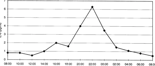
electrophoresis. In the presence of urticaria, serum MIP–1a and SCF (R&D Systems) in serum samples taken on three consecutive days at 7.30, 12.00 and fibrinogen was slightly elevated and the reptilase test
slightly prolonged, while thrombin time I was 18.00 h. Among the cytokines quantified, only IL–6 was elevated. Most striking was the fact that high shortened. Thromboplastin time and activated partial
thromboplastin time were normal. At the climax of levels of IL–6 (up to 91 pg/ml) were found in the evening samples, but not in the samples taken in the the urticaria, cortisol levels were elevated (up to
700 nmol/l) and ACTH levels inversely suppressed morning and at noon. We therefore monitored IL–6 levels over 24 h in samples taken at 2-h intervals, (<1.0 ng/l). The dexamethasone suppression test
with 1 mg revealed a fasting cortisol level of clearly confirming the circadian elevation of IL–6 (Figure 2). During this period the patient exhibited 42.7 nmol/l, ruling out a Cushing syndrome. The
circadian profile of melatonin corresponded to the unusually few symptoms, and the IL–6 levels were accordingly low. Unfortunately, she refused a second patient’s age (Figure 1). The following laboratory
parameters were also within normal limits: urea, session of continuous blood sampling. Her father exhibited a similar isolated IL–6 elevation (up to creatinine, creatinine clearance (74 ml/min),
potas-sium, liver function tests, triglycerides, total choles- 20 pg/ml) in the evening. terol, C3, C4, C3dg and the C1q-binding assay.
Based on the clinical picture and these laboratory
data, our patient fulfilled the criteria of MWS.
Discussion
Because MWS has features of an inflammatorydisorder, and to gain insight into the pathogenesis IL–6 is a pleiotropic cytokine produced by a large variety of cell types and acting on a wide range of of the disease, we measured several cytokines, i.e.
IL–1a, IL–4, IL–6, IL–12, TNF-a, interferon-a, tissues.6 In our patient the increase in IL–6 levels
Circadian IL-6 variation in Muckle–Wells syndrome 491
could explain the increase in acute-phase proteins be stored and released in the pituitary gland and induces the production of proinflammatory cytokines, such as CRP and fibrinogen with associated raised
ESR, as well as leucocytosis and thrombocytosis.7 including IL–6, by monocytes.11 It will be interesting to study MIF levels in MWS patients, once a suitable Since IL–6 is known to function as an osteoclast
activating factor,8 it is conceivable that the sensorin- assay is available.
Since a specific treatment selectively reducing eural deafness results from the destruction of the
Corti-organ, as reported by Muckle and Wells in two IL–6 in vivo is not yet available, short-term thera-peutical trials including different types of antihis-patients.1 However, fever, chills, aching limb pains
and probably also urticaria may be due to an indirect tamines and NSAIDs, nifedipine, pentoxifylline, theophylline, colchizine, dapsone, disodium cromo-effect of IL–6 or an additional unknown mediator.
Little is known about the effect of cytokines on mast- glycate, urso- and chenodeoxycholic acid, prednis-one and even thalidomide have been conducted. cell function and mediator release and accordingly,
the pathogenesis of most forms of chronic urticaria Dapsone and thalidomide delayed, urso- and cheno-deoxycholic acid as well as small doses of prednisone is unknown. Among a large number of cytokines,
including IL–6, tested in vitro, only SCF was able to weakened, but only high doses of prednisone (50 mg/day) suppressed the urticaria and associated activate mast cells.9 We also tested whether
intradermal injection of autologous serum, collected symptoms.
In conclusion, we find elevated IL–6 levels with in the morning and evening, could elicit a weal and
flare reaction, but the results were negative. Although a striking circadian pattern in two patients with MWS. To our knowledge, this is the first report of the elevated levels of IL–6 may be involved in the
pathogenesis of amyloidosis through the chronic such a prominent circadian expression of an isolated cytokine due to a hereditary disorder.
induction of acute-phase proteins, the fact that only about one third of patients with MWS develop amyloidosis could point to additional causative
factors.
Acknowledgements
The production of IL–6 is part of the stress
response of many cell types and is triggered by We would like to thank Dr U. Buergi, Department immunological, chemical and physical stimuli as of Endocrinology, and Dr A. Tobler, Department of well as by several proinflammatory cytokines. The Hematology, University Hospital of Bern, for helpful fact that MWS is a hereditary disorder and that the discussions, Dr P. Spa¨th, Central Laboratory of the symptoms of this patient were present since birth Blood Transfusion Service, Swiss Red Cross, Bern, indicates that the elevation of IL–6 is due to an for complement analyses and Dr C.M. Gerbig for endogenous factor (or factors). Furthermore, the skilled assistance in creating the figures.
striking circadian pattern suggests a disturbance in the neuro-endocrine-immune axis. The melatonin rhythm is produced by the vertebrate pineal gland
References
and is thought to be generated by a biological
1. Muckle TJ, Wells M. Urticaria, deafness, and amyloidosis: a
(circadian) clock in the suprachiasmatic nuclei of
new heredo-familial syndrome. Q J Med 1962; 31:235–48.
the anterior hypothalamus. The rhythm is entrained
2. Fu¨ger K, Fleischmann M, Weber, M, Mann J.
to the 24-h period by the daily light-dark cycle, with
Komplikationen im Verlauf eines Muckle-Wells-Syndroms.
hormone levels increased at night (Figure 1).
Dtsch med Wschr 1992; 117:256–60.
Interestingly, exogenously administered melatonin
3. Muckle TJ. The ‘Muckle-Wells’ syndrome. Br J Dermatol
can entrain circadian rhythms, can change the phase 1979; 100:87–92. of the endogenous melatonin rhythm, and facilitate
4. Prieur AM, Griscelli C, Lampert F, Truckenbrodt H,
reentrainment of circadian rhythms.10 Thus, mela- Guggenheim MA, Lovell DJ, Pelkonnen P, Chevrant-Breton J, tonin may modulate the entrainment process in Ansell BM. A chronic, infantile, neurological, cutaneous
humans and be useful for treating circadian rhythm and articular (CINCA) syndrome. A specific entity analysed in 30 patients. Scand J Rheumatol 1987; 66:57–68.
disorders. In our patient, however, the 24-h
mela-5. Linke RP, Heilmann KL, Nathrath WBJ, Eulitz M.
tonin profile presented a normal circadian pattern
Identification of amyloid A protein in a sporadic
Muckle-for an adult female (Figure 1) and the administration
Wells syndrome. Lab Invest 1983; 48:698–704.
of melatonin for 1 week (5 mg per day, given at
6. Hirano T, Akira S, Taga T, Kishimoto T. Biological and
breakfast) did not influence the time pattern of
clinical aspects of interleukin 6. Immunol Today 1990;
symptoms. Therefore the mechanism inducing the 11:443–9. diurnal elevation of IL–6 could not be identified. An
7. Sun WH, Binkley N, Bidwell DW, Ershler WB. The
attractive candidate linking the neuro-endocrine influence of recombinant human interleukin–6 on blood system with the innate immune system would be the and immune parameters in middle-aged and old rhesus
monkeys. Lymphokine Cytokine Res 1993; 12:449–55.
A.W. Gerbig et al.
492
8. Jilka RL, Hangoc G, Girasole G et al. Increased osteoclast 10. Cavallo A. The pineal gland in human beings: Relevance to pediatrics. J Pediatr 1993; 123:843–51.
development after estrogen loss: mediation by interleukin–6. Science 1992; 257:88.
11. Bucala R. MIF re-discovered: pituitary hormone and 9. Bischoff SC, Dahinden CA. C-kit ligand: a unique glucocorticoid-induced regulator of cytokine production.
potentiator of mediator release by human lung mast cells. Cytokine Growth Factor Rev 1996; 7:19–24. J Exp Med 1992; 175:237–44.