M A J O R A R T I C L E
Oropharyngeal Group A Streptococcal
Colonization Disrupts Latent Epstein-Barr
Virus Infection
Seigo Ueda,1,2,5Satoshi Uchiyama,4Tarik Azzi,1,2Claudine Gysin,2,3Christoph Berger,1,2Michele Bernasconi,1,2 Yasuaki Harabuchi,5Annelies S. Zinkernagel,4and David Nadal1,2
1
Experimental Infectious Diseases and Cancer Research, Division of Infectious Diseases and Hospital Epidemiology,2Children’s Research Center, and
3
Division of Otolaryngology, University Children’s Hospital of Zurich, and4Division of Infectious Diseases and Hospital Epidemiology, University Hospital Zurich, University of Zurich, Switzerland; and5Department of Otolaryngology/Head and Neck Surgery, Asahikawa Medical University, Japan
Epstein-Barr virus (EBV) infects >90% of the human population within thefirst 2 decades of life and establishes reversible latent infection in B cells. The stimuli that lead to switching from latent to lytic EBV infection in vivo are still elusive. Group A streptococci (GAS) are a common cause of bacterial pharyngotonsillitis in children and adolescents and colonize the tonsils and pharynx of up to 20% of healthy children. Thus, concomitant presence of EBV and GAS in the same individual is frequent. Here, we show that EBV carriers who are colonized with GAS shed EBV particles in higher numbers in their saliva, compared with EBV carriers not colonized with GAS. Mes-senger RNA levels of the master lytic regulatory EBV gene BZLF1 were more frequently detected in tonsils from EBV carriers colonized with GAS than from EBV carriers not colonized. Heat-killed GAS, potentially mimicking GAS colonization, elicited lytic EBV in latently infected lymphoblastoid cell lines (LCLs) partially via Toll-like re-ceptor 2 triggering, as did purified GAS peptidoglycan. Thus, colonization by GAS might benefit EBV by increas-ing the EBV load in saliva and thereby enhancincreas-ing the likelihood of EBV spread to other hosts.
Keywords. Epstein-Barr virus (EBV); group A Streptococci (GAS); tonsil; oropharynx; lytic; latent; TLR2; salivary shedding.
More than 90% of the adult population is persistently and asymptomatically infected with Epstein-Barr virus (EBV), a human B-lymphotropic γ-herpesvirus [1]. Primary infection with EBV is acquired mainly in childhood via saliva [1]. The oropharynx is the portal of entry and exit, where the palatine tonsils act as reser-voir for EBV [2–4]. In its latent form in B cells, EBV ex-presses a limited number of genes, does not replicate, and its DNA is propagated to daughter cells during cell division. By contrast, in its lytic form, EBV expresses
genes required for replication and generation of infec-tious virus particles resulting in host cell lysis and virus release [5]. The default state of EBV infection is the latent form, which is reversible, permitting the creation of new viral particles and transmission to other hosts [6]. The mechanisms responsible for switching to the lytic form in vivo are not completely understood.
EBV is associated with Burkitt lymphoma (BL), Hodgkin disease (HD), and posttransplantation lym-phoproliferative disease [7]. These B-cell tumors display distinct patterns of EBV latency gene expression [8]. The oncologic potential of latent EBV is indicated by its unique capacity to growth transform B cells in vitro to lymphoblastoid cell lines (LCLs) [1]. Thus, disruption of latency is not only essential to enable transmission of EBV to other hosts but might be an important factor to limit EBV-induced B-cell lymphoproliferation.
Being immunocompromised increases the risk of EBV-associated B-cell tumors [9]. But nonimmunocom-promised individuals may also develop EBV-associated
Received 30 April 2013; accepted 12 July 2013; electronically published 9 August 2013.
Presented in part: 3rd Swiss Workshop in Fundamental Virology, Thun, Switzerland, 29–30 August 2011.
Correspondence: David Nadal, MD, Division of Infectious Diseases and Hospital Epidemiology, University Children’s Hospital Zurich, Steinwiesstrasse 75, CH-8032 Zurich, Switzerland (david.nadal@kispi.uzh.ch).
The Journal of Infectious Diseases 2014;209:255–64
© The Author 2013. Published by Oxford University Press on behalf of the Infectious Diseases Society of America. All rights reserved. For Permissions, please e-mail: journals.permissions@oup.com.
BL or HD. EBV-associated HD is more likely to develop when primary EBV infection occurs in adolescence and manifests as infectious mononucleosis with an exuberant immune activation [10]. In endemic BL, >95% of the tumors are EBV positive, and they are epidemiologically linked to Plasmodium falciparum malaria, resulting in chronic immune activation [11]. The pattern-recognition receptor Toll-like receptor 9 (TLR9) is abun-dantly expressed in B cells, and it senses DNA and the malaria parasite’s pigment hemozoin [12–14]. We recently demonstrated that TLR9 activation of B cells inhibits lytic EBV during primary infection and inhibits the switching of latent to lytic EBV in chronic infection in vitro [15,16].
Group A streptococci (GAS) colonize tonsils and the pharynx of up to 20% of healthy children [17]. Considering the high prevalence of EBV and GAS, concomitant presence of both microorganisms in the same individual is frequent. TLR9 senses bacterial DNA and is crucial for controlling GAS infec-tions [18]. One may thus reason that the presence of GAS in tonsils may direct the EBV life cycle toward latency, thereby impairing EBV transmission to other hosts via saliva.
Here, we investigated the influence of GAS on EBV’s life cycle and salivary shedding and the mechanisms involved.
METHODS Ethics Statement
This study was conducted according to the principles expressed in the Declaration of Helsinki. The Ethics Commission of the Canton of Zurich approved the study (StV 40/05). All subjects or their caregivers provided written informed consent.
Cell Culture
The EBV-producer cell line B95.8, Akata BL cells, tonsillar mononuclear cells (TMCs), and LCLs were maintained in Roswell Park Memorial Institute 1640 medium (Sigma-Aldrich, Buchs, Switzerland) with 10% heat-inactivated fetal bovine serum (Life Technologies, Zug, Switzerland), 1% L-glutamine, and 1% penicillin-streptomycin, referred to hereafter as R10.
Enzyme-Linked Immunosorbent Assay (ELISA)
Interleukin 6 (IL-6) or interleukin 10 (IL-10) levels were mea-sured using human IL-6 or IL-10 ELISA kits (R&D Systems, Abingdon, United Kingdom) following the manufacturer’s instructions.
DNA Extraction and EBV DNA Detection
Saliva samples were obtained and DNA was extracted as reported elsewhere [19]. EBV DNA levels were determined by quantitative real-time PCR (qPCR) targeting the conserved EBV BamHI W region, as reported elsewhere [20].
RNA Extraction and qPCR
Total RNA was extracted using the RNeasy Mini Kit (Qiagen) following the manufacturer’s instructions. After DNase I treatment, 1 µg of total RNA was used as template for reverse transcription by use of a High-Capacity cDNA Reverse Tran-scription Kit (Life Technologies). qPCR for human and EBV gene messenger RNA (mRNA) was performed using specific primers and probes for IL-6, IL-10, or BZLF1, as reported else-where [15], and for TLR1–10 (Life Technologies). All reactions were performed on a real-time PCR machine (7900HT; Life Technologies) with TaqMan Gene Expression Master Mix (Life Technologies). The relative gene expression was calculated for each gene of interest by using aΔΔCT method, where CT values were normalized to the value for the housekeeping gene hydroxymethylbilane synthase (HMBS) [15].
Isolation of TMCs and EBV Serology
TMCs were isolated from palatine tonsils obtained from pa-tients who underwent routine tonsillectomy, as reported else-where [21]. The EBV serologic characteristics of the TMC donors were determined using the Immunodot-Mono G and Mono M kit according to the manufacturer’s instructions (Ruwag Diagnostics, Bettlach, Switzerland).
Preparation of Stock EBV
Supernatant of B95.8 cells was obtained as reported elsewhere [21] and stored at −80°C. The cell-free supernatants con-tained approximately 1 × 10⁸/mL EBV DNA as evaluated by qPCR [20].
GAS Strains
The well-characterized clinical isolate M1T1 GAS strain 5448 [22] was grown to logarithmic phase in Todd-Hewitt broth (Becton Dickinson, Allschwil, Switzerland) containing 2% yeast extract (THY; Oxoid, Pratteln, Switzerland) and was resuspend-ed in Roswell Park Memorial Institute 1640 mresuspend-edium at afinal concentration of 2 × 10⁹colony-forming units/mL. Bacteria were killed by heating (at 85°C for 60 minutes) or by sonication (Sonoplus HD2070; Bandelin Electronic, Berlin, Germany) with 20 kHz at 70 W (amplitude, 100%) for 15 minutes on ice. Detection of GAS Colonization
Explanted tonsils were rolled over 5% sheep blood agar that was incubated for 48 hours. GAS was identified by latex aggluti-nation for Lancefield group A (Bio-Rad, Cressier, Switzerland) and detection of pyrrolidonyl peptidase production, using L -pyrrolidonyl-beta-naphthylamid (0.7%) disks (Oxoid, Pratteln, Switzerland).
Stimulation of EBV-Infected Cells With GAS
To model acute infection, TMCs plus B95.8 culture superna-tants were used, and to model persistent infection, LCLs and Akata cells [23] were used. LCLs were established from TMCs
by infection with B95.8 EBV. Heat-killed or sonicated GAS at a multiplicity of infection (MOI) of 100 or 20 µg/mL of GAS peptidoglycan (Toxin Technology, Sarasota, FL) was added for 24 hours. Apoptosis was assessed using a PE Annexin V Apoptosis Detection Kit I according to the manufacturer’s in-structions (Becton Dickinson). To neutralize TLR2, LCLs were preincubated with anti-TLR2 polyclonal antibodies (LabForce AG-InvivoGen, Nunningen, Switzerland).
Flow Cytometry
For TLR2 detection,fluorescein isothiocyanate (FITC)–labeled anti-TLR2 monoclonal antibodies (TL2.1; LabForce AG-Inviv-oGen) were used, and FITC-labeled mouse immunoglobulin G2a (Becton Dickinson) was used as isotype control. For TLR2 and BZLF1 detection, following TLR2 staining, cells werefixed with 2% paraformaldehyde (Sigma-Aldrich) and permeabilized using 0.4% Triton X-100 before adding anti-BZLF1 antibodies (BZ1; Santa Cruz Biotechnology, Heidelberg, Germany) followed by PE-labeled rabbit anti-mouse immunoglobulin G1 (Santa Cruz Biotechnology). Data on apoptosis or TLR2 and/or BZLF1 detection were collected using a FACSCanto II (Becton Dickinson) and were analyzed using FlowJo software.
Statistical Analyses
Analyses of statistical significance were based on a 2-tailed paired or unpaired Student t test, Wilcoxon signed rank test, Mann–Whitney U test, or χ2test. Differences with a P value of <.05 were regarded as statistically significant.
RESULTS
Saliva From EBV-Infected GAS-Colonized Individuals Contained More EBV Than Saliva From EBV-Infected Individuals Not Colonized With GAS
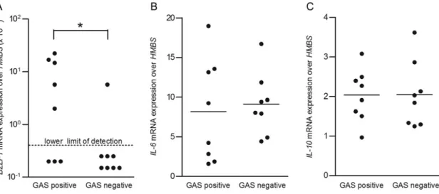
We asked whether salivary EBV shedding is influenced by colo-nization with GAS. Thus, we assayed saliva from EBV-infected individuals who were or were not colonized with GAS by using qPCR targeting EBV DNA to detect viral DNA contained in intact EBV particles. We detected EBV in 7 of 12 GAS-colo-nized individuals (58%; median age, 6.0 years [range, 2.2–15.0 years]; mean age, 6.2 years) and 9 of 15 GAS-negative individu-als (60%; median age, 5.3 years [range, 3.1–13.8 years]; mean age, 6.0 years). The number of EBV DNA copies in saliva from GAS-colonized EBV-infected individuals was higher than that in saliva from noncolonized individuals (P = .03; Figure1). More-Frequent BZLF1 mRNA Expression in Tonsils From GAS-Colonized Than From Noncolonized EBV-Infected Individuals The EBV load in saliva may reflect distinct EBV replication in oropharyngeal epithelial cells, mucosa-associated B cells, or both. Since determination of EBV replication in primary epi-thelial cells is not feasible, we measured BZLF1 mRNA expres-sion in tonsils. BZLF1 is an immediate-early lytic EBV gene,
the expression of which is sufficient to initiate EBV replication. Tonsil tissues from 5 of 8 GAS-colonized individuals expressed BZLF1 mRNA (62.5%; median age, 5.3 years [range, 2.8–6.8 years]; mean age, 4.9 years), in contrast to 1 of 8 GAS-negative individuals (12.5%; median age, 4.8 years [range, 3.1–6.0 years]; mean age, 4.8 years; P = .04; Figure2A). IL-6 and IL-10 mRNA expression levels determined by human specific qPCRs showed no significant differences between GAS-positive and GAS-negative tonsils (Figure2B and2C), suggesting a similar inflammation status.
Heat-Killed but Not Sonicated GAS Induced Lytic EBV In Vitro After EBV Exposure
Heat-killed GAS are intact bacteria that mimic colonizing GAS. In contrast, sonication disrupts GAS, resulting in fragmentation and release of bacterial cell wall components and DNA, mim-icking an acute GAS infection during which GAS are lysed by both the immune system and antibiotics. We inoculated TMCs with EBV B95.8 and concomitantly stimulated them with heat-killed or sonicated GAS at a MOI of 100. We then measured EBV and HMBS mRNA expression levels by qPCR 24 hours after inoculation with EBV. Levels of BZLF1 mRNA in TMCs 24 hours after stimulation with heat-killed GAS were higher than in nonstimulated TMCs or in TMCs stimulated with soni-cated GAS (P = .03; Figure3A). Furthermore, heat-killed GAS did not affect latent EBV (LMP1 and EBNA3C; data not Figure 1. Saliva from Epstein-Barr virus (EBV)–infected group A strepto-coccus (GAS)–colonized individuals contained more EBV particles than saliva from EBV-infected noncolonized individuals. The number of EBV DNA copies per milliliter in saliva samples from EBV carriers who under-went routine tonsillectomy was determined by quantitative real-time poly-merase chain reaction after DNase I treatment. Results for GAS-positive (n = 12) and GAS-negative (n = 15) individuals were compared.P values were calculated using the Mann–Whitney U test. *P = .03.
shown), IL-6, and IL-10 mRNA expression in TMCs, compared with no stimulus (Figure 3B and3C). In contrast, sonicated GAS increased IL-6 and IL-10 mRNA expressions in TMCs, compared with no stimulus or heat-killed GAS (P = .03; Figure3B and3C). Thus, neither heat-killed nor sonicated GAS influenced latent EBV gene expression. In contrast, heat-killed GAS strongly induced lytic EBV infection in TMCs exposed de novo to EBV, whereas sonicated GAS did not. The latter induced strong cytokine gene expression in TMCs, whereas heat-killed GAS did not.
Heat-Killed GAS Induced Lytic EBV and Caused Cell Death in Latently EBV-Infected B Cells
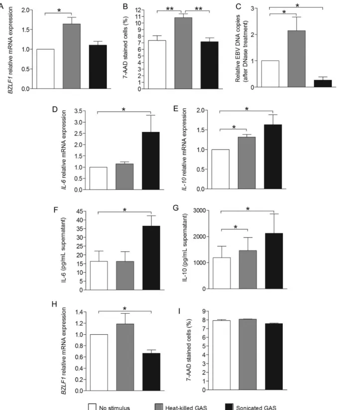
Next, we asked whether GAS modulates EBV persistent B-cellular infection. We used LCLs as a model for persistent (latent) EBV infection and stimulated them with heat-killed or sonicated GAS at a MOI of 100. EBV and mRNA expression levels of human genes were measured by qPCR 24 hours later. BZLF1 mRNA levels in LCLs exposed to heat-killed GAS were higher (P = .02) than in LCLs with or without exposure to sonicated GAS (Figure4A).
Figure 2. BZLF1 messenger RNA (mRNA) in tonsils from Epstein-Barr virus (EBV)–infected group A streptococcus (GAS)–colonized individuals was more frequently detectable than in noncolonized individuals. Levels ofBZLF1 (A), IL-6 (B), and IL-10 (C ) mRNA expression in tonsil tissues from EBV-seropositive patients who underwent routine tonsillectomy were analyzed by quantitative real-time polymerase chain reaction. Results for GAS-positive (n = 8) and GAS-negative (n = 8) individuals were compared.BZLF1 expression values above the lower detection limit were considered positive, and those below the lower detection limit were considered negative.P values were calculated using the χ2test. *P = .04.
Figure 3. Heat-killed but not sonicated group A streptococcus (GAS) induced lytic Epstein-Barr virus (EBV) after de novo in vitro EBV exposure. Tonsillar mononuclear cells (TMCs) were inoculated with supernatants from B95.8 cells (EBV-producer cells). Concomitantly, heat-killed or sonicated GAS were added, and then cell pellets were harvested and subjected to quantitative real-time polymerase chain reaction to determine expression ofBZLF1 (A), IL-6 (B), and IL-10 (C) messenger RNA (mRNA) expression. Results shown were pooled from TMCs from 6 donors and are expressed as mean values ± standard error of the mean.P values were calculated using a Wilcoxon signed rank test. *P = .03.
We asked whether the increased BZLF1 mRNA levels after exposure to heat-killed GAS were associated with B-cell death. We assessed late apoptosis by determining the percentage of 7-aminoactinomycin D (7-AAD)–stained cells by flow cytome-try in LCLs following stimulation or no stimulation. Exposure to heat-killed GAS for 24 hours resulted in higher percentages of 7-AAD–stained cells as compared to exposure to sonicated GAS or controls (P = .008; Figure4B). There were no differenc-es in percentagdifferenc-es of early apoptotic cells as assdifferenc-essed by flow cytometry after staining with Annexin V and 7-AAD. This was confirmed by assessing cell viability, using the Trypan blue exclusion method (not shown). The number of EBV DNA copies after DNase I treatment in the supernatants of LCLs exposed to heat-killed GAS were higher than in controls (P = .02; Figure4C), corroborating our above-described in vivo observations of higher EBV copies in tonsils of GAS-colonized individuals. Exposure to sonicated GAS resulted in lower EBV DNA copies, compared with no exposure (P = .02; Figure4C). This is in line with our previous study showing that TLR9 triggering results in suppression of switching from latent to lytic EBV. Sonicated GAS contain increased levels of GAS DNA, which has been recently demonstrated to trigger TLR9 and subsequently induce proinflammatory cytokine expression [18,24].
Heat-killed GAS did not affect the levels of EBNA3C latent EBV (not shown) or IL-6 (Figure4D), although it slightly upre-gulated IL-10 mRNA expression (Figure4E). Similarly, after de novo EBV exposure, sonicated GAS increased IL-6 (P = .02; Figure 4D) and IL-10 mRNA expression in LCLs, compared with no stimulus (P = .03; Figure4E), as well as IL-6 and IL-10 protein levels (Figure4F and4G).
We wondered whether heat-killed or sonicated GAS exhibit-ed similar effects on EBV-carrying BL cells as on LCLs. We chose Akata cells because they represent a well-established model for studying the switching from latent to lytic EBV in vitro. We analyzed BZLF1 mRNA expression 24 hours after treatment with heat-killed or sonicated GAS by qPCR. In con-trast to LCLs, levels of BZLF1 mRNA in Akata cells exposed to heat-killed GAS were not altered, whereas exposure to sonicat-ed GAS significantly decreased endogenous levels of BZLF1 mRNA expression (Figure 4H). These results suggested that sonicated GAS might trigger TLR9. Sonicated GAS samples contain genomic DNA with unmethylated CpG motifs known to trigger TLR9 [18,24], and triggering of TLR9 results in sup-pression of lytic induction in Akata cells [15,16]. No difference in the percentage of 7-AAD stained cells was observed in the cells exposed to heat-killed or sonicated GAS (Figure4I). Primary B Cells and LCLs Expressed TLR2, in Contrast to Akata Cells
Gram-positive bacteria, such as GAS, are recognized by TLR2 on the cell surface and by TLR9 in endosomes [18,24]. TLRs
are key players in innate immunity and are involved in the rec-ognition of pathogens and microbial products leading to activa-tion of antimicrobial effector pathways [25]. TLR2 generally forms heterodimers with TLR1 or TLR6, which recognize pep-tidoglycan and lipoteichoic acids from gram-positive bacteria [26]. We asked whether the differences in lytic EBV gene ex-pression were due to distinct exex-pression profiles of TLRs. Thus, we analyzed TLR expression in primary B cells, LCLs, and Akata cells (Figure5A). Primary B cells, LCLs, and Akata cells expressed TLR1, TLR6, TLR7, TLR9, and TLR10 mRNA, whereas TLR5 mRNA expression was detected at extremely low levels in primary B cells and was not detected in LCLs and Akata cells. TLR2, TLR3, and TLR4 mRNA was detected at ex-tremely low levels in Akata cells, in contrast to primary B cells and LCLs. Similar to TLR2 mRNA, TLR2 protein was expressed in LCLs but not in Akata cells (Figure5B), and the mean fluo-rescence intensity on LCLs was higher than on Akata cells (Figure5C), as determined by flow cytometry. Since heat-killed Gram-positive bacteria are known TLR2 ligands [27] above results suggested that heat-killed GAS might induce lytic EBV infection in LCLs via TLR2 activation.
Heat-Killed GAS and GAS Peptidoglycan Induce Lytic EBV via TLR2
Exposure of LCLs to either heat-killed GAS, purified GAS pep-tidoglycan used as control for the GAS cell wall component peptidoglycan, or sonicated GAS did not change the percentag-es of TLR2-positive cells as determined by flow cytometry (Figure6A and6B upper panel left). Percentages of BZLF1-pos-itive cells in LCLs exposed to heat-killed GAS were higher than in nonexposed LCLs or in LCLs exposed to sonicated GAS (Figure6A and6B), corroborating mRNA expression findings (Figure 4A), and exposure to GAS peptidoglycan showed results similar to those for the heat-killed GAS. The propor-tions of BZLF1-positive cells showed around a 4-fold higher increase among TLR2-positive cells, compared with TLR2-neg-ative cells, after exposure to heat-killed GAS or GAS peptido-glycan (Figure 6B). We confirmed that exposure of LCLs to GAS peptidoglycan resulted in similar upregulation of BZLF1 and IL-10 mRNA as heat-killed GAS, compared with no expo-sure (Figure6C). Similar results were obtained using 2 other TLR2 ligands, staphylococcal lipoteichoic acid and staphylo-coccal peptidoglycan (Supplementary Figure 1) [28, 29]. Finally, anti-TLR2 polyclonal neutralizing antibodies inhibited upregulation of BZLF1 and IL-10 mRNA expression (Figure6C) in LCLs exposed to heat-killed GAS or GAS peptidoglycan. DISCUSSION
We examined the influence of GAS oropharyngeal colonization on EBV’s life cycle. We found that (1) EBV carriers shed more EBV particles in their saliva when colonized with GAS, (2)
Figure 4. Heat-killed group A streptococcus (GAS) induced lytic Epstein-Barr virus (EBV) and caused increased cell death of latently EBV-infected B cells. Levels ofBZLF1 (A), IL-6 (D), and IL-10 (E) messenger RNA (mRNA) expression in lymphoblastoid cell lines (LCLs) were analyzed by quantitative real-time polymerase chain reaction (qPCR) 24 hours after exposure.B, 7-aminoactinomycin D (7-AAD)–stained LCLs were analyzed by flow cytometry 24 hours after exposure. The numbers of EBV DNA copies in culture supernatants from LCLs were analyzed by qPCR 24 hours after exposure (C). Interleukin 6 (IL-6; F) and interleukin 10 (IL-10; G) concentrations in supernatants from LCLs were measured by enzyme-linked immunosorbent assay (ELISA). The results shown were pooled from LCLs from 7 donors, from 4 donors (done in duplicate), and from 6 donors for qPCR,flow cytometry, and ELISA, respectively, and are expressed as mean values ± standard error of the mean (SEM).P values were calculated by the Wilcoxon signed rank test. *P < .05, **P < .01. H, Levels ofBZLF1 mRNA expression in Akata Burkitt lymphoma (BL) cells were analyzed by qPCR 24 hours after exposure to heat-killed or sonicated GAS. I, 7-AAD–stained Akata BL cells were analyzed by flow cytometry 24 hours after exposure. Results shown were from Akata BL cells pooled from 3 experi-ments and are expressed as mean values ± SEM. TheP value was calculated using a paired Student t test. *P < .05.
EBV’s immediate-early lytic gene BZLF1 mRNA expression was more frequent in tonsils from GAS-positive EBV carriers than from GAS-negative EBV carriers, (3) heat-killed GAS induced lytic EBV in tonsils during primary infection with EBV in vitro and in persistently EBV-infected LCLs resulting in cell death, and (4) heat-killed GAS and purified GAS pepti-doglycan induced lytic EBV in LCLs via TLR2 activation. Thus, GAS colonization of the oropharynx might benefit EBV by in-creasing the salivary EBV load and thereby enhancing the like-lihood of EBV spread to other hosts.
Our observation that concomitant GAS colonization resulted in higher salivary EBV shedding is unprecedented. Salivary EBV shedding lasts at least 6 months after primary EBV infec-tion manifesting as infectious mononucleosis [10, 30, 31]. Frequent and abundant salivary EBV shedding is observed in EBV-infected children with tonsillar hypertrophy [19], a
condition that is associated with more-frequent GAS coloniza-tion [32]. Lytic EBV replication in the oropharynx may occur in epithelial cells, which act as amplifiers [33] after acquiring EBV from tonsillar or other local mucosa-associated B cells [34]. Investigation of lytic EBV replication in oropharyngeal ep-ithelial cells has not been successful. Thus, we assessed the effect of GAS carriage in tonsils from EBV carriers. We found that GAS carriage was associated with significantly higher im-mediate-early lytic gene BZLF1 mRNA expression, which induces switching from latent to lytic EBV [1,6,35]. In vitro, upregulation of BZLF1 is induced by sodium butyrate [36], 12-O-tetradecanoylphorbol-13-acetate [37], cross-linking of B-cell receptor [38,39], or the antiinflammatory cytokine transform-ing growth factorβ [40]. Based on IL-6 and IL-10 expression, the inflammation status of tonsils from GAS-colonized individ-uals did not differ from that of tonsils from noncolonized Figure 5. Primary B cells and lymphoblastoid cell lines (LCLs) expressed Toll-like receptor 2 (TLR2), in contrast to Akata BL cells.A, Messenger RNA (mRNA) expression levels ofTLR1–TLR10 in peripheral blood and tonsillar primary B cells, LCLs (n = 4), or Akata BL cells were analyzed by quantitative real-time polymerase chain reaction. Representative data offlow cytometry for TLR2 on LCLs and Akata BL cells (B) and the mean fluorescence intensity (MFI) from Akata BL cells pooled from 3 experiments and LCLs from 3 donors (C) are shown. The closed histogram, dotted line, and solid line represent un-stained cells, isotype control, and anti-TLR2 antibody, respectively. TheP value was calculated using an unpaired Student t test. *P < .05.
individuals in this study. Thus, enhanced EBV lytic replication was not due to a lower proinflammatory or higher antiinflam-matory state of the tonsils.
Intriguingly, we found that heat-killed GAS and sonicated GAS exhibited contrasting effects on both EBV and immune activation. Whereas heat-inactivated GAS induced lytic EBV, Figure 6. Heat-killed group A streptococcus (GAS) and GAS peptidoglycan induced lytic Epstein-Barr virus via Toll-like receptor 2 (TLR2).A, Representa-tiveflow cytometry findings for TLR2 and BZLF1 in lymphoblastoid cell lines (LCLs) unstimulated or simulated with heat-killed GAS, GAS peptidoglycan, or sonicated GAS.B, Percentages of TLR2-positive cells, BZLF1-positive cells, and BZLF1-positive cells among TLR2-positive cells and TLR2-negative cells fol-lowing no treatment or treatment with heat-killed GAS or GAS peptidoglycan.C, Levels of BZLF1 and IL-10 RNA expression in LCLs following no exposure or exposure to heat-killed GAS, GAS peptidoglycan, or sonicated GAS without or with antecedent anti-TLR2 polyclonal neutralizing antibodies (anti-TLR2 pAb) treatment were analyzed by quantitative real-time polymerase chain reaction. Results shown are from LCLs from 6 donors and are expressed as mean values ± standard error of the mean.P values were calculated using a Wilcoxon signed rank test. *P < .05.
sonicated GAS did not. Sonicated GAS, by contrast, increased the expression of IL-6 in TMCs, whereas heat-inactivated GAS did not. Thus, heat-inactivated GAS—intact GAS bacterial cells that may therefore mimic GAS colonization—seem to activate the innate immunity differently from sonicated GAS and have an opposite influence on EBV. GAS DNA found in sonicated GAS suspensions triggers TLR9 and subsequently induces proinflammatory cytokine expression [18, 24]. Activation of Akata cells was diminished when a TLR9 antagonist was added (Supplementary Figure 2). This is compatible with our observa-tion of increased IL-6 expression in TMCs exposed to sonicated GAS but not in TMCs exposed to heat-killed GAS. Important-ly, we recently demonstrated that TLR9 triggering suppresses switching from latent to lytic EBV [15,16].
The upregulation of BZLF1 following exposure to heat-killed GAS was only approximately 1.5-fold but was highly reproduc-ible, implying that only a small fraction of B cells was provoked to switch to lytic EBV infection. Indeed, we found that around 1% of LCL cells upregulated BZLF1 expression after exposure to heat-killed GAS. An alternative reason for the increase in the level of EBV shedding in GAS-colonized children might be that they have an immunological difference that allows coincident GAS persistence and higher EBV secretion. Nevertheless, no such difference was observed for IL-6, IL-10 (Figure 2B and 2C), and TNF-α mRNA expression levels (Supplementary
Figure 3A). Surprisingly, heat-killed GAS did not induce
switching to lytic EBV in Akata cells, which are commonly used as a model to study the switching from latent to lytic EBV. Thus, we hypothesized that Akata cells may differ from primary B cells and LCLs in their expression of TLRs. Indeed, we found that Akata cells expressed TLR2 at remarkably lower levels, if at all, than primary B cells and LCLs. Exposure of LCLs to GAS peptidoglycan, a known ligand for TLR2, pro-voked similar expression of BZLF1 as heat-killed GAS. Upregu-lation of BZLF1 was around 4-fold higher in TLR2-positive B cells, compared with TLR2-negative B cells. Importantly, anti-TLR2 neutralizing antibodies completely abrogated upregula-tion of BZLF1 following exposure to heat-killed GAS or GAS peptidoglycan. This strongly suggested that heat-killed GAS and GAS peptidoglycan upregulated BZLF1 expression in LCLs via TLR2 triggering. Finally, we showed that other known TLR2 ligands, namely staphylococcal lipoteichoic acid and pep-tidoglycan [28,29], also induced BZLF1 mRNA expression in LCLs, suggesting the engagement of TLR2 results in lytic reacti-vation of EBV.
Our novelfindings imply that TLR2 triggering may disrupt EBV’s default state of latency. This seems to be beneficial for EBV since it allows EBV to spread to other hosts. However, it may put EBV latency and the survival of the host cell at stake. In view that TLR2-expressing EBV-infected cells are likely to undergo switching to lytic EBV, selection of EBV-infected B-cells subsets with lower or no expression of TLR2 may take
place over time. Notably, we found that TLR2 mRNA levels in negative BL cells were at least 5-fold higher than in EBV-positive BL cells (Supplementary Figure 3B), suggesting that cells expressing low TLR2 levels may have been more likely to survive than cells expressing higher levels of TLR2. Alternatively, EBV may induce downregulation of TLR2 expression, thereby preventing switching to lytic infection and securing its latent state. Indeed, the expression of TLR2 in tonsillar B cells de-creased in a time-dependent fashion after inoculation with EBV
(Supplementary Figure 3C). Thus, TLR2 signal transduction
may contribute to balancing latent and lytic EBV infection. Supplementary Data
Supplementary materialsare available at The Journal of Infectious Diseases online (http://jid.oxfordjournals.org/). Supplementary materials consist of data provided by the author that are published to benefit the reader. The posted materials are not copyedited. The contents of all supplementary data are the sole responsibility of the authors. Questions or messages regarding errors should be addressed to the author.
Notes
Acknowledgment. We thank Federica Andreoni, PhD, for help with the manuscript.
Financial support. This work was supported by the Swiss National Science Foundation (grant 310030_135028 to D. N. and grant 31-130748 to A. S. Z.) and the Cancer League of the Canton Zurich (to D. N.).
Potential conflict of interest. All authors: No reports conflicts. All authors have submitted the ICMJE Form for Disclosure of Potential Conflicts of Interest. Conflicts that the editors consider relevant to the content of the manuscript have been disclosed.
References
1. Rickinson A, Kieff E. Epstein-Barr Virus. In: Knipe D, Howley P, eds. Fields virology. 5th ed. Philadelphia, PA: Lippincott Williams & Wilkins, 2007:2604–54.
2. Thorley-Lawson DA. Epstein-Barr virus: exploiting the immune system. Nat Rev Immunol 2001; 1:75–82.
3. Nadal D, Blasius M, Niggli FK, Meier G, Berger C. Epstein-Barr virus (EBV) DNA levels in palatine tonsils and autologous serum from EBV carriers. J Med Virol 2002; 67:54–8.
4. Berger C, Hug M, Gysin C, et al. Distribution patterns of beta- and gamma-herpesviruses within Waldeyer’s ring organs. J Med Virol 2007; 79:1147–52.
5. Cohen JI. Epstein-Barr virus infection. N Engl J Med 2000; 343:481–92.
6. Speck SH, Ganem D. Viral latency and its regulation: lessons from the gamma-herpesviruses. Cell Host Microbe 2010; 8:100–15.
7. Long HM, Taylor GS, Rickinson AB. Immune defence against EBV and EBV-associated disease. Curr Opin Immunol 2011; 23:258–64. 8. Zauner L, Nadal D. Understanding TLR9 action in Epstein-Barr virus
infection. Front Biosci 2012; 17:1219–31.
9. Cesarman E. Gammaherpesvirus and lymphoproliferative disorders in immunocompromised patients. Cancer Lett 2011; 305:163–74. 10. Luzuriaga K, Sullivan JL. Infectious mononucleosis. N Engl J Med
2010; 362:1993–2000.
11. Rochford R, Cannon MJ, Moormann AM. Endemic Burkitt’s lympho-ma: a polymicrobial disease? Nat Rev Microbiol 2005; 3:182–7. 12. Coban C, Ishii KJ, Kawai T, et al. Toll-like receptor 9 mediates innate
immune activation by the malaria pigment hemozoin. J Exp Med 2005; 201:19–25.
13. Parroche P, Lauw FN, Goutagny N, et al. Malaria hemozoin is immu-nologically inert but radically enhances innate responses by presenting malaria DNA to Toll-like receptor 9. Proc Natl Acad Sci U S A 2007; 104:1919–24.
14. Coban C, Igari Y, Yagi M, et al. Immunogenicity of whole-parasite vac-cines against Plasmodium falciparum involves malarial hemozoin and host TLR9. Cell Host Microbe 2010; 7:50–61.
15. Ladell K, Dorner M, Zauner L, et al. Immune activation suppresses ini-tiation of lytic Epstein-Barr virus infection. Cell Microbiol 2007; 9:2055–69.
16. Zauner L, Melroe GT, Sigrist JA, et al. TLR9 triggering in Burkitt’s lym-phoma cell lines suppresses the EBV BZLF1 transcription via histone modification. Oncogene 2010; 29:4588–98.
17. Roberts AL, Connolly KL, Kirse DJ, et al. Detection of group A Strepto-coccus in tonsils from pediatric patients reveals high rate of asymptom-atic streptococcal carriage. BMC Pediatr 2012; 12:3.
18. Zinkernagel AS, Hruz P, Uchiyama S, et al. Importance of Toll-like re-ceptor 9 in host defense against M1T1 group A Streptococcus infec-tions. J Innate Immun 2012; 4:213–8.
19. Hug M, Dorner M, Frohlich FZ, et al. Pediatric Epstein-Barr virus car-riers with or without tonsillar enlargement may substantially contribute to spreading of the virus. J Infect Dis 2010; 202:1192–9.
20. Berger C, Day P, Meier G, Zingg W, Bossart W, Nadal D. Dynamics of Epstein-Barr virus DNA levels in serum during EBV-associated disease. J Med Virol 2001; 64:505–12.
21. Dorner M, Zucol F, Berger C, et al. Distinct ex vivo susceptibility of B-cell subsets to epstein-barr virus infection according to differentiation status and tissue origin. J Virol 2008; 82:4400–12.
22. Chatellier S, Ihendyane N, Kansal RG, et al. Genetic relatedness and superantigen expression in group A streptococcus serotype M1 isolates from patients with severe and nonsevere invasive diseases. Infect Immun 2000; 68:3523–34.
23. Takada K, Horinouchi K, Ono Y, et al. An Epstein-Barr virus-producer line Akata: establishment of the cell line and analysis of viral DNA. Virus Genes 1991; 5:147–56.
24. Uchiyama S, Andreoni F, Schuepbach RA, Nizet V, Zinkernagel AS. DNase Sda1 allows invasive M1T1 group A Streptococcus to prevent TLR9-dependent recognition. PLoS Pathog 2012; 8:e1002736. 25. Medzhitov R. Toll-like receptors and innate immunity. Nat Rev
Immunol 2001; 1:135–45.
26. Kawai T, Akira S. The role of pattern-recognition receptors in innate im-munity: update on Toll-like receptors. Nat Immunol 2010; 11:373–84. 27. Beran O, Potmesil R, Holub M. Differences in Toll-like receptor
expres-sion and cytokine production after stimulation with heat-killed Gram-positive and Gram-negative bacteria. Folia Microbiol (Praha) 2011; 56:283–7.
28. Schwandner R, Dziarski R, Wesche H, Rothe M, Kirschning CJ. Pepti-doglycan- and lipoteichoic acid-induced cell activation is mediated by toll-like receptor 2. J Biol Chem 1999; 274:17406–9.
29. Takeuchi O, Hoshino K, Kawai T, et al. Differential roles of TLR2 and TLR4 in recognition of gram-negative and gram-positive bacterial cell wall components. Immunity 1999; 11:443–51.
30. Miller G, Niederman JC, Andrews LL. Prolonged oropharyngeal excre-tion of Epstein-Barr virus after infectious mononucleosis. N Engl J Med 1973; 288:229–32.
31. Fafi-Kremer S, Morand P, Brion JP, et al. Long-term shedding of infec-tious epstein-barr virus after infecinfec-tious mononucleosis. J Infect Dis 2005; 191:985–9.
32. Jeong JH, Lee DW, Ryu RA, et al. Bacteriologic comparison of tonsil core in recurrent tonsillitis and tonsillar hypertrophy. Laryngoscope 2007; 117:2146–51.
33. Hadinoto V, Shapiro M, Sun CC, Thorley-Lawson DA. The dynamics of EBV shedding implicate a central role for epithelial cells in amplify-ing viral output. PLoS Pathog 2009; 5:e1000496.
34. Laichalk LL, Thorley-Lawson DA. Terminal differentiation into plasma cells initiates the replicative cycle of Epstein-Barr virus in vivo. J Virol 2005; 79:1296–307.
35. Tsurumi T, Fujita M, Kudoh A. Latent and lytic Epstein-Barr virus rep-lication strategies. Rev Med Virol 2005; 15:3–15.
36. Luka J, Kallin B, Klein G. Induction of the Epstein-Barr virus (EBV) cycle in latently infected cells by n-butyrate. Virology 1979; 94:228–31. 37. zur Hausen H, O’Neill FJ, Freese UK, Hecker E. Persisting oncogenic
her-pesvirus induced by the tumour promotor TPA. Nature 1978; 272:373–5. 38. Takada K. Cross-linking of cell surface immunoglobulins induces
Epstein-Barr virus in Burkitt lymphoma lines. Int J Cancer 1984; 33:27–32. 39. Tovey MG, Lenoir G, Begon-Lours J. Activation of latent Epstein-Barr
virus by antibody to human IgM. Nature 1978; 276:270–2.
40. di Renzo L, Altiok A, Klein G, Klein E. Endogenous TGF-beta contrib-utes to the induction of the EBV lytic cycle in two Burkitt lymphoma cell lines. Int J Cancer 1994; 57:914–9.