Prospective Evaluation of Amplification-Boosted ELISA for Heat-Denatured
p24 Antigen for Diagnosis and Monitoring of Pediatric Human
Immunodeficiency Virus Type 1 Infection
David Nadal,1Ju¨rg Bo¨ni,2Christian Kind,3Oliviero E. Varnier,4Felicitas Steiner,1Zuzana Tomasik,2 and Jo¨rg Schu¨pbach2
1Pediatric Infectious Diseases Unit, University Children’s Hospital of Zurich,2Swiss National Center for Retroviruses, University of Zurich,3Division of Neonatology, Kantonsspital St. Gallen, Switzerland;4Molecular Virology Unit, Advanced Biotechnology Center, Department of Surgery, Anesthesiology, Organ Transplantation and Microbiology, School of Medicine, University of Genova, Italy The performance in pediatric human immunodeficiency virus type 1 (HIV-1) infection of
a signal-amplification boosted ELISA for HIV-1 p24 antigen in plasma after heat-mediated immune complex dissociation was prospectively compared with polymerase chain reac-tion–based procedures. Diagnostic sensitivity and specificity of the p24 antigen test were 100% and 99.2%, respectively. Quantification revealed RNA in 85.7% and p24 antigen in 87.4% of 230 samples from 25 infected children. Concentrations of these indices in individual samples correlated (P!.0001). Introduction or modification of antiretroviral treatment showed con-cordant responses of RNA and p24 antigen in 39 (90.7%) of 43 instances. The treatment-induced changes in concentrations of RNA were higher than those of p24 antigen in 11 instances. In 1 instance, however, the concentration change of p24 antigen was greater than that of RNA (P = .002). Variation of RNA concentrations was more marked than that of p24 antigen (P = .002). The p24 antigen test was equivalent to PCR for diagnosing and mon-itoring pediatric HIV-1 infection.
The quantity of human immunodeficiency virus (HIV) type 1 components in the plasma mirrors the replication of the virus in lymphoid tissue [1]. Longitudinal observations of plasma HIV RNA have revealed that the levels correlate with pro-gression of HIV-1 infection [2–4]. In consequence, quantifica-tion of HIV-1 components in plasma has become instrumental in monitoring HIV-1–infected subjects and their response to antiretroviral treatment (ART) [5, 6].
Several methods are being employed for quantification of HIV-1 in plasma, including molecular amplification techniques of HIV-1 RNA or DNA [7, 8] and detection of HIV-1 p24 antigen by ELISA [9]. Amplification of HIV-1 RNA or DNA is hampered by the need for costly technical equipment and expensive reagents and is therefore hardly affordable where financial resources for health care are limited. By contrast, de-termination of HIV-1 p24 antigen can be accomplished by using
Received 2 February 1999; revised 12 May 1999; electronically published 8 September 1999.
Presented in part: 6th Conference on Retroviruses and Opportunistic In-fections, 31 January–4 February 1999, Chicago, IL (abstract 180).
Grant support: Grant 31-39043 of the Swiss Foundation for AIDS re-search and the Swiss National Science Foundation, and by the CKM Stif-tung. O.E.V. is the recipient of a grant from the Italian AIDS Research Program (ISS 30A/0/71).
Reprints or correspondence: Dr. D. Nadal, Pediatric Infectious Diseases Unit, Universita¨ts-Kinderklinik, Steinwiesstrasse 75, CH-8032 Zurich, Swit-zerland (dnadal@kispi.unizh.ch).
The Journal of Infectious Diseases 1999; 180:1089–95
q 1999 by the Infectious Diseases Society of America. All rights reserved. 0022-1899/1999/18004-0021$02.00
simple equipment and at considerably lower costs. Thus, this latter test, if sufficiently accurate and precise, could provide a monitoring tool for a more widespread use.
We present here the results of prospectively determined plasma HIV-1 loads in HIV-1–exposed uninfected and infected children comparing detection of HIV-1 RNA copy numbers with detection of HIV-1 p24 antigen levels. This study shows that the antigen test performs with a high diagnostic sensitivity and specificity and that quantification of HIV-1 expression by p24 antigen measurement is as precise as by commercial quan-titative polymerase chain reaction (PCR) for HIV-1 RNA.
Materials and Methods
Patients and study design. For determination of the diagnostic
sensitivity and specificity, all results of routine HIV-1 testing gen-erated between 1 January 1994 and 31 December 1997 at the Swiss National Center for Retroviruses on samples from neonates, in-fants, or older children born to HIV-1–positive mothers were eval-uated. Most children were enrolled in the Swiss Neonatal HIV Study Cohort [10]. Until December 1994, diagnostic testing was scheduled for age 3 months and thereafter at intervals of 3–6 months up to age 24 months. From January 1995 onward, diag-nostic testing was scheduled for ages 3–7 days, 2 months, and 6 months. The evaluated cohort included 232 blood samples from 61 children with confirmed HIV-1 infection and 643 blood samples from 246 infants considered uninfected. A child was considered infected if a first sample was positive for HIV-1 DNA, RNA, or p24 antigen and if the subsequent sample was positive for at least
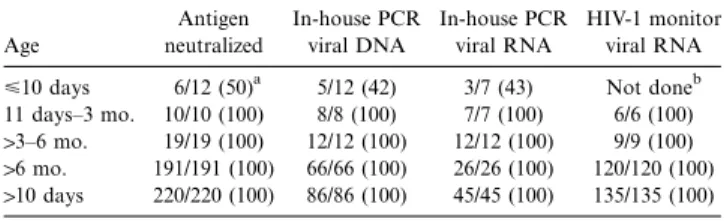
Table 1. Diagnostic sensitivity of human immunodeficiency virus type 1 (HIV-1) detection methods.
Age Antigen neutralized In-house PCR viral DNA In-house PCR viral RNA HIV-1 monitor viral RNA <10 days 6/12 (50)a 5/12 (42) 3/7 (43) Not doneb 11 days–3 mo. 10/10 (100) 8/8 (100) 7/7 (100) 6/6 (100)
13–6 mo. 19/19 (100) 12/12 (100) 12/12 (100) 9/9 (100) 16 mo. 191/191 (100) 66/66 (100) 26/26 (100) 120/120 (100) 110 days 220/220 (100) 86/86 (100) 45/45 (100) 135/135 (100)
NOTE. PCR, polymerase chain reaction.
a
No. samples positive/no. tested (%).
b
The sample that was positive in the antigen assay but negative by in-house PCR for viral DNA. Respective RNA was also negative by the ultrasensitive HIV-1 monitor version 1.5.
HIV-1 DNA or RNA. All infants who did not meet these criteria were considered uninfected, provided that they had tested negative at least twice for DNA or RNA when age12 months and/or had
seroreverted. The infection status of all infants born toward the end of 1997 was verified by further testing during the first 6 months of 1998, resulting in a clear distinction of infected and uninfected children, with no cases remaining indeterminate. The diagnostically relevant age distribution of the children is shown in table 1.
For the prospective evaluation of plasma HIV-1 load levels, 25 HIV-1–infected children followed between June 1996 and Decem-ber 1997 at the HIV outpatient clinic of the University Children’s Hospital of Zurich were enrolled after informed consent. The age of the patients at entry was 0.4–15.5 years (mean 7.9; median 9.3), and stages according to the Centers for Diseases Control classifi-cation [11] were A2 (n = 2), A3 (n = 3), B1 (n = 3), B2 (n = 4), B3 (n = 4), C2 (n = 2), and C3 (n = 7). Most patients were partici-pating in clinical trials of the Pediatric European Network for Treatment of AIDS or of the Pediatric AIDS Group of Switzerland. Blood samples were collected at time points dictated by the trial protocols, before change of treatment, or at regular 3-month intervals.
Circadian variation of plasma HIV-1 levels was studied in 5 patients after informed consent and in no relation to immuno-globulin infusions. Two children (figure 1; patients 2 and 4) were receiving zidovudine and didanosine and 1 child (figure 1; patient 1) zidovudine and zalcitabine, whereas 2 children (figure 1; patients 3 and 5) were without antiretroviral treatment. Blood specimens were drawn from implanted intravascular catheters (used for monthly immunoglobulin infusions) into vacutainer CPT tubes (Becton Dickinson, Basel, Switzerland) at 4 defined time points (at hours 7, 9, 12, and 16) on each of 2 different days. At each time point, the first 5 mL of aspirated blood were discarded, to avoid dilution bias. Plasma was harvested within 30 min and frozen at 2707C until retrospective batch testing was performed.
Tests for viral DNA or RNA. For diagnostic purposes, 1-mg
ali-quots of DNA extracted from peripheral blood mononuclear cells were prospectively tested by an in-house PCR method for HIV-1 in gag and long terminal repeat sequences, as described elsewhere [12]. All tests were done in duplicate reactions for both target regions. A positive result of DNA PCR required reactivity for both target regions, with duplicate reactivity in at least 1 of them. Qual-itative PCR for viral RNA in plasma was performed in some sam-ples that had already been tested for viral DNA. An in-house method based on detection of the same sequences as for DNA PCR was used [13]. A positive result of RNA PCR also required reac-tivity for both target regions, but each region was assessed with only a single reaction. In both methods, DNA and RNA PCR, a single DNA or cDNA copy, respectively, can be detected [9].
For real-time prospective quantification of HIV-1 RNA in plasma, an in-house procedure was not available; the Amplicor HIV monitor version 1.0 kit (Roche Molecular Systems, Basel, Switzerland), with a nominal lower quantification limit of 400 cop-ies/mL, was therefore used for testing according to the manufac-turer’s guidelines. From August to December 1997, the Amplicor HIV-1 monitor kit was used in combination with Roche’s mix-in primers to improve detection of HIV-1 subtypes A and E. No increases of RNA unparalleled by increases in p24 antigen were seen after the introduction of mix-in primers. This indicated that
subtypes A, E, or G (which were not well detected without the mix-in primers) were not present in the studied cohort and that, consequently, sequential data from samples prior to and subsequent to this modification could be merged for each child.
Viral antigen. Detection of p24 antigen in heat-denatured plasma
was done prospectively (real time) and essentially as described else-where [9]. Briefly, 100 mL plasma was diluted with 500 mL of 0.5% Triton X-100 in 1.5-mL Eppendorf tubes, denatured by heating at 1007C for 5 min on a Techne (Cambridge, UK) dry heat block, and tested in duplicate with the NEN/DuPont HIV-1 core profile ELISA in combination with the ELAST ELISA amplification sys-tem (both purchased from NEN Life Science Products, Geneva). The cutoff level for positivity in diagnostic testing was determined for each assay plate by calculating the mean absorbance of 8 HIV-1–negative controls run on the same plate plus 5 SD. For quan-tification, a cutoff corresponding to the mean plus 3 SD was used. Absorbance was read by using a Dynatech MR5000 ELISA reader (Microtech Produkte, Embrach, Switzerland). Antigen was quan-tified with a kinetic analysis by using the Quanti-Kin detection system (Tib Molbiol Srl, Genova, Italy). This permitted quantifi-cation in a range∼500–6,250,000 fg/mL with a single sample di-lution [14].
All reactive samples from children in whom infection with HIV-1 as defined above had not, or not yet, been firmly established were subjected to confirmatory antigen neutralization, by use of heat-denatured plasma and NEN/Du Pont’s confirmatory reagents and procedure.
Data analysis. Analysis of data, figure plotting, and statistics was
done employing the StatView 4.02 program for Macintosh (Abacus Concepts, Berkeley, CA). Values below the limit of detection were set arbitrarily to 100 copies/mL for RNA and 100 fg/mL for p24 antigen, respectively, both in the calculations and in the figures.
For each patient, corresponding levels of HIV-1 RNA and p24 antigen were compared for their respective changes and longitu-dinal evolution in relation to unmodified or modified ART. Re-actions of HIV-1 plasma levels determined by the 2 methods were regarded as concordant when both methods showed increase or decrease of HIV-1 plasma levels by10.5 log10and when both
meth-ods showed HIV-1 plasma levels remaining within a range of50.5 log10, respectively. The change in HIV-1 plasma levels was regarded
as similar in both methods when the changes in concentration did not differ by 10.5 log10. If the response of HIV-1 plasma levels
determined by 1 method became apparent only in blood specimens after the specimen showing a response by the other method, the
Table 2. Diagnostic specificity of p24 antigen in uninfected chil-dren of mothers positive for human immunodeficiency virus.
Age No. children
Antigen initially reactive, no. (%) Antigen neutralized positive, no. (%) <10 days 148 15 (10.1) 2 (1.4) 110 days–3 mo. 116 6 (5.2) 2 (1.7)a 13–6 mo. 68 4 (5.9) 0 16–12 mo. 106 3 (2.8) 0 112 mo. 205 12 (5.9) 1 (0.5)b All ages 643 40 (6.2) 5 (.8) a
Two samples of same infant: 27 pg at 0.5 months, 100% neutralization; 2 pg at 3 months, 72% neutralization.
b
Child was positive for p24-specific IgA antibody at birth (transplacental transfusion).
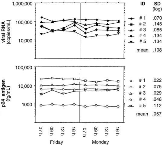
Figure 1. Precision of p24 antigen and human immunodeficiency virus type 1 (HIV-1) RNA quantification in circadian variation of HIV-1 load in 5 children.
response of HIV-1 plasma levels determined by the first method was regarded as delayed. Finally, longitudinal variation of HIV-1 plasma levels by10.5 log
10while ART was not modified was judged
to be increased variation.
Results
Diagnostic sensitivity and specificity. Diagnostic sensitivity of
p24 antigen– and PCR-based tests was determined in a total of 232 samples derived from 61 HIV-1–infected children at dif-ferent ages with no ART (table 1). Because of limited sample volumes in neonates and infants and the high cost of PCR testing, not all samples were tested by all 4 tests, but all were at least tested for p24 antigen. Above the age of 10 days, all tests were positive in all samples tested. Below 10 days, the antigen test was neutralization positive in 6 of 12 samples. DNA PCR, as well as the in-house PCR for viral RNA, missed 1 of the samples positive by antigen assay. This sample was also negative by the ultrasensitive HIV-1 monitor version 1.5. All positive RNA results were confirmed by a positive result in the subsequent sample.
Determination of diagnostic specificity of the p24 antigen
assay was based on testing a total of 643 plasma samples from 246 infants or children born to HIV-1–positive mothers con-sidered uninfected (table 2). Although reactivity in initial testing was relatively high, specificity after neutralization was 99.2%. Two of the positive results were from an infant who exhibited 27 pg/mL of 100% neutralizable antigen at 0.5 months and a decreased amount of 2 pg/mL (72% neutralizable) at 3 months, whereas PCR for viral DNA or RNA were negative in both
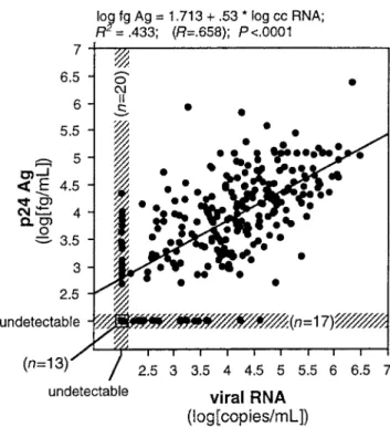
Figure 2. Correlation of p24 antigen and human immunodeficiency virus type 1 (HIV-1) RNA concentrations in 230 plasma samples from 25 HIV-1–infected children.
samples. In a subsequent sample taken at 5.6 months, antigen was also undetectable. A test for particle-associated reverse transcriptase activity, which permits detection of HIV as sen-sitively as PCR for viral RNA but is independent of genomic sequence variation [15], was negative. Despite confirmed evi-dence of transient antigenemia, this child is thus considered uninfected. One positive sample was from an uninfected child with p24-specific IgA antibody at birth [16]. The 2 other positive samples were from the neonatal period, during which p24 an-tigen of maternal origin may be transiently detectable. Among 570 samples tested for viral DNA, there were no false-positive results. Three samples (0.53%) had an indeterminate result (1 of 4 reactions positive) but were negative by the in-house method for viral RNA, and in 2 instances there was no valid result because of a technical failure. Two (1.4%) of 141 samples tested with the in-house method for viral RNA gave false-positive findings, resulting in a diagnostic specificity of 98.6%, and 2 more showed an indeterminate result. Only 5 of the 643 samples from uninfected children that were tested for antigen were tested for RNA with the HIV-1 monitor, and all were negative.
Diurnal variation of plasma HIV-1 load. Variation of plasma
HIV-1 load at different time points of the day was studied in 5 HIV-1-infected children. The results of HIV-1 quantification in blood specimens drawn at 4 defined time points on each of 2 different days are presented in figure 1. The SD for the 8 samples of a single patient ranged from 0.07 to 0.15 log10by
use of HIV-1 RNA quantification and from 0.02 to 0.11 log10
by use of p24 antigen quantification.
Correlation of p24 antigen and HIV-1 RNA copy numbers.
Comparison of real-time HIV-1 RNA and p24 antigen quan-tification in 230 blood samples from the 25 HIV-1–infected children showed detection of HIV-1 RNA in 197 (85.7%) chil-dren and of p24 antigen in 201 (87.4%). Twenty (8.7%) samples negative for HIV-1 RNA were found to be positive for p24 antigen and 16 (7.0%) samples negative for p24 antigen were positive for HIV-1 RNA. Thirteen (5.7%) samples were negative in both tests. The correlation of both tests by Spearman rank correlation and analysis of variance wasR = .658 R = .419( 2 ; ) when values below cutoff were included in the analysis
P!.001
andR = .560 R = .296 P( 2 ; !.0001) when values below cutoff were excluded (figure 2). Although the 2 assays measure dif-ferent HIV-1 products in difdif-ferent units, a surprisingly good correlation was found, as shown in figure 2.
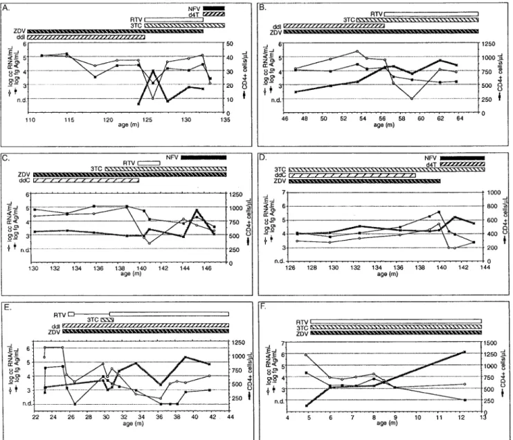
Influence of antiretroviral treatment on HIV-1 load parame-ters. On enrollment into the study, 7 patients were naive for
ART, 15 were receiving nucleoside reverse transcriptase inhib-itors (NRTIs), and 3 were being treated with NRTIs plus a protease inhibitor (PI). During the study period, ART was in-troduced or modified (addition or discontinuation of drug) in 24 patients on 43 occasions. In 13 (30.2%) of these events, an introduction or modification of ART involved only NRTIs (fig-ure 3B, 3C, 3D, 3E), in 17 (39.5%) only PIs (fig(fig-ure 3E), and
in 13 (30.2%) NRTIs plus PIs (figure 3A, 3D, 3F). The mean numbers of viral load determinations after these events (until the next event or conclusion of the study) were 2.5 (range, 1–5), 3.2 (1–7), and 3.2 (1–7), respectively. The first assessment of viral load after introduction or modification of ART showed overall concordant responses of RNA and p24 antigen in 39 (90.7%) events: 13 of 13 for NRTIs plus PIs (figure 3A, 3D, 3F), 15 (88.2%) of 17 for PIs only (figure 3E), and 11 (84.6%) of 13 for NRTIs only (figure 3B, 3C, 3D, 3E). The treatment-induced changes in concentrations were more pronounced for HIV-1 RNA in 11 (25.6%) instances and for p24 antigen in 1 (2.3%) instance (P = .002, Fisher’s exact test, 2-tailed). For ex-ample, in figure 3C, after the addition of ritonavir at month 140, the viral RNA dropped by 2 log10within half a month,
whereas the reduction in p24 antigen was minimal (from 5 to 4.75 log10). Variation of HIV-1 load, as judged by the 24
treat-ment modifications that were assessed by12 follow-up mea-surements, was more marked for HIV-1 RNA than for p24 antigen in 14 (58.3%) instances, whereas variation was more marked for p24 antigen than for RNA in only 3 (12.5%) in-stances (P = .002, Fisher’s exact test, 2-tailed). In figure 3B, for example, the addition of ritonavir induced an only modest but sustained decrease in p24 antigen, whereas RNA quickly dropped from an initial level of almost 5 log to undetectable but bounced as quickly back to 4 log. The net decrease in both markers is thus similar. Interestingly, the sustained increase of CD4 during this period correlated better with that of antigen
Figure 3. Longitudinal courses of human immunodeficiency virus type 1 (HIV-1) load markers and CD41lymphocytes and effects of anti-retroviral treatment in 6 representative children. d4T, stavudine; ddI, dideoxyinosine; ddC, dideoxycytidine; 3TC, lamivudine; NFV, nelfinavir; RTV, ritonavir; ZDV, zidovudine; n.d., not detectable. For further details, see text.
than RNA, and a similar observation could be made in the course represented in figure 3F.
Discussion
This is the first prospective study that compares HIV-1 DNA or RNA and p24 antigen with regard to their performance as a diagnostic test for pediatric HIV-1 infection and as tools for quantification of virus in chronically infected infants. The di-agnostic sensitivity of signal-amplification boosted HIV-1 p24 antigen ELISA after heat-mediated immune complex dissoci-ation was found to be as high as that of PCR detection of viral
DNA or RNA in both untreated (table 1) and treated children (figure 2). The high degree of specificity afforded by a conse-quent use of confirmatory antigen neutralization permits one, as with PCR, to clearly establish the status of HIV-1 infection in the first few weeks or months after birth. As a matter of fact, during the neonatal period, the p24 antigen was correctly positive with a sample that was still negative by PCR methods capable of detecting a single HIV-1 copy in 1 mg of leukocyte DNA or, respectively, about 50 HIV-1 RNA copies/mL of plasma. No sample from an untreated infected child seen in our laboratory during the 4-year study period was positive by PCR but negative by antigen testing. Seventy percent of the
uninfected children tested at age<6 months had perinatal zi-dovudine prophylaxis. Among them, there was 1 child who showed positive RNA but negative antigen and DNA PCR at age 2 months, whereas results at 1 week and 4 months were all negative. Regarding the prospective diagnostic specificity, the 99.2% of the antigen test after neutralization compared well with the in-house method for viral RNA (98.6%) and was al-most as good as the method for viral DNA (no false positives). A direct comparison of specificity with the commercial HIV-1 monitor kit could not be performed in this study, but the recent literature shows that this method, when used diagnostically, may apparently also yield false-positive results [17]. Thus, the p24 antigen test can be used as a screening tool, followed by neutralization tests or, if available, PCR in reactive samples. In contrast, the antigen test may also be used diagnostically for the confirmation of positive PCR results. Used in combi-nation, these 2 tests should provide an optimum of diagnostic safety. Quantification of p24 antigen proved to be as precise and sensitive as quantification of HIV-1 RNA by a commercial PCR kit with a nominal detection limit of 400 copies/mL (figure 2). Levels of p24 antigen in individual patients were correlated and longitudinally concordant with levels of HIV-1 RNA in individual patients but showed less variation than the latter (figure 3).
The precision of p24 antigen quantification was shown by a study of plasma HIV-1 loads at 4 time points each on 2 different days (figure 1). The range of diurnal variation of p24 antigen levels was similar to the range of variation seen when deter-mining HIV-1 load by measuring RNA copy numbers (0.108 vs. 0.057 log10). Kinetics of HIV-1 RNA concentrations in
pe-diatric patients have been determined in an earlier study show-ing variations in a similar log10range [18]. Notably, in our study,
diurnal variation of HIV-1 load, as determined by both meth-ods, was not significantly affected by the time of intake of antiretroviral drugs. This is a substantial observation for prac-tical reasons, in that for each individual patient blood speci-mens need not be drawn at specified time points related to drug intake to allow for comparison of longitudinally determined values.
One significant finding of this study is the good correlation of p24 antigen levels and of HIV-1 RNA copy numbers (figure 2;P!.0001) in children, regardless of whether they were re-ceiving ART. In an earlier study, we clearly showed that the use of p24 antigen detection after heat-mediated immune com-plex dissociation provides a sensitive means for early diagnosis of infection in HIV-1–exposed infants [12]. Similar results were obtained by employing the more widely used acid-dissociated p24 antigen test [19–22]. The sensitivity of antigen detection in the present study was even higher because of the addition of a signal-amplification step to the ELISA, by which the detection limit was lowered to about 0.5 pg/mL. Three samples that were negative in our earlier work [12] now tested positive (data not shown). The excellent diagnostic performance of the
amplifi-cation-boosted p24 antigen assay in vertically HIV-1–exposed children has also been reported by others who used our pro-cedure in a study of African children [23]. Recently, our group showed that heat-mediated immune complex dissociation fol-lowed by this signal-amplification boosted ELISA detects p24 antigen in plasma of HIV-1–infected adults with the same sen-sitivity as does PCR [24]. In a subsequent retrospective analysis conducted in HIV-infected adults, this p24 antigen detection procedure proved to perform comparably to RNA PCR in monitoring the efficacy of ART [9].
An important fact regarding patient monitoring is that, on introduction or modification of ART, the vast majority of re-sponses in the p24 antigen load were concordant with the HIV-1 RNA load responses. Although the reduction of HIV-HIV-1 RNA could be more pronounced and prompt than the reduction of p24 antigen levels, the opposite was also observed. However, p24 antigen levels showed significantly less variation than HIV-1 RNA. This might be caused by p24 antigen’s being more refractory than RNA to intercurrent events, resulting in pro-virus activation—for example, immunizations or intercurrent illnesses—but chart review disclosed no such events (data not shown). Thus, longitudinal quantification of p24 antigen after introduction of highly active ART including PIs provides cli-nicians with information of sufficient accuracy and of less con-fusing variability than quantification of HIV-1 RNA. This is of clinical impact because the recommendations for modifica-tion of ART on the basis of changes of plasma HIV-1 load and the frequency of plasma HIV-1 load assessment after such mod-ification are not yet clear.
In conclusion, quantification of plasma HIV-1 load by signal-amplification boosted ELISA detection of p24 antigen after heat-mediated immune complex dissociation offers a highly precise alternative to RNA PCR for monitoring HIV-1 disease evolution and its response to ART. The considerably less ex-pensive p24 antigen detection (in Switzerland 35 Swiss francs [∼$23 US]/test are charged for screening and 50 Swiss francs for a neutralization or a quantification test) render this tool affordable to a larger community of patients than does RNA PCR (200 Swiss francs/test for a qualitative and 275 Swiss francs for a quantitative test).
Acknowledgment
We thank Antonietta Baumgartner, Soksimon Kaing, Sonja Kreiner, and Lucia Porong for excellent technical assistance.
References
1. Haase AT, Henry K, Zupancic M, et al. Quantitative image analysis of HIV-1 infection in lymphoid tissue. Science HIV-1996; 274:985–9.
2. Mellors JW, Rinaldo CR, Gupta P, White RM, Todd JA, Kingsley LA. Prognosis of HIV-1 infection predicted by the quantity of virus in plasma. Science 1996; 272:1167–70.
in infants infected with the human immunodeficiency virus type 1. N Engl J Med 1997; 336:1337–42.
4. Coombs RW, Welles SL, Hooper C, et al. Association of plasma human immunodeficiency virus type 1 RNA level with risk of clinical progression in patients with advanced infection. J Infect Dis 1996; 174:704–12. 5. Carpenter CCJ, Fischl MA, Hammer SC, et al. Antiretroviral therapy for
HIV infection in 1998. JAMA 1998; 280:78–86.
6. Gazzard B, Moyle G. 1998 revision of the British HIV Association guidelines for antiretroviral treatment of HIV seropositive individuals. Lancet 1998; 352:314–6.
7. Revets H, Marissens D, de Wit S, et al. Comparative evaluation of NASBA HIV-1 RNA QT, Amplicor-HIV Monitor, and QUANTIPLEX HIV RNA assay, three methods for quantification of human immunodeficiency virus type 1 RNA in plasma. J Clin Microbiol 1996; 34:1058–64.
8. Bush CE, Donovan RM, Manzor O, et al. Comparison of HIV typ1 1 RNA plasma viremia, p24 antigenemia, and unintegrated DNA as viral load markers in pediatric patients. AIDS Research and Human Retroviruses 1996; 12:11–5.
9. Bo¨ni J, Opravil M, Tomasik Z, et al. Simple monitoring of antiretroviral therapy with a single-amplification-boosted HIV-1 p24 antigen assay with heat-denatured plasma. AIDS 1997; 11:F47–F52.
10. Kind C, Rudin C, Siegrist C-A, et al. Prevention of vertical HIV transmission: additive protective effect of elective Cesarean section and zidovudine pro-phylaxis. AIDS 1998; 12:205–10.
11. Centers for Disease Control and Prevention. 1994 revised classification system for human immunodeficiency virus infection in children less than 13 years of age. Morb Mortal Wkly Rep MMWR1994; 43(RR-12):1–10. 12. Schu¨pbach J, Bo¨ni J, Tomasik Z, et al. Sensitive detection and early
prog-nostic significance of p24 antigen in heat-denatured plasma of human immunodeficiency virus type 1-infected infants. J Infect Dis 1994; 170: 318–24.
13. Bo¨ni J. Detection of HIV by PCR. In: Clapp J, ed. Methods in Molecular Biology. Vol. 50. Totowa, NJ, USA: Humana, 1995:93–107.
14. Giacomini M, McDermott JL, Giri AA, Martini I, Lillo FB, Varnier OE. A novel and innovative quantitative kinetic software for virological color-imetric assays. J Virol Methods 1998; 73:201–9.
15. Bo¨ni J, Pyra H, Schu¨pbach J. Sensitive detection and quantification of
par-ticle-associated reverse transcriptase in plasma of HIV-1 infected individ-uals by the product-enhanced reverse transcriptase assay. J Med Virol 1996; 49:23–8.
16. Schu¨pbach J, Tomasik Z, Jendis J, et al. IgG, IgM, and IgA response to HIV in infants born to HIV-1 infected mothers. J Acquir Immune Defic Syndr Hum Retrovirol 1994; 7:421–7.
17. Cunningham CK, Charbonneau TT, Song K, et al. Comparison of human immunodeficiency virus 1 DNA polymerase chain reaction and qualitative and quantitative RNA polymerase chain reaction in human immunode-ficiency 1-exposed infants. Pediatr Infect Dis J 1999; 18:30–5. 18. Zeichner SL, Mueller BU, Pizzo PA, Dimitrov DS. Kinetics of HIV-1 RNA
concentration changes in pediatric patients. Pathobiology 1996; 64:289–94. 19. Rich KC, Janda W, Kalish LA, et al. Immune complex-dissociated p24 an-tigen in congenital or perinatal HIV infection: role in the diagnosis and assessment of risk of infection in infants. J Acquir Immune Defic Syndr Hum Retrovirol1997; 15:198–203.
20. Miles SA, Balden E, Magpantay L, et al. Rapid serologic testing with im-mune-complex-dissociated HIV p24 antigen for early detection of HIV infection in neonates. N Engl J Med 1993; 328:297–302.
21. Chandwani S, Moore T, Kaul A, Krasinski K, Borkowsky W. Early diagnosis of human immunodeficiency virus type 1-infected infants by plasma p24 antigen assay after immune complex dissociation. Pediatr Infect Dis J 1993; 12:96–7.
22. Quinn TC, Kline R, Moss MW, Livingston RA, Hutton N. Acid dissociation of immune complexes improves diagnostic utility of p24 antigen detection in perinatally acquired human immunodeficiency virus infection. J Infect Dis 1993; 167:1193–6.
23. Lyamuya E, Bredberg-Raden U, Massawe A, et al. Performance of a modified HIV-1 p24 antigen assay for early diagnosis of HIV-1 infection in infants and prediction of mother-to-infant transmission of HIV-1 in Dar es Sa-laam, Tanzania. J Acquir Immune Defic Syndr Hum Retrovirol 1996; 12: 421–6.
24. Schu¨pbach J, Flepp M, Pontelli D, Tomasik Z, Lu¨thy R, Bo¨ni J. Heat-mediated immune complex dissociation and ELISA signal amplification render antigen p 24 detection in plasma as sensitive as HIV-1 RNA de-tection by polymerase chain reaction. AIDS 1996; 10:1085–90.