Instruments atid Techniques
Production of Chronic Atrioventricular
in Dogs Without Thoracotomy
Block
MARKO
TURINA*,ISTVAN
BABOTAI,
and
WERNERWEGMANN
From Surgical Clinic A and the Institute of Pathology, University of Zurich, Switzerland.
AUTHORS’
SYNOPSIS. Total heart block was produced in dogs without thoracotomy by means of a formaldehyde injection into the bundle of His. The method was successful in 22 out of 24 experiments.It
is
a useful model for haemodynamic studies in the intact animal. Experimental atrioventricular (AV) block in dogs isa versatile research model and numerous methods for its production have been devised. With only two exceptions (Williams and Lambert, 1964; Fisher, Lee, Christianson, and Kavaler, 1966) all these methods use thoracotomy, with or without atriotomy, as the operative approach. Thoraco- tomy, however, produces important cardiovascular changes, for left ventricular volume and cardiac output decline, and heart rate, pulmonary vascular resistance, and ventricular ejection fraction all rise upon opening the chest ((Rushmere, Finlayson, and Nash, 1954; Fermoso, Richardson, and Guy- ton, 1964; Coles, Buttigliero, and Gergely, 1965). All these facts make exact cardiological evaluation of acute experimental AV block difficult, since immediate and late effects of thoracotomy become superimposed on the primary change in cardiac dynamics.
These observations have prompted us to develop
a new method of block production in dogs. This method does not require thoracotomy, produces only a small myocardial lesion, and results in a
permanent AV block. In this paper, which re- presents a further extension of our first report about experimental AV block (Turina and Babotai, 1967), we describe percutaneous production of
chronic AV block in dogs. Stability of the block is confirmed by long-term observations, and the histological changes in the acute and chronic phases of experimental heart block are reported.
Received January 25, 1968.
* Present address: Medical Policlinic of the University,
Kantonsspital, 8006 Ziirich, Switzerland.
Methods
Twenty-two mongrel dogs, weighing between 17 and
33 kg were used. In 11 dogs, the block was produced in an acute experiment under non-sterile conditions. Thirteen dogs were operated upon under sterile condi- tions and allowed to recover; duration of the block and long-term histological changes were studied in these animals.
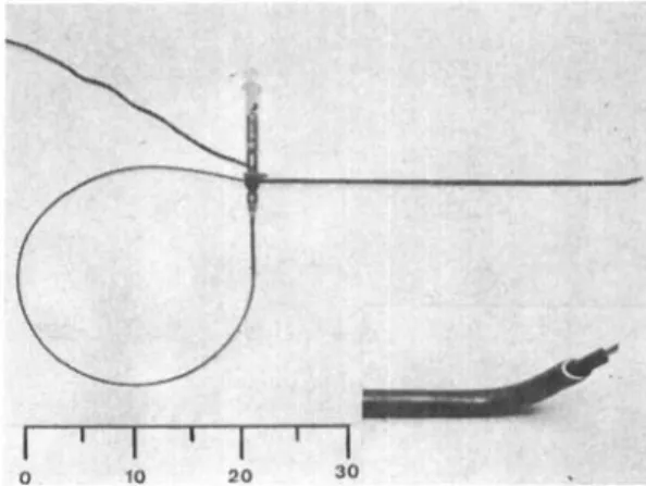
Anaesthesia was induced with sodium pentobarbital (Pentothal) 1&15 mg/kg. Each animal was placed on its left side with the neck slightly elevated, and the right external jugular vein was exposed through a small in- cision. A stainless-steel cannula, 35 cm long (Fig. 1)
was introduced into the right atrium under fluoroscopic guidance, until its tip rested slightly superior to the septa1 tricuspid leaflet, anterior to the coronary sinus.
Fig. 1. Block cannula with the needle catheter inserted; formaldehyde syringe and ECG connection are attached to
the cannula. Insert: cannula tip with the needle catheter urotrudina. Scale: metric.
390
Turina, Babotai, and Wegmann
Electric insulation of the cannula was achieved by cover- ing it with a non-conductive paint. Only the tip of the cannula was left bare of insulation, acting as an ex- ploring electrode to record potentials from the nodal region. The cannula was connected to the precordial lead of a standard ECG recorder (Mingograf 81 by Elema Schonander) ; the extremity leads were recorded simultaneously and displayed on the oscilloscope screen and on a paper strip, together with intra-atrial electro- cardiogram (atriogram). A No. 7 catheter with a needle mounted on its tip (see insert, Fig. 1) was filled with
40% formaldehyde solution and threaded through the cannula until the nedle reached the end of the cannula. Under fluoroscopic guidance and continuous registra- tion of atriograms, characteristic potentials from the bundle of His were looked for with small movements of the cannula tip. According to Pruitt and Essex (1960) and to Hoffman, Moore, Stuckey, and Cranefield (1963) these potentials consist of a fast, predominantly negative deflection of the QRS complex (Fig. 2). Upon locating the bundle, the needle-tipped catheter was advanced through the cannula until the needle became firmly embedded in the myocardium. Under continuous ECG registration 0.2-0.3 ml. formaldehyde was injected into the myocardium at this site. Within a few seconds, sometimes after a burst of ventricular or supraventricular extrasystoles, a total AV block appeared (Fig. 3).
In acute experiments, animals were kept under ob- servation for 6-12 hr after a stable AV block had been produced; after this time they were killed. The follow- up period in long-term experiments ranged between eight and 64 days. Recordings were made in awake animals every two weeks after the block production. Extremity and precordial leads were analysed for heart rate, conduction disturbances, and for origin of the ventricular impulse. A QRS complex wider than 0.08 sec was usually associated with a heart rate of less than 50/min; in precordial leads the ventricular activation time was d,elayed and bundle branch block pattern was recorded over one of the ventricles. This was con- sidered as conclusive of infra-bifurcational rhythm. On the other hand, a normal QRS complex, heart rate of more than 50/min, and a normal ventricular activa- tion time were diagnosed as supra-bifurcational rhythm (Wyss, Holzmann and Schaub 1961).
'
Results
Our overall results of block production are summarized in Table 1. There was one failure o u t of 11 acute experiments a n d one failure out of
13 long-term experiments. In both of these animals no identification of the bundle was possible. One animal was lost 10 min after the block had been produced : intra-atrial manipulation of the cannula gave rise t o a ventricular tachycardia, which progressed t o fibrillation within a few sec- onds. In about half of all the experiments only
the block. In the other half a second injection was
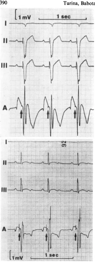
Fig. 2. Bundle potentials, as recorded with the cannula.
I, 11, III=extremity leads; A=atriogram.
Fig. 3. Percutaneous block production in continuous ECG recording. In first seven P waves atriograms show bundle spikes; after formaldehyde injection a 2: I block appears, which after three ventricular extrasystoles progresses to total AV block. Symbols as in Fig. 2.
needed; this happened if the needle was only loosely embedded and the first injection was not properly deposited into the myocardium.
Table 1. Results of A V Block Production
Acute experiments Chronic experiments (no. of animals) (no. of animals) Successful 10
Unsuccessful 1 12 1
In chronic AV block the heart rate varied between 37 and S3/min; nine animals showed an infra- bifurcational and three a supra-bifurcational rhythm pattern. Six dogs were observed for one to two weeks and another six between 50 and 64 days. The total heart block was permanent; none of the animals reverted to sinus rhythm during the observed period.
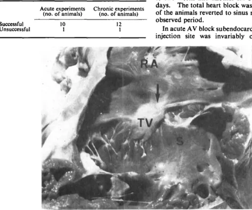
In acute AV block subendocardial bleeding at the injection site was invariably observed. Micro-
Fig. 4. Gross appearance of the heart with an AV block of two months’ duration. R A = right atrium; TV=septal tricuspid leaflet, S=septal portion of the right ventricle. Arrow denotes the site of injection.
392
Turina, Babotai, and Wegmann
scopically an
‘‘
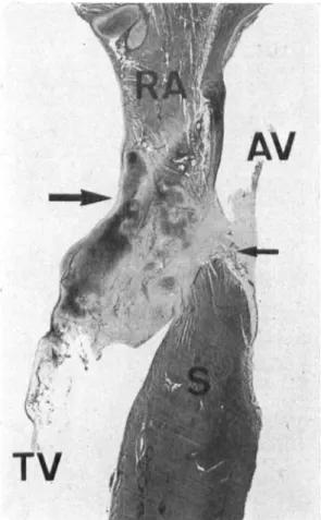
in-uivo formaldehyde fixation” could be noted, consisting of interstitial bleeding and oedema, cellular shrinking, and thrombosis of blood vessels. Within a few days parietal throm- bosis of the atrial wall over the injection site developed, and histiocyte-rich granulation tissue appeared on the edges of formaldehyde-impreg- nated area. Eight to nine weeks after the block production a fibrotic scarring in the region of the bundle was noted (Fig. 4); this reaction sometimes proceeded to the production of cartilage. The bundle of His showed different degrees of fibrosis in all animals. Extension of formaldehyde im- pregnation varied widely: in eight dogs, only apea-sized area was affected; the bundle of His was destroyed, but the AV node and both bundle branches were intact (Fig. 5).
In
five animals aFig. 5. Cut through the AV border of a dog heart with total AV block, two months after the block production. A pea- sized granuloma with cartilaginous transformation is found on the injection site. The base of the septal tricuspid leaflet (TV) is thickened. Bundle of His lies in the granuloma, the left bundle branch (small arrow) can be recognized on the left
more widespread necrosis and fibrosis was found, due to diffusion of formaldehyde into the surround- ing structures. In these dogs fibrotic changes were noted in right atrial wall, AV node, the whole of the bundle of His and proximal parts of both bundle branches, aortic ring, septal tricuspid leaflet, and the uppermost portion of the ventricular septum.
Discussion
Williams and Lambert (1964) were the first to develop a method for AV block production without thoracotomy. They identified AV nodal deflection by means of intracardiac potentials; injection of formaldehyde or procaine was used to block im- pulse conduction. No details of the procedure or
the percentage of successful block productions were given. Fisher et al. (1966) produced the block by means of formaldehyde injection in the region where the bundle of His was presumed to be. These authors used only fluoroscopic guidance for their orientation and their success rate was about 50%. Brutsaert (1966) tried electrocoagulation of the bundle of His via a catheter-tip electrode, but he was also hampered by the lack of identification of the bundle of His. Cruze and Schiebler (1963) devised a two-stage procedure, which eliminated the acute, but not chronic changes due to thora- cotomy ; their method is, however, too complicated for routine laboratory work.
With our method, block production was accom- plished in a very short time and animals recovered rapidly. Widespread fibrotic changes found in five dogs indicate, nevertheless, that the utmost care must be exercised in injecting only the smallest amount of formaldehyde needed; this will limit the necrosis to the bundle of His only. Three possible complications can arise from an excessive formaldehyde injection, namely tricuspid and aortic regurgitation, due to fibrosis, and pulmonary embolism from the atrial wall thrombosis. This last complication was never encountered ; in our
present haemodynamic studies we could not detect any aortic incompetence either.
Deliberate production of an AV block in humans, followed by a pacemaker implantation, has been reported several times in the last few years (Char- dack, 1964; Gianelli, Ayres, Gomprecht, Conklin, and Kennedy, 1967 ; Slama, Blondeau, Aigueperse, Cachera, Degorges, and Abbou, 1967). It was used as a last, admittedly heroic, measure to con- trol an otherwise refractory supraventricular tachycardia. In all these operations open-heart surgery had to be done, and percutaneous block
- .
of the
ventricular
septum (s). Bigger-arrow: injection production could have achieved the same effect in amuch simpler way. If a local anaesthetic agent
Production of Chronic Atrioventricular Block in Dogs
like procaine be used, only a temporary AV block results (Williams and Lambert, 1964), and a
short-term control in selected cases of supraven- tricular tachycardia can thus be envisioned.
Summary
Production of an atrioventricular block in dogs without thoracotomy is based on electrocardio- graphic identification of the bundle of His and its destruction with a small amount of formaldehyde.
A long steel cannula, serving as exploring electrode, is used for identification; it is introduced into the right atrium via the right external jugular vein. When the cannula tip records characteristic bundle potentials, a needle-tipped catheter is pushed through the cannula and advanced into the myo- cardium. Through this catheter 0.2-0.3 ml. 40% formaldehyde is injected, whereupon total heart block appears. The method was successful in 22 out of 24 experiments. This block is permanent; none of the surviving animals reverted to sinus rhythm even after two months of observation. In chronic experiments, endocardial thickening and fibrosis appear in the bundle region; extension of fibrosis varies widely, according to the amount of formaldehyde injected. In eight dogs only a pea-sized granuloma was found in the place where injection had been made; the other five animals showed localized necrosis and a certain degree of fibrosis in the surrounding structures. The utmost care has to be exercised in injecting only the small- est amount of formaldehyde needed; this limits de- struction to the bundle of His only.
References
Brutsaert, D. L. (1966). Comparison of singled and paired electrical heart stimulation on cardiac hemodynamics
a t rest and during exercise in dogs with atrioventricular heart block. Arch. int. Physiol., 14, 9-20.
Chardack, W. M. (1964). Discussion, cardiac pacemakers (part). Ann. N. Y. Acad. Sci., 111, 1109.
Coles, J. C., Buttigliero, J. R., and Gergely, N. F. (1965).
Pulmonary vascular resistance following thoracotomy.
Arch. Surg., 91, 55-57.
Cruze, K., and Schiebler, G. L. (1963). Production of com- plete atrio-ventricular dissociation. A new experi- mental technique. Arch. Surg., 86, 331-333.
Fermoso, J. D., Richardson, T. Q., and Guyton, A. C. (1964).
Mechanism of decrease in cardiac output caused by opening the chest. Amer. J. Physiol., 207, I 1 12-1 116.
Fisher, V. J., Lee, R. J., Christianson, L. C., and Kavaler, F.
(1966). Production of chronic atrioventricular block in dogs without thoracotomy. J. appl. Physiol., 21, 1 1 19-1 12 I .
Giannelli, S . , Ayres, S. M., Gomprecht, R. F., Conklin, E. F. and Kennedy, R. J. (1967). Therapeutic surgical division of the human conduction system. J . Amer.
med. Ass., 199, 155-160.
Hoffman, B. F., Moore, E. N., Stuckey, J. H., and Cranefield, P. F. (1963). Functional properties of the atrio- ventricular conduction system. Circular. Res., 13,
Potential changes attending the excitation process in the atrioventricular conduction system of bovine and canine hearts.
Circulat. Res., 8, 149-114.
Rushmere, R. F., Finlayson, B. L., and Nash, A. A. (1954).
Shrinkage of the heart in anesthetized, thoracotomized dogs. Circulat. Res., 2, 22-27.
Slama, R., Blondeau, Ph., Aigueperse, J., Cachera, J., Degorges, M., and Abbou, E. (1967). 'Creation chirurgicale d'un bloc auriculoventriculaire et implanta- tion d'un stimulateur dans deux cas de troubles d u rhythme irreductibles.
Turina, M., and Babotai, I. (1967). Erzeugung eines totalen atrio-ventrikularen Blocks beim Hund ohne Thora- kotomie. Thoraxchirurgie, 15, 601-608.
Williams, J. C. P., and Lambert, E. H . (1964). Production of heart block in dogs without thoracotomy. Fed.
Proc., 23, 4 13.
Wyss, S., Holzmann, M., and Schaub, F. (1961). Der totale Atrioventrikular-Block (Av-Block). Klinische und elektrokardiographische Beobachtungen bei 90 Fallen.
Arch. Kreis1.-Forsch., 36, 148. 308-328.
Pruitt, R. D., and Essex H. E. (1960).