Characterization and Improvement of the Clinical Assessment of
Vocal Hyperfunction
by -MASSACHUSE3TS INS E
by
OF TECHNOLOGYCara Elizabeth Stepp O "
OCT 0
2
2009
S.B. Engineering Science
Smith College, 2004
LIBRARIES
S.M. Electrical Engineering and Computer Science Massachusetts Institute of Technology, 2008
SUBMITTED TO
THE HARVARD-MIT DIVISION OF HEALTH SCIENCES AND TECHNOLOGY IN PARTIAL FULFILLMENT OF THE REQUIREMENTS FOR THE DEGREE OF
DOCTOR OF PHILOSOPHY IN BIOMEDICAL ENGINEERING AT THE
MASSACHUSETTS INSTITUTE OF TECHNOLOGY SEPTEMBER 2009
ARCHVES
©2009 Massachusetts Institute of Technology All rights reserved.
Signature of Author ... . .. ...
Harvard-MIT Division of Health Scidacesa Technology July 29, 2009
Certified by ... ..
James T. Heaton, Ph.D. Assistant Professor, Harvard Medical School Thesis Supervisor
Accepted by ...
/rS ,tosisekharan, Ph.D. Director, Harvard-MIT Division of Hea Sciences and Technology Edward Hood Taplin Professor of Health Sciences and Technology and Biological Engineering
Characterization and Improvement of the Clinical Assessment of
Vocal Hyperfunction
by
Cara Elizabeth Stepp
Submitted to the Division of Health Sciences and Technology on July 31, 2009
in Partial Fulfillment of the Requirements
for the Degree of Doctor of Philosophy in Biomedical Engineering
ABSTRACT
Vocal hyperfunction refers to "conditions of abuse and/or misuse of the vocal mechanism due to excessive and/or 'imbalanced' muscular forces" (Hillman, Holmberg, Perkell, Walsh, & Vaughan, 1989), characterized by excessive laryngeal and paralaryngeal tension (Aronson, 1980; M. D. Morrison, Rammage, Belisle, Pullan, & Nichol, 1983; N. Roy, Ford, & Bless, 1996). There is no widely accepted diagnostic measure of the presence and degree of vocal hyperfunction, and currently, assessment during diagnosis is often primarily based on subjective impressions given the patient's history and
presentation of symptoms such as auditory-perceptual and visual or tactile discrimination of muscle tension (e.g., laryngeal palpation). Clinical care is hindered by the lack of a "gold standard" objective measure for the assessment of vocal hyperfunction.
The first study in this thesis evaluated a novel experimental design for the study of vocal hyperfunction, making use of the established clinical procedure of injection
laryngoplasty. This work found that the use of injection laryngoplasty as a platform for the study of some types of vocal hyperfunction is limited, but may offer a convenient opportunity to study selected associated parameters. Particular promising objective measures were investigated in the remaining four studies: kinematics of the vocal folds, root-mean-squared (RMS) measures of surface electromyography (sEMG), and spectral characteristics of sEMG. Kinematic features of vocal fold abduction and adduction were shown to discriminate between individuals with muscle tension dysphonia and controls. RMS measures of sEMG were investigated through correlation with current clinical neck palpation techniques in voice therapy patients and via a cross-sectional study of
individuals with vocal fold nodules. Correlations between RMS neck sEMG and palpation ratings were low, and although some individuals with nodules displayed RMS neck sEMG patterns that were inconsistent with those seen in controls, overall the RMS measures were unable to discriminate between disordered and control groups. Mean coherence between two neck sEMG locations in individuals with vocal nodules was significantly lower in the 15 - 35 Hz band relative to controls, possibly agreeing with past subjective accounts of "imbalanced" muscle activity.
Thesis Supervisor: James T. Heaton, Ph.D.
Acknowledgments
First, James T. Heaton, for being the best advisor imaginable - smart, caring, and fun. Thanks for pushing me when I needed to be pushed, backing off when I needed to be left alone, and listening to me when you could have been doing something more important. Dr. Hillman, thank you for the opportunity to work at the Voice Center, for all of your advice on my research, and also my professional growth and career. Your guidance was hugely helpful. Thanks to my committee members Dr. Roy and Dr. Howe for graciously agreeing to be a part of this. I hope you know how much I appreciate your time and expertise. The assistance of Drs. Joseph Perkell, Jim Kobler, Steve Massaquoi, and Neville Hogan was also essential to this endeavor, and I am very grateful for it. Thanks to all of the Voice Center staff for the constant support, including Dr. Zeitels, Meredith, Marie, Tara, Harold, Kathryn, Selena, Ashley, and Matt. In particular, Dr. Bums and Maia really went above and beyond to find me subjects, Jennifer made
everything easier, and Janet had the best advice. Thanks to the voice center grad students, especially Asako, Daryush, and Prakash (my guardian tech angel). Every day was easier because of you. Asako, consider this thesis mention as a replacement for the one you should have received. You are absolutely loved and appreciated by me. And much thanks to my two favorite professors, Yoshi and Gerardo. Yoshi, thank you for many morning meetings - I hope you give this thesis an A+. Gerardo, thanks for being there. I like you.
To my dear friends, thanks for many nights across the street: Melanie and Mike, Hannah, Maia and Jason, and KC. Mel, thanks for taking the time to listen to me blow off steam no matter how busy you are! KC, how could I get along without my career guide and my go-to (not back-up) date? Ann, Naomi, Adam, Ronit, and Bennett, thanks for all the fun times. The 2004 SHBT class, in order of their attendance at class outings, with (#) indicating their responsiveness during email discussions: wendy(6), nancy(5), erik(2), thomas(7), ted(4), tom(l), manny(3). We had great times, and I'm thankful for all of you. Tom especially, thanks for being 'my person' when I needed it.
My greatest thanks go to my family. My Mom and Dad have always given me completely
unconditional love and support, and the expectation that I should do my best. Mom, you are inspirational - you are such a smart, giving woman. I can only aspire to become what you are. Dad, thanks for the permission to slow down sometimes. You have no idea how much it helps to have someone telling you that you are already great, and that it's ok to take a break. Brandon, thanks for always being there, being the brother I didn't have, forgetting my birthday, and always ringing the doorbell. There's no one I'd rather eat cheese with. To all of my extended family, thanks for all of the love and support and card games. It means more than you know.
Contents
A bstract ...
3
Acknowledgments ...
5
Contents
... 7
Thesis Organization ...
11
Chapter 1: Background ...
13
Voice Production ... 9Relevant Anatomy and Physiology. ... 10
Role of Extrinsic Laryngeal Musculature ... ... 11
Voice Disorders ... .. ... ... 10
Clinical Assessment of Vocal Function ... ... 11
Vocal Hyperfunction ... 16
Symptoms / Current State of Assessment ... ... .. 10
Organic Developments and Interactions ... 11
Treatment Options ... ... 11
Surface electromyography ... 16
Theory.. ... 10
Recording and Analysis Recommendations ... . ... 10
M otivation ... 16
Chapter 2: Use of Injection Laryngoplasty as a Platform for the
study of Objective Measures of Vocal Hyperfunction ... 9
Introduction ... 9
M ethod ... 10
Results... ... 11
Chapter 3: A Virtual Trajectory Model Predicts Differences in
Vocal Fold Kinematics in Individuals with Vocal Hyperfunction
..
9
Introduction... 10
Vocal Hyperfunction... 10
Vocal Fold Kinematics ... ... ... 11
Modeling Vocal Fold Abduction and Adduction ... 11
Model Formulation ... ... 11
Modeling Results and Predictions ... ... 11
Vocal fold kinematics in individuals with vocal hyperfunction ... 12
Experimental Methods ... ... ... 12
Experimental Results ... ... ... 13
D iscussion ... 13
Average Abduction and Adduction Velocities of Individuals with Healthy Normal Voice .. 10
Experimental Confirmation of the Modeling Hypothesis in Individuals with MTD ... 11
Vocal Fold Kinematics in Individuals with Nodules ... 10
Use of Vocal Fold Kinematics as a Measure of Vocal Hyperfunction ... 11
Summary and Future Work ... ... ... 10
Chapter 4: Comparison of Neck Palpation Rating Systems with
Surface Electromyographic and Acoustic Measures in Vocal
Hyperfunction ...
9
Introduction...10
M ethods ... 11
Participants ... ... 11
Clinical Palpation Methodology ... ... ... ... 11
sEMG and Acoustic Recording Methodology ... 11
Data Analysis ... 11
Results ... . 12
Inter-rater Reliability ... ... 12
Correlation between Palpation Ratings and sEMG ... ... 13
Relationship between Perceived Laryngeal Height and the third formant. ... 13
D iscussion ... 13
Inter-rater Reliability ... 12
Correlations between Palpation Ratings and Objective Measures ... ... 13
Issues with Respect to Clinical Adoption of Palpation Rating Scales ... 13
Chapter
5:
Acoustic, Aerodynamic, and Electromyographic
Characteristics of Phonatory Behaviors in Individuals with
Vocal Fold Nodules ...
9
Introduction ... 10 Method ... 11 Participants ... ... . ... ... 11 Recording Procedure ... ... 11 Data Analysis ... ... 11 Results ... 12
Mean Anterior Neck sEMG and Acoustic Rise Time in All Participants ... 12
Airflow, Vocal Efficiency, and measures of Nodule Size and Location in Singers and Non-Singers with Vocal Nodules ... 13
Correlations between measures in Singers and Non-Singers with Vocal Nodules ... 12
D iscussion ... 13
Anterior Neck sEMG and Acoustic Rise Time do Not Differentiate Individuals with Nodules from those with Healthy Normal Voice ... 12
Differences between Singers and Non-Singers with Nodules ... 13
Use of objective Measures in Clinical Assessment of Vocal Hyperfunction ... 12
C onclusions ... 13
Chapter 6: Use of Neck Strap Muscle Intermuscular Coherence as an
Indicator of Vocal Hyperfunction ...
9
Introduction ... 10 Methods ... 11 Participants ... .... ... ... 11 Tasks... . ... ... ... ... 11 Analysis ... 11 R esults ... 12 D iscussion ... 13
Bilateral EMG-EMG Beta Band Coherence is Reduced in Individuals with Nodules Relative to Healthy Controls ... 11
Speech Type has No Effect on Bilateral EMG-EMG Beta Band Coherence ... 11
Summary and Indications for Future Work ... 11
Sum m ary ...
9
Thesis organization
This thesis contains five studies with a common underlying goal of improving the clinical assessment of vocal hyperfunction. The thesis is organized as five self-contained
manuscripts (Chapters 2 - 6), preceded by a common foreword (Chapter 1). Chapter 1
provides the motivation for the current studies and acts as a general primer for
understanding the work performed for naYve readers. More familiar readers may prefer to skip this section and begin with Chapter 2.
Each of the five manuscripts (Chapters 2 - 6) is written in preparation for publication in a range of different journals. Given the varied audiences of the associated journals, the chapters vary in the amount and type of background information provided. In some manuscript chapters there is significant overlap with the information provided in the common background, whereas in others there is not.
Specifically:
Chapter 2: C.E. Stepp, J.T. Heaton, M.E. Jett6, J.A. Burns, R.E. Hillman. "Use of injection laryngoplasty as a platform for the study of objective measures of vocal hyperfunction," to be submitted to the Journal of Speech Language and Hearing Research.
Chapter 3: C.E. Stepp, R.E. Hillman, J.T. Heaton. "A virtual trajectory model predicts differences in vocal fold kinematics in individuals with vocal hyperfunction," submitted to the Journal of the Acoustical Society of America (JASA).
Chapter 4: C.E., Stepp, J.T. Heaton, m.N. Braden, M.E. JettY, T.K. Stadelman-Cohen, R.E. Hillman. "Comparison of neck palpation rating systems with objective measures," submitted to the Journal of Voice.
Chapter 5: C.E. Stepp, J.T. Heaton, T.K. Stadelman-Cohen, M.N. Braden, M.E. Jett6,
R.E. Hillman. "Acoustic, aerodynamic, and electromyographic characteristics of phonatory behaviors in individuals with vocal fold nodules," to be submitted to the Journal of Speech Language and Hearing Research.
Chapter 6: C.E. Stepp, R.E. Hillman, J.T. Heaton. "Use of Neck Strap Muscle
Intermuscular Coherence as an Indicator of Vocal Hyperfunction," submitted to the IEEE Transactions in Neural Engineering and Rehabilitation Engineering.
Voice Production
The classic theory of speech production is the source-filter model (Fant, 1960; further described by Stevens, 2000). This model separates functionally and physically the source, or vocal stimulus, from the filter, which consists of the articulatory apparatus. The source, then, of most human speech is created through use of the larynx.
RELEVANT ANATOMY AND PHYSIOLOGY
The larynx is a system of suspended cartilages, lined with folds, acting as a valve between the airway and the pharynx (see Figure 1-1). Nearby muscles can alter the position and shape of these folds. The area of the larynx at the level of the vocal folds is referred to as the glottis. The muscles of the larynx can be divided into two main groups: intrinsic and extrinsic muscles. While intrinsic laryngeal muscles connect different parts of the larynx to each other, the extrinsic laryngeal muscles (also referred to as strap muscles) connect the larynx to outside structures (Fink & Demarest, 1977).
The larynx transforms airflow from the lungs into a series of air puffs which constitute the voice, and thus the source for the articulatory filters of the upper airway. Airflow from the lungs drives through the vocal folds, causing them to open; the folds are then pulled back together due to Bernoulli forces and the elastic properties of the folds, cutting off the airflow and creating an air puff. These forces may be manipulated to change the characteristics of phonation. Forces created by airflow from the lungs may be
manipulated by using higher driving subglottal pressures, whereas the vocal fold tension and length characteristics are primarily controlled by the intrinsic laryngeal muscles. In addition, the intrinsic laryngeal muscles are also the primary actors in vocal fold
adduction, thereby positioning the vocal folds to allow generation of voice. The extrinsic laryngeal muscles also have a role in vocal control, including a part in directing
abduction, adduction, and pitch changes (Erickson, Baer, & Harris, 1981; Hirano, Koike, & von Leden, 1967; Hong, Ye, Kim, Kevorkian, & Berke, 1997; Konrad, et al., 1984; Roubeau, Chevrie-Muller, & Lacau Saint Guily, 1997), although their specific
mechanical action in vocal control is not well understood.
The human vocal fold (informally referred to as the vocal cord) has a complex structure that allows it to perform not only its respiratory function, but also that as the primary phonatory instrument. The vocal fold has a number of layers which become less pliable as you move deep to the surface of the fold (Carole T. Ferrand, 2001). The topmost layer of the vocal fold is a thin epithelium, under which lies a basement membrane (Carole T. Ferrand, 2001; Hirano, 1988). Underneath these outermost layers is the lamina propria. The lamina propria can be divided into three layers: the superficial layer (Reinke's space), the intermediate layer, and the deep layer. Reinke's space is quite pliable and plainly moves during vocal fold vibration. The intermediate layer is somewhat less
pliable, although it is made up of elastic fibers. The deep layer of the lamina propria is much less pliable and consists of collagenous fibers. Together, the deep and intermediate layers may be called the vocal ligament. Beneath the lamina propria lies the
thyroarytenoid (vocalis) muscle, which constitutes the main body of the vocal fold (Hirano, 1988).
There are five intrinsic muscles of the larynx, all of which have both their origin and insertion within the confines of the laryngeal cartilages (C.T. Ferrand, 2001), and are usually defined as abductors, adductors, and tensors (Choi, Berke, Ye, & Kreiman, 1993). Figure 1-2 shows a superior view of the glottis with labeled intrinsic muscles. Tensors are thought to regulate the length and tension of the vocal folds; adductors contract to close the glottis, while the sole abductor opens the glottis (Choi, et al., 1993). The three adductors are the lateral cricoarytenoid (LCA), the thyroarytenoid (TA) and the
interarytenoid (IA). The LCA is a paired muscle and adducts the vocal folds by pulling the vocal processes toward each other in an inward and downward movement via its contraction. The thyroarytenoid (TA) forms the muscle mass of the vocal folds; it is a paired muscle and its medial-most region is sometimes referred to as the vocalis. The TA is subject to abduction, adduction, and stretching via the contraction of the surrounding intrinsic muscles; however, it can also exert its own internal tension. This tension is thought to stiffen the TA, contribute to adduction of the vocal folds, and increase the rate of vibration of the vocal folds (C.T. Ferrand, 2001). The IA is unpaired and consists of two bundles of muscle fiber, the transverse and oblique portions. Contraction of the IA moves the arytenoid cartilages medially, closing the posterior portion of the glottis. The posterior cricoarytenoid (PCA) is the only abductor of the vocal folds (C.T. Ferrand, 2001; Tucker, 1987); it is large and fan-shaped. Its contraction causes the vocal
processes to be pulled away from each other (C.T. Ferrand, 2001). The most important function of the PCA is to open the glottis during respiration; however, the function of the PCA is not limited to respiration, nor does it contribute with merely passive control of voice as has been previously supposed. The PCA is known to contribute to fine control of subglottic pressure, frequency, and intensity (Choi, et al., 1993) and controls active devoicing. The cricothyroid (CT), considered to be the main actor in pitch change, elongates and stretches the vocal folds with its two sets of muscle fibers, the pars recta and the pars oblique. Contraction of the CT is known to elongate the vocal folds, decrease their mass per unit area, and increase their tension, thus resulting in a higher frequency of vibration (C.T. Ferrand, 2001). The cricothyroid joint movement is caused by contraction of the two bellies of the CT muscles. The pars recta displaces the joint vertically, while the pars oblique displaces the joint horizontally (Hong, et al., 1998). The motor innervation of the intrinsic muscles of the larynx is exclusively supplied by the vagus nerve; further, with the exception of the cricothyroid, all of the intrinsic muscles are supplied by the same branch of the vagus nerve, the recurrent laryngeal nerve. The cricothyroid muscle, however, receives innervation via the external division of the
superior laryngeal nerve of the vagus (Fink & Demarest, 1978).
The extrinsic muscles of the larynx are those which have only one point of attachment to the larynx; they are also referred to as the strap muscles (see Figure 1-3). These muscles may be subdivided into two classes: infrahyoid and suprahyoid muscles. The infrahyoid
A
B
S" ' ' ' Aryepiglottic muscle Thyroarytenoid muscle Interarytenold muscles Lateral -- cricoarytenoid muscle Posterior cricoarytenold muscle NPosterior cricoarytenoid muscle
C
Pars recta of thecricothyroid muscle
Pars obliqua of
the cricothyroid
muscle
D
Figure 1-1. Anatomy of the larynx. Anterior and lateral views of the laryngeal
cartilages and the hyoid bone are shown in panels A and B, respectively. Posterior-lateral and Posterior-lateral views of the intrinsic laryngeal muscles are shown in panels C and D, respectively. (Adapted from Titze, 1994b)
Thyroarytenoid Vocal Proces Muscular Process Lateral Posterior Cricoarytenoid Cricoarytenoid
Figure 1-2. Superior view of the glottis. Mode of operation of the four intrinsic laryngeal muscles associated with abduction and adduction are indicated with arrows. The TA, IA, and LCA contribute to adduction, whereas the PCA is
responsible for abduction. Blue arrows indicate the vocal and muscular processes of the arytenoid cartilage.
muscles have their point of attachment at structures inferior to the hyoid bone and contract to lower the larynx, while the suprahyoid muscles have their point of attachment to structures superior to the hyoid bone and contract to elevate the larynx. The
suprahyoids include the anterior and posterior digastric, stylohyoid, mylohyoid, geniohyoid, and hyoglossus, while the infrahyoids include the sternohyoid, omohyoid, sternothyroid, and thryohyoid (C.T. Ferrand, 2001). The motor innervation of the extrinsic muscles is more diverse and includes the glossopharyngeal nerve, the pharyngeal plexus of the vagus, and the cervical plexus (Fink & Demarest, 1978).
... .r . i .. " ; -: .... /... . ... Posterior
digastric
Stylohyoid Hyoglossus Anterior digastric Geniohyoid Mylohyoid Thyrohyoid Omohyoid Sternohyoid SternothyroidSternum
I""'41Iv::
Figure 1-3. The extrinsic laryngeal muscles. (Adapted from Titze, 1994b)
, .. ..
i
r
,i .o... ... .... ... ... ... "/. .. .. .. . . . .. . . . S .' ... .. .. '.. .... ... .."... ....;.. .-.'. '"...''
ROLE OF EXTRINSIC LARYNGEAL MUSCULATURE
By the very nature of their anatomical connections, it is apparent that the extrinsic
laryngeal muscles are positioned to have some effect on phonation control. For example, in cases of thyroidectomy, the strap muscles are routinely cut and/or damaged, with patients generally reporting transient and in some cases more permanent changes in voice; (Debruyne, Ostyn, Delaere, & Wellens, 1997; Debruyne, Ostyn, Delaere, Wellens, & Decoster, 1997; Hong & Kim, 1997; McIvor, Flint, Gillibrand, & Morton, 2000; Reynere, 1974; Stojadinovic, et al., 2002).
The extrinsic laryngeal muscles have direct control over the vertical position of the larynx, which in turn affects the length and tension of the vocal folds due to rotation of the cricoid cartilage caused by cervical lordosis (Honda, Hirai, Masaki, & Shimada,
1999). Direct forces likely to affect phonation include tracheal pull, and tension from the following muscles: sternothyroid (ST), thyrohyoid (TH), sternohyoid (SH),
cricopharyngeal (CP), thyropharyngeal (TP). More indirect influences on voice production include the following suprahyoid and infrahyoid muscles: digastric, mylohyoid, geniohyoid (GH), hyoglossus, genioglossus, and omohyoid. The large number of factors affecting the biomechanics of the larynx creates a redundancy in this system and may play a role in the apparent contradiction across studies of the
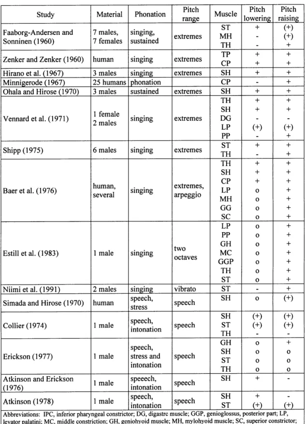
contributions of various muscles to phonation tasks. Specifically, a 1996 review by Vilkman and colleagues of 15 EMG studies of the contributions of extrinsic laryngeal muscles to phonation in humans found lack of consistency within studies as well as conflicting results between them (Vilkman, Sonninen, Hurme, & Korkko, 1996). Muscles examined in multiple studies were the ST, TH, CP, and SH, none of which yielded conclusive results for pitch lowering or raising. Table 1-1 summarizes the results of this review. Although many models of pitch control via extrinsic factors exist
(Kenyon, 1927; Sonninen, 1968; Vilkman, et al., 1996), the conclusion of this review was that current data are too disparate to corroborate any comprehensive model extrinsic laryngeal pitch control.
More recently, Roubeau and colleagues measured the normalized EMG of the TH, ST, and SH in one female and one male with healthy normal voice while performing ascending and descending glissandos (Roubeau, et al., 1997). Their results are
reproduced in Figure 1-4. During ascending glissando from 120 Hz to 600 Hz, the SH was found to be generally active throughout, whereas the ST and TH were modestly active (10% of the maximum recorded) between 120 Hz - 150 Hz and then 500 Hz - 600 Hz. During descending glissando, however, the female participant showed most
pronounced differences in ST and TH activity across frequency. Here, the ST and TH were again modestly active in the 500 Hz - 600 Hz range, but in the range from 120 Hz
-200 Hz showed more activity (25% of the maximum recorded). The SH again showed moderate activity over all frequencies except at the extremes (0% of the maximum recorded at 600 Hz, 15% of the maximum recorded in the 120 Hz - 200 Hz range). For the ascending glissando, the male participant showed far more variation than the female participant. The TH, ST, and SH all showed high activity during both low and high frequencies (values of 20 % -50% of the maximum recorded). For the descending glissando, the male participant showed tonic activity in the TH (30% of the maximum
recorded), with maximum activity at the lower frequencies (50% of the maximum recorded). The ST and SH showed fairly stable levels (around 10% of the maximum recorded) at most frequencies, with some increase in activity at the lower frequencies (15% - 25% of the maximum recorded). The differing results between ascending and descending pitch change noted by Roubeau imply that activity of the extrinsic laryngeal muscles is more closely tied to dynamic aspects of pitch than to specific pitch production. VOICE DISORDERS
The term "voice disorder" has been defined as "a problem in producing voice that is primarily caused by a disturbance or loss of normal laryngeal function" (Hillman, Gress, Hargrave, Walsh, & Bunting, 1990). Studies have shown that a voice disorder does not only affect the economic well-being of the patient, but that it also may cause a disordered self-image that negatively affects the patient socially (Ramig & Verdolini, 1998).
One school of thought identifies four general components to voice disorder: 1) voice-related muscular skill including posture, 2) behavior pertaining to lifestyle and
personality, 3) gastro-esophageal reflux disease, and 4) psychological factors (M. Morrison, 1997a). Further, it is thought that pathological process may overlie the disorder; these processes being broadly divided into two groups: neurological disease or organic changes to the vocal folds (M. Morrison, 1997a). The poor muscle usage (or low level of vocal skill) is commonly referred to as muscle and/or voice misuse. This misuse is correlated in a nontrivial way to fundamental frequency, sound pressure level, and particular vocal behaviors of the patient with respect to their "normal" voice (Sander & Ripich, 1983).
CLINICAL ASSESSMENT OF VOCAL FUNCTION
Current clinical assessment of vocal function is a multidimensional process consisting of perceptual and objective measures. Current practice includes patient interview for voice and psychosocial history and status, perceptual assessment of voice quality by the
clinician and patient, observation of positioning and posture of the upper body, elicitation of reflexes, direct (oral) endoscopy, flexible (nasal) endoscopy, acoustic analysis of the speech signal, electroglottography, aerodynamic assessment, evaluation of respiratory kinematics, and in some cases laryngeal electromyography (Andrews, 1999; Colton, Casper, & Leonard, 2006).
These general categories of voice-related information can provide overlapping and complementary information regarding the vocal health of the patient. The patient
interview includes discussion of the history, effect, onset, and duration of his or her voice issues. Further, the patient interview can supply information about the voice use, general health, and social/psychological status of the patient (Colton, et al., 2006). The state-of-the-art in clinician perceptual assessment of voice quality is the Consensus Auditory-Perceptual Evaluation of Voice (CAPE-V), a visual-analog scale ratings system of six perceptual attributes of voice: overall severity, roughness, breathiness, strain, pitch, and loudness (Colton, et al., 2006). Patient perception of their voice and the impact of any
Pitch Pitch Pitch
Study Material Phonation Muscle
range lowering raisin
ST + (+)
Faaborg-Andersen and 7 males, singing, extremes MH - (+)
Sonninen (1960) 7 females sustained TH +
TP + +
Zenker and Zenker (1960) human singing extremes TP + +
CP + +
Hirano et al. (1967) 3 males singing extremes SH + +
Minnigerode (1967) 25 humans phonation CP - +
Ohala and Hirose (1970) 3 males sustained extremes SH + +
TH + +
SH + +
1 female SH + +
Vennard et al. (1971) 2 male singing extremes DG
LP (+) (+)
PP - +
ST + +
Shipp (1975) 6 males singing extremes TH - +
TH + +
TH + +
SH + +
CP + +
human, extremes,
Baer et al. (1976) human, singing extremes, LP +
several arpeggio MH o+ GG o + SC o + LP o + PP o + GH o + two
Estill et al. (1983) 1 male singing MC o +
octaves
GGP o +
TH o +
ST o +
Niimi et al. (1991) 2 males singing vibrato ST - +
Simada and Hirose (1970) human speech, speech SH (+)
stress
SH (+) (+)
speech, ST () (+)
Collier (1974) 1 male intonation speech ST (+)
TH
GH o +
speech, SH
Erickson (1977) 1 male stress and speech ST o o
intonation
TH o o
Atkinson and Erickson speeech, SH +
(1976) intonation
speech, SH +
Atkinson (1978) 1 male intonati speech ST +
intonation ST N N
Abbreviations: IPC, inferior pharyngeal constrictor; DG, digastrc muscle; GGP, genioglossus, posterior part; LP, levator palatini; MC, middle constriction; GH, geniohyoid muscle; MH, mylohyoid muscle; SC, superior constrictor;
CP, crycopharyngeal muscle; TP, thyropharyngeal muscle; SH, sternohyoid muscle; TH, thyrohyoid muscle; GG,
genioglossal muscle; PP, palatopharyngeal muscle. +, effect; -, no effect; o, not studied; (+), inconsistent.
Table 1-1. Reproduction of the summary of Vilkman et al. (1996) of 15 EMG studies of the contributions of extrinsic laryngeal muscles to phonation in humans.
disorder is addressed using perceptual "quality of life" scales such as the Voice Handicap Index (VHI) and the Voice-Related Quality of Life (V-RQOL) (Colton, et al., 2006). Observation of the head, neck, and torso can be vital in a complete evaluation of vocal health. In particular, clinicians look for anterior/posterior weight bearing, head tilt, deviated larynx (abnormally rotated), asymmetrical stemocleidomastoid muscles, an abnormally held larynx (high or low), and high muscle tone / reduced flexibility
surrounding the larynx (see Lieberman, 1998, for a comprehensive tutorial). Elicitation of oral, laryngeal, and gag reflexes can be used to rule out or suggest neurological impairment (Andrews, 1999; Colton, et al., 2006).
For the most part, current clinical practice utilizes two main types of endoscopy: rigid (oral) and flexible (nasal). Rigid endoscopy is performed by passing a rigid endoscope over the top of the tongue and into the pharyngeal cavity. This procedure allows for larger, brighter images of the vocal folds, but requires the clinician to pull the patient's tongue to get an adequate view, manipulating his or her natural vocal behaviors. In flexible endoscopy, the endoscope is passed through the nose and down into the pharynx in order to observe the vocal folds and supraglottic space during more natural speech and singing tasks. Figure 1-5 shows example stills of the same larynx taken from both exam types. Stroboscopy can be utilized with both methods of endoscopy, and involves flashing light at a rate slightly different than the vibrating rate of the vocal folds. This technique allows the vocal folds to be illuminated at different phases of their vibratory cycle, creating the illusion of a slowing of the vibratory motion of the folds and enabling the clinician to view representative repeated cycles of vocal fold motion.
Acoustic signs typically indicative of vocal fold health are fundamental frequency (mean, variability, range, and perturbation), amplitude (average, variability, dynamic range, and perturbation), signal-to-noise ratio, and maximum phonation time. Electroglottography (EGG) uses two electrodes placed on the surface of the each side of the neck at the larynx to measure the current flow through the neck tissues between the two, giving an indirect measure of the closure of the vocal folds. Aerodynamic assessment can include measures of airflow, subglottal pressure, and phonation threshold pressure, all of which may provide information about phonation production. Respiratory kinematics can highlight the patient's control over respiratory movements and are usually measured using an induction plethysmograph or magnetometer, devices that can non-invasively monitor the movements of the chest and abdomen during speech tasks. Laryngeal electromyography (EMG) is an invasive procedure during which electrodes are inserted through the neck surface or brought through the mouth and inserted into the intrinsic laryngeal muscles to record their electrical activity. Due to the invasive nature of this procedure and the neurological expertise required to interpret the test findings, it is not common clinical practice. However, it can be used to support diagnosis of paralysis or to guide injections of botulinum toxin for treatment of spasmodic dysphonia. Of the measures currently available for clinical assessment of voice, objective measures include EMG, aerodynamic assessment, EGG, and acoustic assessment. Currently, these objective measures are not used as primary diagnostic procedures, but as supplemental tools to enhance diagnosis quality (Hillman, Montgomery, & Zeitels, 1997).
, A A
,1 _ 0,1
0,
50 150 250 350 450 550 850 $se s o o 2s 0se iso so
"AgMAWAL PRAa OOcy rumnAunAr"Qnoumef t)
ASCDM GUSSANDOMALE S lCt DCSCVWO oussANO MLSUS.MCt
0,3 0,2
10,2
so0 100 SO 200 250 300 300 250 200 1SO 100 so
Figure 1-4. Reproduction of the results of Roubeau et al. (1997). Data for the ascending glissando are shown in the left panels, descending in the right panels. Data from the female and male participants are shown in the upper panels and
lower panels, respectively.
Rigid
Exam
Flexible Exam
Figure 1-5. Example images of the same set of vocal folds as viewed by rigid (left panel) and flexible (right panel) endoscopy.
Vocal Hyperfunction
Functional dysphonia is an "umbrella diagnosis" for impairment of voice production in the absence of structural change or neurogenic disease of the larynx (Altman, Atkinson,
& Lazarus, 2005). While this type of functional disorder can have fairly homogenous
clinical symptoms, the etiology is more heterogeneous (Kinzl, Biebl, & Rauchegger,
1988). There is thought to be some psychogenic nature to this etiology, however, which
is supported by the finding that some 75% of functional voice patients have other psychosomatic functional disturbances in their histories, such as anorexia nervosa, bulimia nervosa, cardiac neurosis, migraine, bronchial asthma, and diffuse abdominal complaints (Kinzl, et al., 1988). While functional voice disorders are by definition primary muscle tension dysphonias, many patients who display characteristics of
functional voice disorders have underlying organic conditions. Here, the functional voice aberrations are secondary and may result from improper compensatory muscular activity (Koufman & Blalock, 1991a). Thus, most voice disorders can be viewed as completely or partly functional (Koufman & Blalock, 199 l1a).
Vocal hyperfunction is a common functional voice disorder referring to "conditions of abuse and/or misuse of the vocal mechanism due to excessive and/or 'imbalanced' muscular forces" (Hillman, et al., 1989), characterized by excessive laryngeal and
paralaryngeal tension (Aronson, 1980; Dworkin, Meleca, & Abkarian, 2000; Koufman & Blalock, 1991b; M. D. Morrison, et al., 1983; N. Roy, et al., 1996). Individuals with vocal hyperfunction and no other known cause of voice disorder (e.g., some type of glottal insufficiency) are often diagnosed with muscle tension dysphonia (MTD; also known as hyperfunctional dysphonia, hyperkinetic dysphonia, muscular tension dysphonia, vocal hyperfunction, muscular tension dysphonia, vocal fatigue, laryngeal tension-fatigue syndrome, and functional hypertensive dysphonia (Hsiao, Liu, Hsu, Lee,
& Lin, 2001)). This is a type of functional dysphonia making up some 50% of the cases
seen (Koufman & Blalock, 1982). Other estimates of the prevalence of vocal
hyperfunction indicate that the condition may account for 10 - 40 % of cases referred to multidisciplinary voice clinics (N. Roy, 2003). Hyperfunction of the laryngeal and paralaryngeal muscles could be due to psychological and/or personality factors, misuse of muscles, learned adaptation based on short-term illness, or compensation for underlying disease (Aronson, 1980; Hsiung & Hsiao, 2004; M. D. Morrison, Nichol, & Rammage,
1986; M. D. Morrison & Rammage, 1993; M. D. Morrison, et al., 1983; Rammage,
Nichol, & Morrison, 1987).
SYMPTOMS / CURRENT STATE OF ASSESSMENT
The manifestation of muscle tension dysphonia / vocal hyperfunction has been typified as the incorporation of the following symptoms: obvious muscular tension in paralaryngeal musculature, high larynx position, vocal fry, phase asymmetry, low pitch, diplophonia,
rough voice quality, exaggerated posterior glottic chink (gap), lateral compression in the membranous vocal folds, breathiness, glottic and supraglottic compression, adduction spasms, and strained voice quality (M. Morrison, 1997b; M. Morrison, Rammage, & Emami, 1999; M. D. Morrison, et al., 1986; M. D. Morrison, et al., 1983). Also, patients are often pitch- and loudness-locked with reduced dynamic range, reporting with aches and tightness of the neck, larynx, and shoulder regions, as well as episodic neck swelling and ear "fullness"(Aronson, 1980; Nelson Roy & Bless, 1998).
Videostroboscopic evaluation of MTD patients may also show characteristic signs including an uneven mucosal layer (Hsiao, et al., 2001; Hsiao, Liu, & Lin, 2002), abnormal glottal closure, phase or amplitude asymmetry, and an irregular mucosal wave (Hsiao, et al., 2001). A further symptom visible via flexible endoscopy includes
supraglottic compression. Supraglottic compression describes obstruction of part or all of the view of the vocal folds during indirect laryngoscopy and videoendoscopy (Stager, Bielamowicz, Regnell, Gupta, & Barkmeier, 2000). There are two primary components: anterior-posterior (AP) compression, in which the arytenoids cartilages are drawn toward the petiole of the epiglottis, and medial or false vocal fold (FVF) compression, which is caused by adduction of the false vocal folds (Stager, et al., 2000). Studies have cited this supraglottic activity as a symptom characteristic of MTD (M. Morrison, et al., 1999), but without comparisons to its prevalence in populations with healthy normal voice. Stager and colleagues compared the incidence of AP and FVF compression in hyperfunctioning, vocal nodule, and healthy normal voice participants, finding that the incidence in
individuals with healthy normal voice was less than that in the voice patients, but was not absent (Stager, et al., 2000). However, the control group in the study consisted of
patients with allergies and/or reflux, which may have been a confounding factor. Specifically, reflux patients have been described as likely to display supraglottic compression (Koufman & Blalock, 1991b). A retrospective study by Morrison and colleagues recorded the laryngoscopic examination of patients diagnosed with vocal hyperfunction (M. D. Morrison, et al., 1986). Of diagnoses of vocal hyperfunction: 11% presented with no mucosal change, 90% presented with an open posterior chink, 49% presented with visible suprahyoid tension. While 90% of MTD diagnoses present with a glottal posterior chink (M. D. Morrison, et al., 1986), this symptom is also prevalent in young women with normal voice (Linville, 1992). In a videostroboscopic study of young adult women with normal voice, a posterior-chink glottal configuration was noted 42% of the time, compared to 13% in a population of elderly adult women with healthy normal voice (Linville, 1992). Rammage and colleagues found a similar result: "virtually all" of their 50 participants identified as having bilateral vocal nodules presented with a
posterior glottal chink during phonation; further, a majority of the 20 participants with healthy normal voice (women, aged between 17 and 36) also presented with a posterior glottal chink (Rammage, Peppard, & Bless, 1992).
Compared to individuals with healthy normal voice, muscle tension dysphonia patients have also been demonstrated to have a higher prevalence of abnormal glottic closure during inspiration (similar to PVFM, paradoxical vocal fold movement) (Vertigan, et al., 2006). PVFM is also referred to as episodic paroxysmal laryngospasm (EPL) and is characterized by involuntary paradoxical adduction of the vocal folds and ventricular folds during inspiration (Andrianopoulos, Gallivan, & Gallivan, 2000). It is frequently mistaken for asthma, acute respiratory distress, or other laryngeal problems, including
MTD (Andrianopoulos, et al., 2000). Morrison and colleagues have postulated that PVFM and MTD, as well as chronic cough, throat clearing, and globus pharyngeus are all forms of hyperkinetic laryngeal dysfunction, which they have termed irritable larynx syndrome (ILS) (M. Morrison, et al., 1999). Highlighting the interplay between vocal hyperfunction and other laryngeal irritation, a series of three case studies from Japan showed that there was subjective improvement in the voice and glottal closure of MTD patients following treatment via proton pump inhibitors, indicating that reflux may have been a major causative factor in the disorder (Mesuda, et al., 2007).
Commonly associated symptoms of vocal hyperfunction are not limited to the larynx. Many muscles in the neck that attach to the larynx and/or hyoid bone have voice and speech-related contractions due to their role in controlling the vertical position of the larynx in the neck and, to some degree, the position of the tongue. When individuals demonstrate an inappropriate degree of intrinsic laryngeal muscle contraction
(hyperfunction), it is thought that they often simultaneously contract the extrinsic
laryngeal muscles and other superficial neck muscles in a similar hyperfunctional manner (Aronson, 1980). A study by Altman and colleagues looked at 150 patients who had been diagnosed with muscle tension dysphonia; based on a speech pathology evaluation of these patients, 70% were found to have "obvious cervical neck tension visible"
(Altman, et al., 2005). Clinically, excessive tension has been noted via palpation over the major horns of the hyoid bone, over the superior cornu of the thyroid cartilage, along the anterior border of the sternocleidomastoid muscle, and throughout the suprahyoid
musculature (N. Roy, et al., 1996). Strap muscle tension can be noted in the MTD patient through both visual and tactile inputs. Particularly, observation of the inferior bellies of the omohyoid muscle crossing the supraclavicular fossae easily shows them to be tense and prominent during speech (M. Morrison, 1997b). Further information about the extent of muscle tension found in the patient can be gained by palpation of the larynx at rest and during voicing (M. Morrison, 1997b).
Further known symptoms of MTD include subjective observations of perceived breathiness (Aronson, 1980; M. Morrison, et al., 1999; M. D. Morrison, et al., 1986), which are supported in part by findings of increased flow rates in affected patients (Eustace, Stemple, & Lee, 1996). Another aspect of aerodynamic assessment cited by Hillman and colleagues as specific evidence of vocal hyperfunction is the ratio of
subglottal pressure and sound pressure level with respect to age-and gender-specific normative data. Specifically, the use of excessive subglottal "driving pressure" to produce sound pressure levels is a feature seen in many patients with vocal hyperfunction
and can be measured objectively (Hillman, et al., 1997). Morrison and colleagues found that of individuals with vocal hyperfunction: 81% presented with breathiness, 50% presented with glottal fry, 65% with hard glottal attack (M. D. Morrison, et al., 1986).
ORGANIC DEVELOPMENTS AND INTERACTIONS
For many years, organic developments on the vocal fold surface have been assumed to be related to hyperfunctional behavior or phonotrauma (Hillman, et al., 1990). For instance, in 1962, Godrey Arnold wrote that "vocal nodules and polyps represent a local tissue reaction to the mental strain imposed by inappropriate emotional adjustment to the
demands made by society" (Arnold, 1962). However, much is still unknown about the underlying mechanisms of vocal hyperfunction and its role in developing organic disorders (Hillman, et al., 1990).
It has been argued that a patient's vocal hyperfunction begins with a structurally normal larynx, in which the imbalanced muscular activity causes an open posterior glottic chink between the arytenoids cartilages upon phonation (M. D. Morrison, et al., 1986).
Prolonged voicing in this arrangement leads to excessive mechanical stress and trauma that may eventually cause mucosal changes to develop, including nodules, diffuse
erythema and edema, and polyps (Hillman, et al., 1989; Johns, 2003; M. D. Morrison, et al., 1986). While the vocal ligament can protect softer tissues of the lamina propria from the high tensile stress felt at high pitches, impact stress as a result of vocal fold collision may cause damage in the form of vocal nodules, which occur at the point along the edge
of the vocal fold that experiences the maximum impact stress (Titze, 1994a).
Clinically, a nodule is defined as a small protuberance located between the anterior and middle third of the vocal fold (Aronson, 1980; Marcotullio, Magliulo, Pietrunti, & Suriano, 2002). It is described as being gray/white or pearl in color and is usually bilateral (Aronson, 1980; Marcotullio, et al., 2002). Vocal fold nodules are benign lesions characterized as being bilateral, symmetrical, and involving minimal disruption of the mucosal wave on stroboscopy. The basement membrane zone is thickened, having increased fibronectin. In vocal fold polyps, there are focal depositions of gelatinous material comprised of amorphous, disorganized extracellular matrix. The lesions are exophytic (growing outward from the surface epithelium) and may be associated with an enlarged blood vessel or hemorrhage. They are not typically associated with a thickening of the basement membrane (Dikkers & Nikkels, 1995; Verdolini, Rosen, & Branski, 2006). Some of the confusion in determining differences between various laryngeal lesions may arise from differences in diagnoses. Dikkers and Schutte found a large discrepancy among a large group of clinicians with respect to the name used for various laryngeal lesions (Dikkers & Schutte, 1991). Based on their study, they offered basic clinical definitions. Figure 1-6 is a pictorial summary of the basic categories of benign fibrovascular lesions along with their defining characteristics as described by Dikkers and Schutte (1991). They termed a cyst as a unilateral lesion with a smooth surface that is usually found on the middle third of the vocal fold, and is immobile during phonation (Dikkers & Schutte, 1991). They define Reinke's edema as a condition with unilateral or bilateral white swelling of the vocal fold, in which the lesion is filled with fluid, sessile (adhering closely to the surface), and mobile during phonation (Dikkers & Schutte,
1991). A polyp, they define as a unilateral lesion on the anterior third of the vocal fold, often on the free edge, and either pedunculated (attached by a thin process of tissue rather than a large base) or sessile (Dikkers & Schutte, 1991). Further, vocal fold nodules are described as small lesions on both sides of the larynx, symmetrical on the border of the anterior and middle third of the vocal folds, which are usually immobile during phonation (Dikkers & Schutte, 1991). Also noted in the clinic is the diagnosis of "vocal fold
thickening." This is widely viewed as a antecedent to the formation of vocal nodules, but this hypothesis has not been proven (Goldman, Hargrave, Hillman, Holmberg, & Gress,
The form in which mucosal changes occur seems to depend upon the patient's age, sex, particular voice use, and whether or not the patient smokes (M. D. Morrison, et al., 1986). Particularly, vocal nodules usually occur in young to mid-aged females, whereas more diffuse mucosal changes are seen mostly in men (M. D. Morrison, et al., 1986; M. D. Morrison, et al., 1983). Polyps are commonly found in adult females, of whom almost all smoke cigarettes (M. D. Morrison, et al., 1986; M. D. Morrison, et al., 1983). Vocal nodules also seem to be more common in young larynges; a study of children with muscle tension dysphonia found coexisting vocal nodules in 7 of the 8 participants (Lee & Son, 2005). This is not surprising, given the results of a recent study by Marcotullio and colleagues, who argue that there are no substantial histologic differences between nodules and polyps, but that pathologic manifestations clinically classified as nodules are merely "younger" lesions than those classified as polyps (Marcotullio, et al., 2002). This correlation between age and sex and the development of vocal nodules appears to be routed in laryngeal morphology (Pontes, Kyrillos, Behlau, De Biase, & Pontes, 2002).
Specifically, vocal nodules are found in larynges with a morphology associated with young women, one that reduces the functional opening angle during abduction of the vocal folds (Pontes, et al., 2002). The idea that lesion formation is a function of morphology and not specifically behavior is supported by the work of Andrade and colleagues. They found that hard glottal attacks occur significantly more frequently in MTD patients, but that this possibly causal behavior was found in the same frequency in all subgroups of MTD patients, regardless of the existence of vocal fold lesions or the
lack of such lesions (Andrade, et al., 2000). Titze postulates that, given that vocal nodules are the result of repeated collision, the risk of developing nodules is higher in high-pitched voices, such as children and young women (Titze, 1989). It should be mentioned that the work of Dikkers and Nikkels opposes the idea that there is a lack of histologic difference between nodules and polyps, with work and hypotheses supporting a
lack of clinical continuum between possible laryngeal lesions (Dikkers & Nikkels, 1995, 1999). Also contrary to the work of Marcotullio and colleagues, their work classifies a polyp as a "younger" lesion than a nodule (Dikkers & Nikkels, 1999). Thibeault and colleagues have examined the transcription-level profiles of extracellular matrix proteins
in vocal fold polyps and Reinke's edema, finding differing extracellular matrix regulation (Thibeault, et al., 2002). Compared to polyps, fibronectin (interstitial proteoglycan necessary for the development of fibrosis) transcription levels were found to be down-regulated in Reinke's edema whereas fibromodulin (interstitial proteoglycan whose
absence leads to disorganized collagen fiber bundles) transcription levels were found to be higher. These results support the clinical findings of increased stiffness in polyps relative to Reinke's edema and a lack of continuum between the two lesions.
Some 92% of cases of vocal nodules are coincident with vocal hyperfunction (M. D. Morrison, et al., 1983). Further, of individuals with vocal hyperfunction, Morrison et al. (1986) found that 51% presented with vocal nodules, while Altman et al. (2005) found that 13% presented with vocal nodules, the development of vocal nodules appears to be highly correlated with the disorder. Nodules found in patients that do not present with vocal hyperfunction tend to be more firm and less likely to resolve with voice therapy
Nodules: small, bilateral, symmetrical, located on the border of the anterior and middle thirds of the vocal fold
Cyst: unilateral, smooth surface, typically immobile during phonation, located on the middle third of the vocal fold
Polyp: unilateral, often on the free edge, sessile or pedunculated, located on the anterior third of the vocal fold Reinke's Edema: unilateral or bilateral white swelling of the vocal fold, fluid, sessile, and mobile during phonation
Figure 1-6. Pictorial summary of the basic categories of benign fibrovascular
lesions along with their defining characteristics as described by Dikkers and Schutte (1991).
TREATMENT OPTIONS
Voice therapy has been shown to decrease nodules and improve voice quality (Carding, Horsley, & Docherty, 1999; Holmberg, Hillman, Hammarberg, Sodersten, & Doyle, 2001) and to be generally effective in treating non-organic voice disorders (Carding & Horsley, 1992). A recent study found that even polyps and cysts were effectively treated via voice therapy (Cohen & Garrett, 2007). However, it is generally agreed that time and energy must be devoted to "carryover" of behaviors learned in behaviorally based voice therapy, in order to carry over those vocal behaviors from the clinic to "real life"
situations outside the therapy setting (Holmberg, et al., 2001).
Most voice therapies for MTD include the aim of reducing excess laryngeal
musculoskeletal tension. Most of these therapies such as progressive relaxation, the accent method, yawn-sigh therapy, and resonant voice therapy do this indirectly (Nelson Roy & Bless, 1998). Another typical part of MTD voice therapy that addresses this abnormal tension directly is the practice of "manual laryngeal tension reduction" which includes manually lowering an abnormally elevated larynx, as well as performing circumlaryngeal massage to reduce muscle tension (N. Roy, et al., 1996; N. Roy & Leeper, 1993). This practice is meant to raise the patient's awareness of neck tension (Nelson Roy & Bless, 1998). This practice has been shown to produce meaningful perceptual and acoustic results in short-term scenarios (N. Roy, Bless, Heisey, & Ford,
(N. Roy, et al., 1997). These studies recommend that this practice may be an integral part of voice therapy for MTD, but is an "incomplete remedy" (N. Roy, et al., 1997).
Specifically, patients have trouble relying on their own current feedback systems to learn long-term improved vocal habits (Nelson Roy & Bless, 1998). An extension of manual tension reduction techniques employed through voice therapy has been explored in part. Particularly, some success has been found in limited studies of multidisciplinary
treatment of MTD; in this scenario, voice therapy is supplemented by physical therapy to directly invoke relaxation of head, neck, and back musculature (Jones, Murry, & Rosen,
1998). A Finnish version of circumlaryngeal massage known as "voice massage" was performed on individuals with healthy normal voice with no immediate significant post-treatment effects (Laukkanen, Leppanen, Tyrmi, & Vilkman, 2005); however, this study has not been reproduced in disordered speakers. Dworkin and colleagues offer a possible supplement to typical voice therapy techniques. Their study found that in several case studies of patients with therapy-resistant MTD, a topical lidocaine bath was able to break the hyperfunctional vocal fold behavior with good, and sustained effect (Dworkin, Meleca, Simpson, & Garfield, 2000).
It is possible that some vocal nodules cannot be treated with voice therapy alone, even if voice abuse/misuse is arrested (Gray, Hammond, & Hanson, 1995). If improper healing of the vocal fold has occurred, this can result in increased fibronectin deposition, which is usually a permanent event (Gray, et al., 1995). In these cases, a combined surgical and voice therapy treatment is likely to yield the best clinical result (Gray, et al., 1995).
Surgical treatment without appropriate voice therapy is not recommended; characteristic features of MTD have been noted in non-dysphonic postoperative populations, indicating that the inappropriate vocal behaviors leading to surgery are still present and may lead to new lesion formation (Hsiung & Hsiao, 2004).
While patients with MTD appear to respond well to voice therapy, some findings indicate that there is limited success in the long term (Van Lierde, Claeys, De Bodt, & van
Cauwenberge, 2006). A Belgian study looked at 27 respondents of an original 184 hyperfunctional voice patients an average of 6.1 years after a voice therapy program
(Van Lierde, et al., 2006). In this subgroup, approximately 50% still showed pathological laryngological findings, and the average dysphonia severity index (DSI) was less than the average before treatment (Van Lierde, et al., 2006). However, these results are based on
a small voluntary response sample, a type of sample is often biased, because people with strong (especially negative) options are more likely to respond (e.g., Moore & McGabe, 1998). A more general study of efficacy of speech therapy in 109 patients diagnosed with functional voice disorder found that only 56% were cured by speech therapy, and that therapy was necessary for three months or more in nearly half of patients who were
eventually "cured" (Bridger & Epstein, 1983).
The majority of MTD patients respond well to speech therapy; however, in some cases it is inadequate (Bhalla, Wallis, Kaushik, & Carpentier, 2005). Another option for MTD patients resistant to speech therapy is the use of intravenous midazolam (Bhalla, et al.,
2005). This sedative is used in conjunction with speech therapy to confirm the psychogenic nature of symptoms and to "prove" to the patient that a better voice is
possible (Bhalla, et al., 2005). While this is the drug most commonly used for sedation in children and adults for procedures (Krauss & Green, 2000) and is generally felt to be safe
and effective (Parker, Mahan, Giugliano, & Parker, 1997), the use of any pharmaceutical adds extra risk and cost to patient treatment. Similarly, lidocaine bath was used on a group of MTD patients, finding a temporary reduction of symptoms (N. Roy, Smith, Allen, & Merrill, 2007). The need for more drastic methods to attempt to alleviate vocal hyperfunction supports the idea that there is a need for increased knowledge about the disorder to inform treatment decisions.
Surface electromyography
THEORY
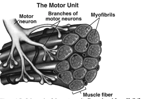
As a nerve impulse reaches the motor end plates from an alpha motor neuron, all muscle fibers innervated by that axon comprise a single motor unit and are discharged nearly synchronously (see Figure 1-7). The electric potential field generated by the
depolarization of the outer muscle-fiber membranes is essentially an amplified version of the alpha motor neuron activity; the electromyogram (EMG) is a representation of this
"myoelectricity" as measured at some distance. Tissues separating the EMG signal sources (depolarized zones of the muscle fibers) act like spatial low-pass filters on the potential distribution, and constitute a volume conductor. Therefore, the EMG may be
measured intramuscularly or at the surface of the skin, yielding different information based on the distance of the observation site from the muscle fibers. For surface detection particularly, the effect of the separating tissues becomes significant.
In order to remove interference sources and to compensate of the low-pass filtering effect of the tissue, surface signals are typically detected using a linear combination of different electrodes, the simplest of which is a differential electrode (Farina, Merletti, & Stegeman, 2004). Bipolar surface EMG (sEMG) is dependent upon on the inter-electrode distance (Roeleveld, Stegeman, Vingerhoets, & Van Oosterom, 1997). In measuring the sEMG, filtering is introduced by finite electrode size, inter-electrode distance, electrode
configuration, electrode location, and characteristics of the front-end amplifier.
The Motor Unit
Motor Branches of Myofibrils
motor neurons neuron
Muscle fiber
RECORDING AND ANALYSIS RECOMMENDATIONS
The European Union sponsored a project termed SENIAM (Surface Electromyography for the Noninvasive Assessment of Muscles), one outcome of which was a set of recommendations for sEMG recording. In general, SENIAM recommends a maximum electrode size of 10 mm in the muscle fiber direction, with an interelectrode distance of approximately 20 mm or /4 the length of the muscle fiber, whichever is smaller
(Hermens, et al., 1999). Other recent recommendations include the assertion that smaller electrodes (diameter less than 5mm) are preferred for sEMG, as the larger electrodes introduce temporal low-pass filtering (Merletti & Hermens, 2004).
Skin preparation techniques can enhance electrode-skin contact, resulting in a reduction of artifacts and less noise. SENIAM recommends shaving the skin surface if it is covered with hair, and cleaning the skin in question with alcohol (Hermens, et al., 1999). Also preferred is the practice of slight skin abrasion or "peeling" with adhesive tape; this practice is known to reduce electrode-skin impedance, noise, DC voltages, and motion artifacts (Merletti & Hermens, 2004).
Recommendations for electrode placements are that the differential electrodes are applied between the innervation zone and a tendon. In the past, sensors have been placed over the belly or over the innervation zone (motor end plate zone), since this was the best location to record "large" monopolar sEMG signals. It is now well known that this location is not suitable for differential recordings; it is not stable or reproducible because relatively small displacements of the sensors with respect to the innervation zone cause large effects on the amplitude of the sEMG signal (Merletti & Hermens, 2004). Thus, in order for sEMG signals to be accurate and repeatable, there must be a clear definition of electrode position relative to the innervation zones (Hermens, et al., 1999). When the locations of innervation zones are unknown, use of double differential electrodes can diminish the effects of an ill-placed sensor (Farina, Merletti, & Disselhorst-Klug, 2004). Ideal sEMG recording procedures would first identify the innervation zones and find the optimal electrode position on a subject by subject basis, using multi-channel electrode arrays. Falla et al. (2002), for example, examined the sternocleidomastoid (SCM) muscles in this way in 11 healthy normal individuals. Based on their findings, they have offered the following recommendations to optimize sEMG recordings from SCM
muscles: the electrode should be placed 1/3 of the distance from the sternal notch to the mastoid process, in the direction of the line from the sternal notch to the mastoid process
(Falla, et al., 2002). Recommendations of this type are not available for sEMG recordings of the extrinsic laryngeal muscles. With regard to ground locations, SENIAM
recommends the wrist, the spinous process of C7, or the ankle as appropriate locations (Hermens, et al., 1999).
Because of the variability surrounding neck surface electrode contact and participant neck mass, sEMG signals should be normalized to a reference contraction before they are compared between conditions and/or participants (Netto & Burnett, 2006). Most
especially, the layers of subcutaneous fat present can have attenuating and widening effects on the signal seen at the surface (Farina & Rainoldi, 1999). Common references include maximal voluntary contraction (MVC) and some percentage of the MVC (usually