Cellular response to 11 3-dichloro, a novel aniline
mustard-estradienone, in various prostate cancer cell lines
By
Francis Heber Gonzilez S.B. Chemistry
University of Puerto Rico - Mayagilez Campus, 2005
Submitted to the Department of Chemistry in partial fulfillment of the requirements for the degree of
MASSACHUSETTS It OF TECHNOLC
Master of Science in Biochemistry
[ jjjj
.jat the
Massachusetts Institute of Technology L
September 2008
C 2008 Massachusetts Institute of Technology. All rights reserved.
4STITUTE )G3Y Signature of Author ... ... ... nz'Iez Chemistry Department September 17, 2008 Certified by...
t.
.. .
- ---..
...--
.... ...
... ... ...
...
K)
UJohn EssigmannProfessor of Chemistry and Biological Engineering Thesis Advisor
Accepted by ...
Timothy Swager Chair, Chemistry Department
ARCHIVES
EB 1 J ZUU9
.IBRARIES
Acknowledgements
First of all, I would like to thank my advisor John Essigmann for allowing me to be part of his research group and carry out the work presented in this thesis. John facilitated the resources and research advice that allowed me to move my career forward within the lab. John was also very supportive during the tough times and always had words of encouragement when faced with difficult situations.
My experience in the Essigmann lab was greatly enhanced by the people that were around me and helped me navigate the intricacies of research at MIT. Specifically,
Bob Croy, Kyle Proffitt, and Sreeja Gopal who provided words of advice and shared long and deep scientific discussions regarding the best way to move forward in this project. I also want to thank present and past members of the lab who provided their support and encouragement and were always willing to listen to my ideas and give me feedback; Bogdan, Nidhi, Charles, Sarah, Lauren, Yuriy, Eunsuk, Peter, Will, Jeannette, Jim, and Alfio. The experience would have been very different and much tougher without you.
Lastly, I want to thank my friends and family for their support and motivation. My parents and my extended family that always believed in me and my potential to achieve great and many things and who are the main motivations to push forward, even in the difficult times. Also, to my closest friends and housemates here in Boston, Jose and Nelly, who kept tabs on me and always believed in me and encouraged me to keep going. And finally, to the person that was closest to me during the last year and a half and
without whose patience, encouragement, and unconditional love and support this last year would have been even more challenging; thank you Isabel for all the good things you brought into my life and for sharing with me so many special moments that enabled me to keep moving forward in this unpredictable journey we call life. You are really an
Abstract
Cancer is a group of diseases characterized by uncontrolled cell growth and abnormal cell cycle and apoptosis regulation. Among the types of cancer, prostate cancer is the leading cause of death in men. Although many advances have been made in the last decade through anti-hormonal treatment, chemotherapy, and early screening, acquired chemotherapeutic resistance has presented a challenge to treating more advanced forms of prostate cancer. To meet this challenge a new compound, 113-dichloro, was designed that would integrate lessons learned from previous studies with other chemotherapuetics, specifically cisplatin. 11P -dichloro is a novel aniline mustard-estradienone that targets both DNA and the androgen receptor (AR), which is over expressed in many types of prostate cancer. The working hypothesis is that by causing substantial amount of un-repairable DNA damage and sequestering the transcription factor function of the androgen receptor it would result in a more effective treatment when compared to existing treatments. Hence, 11 3-dichloro is expected to affect AR-positive prostate cancer cells more robustly than AR-negative cells, based on our repair
shielding and transcription factor hijacking hypotheses.
To assay differential toxicity, five different prostate cancer cell lines were used, namely, LNCaP positive, T887A), PC3 negative, p53 inactive), DU145 negative, p53 null), and the isogenic pair PC3-AR positive) and PC3-neo (AR-negative), which only differ in the AR status. Growth inhibition with LNCaP, PC3, and DU 145 cells showed that LNCaP cells are more sensitive to 11 -dichloro at
concentrations higher than 5.0 tM. To better assay the effect of the AR in cell survival a clonogenic assay was developed using PC3-AR and PC3-neo cells. Preliminary growth inhibition studies with this isogenic pair showed differential toxicity towards AR-positive PC3-AR cells up to 10 pM 11 3-dichloro. However, the series of clonogenic assay
experiments showed no differential toxicity between these two cell lines. Nevertheless, these experiments are not conclusive because they were not carried out using a clonal population of cells.
11 3-dichloro has been shown in previous studies to cause up regulation of certain proteins involved in cell cycle arrest and apoptosis regulation. By looking more closely at the effect of 11 3-dichloro treatment on these proteins, potential biomarkers for the effectiveness of cancer treatment can be developed. In this work special attention will be given to p21, a cyclin-dependent kinase inhibitor that was significantly down regulated at early time points after treatment with 11 3-dichloro, but which showed increased levels at later time points. Levels of p21 were assayed through Western blotting using the five different prostate cancer cell lines specified above after treatment with 10 jM 113-dichloro up to 15 hours. LNCaP cells showed decreased levels of p21 throughout the time course, but more significantly from the 3-9-hour time points. DU145 cells showed increased levels p21 after just 3 hours of exposure with a subsequent decrease to
untreated levels at the 15-hour time point. PC3 cells showed increased levels of p21 after 6 hours, with larger increases (6- and 7-fold) at the 9- and 15-hour time points. The isogenic pair PC3-AR and PC3-neo also showed a different expression profile for p21 expression with PC3-neo cells showing increased levels of p21 all through out the time
course ranging from 1.8- to 2.5-fold increase. However, PC3-AR cells did not exhibit any p21 at any of the time points except for the 15-hour time point where a faint band could be appreciated. Although qualitatively these observations maybe accurate the quantitative aspect of the analysis will have to be reproduced more thoroughly as p21 levels were not normalized to actin expression for each sample.
Taken together these observations suggest that our candidate compound 11P-dichloro exerts its effects on prostate cancer cells differently depending on the presence or absence of the androgen receptor and potentially p53 status. The model suggests that different mechanisms might be used in order to repair the DNA lesions caused by 11 3-dichloro and that these mechanisms are dependent on the presence or absence of the AR since the AR can potentially block DNA repair proteins that are recruited to excise the lesion.
Table of Contents
Acknowledgements ... 2
A bstract .. ... . ... 3
Introduction ... ... 6
Prostate cancer... 6
Lessons learned from cisplatin... 7
Developm ent of 110 ... 9
Characterization of ip ... 11
p21: a cyclin-dependent kinase inhibitor and potential biomarker ... 11
1113 inhibits further tumor growth in mice carrying LNCaP xenograft tumors... 12
113 form s N 7-guanine adducts ... 13
Materials and Methods ... 15
Prostate cancer cell lines ... . 15
Growth inhibition assay... 15
Clonogenic assay ... 16
W estern Blots ... ... 16
PC3-AR and PC3-neo growth rate experiment... ... 17
111-DNA adduct studies using accelerator mass spectrometry (AMS) - drug treatment and DNA isolation ... 18
Results and Discussion ... 19
LNCaP cells are more sensitive to 11i at concentrations higher than 5 tM after 24 hours... ... 19
PC3-AR cells show differential toxicity towards 11ip in a growth inhibition assay, but not in a clonogenic assay ... 20
Immunoblot analyses of p21 levels in various prostate cancer cell lines ... 22
Protein loading curve ... ... 23
p21 levels in LNCaP cells treated with chlorambucil and 113 ... 23
p21 levels in DU145 cells treated with chlorambucil and 1113 ... 24
p21 levels in PC3 cells treated with chlorambucil and 113 ... 24
p21 levels in PC3-AR and PC3-neo isogenic cell lines treated with chlorambucil an d 1 113 ... 2 5 p21 degradation kinetics and mechanism... ... 26
DNA isolation and preliminary growth rate studies on PC3-AR and PC3-neo cells for 110-DNA adduct studies... ... 28
Future Directions...31
Bibliography ... 35
Introduction
Prostate cancer
Broadly defined, cancer is a group of diseases characterized by uncontrolled growth and spread of abnormal cells, which if not treated can result in death. It is
expected by the American Cancer Society that more than half a million people will die of cancer in 2008, making it second only to cardiovascular disease as a cause of mortality (1). Cancer can be caused by external factors, such as tobacco, chemicals, radiation, and infectious organisms, as well as host factors like inheritance, mutations from DNA-interactive metabolic intermediates, hormones, and immune system activation. These factors can act together or in sequence in order to promote carcinogenesis. Some of the most common treatments for cancer are surgery, cytotoxic chemotherapy, radiation, and anti-hormonal therapy (1).
In essence cancer cells have deficiencies in the circuits that govern cell
proliferation and homeostasis. Although many types of cancer exist with greatly varied genetic alterations, six alterations in cell physiology have been identified that are said to control malignant cell growth. These six factors are: self-sufficiency in growth signals, insensitivity to cell-growth inhibitory signals, evasion of programmed cell death
(apoptosis), limitless replicative potential, sustained angiogenesis, and tissue invasion and metastasis. These six factors are shared by most, if not all, human tumors (2).
Prostate cancer is the most commonly diagnosed cancer in men. An estimated 186,000 new cases will be reported in the USA during 2008, resulting in approximately 28,600 deaths (1). However, great advances in survival rate for prostate cancer patients have been made in the last fifteen years. Screening through the prostate specific antigen
(PSA) blood test and the digital rectal examination has increased early detection and hence survival rate.
The only well-established risk factors for prostate cancer are age, ethnicity, and family history. More than half of the new cases are in men over sixty-five years of age; African American men and Jamaican men from African descent have the highest rate of prostate cancer in the world and strong familial predisposition may account for 5-10% of the cases (1). The most common treatments for prostate cancer include surgery, external beam radiation, or radioactive seed implants (brachytherapy) during early stages of the disease. In more advanced stages anti-hormonal therapy may be also used. The five-year relative survival rate for prostate cancer discovered in the local or regional stages is almost 100% owing to advances in early detection and treatments in the last few years (1). However, the treatment success rate for long-term survival of metastatic prostate cancer is very low. Most prostate cancer tumors are dependent on androgens for their growth but sometimes even after androgen ablation therapy, highly aggressive hormone refractory tumors invariably develop that are resistant to commonly used
chemotherapeutic agents (3). Acquired chemotherapeutic resistance is one of the reasons for the low success rate for metastatic prostate cancer treatments and thus there is a need for a new type of chemotherapeutic approach to treat this type of cancer.
Lessons learned from cisplatin
One way to develop efficient chemotherapeutic compounds is to take the lessons learned from the study of well-established, effective agents and try to design a new series of compounds that work through similar mechanisms while incorporating characteristics
that are unique to the new compound. The mechanistic study of cisplatin during the last two decades yielded a wealth of knowledge regarding how this serendipitously
discovered drug became the frontline therapeutic for testicular cancer, having a >90% cure rate (4).
Cisplatin was identified as a potential anti tumor agent in the 1960's when performing an experiment on the effects of electric fields on cell division in E. coli. Researchers noticed that something was halting cell divisions in the cells exposed to the electric field. It was discovered that some secondary platinum compounds were being formed from the electrolysis of the platinum electrodes and those compounds might have been responsible for this effect. Cisplatin was later identified as one of these compounds (5).
Many studies have been done since then trying to identify how and why cisplatin works so effectively. Although many questions have already been answered there is continuing research in this area. Recent studies have shown that uptake of cisplatin has been linked to the copper metabolic pathway through a high affinity copper transporter protein found in yeast and mice, Ctrl. The findings lead to the hypothesis that copper and cisplatin might interfere with their mutual transport (6).
Cisplatin requires aquation inside the cell, to become reactive to cellular targets, including its primary target, DNA (7). Cisplatin mainly interacts with purine bases within DNA (adenine and guanine). More specifically, the platinum ion of cisplatin interacts with the N7 position of purines to form mainly 1, 2- or 1, 3-intrastrand crosslinks
and a lower number of interstrand crosslinks. Cisplatin modifications also distort the structure of DNA. In the case of the 1, 2-intrastrand crosslink the DNA is bent toward
the major groove exposing a wide, shallow minor groove to which different classes of proteins bind, including high mobility group (HMG) box proteins, repair proteins, and transcription factors, among others, which mainly recognize the 1, 2-intrastrand crosslinks (7; 8). These interactions are believed to mediate some of the known
mechanisms of action for cisplatin, as explained later. Some transduction pathways that control the cell cycle are also activated and because of the DNA damage induced, DNA repair enzymes are also recruited. Among them mismatch repair plays a special role because it is thought that mismatch repair proteins might act on the cisplatin-DNA guanine adduct by entering a futile cycle in which they continuously remove and resynthesize the cytosine opposite the cisplatin-DNA guanine adduct. Nicks or gaps resulting from futile cycling can possibly trigger cell death (7; 9). The Essigmann laboratory at the Massachusetts Institute of Technology (MIT) developed 11 P-dichloro, (3-{4-[bis-(2-chloro-ethyl)-amino]phenyl}-3-propyl)-carbamic acid 2-[6-(17-hydroxy-
13-methyl-3-oxo-2,3,6,7,8,11,12,13,14,15,16,17-dodecahydro-1H-cyclopenta[a]phenanthren- 11 -yl)-hexylamino]-ethyl ester, a novel bifunctional
compound that would specifically target prostate cancer tumors by using a mechanism reminiscent of that of cisplatin (10).
Development of
II fl
There are three hypotheses stemming from the previous work on cisplatin that led to the design of this novel compound: 1) cisplatin DNA adducts attract proteins, some of which are expressed in cancer cells, that block DNA repair; (2) its DNA adducts "hijack" specific HMG-domain transcription factors away from their promoters, resulting in
diminished expression of certain genes; and (3) mismatch repair proteins bind cisplatin adducts and apparently sensitize cells to the drug (11-13).
The design parameters of 11P3 include the ability to form covalent DNA adducts that have a high affinity for a protein essential for tumor growth and survival (10). The original studies targeted the estrogen receptor (ER), a transcription factor that is over expressed in many breast and ovarian tumors. A series of compounds was tested, including derivatives of 2-phenyindole, which interact well with the ER, and the natural ligand for the ER, estradiol, linked to an aniline mustard, a well understood DNA alkylating agent (14; 15). An important difference between the compounds being
developed and existing clinically used drugs is that after being introduced into the cell the compound must stay intact and not undergo any type of cleavage in order to have the desired effect. To enable this, a very stable linker had to be developed. Some of the important features of this linker include a six carbon alkyl chain that attaches to the ligand in a manner that allows the binding of the ligand and the receptor; a secondary amine that increases the solubility of the molecule and promotes the interaction with DNA; and a carbamate group, which provides a rigid connection between the ligand and aniline mustard moieties and makes the molecule resistant to cleavage by hydrolytic enzymes (15; 16).
After these initial observations and experiments targeting the ER-expressing breast cancer cells, a compound to target prostate cancer and the androgen receptor was synthesized in which the ligand part of the molecule would be an 11 -substituted estradien-3-one instead of an estradiol. This compound became known as 113-dichloro
or simply 110. It contained the same linker and the same aniline mustard DNA damaging moiety as the previously examined compounds (10).
Characterization of
I I fl
Recent studies have shown that 11 can induce apoptosis in AR-positive LNCaP human prostate cancer cells in culture. It was also shown that 11i strongly increases p21
and p27 after 15 hours with a concomitant decrease in Skp2 levels, which is part of the E3 ubiquitin ligase complex responsible for p27 degradation (10). Activation of p21 and p27, which are cyclin dependent kinase (CDK) inhibitors, usually results in cell cycle arrest, which can lead to apoptosis when DNA damage is extensive. In the case of p21 an initial decrease was seen at 3 hours, but by 15 hours its expression had increased several-fold when compared to untreated cells. According to the results previously reported, upon treatment with the 11i analog 11 P-dimethoxy, which lacks the DNA damaging moiety, an initial decrease is also observed in p21 levels which might point to other
characteristics of the compound responsible for triggering the p21 response, in addition to DNA damage (10).
p21: a cyclin-dependent kinase inhibitor and potential biomarker
The role of p21 in apoptosis is not fully understood, as it has been implicated with both pro- and anti-apoptotic effects in different tissues (17; 18). However, most of the evidence agrees with the following paradigm: normal and increased levels of p21 promote cell cycle arrest and protect against apoptosis, while decreased levels are a sign of apoptotic onset (19; 20).
Also, p21 has been implicated in different roles regarding DNA replication and different types of DNA repair pathways because of the presence of a carboxyl-terminal binding site for PCNA (21). Through its direct interaction with PCNA, p21 can block DNA synthesis by polymerase 6 (22). Its role on nucleotide excision repair has not been clearly established but it has been shown to inhibit mismatch repair and long patch excision base repair (23; 24).
The expression of p21 is largely regulated at the transcriptional level by p53-dependent and inp53-dependent mechanisms. The p21 promoter contains two p53 binding sites, one of which is essential for the response of p53 to DNA damage (25).
Furthermore, transcription factors induced by different signaling pathways can activate p21 transcription, including Spl, Sp3, and STATs, in addition to growth factors like TGFfl, and deacetylase inhibitors (19). In addition, p21 can also be regulated post-transcriptionally by ubiquitin-dependent and -independent proteosome mediated degradation (26; 27).
Although p21 patterns can be difficult to analyze, it can also potentially be a very effective biomarker for disease, which is why focus will be given in this work to the role of p21 in LNCaP and other cell lines of interest, after treatment with our candidate compound 11 p.
I
13
inhibits further tumor growth in mice carrying LNCaP xenograft tumorsPerhaps the most promising result in recent publications on the compound 1113 is the one by Marquis (10), which showed that physiologically relevant doses of 1113 administered to mice carrying LNCaP xenograft tumors inhibited further tumor growth. Briefly, as part of preliminary toxicology studies, non-tumor bearing mice were
administered a consecutive 5-day schedule of 30 mg/kg and it was found that this regimen was well tolerated by the animals, which exhibited minimal weight loss and lacked elevated levels of liver transaminase. Tumor bearing mice were exposed to seven consecutive weekly 5-day cycles with a daily dose of 30 mg/kg given through
intraperitoneal injection. It was shown that this 1113 regimen resulted in a >90%
inhibition of tumor growth by the end of the study (45 days) (10). The results reported in Marquis et al (10) demonstrated that 11 3 has good bioavailability and is effective in preventing cancer progression.
I
13
forms N7-guanine adductsMore recently, 11 -induced DNA adducts were measured, both in vitro and in vivo (3). Electrospray ionization mass spectrometry (ESI-MS) was utilized to identify the
11 P-DNA adduct that resulted from treatment of salmon testes DNA, LNCaP cells in culture, and tumor cells isolated from xenograft mouse liver. The adduct 11 -N7-guanine was identified in all three settings. Accelerator mass spectrometry (AMS) was then used to quantify the levels of adducts in LNCaP cells in culture. A direct, positive relationship between administered 1113 concentration and adduct levels in DNA was observed. The rate of formation for the 1 13-DNA adducts was calculated to be 0.25 adducts per million bases per hour when treated with 10 jiM 1113. A growth inhibition assay was also performed that showed that 1113 is growth inhibitory in LNCaP cells at different doses, ranging from 2.5-10 jtM (3).
The goal of this project is to shed light upon the mechanism(s) by which 1113 is exerting its cytotoxic properties on prostate cancer cells triggering cell death. It will also
examine if there is a differential toxicity between AR-positive and AR-negative cell lines and furthermore, clarify the possible signaling pathways involved. Ultimately, it will attempt to explore the chemical reactivity of the compound with DNA, its ability to form adducts, and how this plays a role in the physiological effects observed thus far in cultured tumor cells and mice.
Materials and Methods
Prostate cancer cell lines
The cell lines used for the experiments were: LNCaP (AR-positive, T877A), PC3 (AR null; p53 inactive), DU145 (AR and p53 null), PC3-AR (AR-positive), and PC3-neo (AR-negative). LNCaP, PC3, and DU145 cells were obtained from American Type Culture Collection (ATCC) and maintained in Sigma-Aldrich RPMI 1640 + Glutamax media supplemented with 10% fetal bovine serum (FBS), 5% Glucose, 10 mM sodium pyruvate and 100 mM HEPES buffer. PC3-AR and PC3-neo cells were obtained from Dr. Mien-Chie Hung at the University of Texas, M.D. Anderson Cancer Center (28) and maintained in the same conditions outlined above. Cells were grown in a humidified 5% CO2/air atmosphere at 370C.
Growth inhibition assay
For the growth inhibition assay LNCaP, PC3, and DU145 cells were utilized. Cells were maintained as described above. Cells were plated in 6-well plates at a density of 40,000 cells per well. After 36-48 hours cells were treated with different
concentrations of 1103: 2.5, 5.0, 7.5, and 10 pM. At either 24 or 48 hours after treatment cells were rinsed with PBS and trypsinized and later counted using a Coulter particle counter.
Clonogenic assay
For the clonogenic assay PC3-AR and PC3-neo cells were used and maintained as described above. Cells were plated in 100 mm (p100) plates at a density of 800,000 cells per plate. After 24-48 hours cells were treated with our candidate compound 11 at different concentrations, 5.0 or 10 jM, up to 2, 12, or 24 hours. After each time point cells were rinsed with PBS, trypsinized, and counted using a Coulter particle counter. Surviving cells were then re-plated in 6-well plates at a density of 1,000 cells per well and were left growing for approximately seven days or until colonies were easily
distinguishable so that individual colonies could be counted. After this point was reached cells were washed twice with cold PBS and a 9:1 methanol/acetic acid plus Coomassie blue staining solution was added to each well. Plates were then washed with ethanol, water, and ethanol again and later air-dried. Colonies were labeled with a marker and counted using the Alpha-imager.
Western Blots
Immunoblot analysis was carried out after treatment with 10 IM 11 and 20 tM1
chlorambucil. LNCaP, PC3, DU145, PC3-AR, and PC-neo cells were all treated the same during the cell extract procedure. After each treatment cells were harvested into the media and scraped with PBS once. After centrifugation, the pellet was re-suspended in lysis buffer containing RIPA buffer as the matrix and supplemented with 10 mM sodium vanadate (Na2VO3), 20 mM sodium fluoride (NaF), and 10 pL/mL of E (sigma) protease
inhibitor cocktail. Protein concentration was determined using the Bradford assay. Samples for loading were prepared by adding RIPA buffer and 10OX reducing agent.
Twenty micrograms from each sample were loaded unto each well of a 10% Bis-Tris pre-cast gel. Prestained blue and magic marker ladders were used for size identification. MOPS buffer was used as running buffer. Proteins were resolved through SDS-PAGE, and then transferred to a polyvinylidene fluoride (PVDF) membrane. Membranes were blocked using 5% non-fat milk in Tris-buffered saline (TBS-T) solution containing 10 mM Tris [pH 7.4], 150 mM NaCl, and 0.1% Tween 20. Primary antibodies against the protein of interest were prepared and secondary antibodies containing horseradish peroxidase (HRP) were used for visualization, which was made possible by using chemoluminescent reagents.
PC3-AR and PC3-neo growth rate experiment
A growth rate experiment was setup in order to establish what number of cells had to be plated from each cell line to obtain similar number of cells at the time of treatment. Cells were maintained as established above. Both cell lines were plated at four different densities, namely, 50,000, 100,000, and 200,000 cells per plate, and a calculated density for each cell line based on previous observations that would result in similar number of cells at 48 hours, the time at which cells are treated after plating. This calculation resulted in a cell density of 57,500 cells per plate for PC3-AR and 81,400 cells per plate for PC3-neo cells. Time points were taken at 31, 50 and 78 hours (approximately 24, 48, and 72 hours).
I I-DNA
adduct studies using accelerator mass spectrometry
(AMS) - drug treatment and DNA isolation
PC3-AR and PC3-neo cells were maintained as described above. 14C 113 solutions were prepared through successive dilutions at the following concentrations: 0.5, 1.0, 1.5, and 2.0 tM. Cells were plated in triplicates at a density of 200,000 cells per well in 6-well plates. Each pair of wells corresponded to one full sample so that there would be enough DNA to extract; that is, cells from two wells equally treated were combined during the harvesting procedure. After 2 hours of 14C 11 treatment cells were rinsed with PBS and trypsinized. Pellets were resuspended in lysis buffer containing 50 mM HEPES, 100 mM NaC1, and 10 mM EDTA. RNase at a concentration of 10 mg/mL was added to the lysate followed by 20% SDS. After a 30-minute incubation at 370C, 4
mg/mL of Proteinase K was added and the solution was incubated for an additional two hours also at 370C. Successive extractions with phenol/chloroform/isoamyl alcohol and
chloroform/isoamyl alcohol were carried out. Ethanol precipitation of DNA was
achieved by adding 7.5 M of ammonium acetate and 100% ice-cold ethanol. After DNA strands were visible they were transferred to new eppendorf tubes and centrifuged. Pellets were air dried and dissolved in deionized water.
Results and Discussion
LNCaP cells are more sensitive to
II /
at concentrations
higher than 5
iM after
24 hours
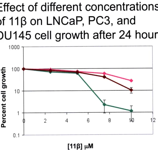
Published data has shown that LNCaP cells (AR-positive) are very susceptible to apoptosis after treatment with our candidate compound, 11 3, at a concentration of 10 p~M (10). It was anticipated that AR-positive cell lines would be more sensitive to 113 than AR-negative cell lines. In order to test this hypothesis, growth inhibition assays were performed with various cell lines that differed in AR and p53 status. The cell lines used were LNCaP (AR-positive, T877A), PC3 (AR null; p53 inactive), and DU145 (AR and p53 null). Experiments were carried out using the following concentrations of 11 3: 2.5,
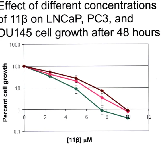
5.0, 7.5 and 10 pM. Percent control growth was assayed in cells that were treated for 24 or 48 hours with 113. Figure 3 shows the results for the 24-hour dose response
experiment. When cells were treated for only 24 hours, a one-log difference in the percent control growth was observed in favor of the AR-negative cells (PC3 and DU145). At certain doses, namely 7.5 and 10 jtM, AR-positive LNCaP cells are more sensitive to 11 treatment after 24 hours. However, when treated for 48 hours (Figure 4), there is no evidence of differential toxicity between the AR-positive and AR-negative cell lines. A possible explanation for these results is that 11 3 is able to induce apoptosis more quickly in cells expressing the AR because the AR could bind to the 11 3-DNA adducts generated and shield them from repair. Persistent adducts can then further react, creating more toxic crosslinks that can eventually trigger apoptosis, even in AR-negative cells (7). From these results it can be hypothesized that the compound is equally toxic to
AR-positive and AR-negative cell lines although the former show higher sensitivity. More experiments are needed to confirm this prediction.
PC3-AR cells show differential toxicity towards
I fl/
in a growth
inhibition assay, but not in a clonogenic assay'
Growth inhibition assays count cells as particles observed in solution, and does not distinguish between viable, healthy cells and cells that, even when they remain attached to the plate, will be unable to divide and are thus functionally dead. For this reason it was decided to use a clonogenic assay to probe differential toxicity in AR-positive and AR-negative cells. The advantage of this type of assay is that it counts aggregates or colonies of cells that form around surviving cells after treatment. Non-viable cells will not form colonies affording us a more reliable measure of cell survival when compared to the growth inhibition assay. While difficult to perform, this is a more robust assay for determining cell viability following drug treatment. Also, as noted above, the cells used to carry out the growth inhibition assay differed in AR and p53 status, which could affect other pathways that can potentially trigger the onset of apoptosis.
In order to test the role of the AR in prostate cancer cell survival after treatment with our candidate drug 11 3 a clonogenic assay was performed using AR and PC3-neo isogenic cell lines (28). This pair of cell lines only differs in the presence or absence of the AR receptor, eliminating any other possible confounding factors and focusing on the effect of the AR on cell survival. After these studies were completed Kyle Proffitt 'Mr. Kyle Proffit has continued to examine the differential toxicity of PC3-AR and PC3-neo cell lines. This work will be described later. He has found, using a clonal population of cells, that some of the conclusion below may need to be qualified.
isolated clonal cell populations and re-did many of the experiments below (this
experiment was not done with clonal populations). In the work presented here, the cells were plated at high density, allowed to grow for 24 hours, and treated immediately thereafter with 5.0 or 10 M 11 . After 2, 12, or 24 hours of exposure to the compound
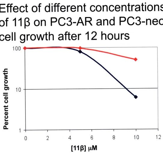
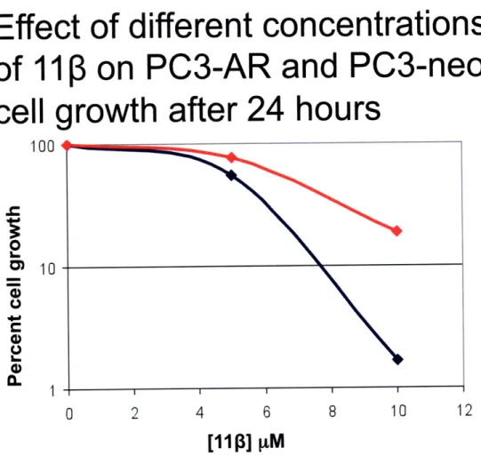
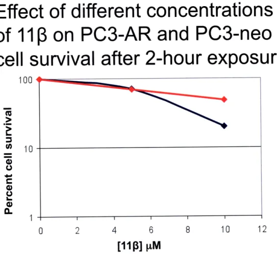
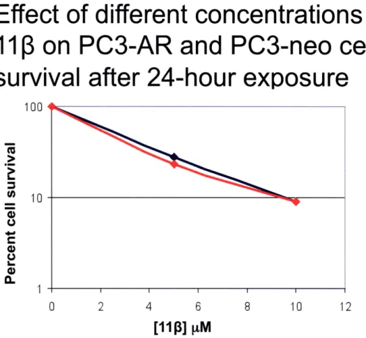
the cells were washed with PBS, counted, and re-plated in triplicate in 6-well plates at a low density. Cells were then monitored for colony formation up to seven days post treatment. This experiment also provided the opportunity to compare the results of the growth inhibition and clonogenic assays because attached cells were counted prior to re-plating. From the growth inhibition data collected, differential toxicity was observed between PC3-AR and PC3-neo cells at a 10 pM dose of 113 at 12 and 24 hours, but not at 2 hours post-treatment (Figures 5-7) This finding can best be explained by the fact that
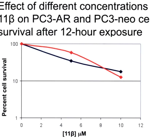
after 2 hours the compound might have not been able to exert its full effect on cells. In addition, it has been observed that it takes 15-20 hours for cells (LNCaP) to experience morphological changes indicative of apoptosis (10). From the clonogenic assay data no differential toxicity was observed at any of the doses or time points (Figures 8-10). Although there was a decrease in the percent cell survival at each concentration as time goes by, the rate at which cells are dying is not significantly different for PC3-AR and PC3-neo cells. This experiment has only been performed once so there are still
conditions that could be optimized in order to get a more accurate result that can address the issue of differential toxicity of 110 in AR-positive and AR-negative cell lines.
A possible confounding factor in this experiment is that some cells were lost during the 12 or 24-hour treatments. Only those remaining attached to the plate post
treatment were re-plated for the clonogenic assay. Any differential loss that occurred during treatment would not be reflected in the assay.
Immunoblot analyses of
p2
I levels in various
prostate
cancer
cell lines
Another important approach used to gain insight into possible mechanisms of action is to probe for proteins whose levels can be used as biomarkers for disease response to treatment. As established above in growth inhibition assays 110 has a selective sensitivity towards AR-positive cells resulting in cell cycle arrest and apoptosis (10). Published data has shown that p21, a CDK inhibitor, experienced dramatic changes upon treatment with 11 3 in LNCaP cells. p21 is transcriptionally activated by p53 in most cells, but this is not the only way by which p21 could be regulated (19; 20). Considering this, p21 levels were examined in the LNCaP, PC3, and DU145 cell lines, which represent different stages of prostate tumor progression. In addition to different AR status, they also differ in p53 status (active, inactivated, and null, respectively) (29).
Briefly, cells from each cell line were treated with 10 pM 11 and 20 tM
chlorambucil (which contains the DNA damaging moiety of 113) and whole cell extracts were prepared after various treatment times: 3, 6, 9, and 15 hours. Protein concentration for each extract was determined using the Bradford assay. Western blot analyses were carried out for samples of the three cell lines. Untreated cells in each cell line were used as control in each gel. Twenty micrograms of each protein sample were loaded per well and proteins were separated by SDS-polyacrylamide gel electrophoresis (PAGE), transferred to polyvinylidine fluoride (PVDF) membrane, and probed with monoclonal antibodies against p21 Wafl/Cipl and anti-mouse secondary antibodies conjugated to
horseradish peroxidase (HRP). Chemiluminescence reagents were added, and the membrane was exposed to film. After developing the film it was photographed and
bands were quantified by spot densitometry (Alpha-imager 2000 software). Membranes were also probed with actin antibody to ascertain that the same amount of protein was being loaded into each well and the changes in protein levels were not an artifact.
Protein loading curve
Before carrying out the 113 treatments on the different cell lines we wanted to make sure that our control compound chlorambucil was working properly and would elicit higher levels of p21 if higher amounts of protein were loaded per well. Also, in order to prove that our assay conditions were accurate a protein loading calibration curve was constructed using LNCaP cells treated with 20 ptM chlorambucil up to 15 hours,
since it was at this time point that the p21 levels in LNCaP cells were the highest,
according to previous experiments (10). Cells were treated as described above. After the cell extracts were prepared different amounts of protein were loaded into each well, namely: 10, 20, 30, 40, 50, and 60 ftg. Linear regression analysis showed that the amount of protein loaded into each well corresponded to the intensity of the p21 band in the gel with an R2 of 0.94, which is acceptable for the semi-quantitative analysis intended to be carried out (Figure 11).
p21 levels in LNCaP cells treated with chlorambucil and I I1
In the LNCaP cell line, treatment with chlorambucil resulted in a fluctuation of the p21 levels from 3-15 hours, experiencing the lowest level at 3 hours followed by an
12). In 11 3 treated cells, p21 levels decreased to one fifth of untreated levels after 3 hours of treatment and remained similar up to 9 hours. At 15 hours post treatment a
3-fold increase from the p21 levels measured at 9 hours was observed but this was still under untreated levels2. Actin blots showed that unequal amounts of protein were loaded, which could potentially change the results significantly. The results herein presented are not in accord with those previously reported in the literature in terms of the magnitude of the increase in the levels of p21, but when comparing the patterns observed for p21 expression both results match up (10).
p21 levels in DU 145 cells treated with chlorambucil and I IP
DU 145 cells showed a different expression profile for p21 after treatment (Figure 13). After treatment with 20 M chlorambucil p21 levels increased approximately 2-fold up to 6 hours of treatment, but by 9 hours levels had dropped to less than half of the untreated levels. When treated with 11, levels of p21 increased over 2-fold at the 3-hour time point when compared to the untreated sample. However, at 6 3-hours there was a sharp decrease in these levels and they continued to decrease up to 15 hours reaching a level that was less than half of the untreated levels. Actin blotting again showed unequal amounts of protein loaded into each well. Gels will be run again in optimized conditions.
p21 levels in PC3 cells treated with chlorambucil and I 13
PC3 cells showed yet a different pattern of p21 expression after chlorambucil and 1113 treatment (Figure 14). After treatment with chlorambucil there was only a slight increase in p21 levels up to 9 hours but after 15 hours levels fell back down to untreated
levels. For 11 3 treatment results are different. After remaining equal to untreated levels after 3 hours, a two-fold increase was observed at 6 hours post treatment followed by a seven-fold and -six-fold increase at the 9- and 15-hour time points, respectively. It is important to note that untreated levels of p21 in PC3 cells were very low, almost barely noticeable, which made any increase in p21 levels a rather large one (fold-wise), as was seen at the 9- and 15-hour time points. The fact that there is almost no p21 normally in PC3 cells might point to different pathways involved in the onset of apoptosis when compared to LNCaP and DU145 cells which contained similar levels of p21 in untreated conditions. Also, this might point to a p21-independent pathway, which triggers
apoptosis and may or may not be present or active in the other cell lines examined.
p21 levels in PC3-AR and PC3-neo isogenic cell lines treated with chlorambucil and I 10
As mentioned before, the cell lines examined until now differ in various biochemical factors including AR and p53 status. In order to isolate the variables and further characterize the role of the AR in p21 levels, if any, the PC3 isogenic pair, PC3-AR and PC3-neo, was used. Note, however that a clonal population was not used, which might affect the results. Nevertheless, in this set of experiments PC3-neo cells showed a similar p21 expression profile to DU 145 cells when treated with chlorambucil (Figure 15). After an increase at the 3-hour time point, three-fold in this case, p21 levels decreased gradually up to 15 hours when the levels observed were less than half of the untreated levels. Nonetheless, in this case p21 levels stayed above untreated up the 9-hour time point; they only went below at the 15-9-hour time point. After treatment with 11 3, p21 levels in PC3-neo cells rose above untreated levels but fluctuated anywhere
from 1.8 to 2.5-fold increase during the 15-hour time course, without any identifiable pattern being observed. On the other hand PC3-AR cells (Figure 16) lacked expression of p21 at all time points and treatments except at the 15-hour 11 P time point where a faint band could be appreciated. Since there was no signal on the rest of the lanes, spot
densitometry analysis was not performed on this gel. Nevertheless, it is an important observation because according to previously published data, AR-positive LNCaP cells also show a significant increase 15 hours after treatment with 1113. In this case as with PC3 cells, the levels of p21 in untreated cells were very low or there was absence of the protein, whereas the PC3-neo cells showed reasonable levels of the protein. In order to
corroborate the results presented here the experiment would have to be repeated. Given the results obtained in the previous experiments, which included various cell lines and other variables that complicate the analysis and future conclusions that could be drawn from them it was decided to focus on the LNCaP cell lines and characterize the changes in p21 expression levels that had already being published.
p21
degradation kinetics and mechanism
The original observation published in Marquis showed p21 levels decreasing sharply after only 3 hours of treatment with a subsequent increase several-fold above untreated levels at the 15-hour time point. It was decided to focus on this decrease and
determine what was causing it. A series of experiments were setup in order to assess the possibilities. The first experiment carried out had the purpose of providing cell extracts of LNCaP cells treated with sub-toxic concentrations of the candidate compound 11P3 (2.5 and 5.0 jtM) at different time points, up to 24 hours. These cell extracts would then be
probed for p21 expression in order to observe any changes in p21 levels at these lower concentrations of 11 . The second experiment was aimed and narrowing down the kinetics of this decrease in p21 levels by treating LNCaP cells with 10 iM 11 but the time points would be a on a shorter time scale, namely, 1, 2, 3, 4, and 6 hours in order to assay at which point p21 levels start going down or if it is a sudden decrease, and then be
able to narrow even further this time frame. Potentially these results could be correlated to morphological changes in the cell, or changes in cell growth or survival through
growth inhibition and clonogenic assays. The third and last experimental setup tried to answer the question of decreased p21 levels from a mechanistic standpoint. To this purpose the translation-blocker cyclohexamide (CHX) was used as a co-treatment in
LNCaP cells along with the already established doses of 10 [tM 11 and 20 iM chlorambucil. Cells were either treated with 10 tg/mL cyclohexamide (CHX) only, CHX and 11 , or CHX and chlorambucil, and extracts were prepared at 1, 2, 3, 5, 7, 9,
and 14 hours. This experiment would shed light on whether the p21 levels were being decreased as a result of increased degradation or if it was a result of a post-translational pathway being activated after treatment with 11 . All the experiments mentioned above were carried out and samples prepared. Only the immunoblots for the latter experiment were carried out. Unfortunately, there were some problems with the Western blot
technique and no results can be shown at this time. Regarding the other two experimental setups, due to time constraints, the Western blot analyses for these samples could not be carried out, but it will be certainly performed in the near future.
DNA isolation and preliminary growth rate studies on PC3-AR
and PC3-neo cells for
I I -DNA
adduct studies
Another important part of the project when it originated was to analyze the ability of 11p to form DNA adducts and how these adducts affect cell survival and apoptosis pathways. To this end, DNA adduct studies using accelerator mass spectrometry (AMS) were set up to examine the levels of 11 P-DNA adduct formation at different
concentrations of 11p, namely: 0.5, 1.0, 1.5, and 2.0 pM. These concentrations were
chosen because they are well below sub-toxic levels and would cause less interference with our study in terms of triggering unwanted effects or pathways in the cells studied.
The isogenic cell lines PC3-AR (AR-positive) and PC3-neo (AR-negative) were chosen to minimize variables presented by the use of different non-isogenic cell lines. Cells were treated with the aforementioned doses for 2 hours and cell lysates were prepared.
DNA isolation was difficult due to the small number of cells being used, which affected the final yield of DNA. The conditions for the DNA isolation have been since optimized but due to time constraints this study was postponed. This AMS DNA adduct study will also be performed in the other two AR-negative cell lines (PC3 and DU145) in the near future. By quantifying the adducts formed by different doses of 11p at various time points and analyzing the persistence of these adducts, a better understanding of the
observed physiological effects will be reached. Also, depending on the results obtained, the repair-shielding hypothesis could be validated. That is, if adducts persist longer in AR-positive cells it would imply that the AR is binding to the estradienone part of the molecule and is thus inhibiting quick and efficient repair of the adduct.
Before carrying out the 11 -DNA adduct study again, it was observed that PC3-AR and PC3-neo cells had different doubling times which would result in different cell
densities at the moment of treatment if both cell lines were plated at the same density at the same time. To this end, taking into account previous observations from other lab
members, a calculation was performed to obtain the amount of cells to be plated for each cell line in order to end up with a similar number of cells 48 hours after plating. The numbers obtained were 57,500 cells per plate for PC3-AR and 81,400 cells per plate for PC-neo, since PC3-AR cells have a faster doubling time than the PC3-neo cells. Even though our goal was to corroborate that at 48 hours there would be the same number of cells it was decided to include a 24- and 72-hour time point in order to assess the growth rate of these cells at different time points and gain a better understanding of their
doubling behavior. With this in mind it was decided to plate 50,000, 100,000, and 200,000 cells per plate in addition to the pre-calculated values established above for each cell line. Experiments were performed in triplicates for each of the established time points and each cell line. Results showed that after 31 hours (-24 hours) cell densities at plating of 50,000 and 100,000 cells per plate resulted in similar number of cells (Figure
17). At 50 hours (-48 hours), the pre-calculated densities of 57,500 cells per plate for PC3-AR and 81,400 cells per plate for PC3-neo resulted in very similar number of cells for the two cell lines at that time point (Figure 18). Nevertheless, it was at 78 hours (-72 hours) that these pre-calculated densities at the time of plating resulted in the closest number of cells of all the time points examined with a percentage difference of less than 2.5% (Figure 19). These results showed that the previous observations were accurate and that starting treatment between 48 and 72 hours after plating would result in a very
similar number of PC3-AR and PC3-neo cells. This in turn would yield more accurate results when measuring the 11 -DNA adducts since the same number of cells from each
cell line will be exposed to the same amount of nutrients from the new drug-containing media and of course the same amount of drug per cell, minimizing hence some of the possible confounding factors. As mentioned already, this study will be carried out in the near future.
Future Directions
The long-term objectives of this project are to assess the repair-shielding hypothesis and probe the signaling pathways that might be involved in triggering apoptosis following 113 treatment in prostate cancer cells. To this end a number of experiments have been planned to try to elucidate and clarify these issues.
As explained earlier, working with so many different cell lines can lead to the incorporation of confounding factors, which could make drawing conclusions an unreliable undertaking. The first step would be to concentrate on a well characterized cell line like the LNCaP cell line on which there is plenty of literature available including work done by our lab on our candidate compound, 113. This action will greatly simplify our problem so that accurate conclusions can be reached. As described in the Results and Discussion section, there are different questions that can be asked. One would be the effect of sub-toxic doses of 11 (2.5 and 5.0 jM) on p21 expression in LNCaP cells at
different time points, up to 24 or 36 hours. Also, a shorter time course can be designed
trying to narrow down the kinetics of p21 decrease, with time points every hour up to 6 hours. After identifying the range at which p21 levels start to go down, then it could be narrowed even further to examine more precisely at what time the ablation of p21 takes place and if it is a gradual or sudden change. In addition, it will also be interesting to process the samples already prepared in the experimental setup with cyclohexamide. By analyzing these results it can be preliminarily established if the decrease in p21 levels is due to increased degradation or if some other pathways are activated as the result of 11 3 treatment. Once this is answered it would be interesting to examine if lower
concentrations of 11
P
cause the same effect on p21 levels by the same mechanism, and in what time frame. Some of this will be answered through one of the experimental setups described above but further experiments would be needed in order to elucidate the whole mechanism, especially linking the changes in p21 to the apoptotic effects of 11 p on LNCaP cells. After this is done a closer look at the other cell lines (PC3, AR, PC3-neo, and DU145) can be taken to elucidate by which mechanism(s) 11 exerts its toxic effect, albeit on a different time scale or dose.To identify the main signaling pathways being impacted by treatment with 11 , a series of proteins that have been shown in preliminary studies to increase or decrease in expression levels after I 1i treatment will be assayed. The levels of these various proteins in different cell lines will be analyzed by immunoblotting. The cell lines to be used will be the five already referred to throughout the thesis. Other proteins to be assayed in the future that could be used as potential biomarkers for treatment response and disease prognosis are: p27, p45, Bcl-2, Bax, PARP, c-Myc, and p70S6K. By examining this set of proteins insight can be gained into which pathways are being affected, to what extent, and at what time after treatment these changes are being
observed. All of these experiments would address the mechanistic aspect of the decrease in p21 and tackle the question of other pathways that may be activated in response to 11p treatment.
Another important part of this project was studying the ability of 11 3 to create DNA adducts and if these adducts were higher in quantity or more persistent in AR-positive versus AR-negative cells. AMS technology can be employed to probe for 113-induced adduct formation and persistence in various cell lines, including AR-positive
(LNCaP and PC3-AR) as well as AR-negative cell lines (PC3, PC3-neo and DU145). The purpose of these experiments is to clarify the effect of 11 P-induced DNA damage on the overall effects observed in cells after treatment and examine if the AR plays any role
in the persistence or accumulation of these adducts. At the same time, by measuring repair kinetics for adducts formed, the repair shielding hypothesis can be tested and the
extent to which it is important in the whole scheme of effects seen in 110 treated cells can be assessed. Similar experiments can be performed in repair-deficient cells, which lack specific components of some of the repair pathways possibly involved in removing the 11 P-N7 adduct (i.e., nucleotide excision repair). The results could shed light into the
exact mechanism by which the adduct is forming and is potentially being removed in AR-negative cells, if the repair shielding hypothesis holds true.
AMS technology can also be used to evaluate the formation of 11 P-DNA adducts in vivo by isolating DNA from tumor tissues of xenograft mice. Briefly, nude,
immunodeficient mice will be injected with AR-positive and AR-negative cells (LNCaP, DU145, PC3, PC3-AR, and PC3-neo) subcutaneously into the rear flank. Depending on what results are obtained from the previous AMS studies as well as the apoptotic pathway study, priority will be given to the cell lines that show the most interesting and relevant results and can potentially provide the most insight into the hypothesis of repair
shielding. Without having those results available, the isogenic pair of AR and PC3-neo would be a logical choice to start with since it will directly target the repair-shielding hypothesis by virtue of the presence of absence of the AR, which is the basis for this proposed effect. Two to six weeks after implantation of tumor cells small nodules will become apparent near the injection site. After tumor size has reached a threshold of 1-2
mm in diameter 11p treatment will be initiated through intraperitoneal injection and tumor growth will be monitored. After approximately six weeks mice will be treated with 11 3 and DNA from tissues will be isolated four hours later. Accelerator mass spectrometry will then be used to measure formation and abundance of adducts in vivo.
By clarifying these aspects, a more complete picture could be generated regarding the effect of DNA damage, repair of DNA adducts (or lack thereof), and cell cycle and apoptotic pathways being affected by 11 in prostate cancer cells.
Bibliography
1. American Cancer Society. Cancer Facts and Figures 2008.
http://www.cancer.org/docroot/STT/stt_0.asp. Accessed: September 3, 2008. 2. Hanahan, D and Weinberg, RA. 2000. The Hallmarks of Cancer. Cell 100:57-70. 3. Hillier, SM, Marquis, JC, Zayas, B, Wishnok, JS, Liberman, RG, Skipper, PL,
Tannenbaum, SR, Essigmann, JM and Croy, RG. 2006. DNA Adducts Formed By a Novel Antitumor Agent 1 lbeta-Dichloro in Vitro and in Vivo. Mol Cancer Ther 5:977-984.
4. Huddart, RA and Birtle, AJ. 2005. Recent Advances in the Treatment of Testicular Cancer. Expert Rev Anticancer Ther 5:123-138.
5. Rosenberg, B, Van Camp, L, Grimley, EB and Thomson, AJ. 1967. The Inhibition of Growth Or Cell Division in Escherichia Coli By Different Ionic Species of Platinum(Iv) Complexes. Journal of Biological Chemistry 242:1347-1352.
6. Ohashi, K, Kajiya, K, Inaba, S, Hasegawa, T, Seko, Y, Furuchi, T and Naganuma, A. 2003. Copper(Ii) Protects Yeast Against the Toxicity of Cisplatin
Independently of the Induction of Metallothionein and the Inhibition of Platinum Uptake. Biochemical and Biophysical Research Communications 310:148-152. 7. Jamieson, ER and Lippard, SJ. 1999. Structure, Recognition, and Processing of
Cisplatin-DNA Adducts. Chem Rev 99:2467-2498.
8. Kartalou, M and Essigmann, JM. 2001. Recognition of Cisplatin Adducts By Cellular Proteins. Mutat Res 478:1-21.
9. Wang, D and Lippard, SJ. 2005. Cellular Processing of Platinum Anticancer Drugs. Nat Rev Drug Discov 4:307-320.
10. Marquis, JC, Hillier, SM, Dinaut, AN, Rodrigues, D, Mitra, K, Essigmann, JM and Croy, RG. 2005. Disruption of Gene Expression and Induction of Apoptosis in Prostate Cancer Cells By a DNA-Damaging Agent Tethered to an Androgen Receptor Ligand. Chem Biol 12:779-787.
11. Treiber, DK, Zhai, X, Jantzen, HM and Essigmann, JM. 1994. Cisplatin-DNA Adducts Are Molecular Decoys for the Ribosomal RNA Transcription Factor Hubf (Human Upstream Binding Factor). Proceedings of the National Academy of Sciences of the United States of America 91:5672-5676.
12. Trimmer, EE and Essigmann, JM. 1999. Cisplatin. Essays Biochem 34:191-211. 13. Mello, JA, Acharya, S, Fishel, R and Essigmann, JM. 1996. The Mismatch-Repair
Protein hMSH2 Binds Selectively to DNA Adducts of the Anticancer Drug Cisplatin. Chem Biol 3:579-589.
14. Mitra, K, Marquis, JC, Hillier, SM, Rye, PT, Zayas, B, Lee, AS, Essigmann, JM and Croy, RG. 2002. A Rationally Designed Genotoxin That Selectively Destroys Estrogen Receptor-Positive Breast Cancer Cells. Journal of the American Chemical Society 124:1862-1863.
15. Rink, SM, Yarema, KJ, Solomon, MS, Paige, LA, Tadayoni-Rebek, BM,
Essigmann, JM and Croy, RG. 1996. Synthesis and Biological Activity of DNA Damaging Agents That Form Decoy Binding Sites for the Estrogen Receptor. Proceedings of the National Academy of Sciences of the United States of America
16. Sharma, U, Marquis, JC, Nicole Dinaut, A, Hillier, SM, Fedeles, B, Rye, PT, Essigmann, JM and Croy, RG. 2004. Design, Synthesis, and Evaluation of Estradiol-Linked Genotoxicants as Anti-Cancer Agents. Bioorg Med Chem Lett
14:3829-3833.
17. Gartel, AL. 2006. Is P21 an Oncogene? Mol Cancer Ther 5:1385-1386.
18. Gartel, AL. 2005. The Conflicting Roles of the Cdk Inhibitor P21(Cipl/Wafl) in Apoptosis. Leukemia Research 29:1237-1238.
19. Gartel, AL and Tyner, AL. 1999. Transcriptional Regulation of the P21((Wafl/Cipl)) Gene. Exp Cell Res 246:280-289.
20. Gartel, AL and Tyner, AL. 2002. The Role of the Cyclin-Dependent Kinase Inhibitor P21 in Apoptosis. Mol Cancer Ther 1:639-649.
21. Zhang, H, Xiong, Y and Beach, D. 1993. Proliferating Cell Nuclear Antigen and P21 Are Components of Multiple Cell Cycle Kinase Complexes. Molecular Biology of the Cell 4:897-906.
22. Flores-Rozas, H, Kelman, Z, Dean, F B, Pan, Z Q, Harper, J W, Elledge, S J,
O'Donnell, M and Hurwitz, J. 1994. Cdk-Interacting Protein 1 Directly Binds With Proliferating Cell Nuclear Antigen and Inhibits DNA Replication Catalyzed By the DNA Polymerase Delta Holoenzyme. Proceedings of the National Academy of Sciences of the United States of America 91:8655-8659.
23. Tom, S, Ranalli, T A, Podust, V N and Bambara, R A. 2001. Regulatory Roles of P21 and Apurinic/Apyrimidinic Endonuclease 1 in Base Excision Repair. The Journal of biological chemistry 276:48781-48789.
24. Umar, A, Buermeyer, A B, Simon, J A, Thomas, D C, Clark, A B, Liskay, R M and Kunkel, T A. 1996. Requirement for Pcna in DNA Mismatch Repair At a Step Preceding DNA Resynthesis. Cell 87:65-73.
25. el-Deiry, W S, Tokino, T, Waldman, T, Oliner, J D, Velculescu, V E, Burrell, M, Hill, D E, Healy, E, Rees, J L and Hamilton, S R. 1995. Topological Control of P21Wafl/Cipl Expression in Normal and Neoplastic Tissues. Cancer Research 55:2910-2919.
26. Maki, C G and Howley, P M. 1997. Ubiquitination of P53 and P21 is
Differentially Affected By Ionizing and Uv Radiation. Molecular and Cellular Biology 17:355-363.
27. Sheaff, R J, Singer, J D, Swanger, J, Smitherman, M, Roberts, J M and Clurman, B E. 2000. Proteasomal Turnover of P21Cipl Does Not Require P21Cipl
Ubiquitination. Molecular Cell 5:403-410.
28. Cha, Tai-Lung, Qiu, Lin, Chen, Chun-Te, Wen, Yong and Hung, Mien-Chie. 2005. Emodin Down-Regulates Androgen Receptor and Inhibits Prostate Cancer Cell Growth. Cancer Research 65:2287-2295.
29. Radhakrishnan, Senthil K and Gartel, Andrei L. 2005. The Ppar-Gamma Agonist Pioglitazone Post-Transcriptionally Induces P21 in Pc3 Prostate Cancer But Not in Other Cell Lines. Cell cycle (Georgetown, Tex) 4:582-584.
1113
and chlorambucil structures
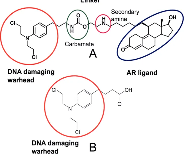
Linker
DNA damaging
AR ligand
warhead
DNA damaging
B
warhead
Figure 1. A, Structure of 1103 showing DNA damaging warhead (red oval), linker with carbamate moiety for stability (green oval) and secondary amine (purple oval) for solubility, and AR ligand (blue oval) portions of the molecule. B, structure of chlorambucil showing similar DNA damaging moiety as 113.
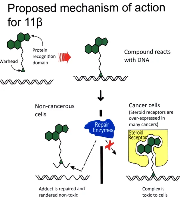
Proposed mechanism of action
for 1113
Protein
Compound reacts
recognition
Warhead domain
with DNA
Non-cancerous
Cancer cells
cells (Steroid receptors are
_- over-expressed in
many cancers) Steroid
Recetor\
I
Adduct is repaired and Complex is rendered non-toxic toxic to cells
Figure 2. Proposed mechanism of action for 11. 11p is a bifunctional agent designed to target DNA and the androgen receptor (AR) which is over-expressed in prostate cancer cells. The proposed mechanism establishes that the molecule will first react with DNA through its aniline mustard moiety. In non-cancerous cells DNA repair enzymes will come in and excise the lesion rendering the 11 p-DNA adduct non-toxic. On the other hand if thell
3-DNA adduct is formed within a prostate cancer cell expressing the AR, this steroid receptor
will potentially bind to the AR-ligand moiety of 110 blocking the DNA repair enzymes. The
11P -DNA adduct will persist and due to the cell's inability to repair the adduct it will
automatically trigger cell death or apoptosis.
Repair
Enzymes
Effect of different concentrations
of 1113
on LNCaP, PC3, and
DU145
cell growth after 24 hours
1000 -L. 1.0 I= 100 10 1
[11 P]
PM
Figure 3. LNCaP cells are more sensitive to 11ip at
concentrations higher than 5.0 pM. Growth inhibition assays were performed to examine the effects of 110 on AR-positive and AR-negative cells. Three different cell lines were used: LNCaP
(AR+, T877A), PC3 (AR null; p53 inactive) and DU145 (AR and p53 null). Cells were exposed for 24 hours to different concentrations of
11P: 0, 2.5, 5.0, 7.5 and 10 [iM.
LNCaP: AR+, p53+
PC3:
AR-,
Effect of different concentrations
of
11r3
on LNCaP, PC3, and
DU145
cell
growth
after 48 hours
10001 M 0 (I) 0.
n
n 100 10 1 0.1[11P]
p~M
Figure 4. LNCaP cells show no differential toxicity after 48 hours of 11 exposure. Using the same experimental setup
described in Figure 3, results showed that after 48 hours of exposure
1113 is equally toxic to LNCaP, PC3, and DU145 cells at all 111 concentrations examined.
LNCaP: AR+,
p53+
PC3:
AR-,
Effect of different concentrations
of
1113
on PC3-AR and PC3-neo
cell growth after
2
hours
100 0 LM (I) I.0 I,,, 0"1 Q) e .4
[111P] iM
Figure 5. PC3-AR and PC3-neo cells show no differential toxicity after 2 hours of 11P treatment. A growth inhibition assay
was also performed using an isogenic cell pair, namely PC3-AR (AR-positive) and PC3-neo (AR-negative). These cell lines only differ in the presence or absence of the AR receptor. Cells were treated with 5 pM and 10 pM 110, or left untreated. After being
exposed to 110 for 2 hours no significant difference in cell growth was seen between PC3-AR and PC3-neo cells.
PC3-AR: AR+
PC3-neo:
AR-Effect of different concentrations
of
1113
on PC3-AR and PC3-neo
cell growth after
1
100
'
0.o
iilU
a-hours
0 2 4 6 8 10 12[1113]
IM
Figure 6. PC3-AR cells are more sensitive to 10 pM 11ip than PC3-neo cells after 12 hours. Using the same experimental setup
discussed in Figure 5, PC3-AR and PC3-neo cells were exposed to different concentrations of 1113. After 12 hours of treatment, PC3-AR cells exhibited differential toxicity only at the 10 pM
concentration with less than 10% cell growth.