ORIGINAL ARTICLE
Targeted alpha-radionuclide therapy of functionally
critically located gliomas with
213
Bi-DOTA-[Thi
8
,Met(O
2
)
11
]-substance P: a pilot trial
D. Cordier&F. Forrer&F. Bruchertseifer&
A. Morgenstern&C. Apostolidis&S. Good&
J. Müller-Brand&H. Mäcke&J. C. Reubi&A. Merlo
Received: 24 September 2009 / Accepted: 5 January 2010 / Published online: 16 February 2010 # Springer-Verlag 2010
Abstract
Purpose Functionally critically located gliomas represent a challenging subgroup of intrinsic brain neoplasms. Stan-dard therapeutic recommendations often cannot be applied, because radical treatment and preservation of neurological function are contrary goals. The successful targeting of gliomas with locally injected beta radiation-emitting 90 Y-DOTAGA-substance P has been shown previously. How-ever, in critically located tumours, the mean tissue range of 5 mm of90Y may seriously damage adjacent brain areas. In contrast, the alpha radiation-emitting radionuclide 213Bi with a mean tissue range of 81 µm may have a more favourable toxicity profile. Therefore, we evaluated locally injected213Bi-DOTA-substance P in patients with critically located gliomas as the primary therapeutic modality.
Methods In a pilot study, we included five patients with critically located gliomas (WHO grades II–IV). After diagnosis by biopsy, 213Bi-DOTA-substance P was locally injected, followed by serial SPECT/CT and MR imaging and blood sampling. Besides feasibility and toxicity, the functional outcome was evaluated.
Results Targeted radiopeptide therapy using 213 Bi-DOTA-substance P was feasible and tolerated without additional neurological deficit. No local or systemic toxicity was observed. 213Bi-DOTA-substance P showed high retention at the target site. MR imaging was suggestive of radiation-induced necrosis and demarcation of the tumours, which was validated by subsequent resection.
Conclusion This study provides proof of concept that targeted local radiotherapy using 213Bi-DOTA-substance P is feasible and may represent an innovative and effective treatment for critically located gliomas. Primarily non-operable gliomas may become resectable with this treat-ment, thereby possibly improving the prognosis.
Keywords Glioma . Substance P. Targeted therapy .213Bi
Introduction
Intracranial gliomas are intrinsic brain tumours that are classified according to their histological features by the WHO grading system. WHO grade IV gliomas (glioblastoma multiforme, GBM) carry the worst prognosis with an overall survival of often less than 12 months [1]. WHO grade II gliomas feature a better prognosis with a median survival of 8–10 years. WHO grade III gliomas (anaplastic gliomas) exhibit a prognosis closer to GBM than to the grade II gliomas; grade I gliomas are regarded as potentially curable.
D. Cordier (*)
:
A. MerloDivision of Neurosurgery, University Hospitals, Basel, Switzerland
e-mail: [email protected]
F. Forrer
:
S. Good:
J. Müller-Brand:
H. Mäcke Institute of Nuclear Medicine, University Hospitals, Basel, SwitzerlandF. Bruchertseifer
:
A. Morgenstern:
C. Apostolidis European Commission, Joint Research Centre, Institute for Transuranium Elements,Karlsruhe, Germany H. Mäcke
Nuclear Medicine and Radiological Chemistry, University Hospital Basel,
Basel, Switzerland J. C. Reubi
Institute of Pathology, University of Berne, Berne, Switzerland
The resection of GBM, followed by combined radio-chemotherapy, is accepted as the current therapeutic standard [2]. A high extent of resection was found to be positively correlated with prolonged time to tumour recurrence [1,3,4]. Different technical developments such as 5-aminolevulinic acid for intraoperative tumour visual-ization [5], navigational systems or intraoperative MRI [6,
7] allow a high extent of resection to be achieved. However, if the tumour is critically located in brain areas that are responsible for functions such as speech or motor function, this aggressive therapeutic approach often cannot be efficiently applied. Preservation of functions is a major goal, and so the extent of resection needs to be limited. This results in higher residual tumour volumes, which represent suboptimal prerequisites for adjuvant radioche-motherapy. In the less aggressive low-grade gliomas (WHO grade II), the situation is more complex: therapeu-tic recommendations range from the “wait and see” strategy over radiotherapy and/or chemotherapy to an initial radical resection [8–12]. If low-grade gliomas are functionally critically located and affect important neuro-logical functions of the patient, there is even more therapeutic insecurity whether taking a higher risk for functional deterioration improves the patient’s overall outcome.
Besides treatment of the main tumour mass, therapy of the infiltration zone needs to be addressed as a major goal, because 95% of gliomas exhibit local recurrence [13].
WHO grades II–IV gliomas have been shown to consistently overexpress the transmembraneous neuro-kinin type-1 receptor (NK-1). NK-1 receptors have also been detected on tumour cells infiltrating the intra- and peritumoural vasculature [14]. The physiological ligand of NK-1 receptors is substance P. The local intratumoural injection of radiolabelled substance P exploits this overexpression of the NK-1 receptor. Substance P can be radiolabelled with various radionuclides for diagnos-tic or therapeudiagnos-tic applications using the chelators DOTAGA (1,4,7,10-tetraazacyclododecane-1-glutaric acid-4,7,10-triacetic acid) or DOTA (1,4,7,10-tetraazacy clododecane-1,4,7,10-triacetic acid). Radiolabelled DOTAGA-/DOTA-substance P exhibits a preserved affinity to the NK-1 receptor in the low nanomolar range; the in vivo stability has been confirmed [15]. Its molecular weight of only 1.8 kDa facilitates sufficient and rapid intratumoural distribution after local application. Treat-ment with90Y-labelled substance P led to good functional outcome and a trend toward improved survival when evaluated as a second-line treatment for recurrent gliomas [15]. Tumour resection following this treatment was signif-icantly facilitated by radiation necrosis and pseudoencapsu-lation of the tumour. We conducted another study to evaluate this treatment as a neoadjuvant modality: 90Y-labelled
substance P was locally injected after biopsy confirmation of GBM, followed by resection of the pretreated glioma. This concept was feasible without signs of decompensating intracranial pressure and relevant toxicity. Of 16 patients, 14 stabilized or improved their neurological function; the pseudocapsule-like tumour demarcation allowed a high extent of resection to be achieved in subsequent surgery (submitted).
The radionuclide used in these studies, 90Y, has several advantageous characteristics:β-particle emitter with a mean energy of 2.1 MeV, mean tissue range of 5 mm, commer-cially available and standardized handling. However, in functionally critical areas of the brain, the tissue range of
90
Y with the resulting “cross-fire effect” [16] potentially damages adjacent brain areas. Alternatively, α-particle-emitting radionuclides may be powerful candidates: 213Bi as an example has a mean tissue range of only 81 µm with a high mean energy of 5.8 MeV, allowing a highly cytotoxic radiation dose to be delivered to targeted cells while sparing adjacent healthy tissue.
Targeted alpha therapy using 213Bi has been shown to exhibit a favourable toxicity profile and therapeutic potential in phase 1 trials in leukemia, non-Hodgkin’s lymphoma and melanoma [17–19]. The short-lived alpha emitter213Bi (T1/2=46 min) is available from a radionuclide
generator loaded with its longer-lived mother nuclide225Ac (T1/2=10 days), which can be produced via radiochemical
extraction from 229Th [20] or cyclotron-driven methods [21]. In this pilot study, we present the results of five patients with WHO grades II–IV gliomas who have been treated with locally injected213Bi-DOTA-substance P as the primary therapeutic modality. Besides feasibility and toxicity as primary end-points, the functional outcome was examined as a secondary end-point.
Materials and methods
Patients
Study patient data are summarized in Table 1. The study protocol had been approved by the Ethics Committee of the University Hospitals of Basel, Switzerland. Patients meet-ing the inclusion criteria were informed about current therapy options. The rationale for inclusion of patients with WHO grade II tumours is, despite the better prognosis than the high-grade gliomas, the still life-limiting character of the disease. Informed consent was obtained before inclu-sion. The functional status was assessed using the Barthel Index, which evaluates neurological function by evaluating the impairment in activities of daily living [22]. Tumour volumetry was performed using the software IPlan2.6 (BrainLAB, Feldkirchen, Germany).
Inclusion criteria
The criteria for inclusion were: newly diagnosed and histopathologically confirmed unifocal glioma (WHO grades II–IV); tumour diameter max. 5 cm; no evidence for obstruction of CSF circulation or decompensating intracranial pressure; Karnofsky performance score ≥70; age 18–75 years; absence of psychological, familial, sociological or geographical conditions potentially hamper-ing compliance with the study protocol.
Study protocol
Week 1: stereotactic biopsy, intratumoural implantation of catheter systems (Fig.1). In tumours≤3 cm in diameter one catheter was implanted and in larger tumours or those with complex configuration two or three catheters. Week 2: local test injection with 111In-DOTA-substance P for confirma-tion of positive NK-1 receptor expression and orthotopic dose distribution. Week 3: intratumoural injection of213 Bi-DOTA-substance P. Depending on clinical and radiographic
findings: repetition of intratumoural injection, otherwise post-therapeutic evaluation as depicted below. Tumour resection after radiopeptide treatment was offered if the operation was feasible and of probable benefit. In cases of tumour progression/recurrence: histology by biopsy or, if feasible, by tumour resection; combined with chemotherapy and/or external beam radiotherapy.
DOTA-substance P
DOTA-[Thi8,Met(O2)11]-substance P was synthesized as
described for the corresponding DOTAGA derivative [15].
Production of 225Ac/213Bi, radiolabelling and stability testing
225
Ac/213Bi was produced at the Institute of Transuranium Elements (Karlsruhe, Germany) by radiochemical extrac-tion from a229Th source [20,23].213Bi was eluted from a
225
Ac/213Bi-generator using 600 µl 0.1 M NaI/HCl (Merck, Darmstadt, Germany, suprapure grade) solution. Addition of 50 µl of 20% ascorbic acid (Prolabo, VWR International, Fontenay sous Bois, France, normapure grade) as radio-protectant, pH adjustment to 8.5–8.7 with 70 µl of 2 M Na2CO3 (Merck, Darmstadt, Germany, suprapure grade).
Incubation of buffered 213Bi eluate (270–1174 MBq) with 10 µg (n=5, patient 1) or 30 µg (n= 22, patients 2–5) DOTA-substance P for 5 min at 95°C in a microwave oven (Biotage Initiator, Uppsala, Sweden) and addition of 30 µl 1 mM Ca-DTPA to complex-free213Bi [24]. Quality control by standard gradient radio-HPLC using a Chromolith Speed ROD RP-18 endcapped 50-4.6 mm column (Merck, Darmstadt, Germany) with acetonitrile-water gradient and ion thin-layer chromatography (ITLC-SG, Pall Inc., New York, NY, USA) using 0.05 M sodium citrate solution (pH 5.5) as mobile phase, followed by sterile filtration. Synthesis, quality control and sterile filtration of 213
Bi-Brain Skull Skin Intratumoral Catheter Tumor Port
A
B
Fig. 1 Intratumoural catheter system. a Schematic drawing of implanted catheter system. b Catheter system, consisting of a bottom outlet port capsule and a connected standard intraventricular catheter Table 1 Patients and characteristics
Pat. No. Age at Dx (years) Diagnosis/location of tumour Cycles/activity (GBq) Tumour volume (cm3)
Barthel Index pre-/post-therapeutic PFS (months) OS (months) 1 60 GBM frontal L callosal 1/1.07 41.6 75/ 90 2 16 2 40 GBM frontal L (SMA precentral) 1/1.92 76.0 80/ 90 11 19
3 55 Astro WHO grade III
fronto-opercular L
4/7.36 74.3 100/100 24+ 24+
4 33 Astro WHO grade II frontal
R (SMA)
1/1.96 12.0 100/100 23+ 23+
5 39 Astro WHO grade II
occipital R
1/2.00 17.1 100/100 17+ 17+
PFS progression-free survival, OS overall survival, + ongoing, SMA supplemental motor area, L left, R right, Astro astrocytoma, GBM glioblastoma multiforme, Dx diagnosis
DOTA-[Thi8,Met(O2)11]-substance P was performed in less
than 15 min to minimize loss of activity. The radiochemical purity of the final product was 98.0±1.4% at specific activities of 20.2±3.9 MBq213Bi/µg peptide.
The stability of213Bi-DOTA-[Thi8,Met(O2)11]-substance
P was tested in human blood serum. Serum samples were prepared from blood samples taken from healthy volun-teers. An aliquot of 213Bi-DOTA-[Thi8,Met(O2)11
]-sub-stance P (18.5 MBq 213Bi/µg peptide) was added to a tenfold excess of blood serum and incubated at 37°C. At various time points, an aliquot was analysed using ion thin-layer chromatography (ITLC-SA, Pall Inc., New York, NY, USA) with 0.05 M sodium citrate solution (pH 5.5) as mobile phase. Under these conditions, intact213 Bi-DOTA-[Thi8,Met(O2)
11
]-substance P radioconjugate remains at the bottom of the ITLC strip (Rf=0), while free213Bi released
from the radioconjugate moves with the solvent front (Rf=1). The radioconjugate was found to be very stable
towards dissociation, as within 5 h, corresponding to 6.5 half-lives of 213Bi, no release of 213Bi could be observed within the uncertainty of the measurement (<2%). This is in good agreement with a previous study investigating the stability of 213Bi-DOTA-[Thi8,Met(O2)11]-substance P in
the presence of a 250-fold excess of DTPA as competing ligand using radio-HPLC. In this study, a very slow release of213Bi with a half-life of 185 h was found.
Injection of the radiopharmaceutical
Intratumoural injection of the radiopharmaceutical is performed via one to three implanted catheters [25–27]. The catheter is connected to a subcutaneous port, which is punctured for injection (Fig. 1). Application of 12 mg dexamethasone and, immediately before injection, 60 g mannitol transiently reduces intratumoural pressure. Before injection, the system is flushed with 1.5 ml human albumin 5% to coat the plastic surface. The active drug is injected in a volume of 1 ml. Finally, the system is flushed again with 1.5 ml human albumin. For each therapeutic cycle, three to five injections were performed over 2 days.
Evaluation of receptor expression and dose distribution
Each patient was injected with 1 MBq of 111 In-DOTA-[Thi8,Met(O2)11]-substance P per catheter as test injection
before therapy. Images were acquired immediately after injection and 4 and 24 h after injection to confirm stable and orthotopic dose distribution. The images were acquired as 3-D data set (SPECT and low-dose CT) using an integrated dual-head SPECT/CT camera (SYMBIA T2,
Siemens, Malvern, PA, USA) and automatic fusion.
111
In SPECT images were acquired in continuous scan mode (25 min/rotation). The windows were centred over
both 111In photon peaks (245 and 172 keV, width ±20%). Therapeutic activities were administered only if the activity was found stable within the tumour, i.e. no dose deposition outside the tumour margins on SPECT/CT over 24 h which was interpreted as specific binding.
After every application of 213Bi-DOTA-[Thi8,Met (O2)11]-substance P, SPECT/CT was acquired to confirm
orthotopic dose deposition. For SPECT, the window was centred at 440 keV (width ±20%). SPECT was acquired with 128 views; acquisition time was 10 s/view.
Blood sampling was performed until 4 h after injection. Aliquots of 3 ml were analysed for 213Bi activity using a NaI(Tl) bore well counter (COBRA II, D 5003 γ-system, Canberra Packard, Melbourne, Australia). The percentage of injected213Bi in the blood pool was determined based on calculated total blood volumes.
Post-therapeutic evaluation
Patients were clinically evaluated weekly within the first 8 weeks after radiopeptide application, afterwards monthly for the grade III and IV tumours and in 3-month intervals for the grade II tumours. MR imaging was performed monthly after injection of the radiopharmaceutical for the first 3 months, thereafter in 3-month intervals for the grade III and IV tumours and in 6-month intervals for the grade II tumours.
Results
Patient characteristics and functional status
Two patients with low-grade astrocytomas (WHO grade II), one patient with anaplastic glioma (WHO grade III) and two patients with GBM (WHO grade IV) in functionally critical locations were treated with intratumourally injected
213
Bi-DOTA-substance P as the primary therapeutic mo-dality (Table 1). The preliminary diagnosis from the fresh-frozen sections was confirmed by definite histology in all cases. Four patients received 1.07–2.00 GBq213
Bi-DOTA-substance P within one therapeutic cycle, and one patient received four therapeutic cycles with a total activity of 7.36 GBq. The pre-therapeutic functional score, evaluated by the Barthel Index [22], was 100 out of 100 in the patients with the WHO grade II and III tumours (patients 3, 4 and 5). In these patients, this unimpaired functional status did not change during or after treatment. The two GBM patients displayed pre-therapeutic Barthel scores of 75 (patient 1) and 80 (patient 2), which means moderate restrictions in activities of daily living. Both GBM patients improved to a score of 90 after treatment, being consistent with minor impairment in daily living (Table1).
Biodistribution and pharmacokinetics
Positive NK-1 receptor expression was demonstrated by test injection of 111In-DOTA-substance P in all patients. The dose distribution of the test injection was congruent with tumour morphology in all cases. The sufficient dose distribution expected from pre-therapeutic test injections could be verified in all cases by post-therapeutic assessment
by SPECT/CT imaging ( shown as examples in Figs.4d–f and 6m–n). High retention of213Bi-DOTA-substance P at the target site was furthermore confirmed by low percent-age values of the injected dose (%ID) in the blood pool. Maximum values of < 4%ID were found at 60–90 min after injection (Fig. 2a). At these time points 50–75% of the
injected activity of short-lived 213Bi (T1/2 = 46 min) have
already decayed. Consequently,213Bi activities found in the
0.0 0.5 1.0 1.5 2.0 0 50 100 150 200 250
Time post application (min)
213 B i ac ti v it y in blood ( % I .D .) 0.0 0.5 1.0 1.5 2.0 2.5 3.0 3.5 4.0 0 50 100 150 200 250
Time post application (min)
213 B i ac ti v it y in blood ( % I .D .) .
A
B
Fig. 2 Activity of 213Bi in blood after local intratumoural injection of213 Bi-DOTA-sub-stance P. Patient 1: closed tri-angles, patient 2: open tritri-angles, patient 3: open circles, patient 4: closed circles, patient 5: closed squares. a Percentage of injected dose after decay cor-rection. b Percentage of injected dose without decay correctionA
B
C
D
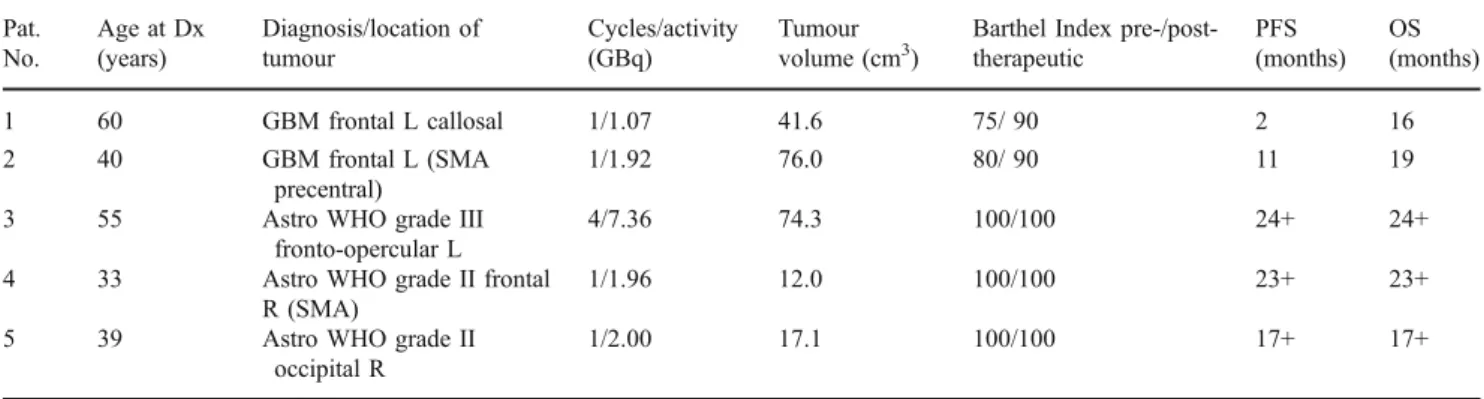
E
Fig. 3 Patient 1, left frontal GBM, CT and T1-weighted contrast-enhanced MR imaging. a Initial CT imaging before stereotactic biopsy and catheter placement. b T1-weighted contrast-enhanced MR imaging 2 weeks after intratumoural radiopeptide application, c
4 weeks after radiopeptide application and after partial resection of the tumour, d 6 weeks after radiopeptide application highly suspicious for tumour progression and e 8 weeks after radiopeptide application with evident tumour progression
blood pool corresponded to less than 2% of the activities injected intratumourally (Fig.2b).
Patient characteristics and response
Patient 1 exhibited a bifrontal GBM; due to the limited prognosis she was initially not considered as an operative candidate. Local injection of 1.07 GBq 213 Bi-DOTA-substance P resulted in intratumoural disseminated changes strongly suggestive for radionecrosis in MR imaging, but on the other hand, volumetric expansion of the tumour was noted. Subsequent partial resection confirmed the radio-graphic suspicion of radionecrosis and relieved the patients’ mental status changes. MR imaging revealed tumour recurrence after only 1 month after the operation (i.e. 2 months after radiopeptide application), whereas the clinical improvement was stable for 10 months (Fig.3).
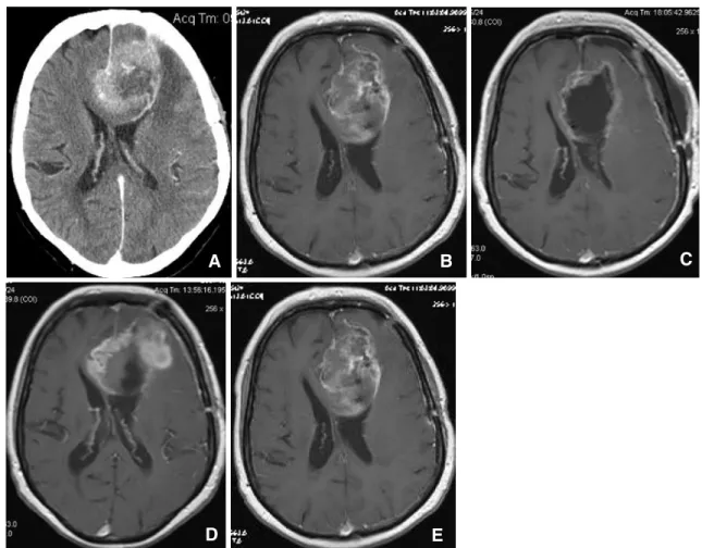
Because only about 50% of the tumour volume in patient 1 displayed radionecrotic changes in MR imaging and due to the absence of any signs of toxicity, the dose in patient 2 was increased to 1.92 GBq. Patient 2 initially presented with right-sided hemiparesis and motor dysphasia due to a
left frontal GBM, affecting the precentral gyrus and the supplemental motor area (SMA). She clinically improved after radiopeptide treatment, becoming ambulatory again. Radiologically, the tumour demarcated with findings suspicious for radionecrosis (Fig. 4a–c). According to the
patient’s wish, the pretreated tumour was resected. A “radical” resection was, due to the location of the tumour, not possible. First clinical and radiographic signs of tumour progression were observed 11 months later. The GBM patients (patients 1 and 2) were postoperatively treated with external beam radiotherapy and temozolomide chemother-apy according to the current therapeutic standard.
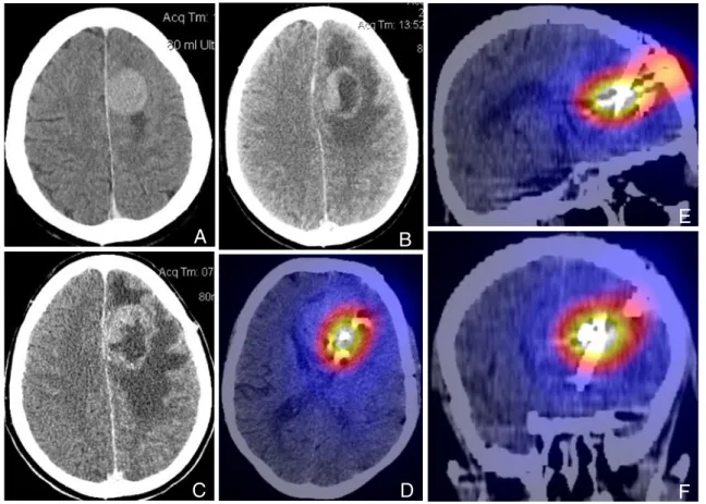
The anaplastic left fronto-opercular astrocytoma (WHO grade III) of patient 3 exhibited a diameter of 5 cm, causing dysphasia. Radiographic changes after injection of 213 Bi-DOTA-substance P, suggestive for radionecrosis, were observed mainly in proximity to the catheters in cortical and apical parts of the tumour, only to a much lesser extent in deep and basal parts (Fig. 5). The catheters were re-positioned and patient 3 received further therapeutic cycles. The distribution of radiological signs of radionecrosis did not significantly change in the further course, but
interest-A
C
D
F
E
B
Fig. 4 Patient 2, left frontal GBM, contrast-enhanced CT imaging (a– c) and SPECT/CT (d–f). a Initial CT scan before stereotactic biopsy and catheter placement. CT scan at b 6 weeks and at c 10 weeks after radiopeptide treatment. Besides the intratumoural changes, please note
the increasing perifocal oedema. SPECT/CT in d axial, e sagittal and f coronal planes with orthotopic dose distribution immediately after intratumoural application of213Bi-DOTA-substance P
ingly there is no clinical or radiographic evidence of progression in the following 24 months.
Patient 4 initially complained about fatigue, accompa-nied by left-sided partial brachiofacial seizure equivalents and facial paresis. MR imaging displayed a right-sided and non-contrast enhancing mass of the SMA extending to the precentral gyrus (Fig.6a, b); biopsy revealed a WHO grade II astrocytoma. Injection of 1.96 GBq 213 Bi-DOTA-sub-stance P resulted in extensive demarcation of the tumour (Fig.6c–f). According to the patient’s wish, the tumour was
subsequently resected. Intraoperatively, the lesion appeared as a necrotic and well-defined encapsulated mass, enabling macroscopic complete tumour removal. Follow-up imaging is so far without evidence for recurrent tumour (Fig.6g–l).
The patient’s fatigue improved and the facial paresis has resolved; anticonvulsive prophylaxis was continued be-cause of electroencephalographic findings of focal epilepsy-specific potentials.
Patient 5 presented with fatigue and headache, which led to the diagnosis of an occipital astrocytoma (WHO grade II). After local injection of 2.00 GBq 213 Bi-DOTA-substance P there is evidence for extensive radionecrotic demarcation of the tumour in repetitive MR imaging in the post-therapeutic follow-up (Fig. 7). The clinical condition of this patient did not change in the course of treatment.
Side effects and toxicity
Intratumourally injected 213Bi-DOTA-substance P was tolerated well by all patients; no relevant acute local or systemic toxicity could be found according to the National Cancer Institute (NCI) Common Toxicity Criteria (CTC) scale (version 2.0). In the long-term follow-up, no further substantial local or systemic toxicity was found. Cortico-steroid therapy generally was tapered 2 weeks after radio-peptide application.
Fig. 5 Patient 3, left opercular anaplastic astrocytoma, contrast-enhanced T1- and T2-weighted MR imaging. a–c Initial imaging before stereotactic biopsy and catheter placement. d–f Status after radiopeptide treatment as described in the text. All three implanted
catheter systems are visible in the axial planes; one catheter with its injection port is visible in the coronal plane. Note the inhomogeneous radionecrotic changes mainly concentrated around the catheters
Fig. 6 Patient 4, right frontal WHO grade II astrocytoma, contrast-enhanced T1- and T2-weighted MR imaging and SPECT/CT. a, b Initial imaging before stereotactic biopsy and catheter placement. c, d Status 2 weeks after intratumoural213Bi-DOTA-substance P injection.
e, f Status 6 weeks after radiopeptide injection; note that the lesion appears more compact with less oedema. g, h Imaging after resection
of the tumour. i, j Status 6 months after resection without evidence for residual or recurrent tumour. k, l Unchanged status 1 year after resection. m, n SPECT/CT with orthotopic dose distribution 30 min after intratumoural application of 213Bi-DOTA-substance P in two
axial planes Sep. 09, 2009 Mar. 29, 2008 Jan. 24, 2008 Sep. 27, 2008
A
C
E
B
D
F
H
G
Fig. 7 Patient 5, left occipital WHO grade II astrocytoma, contrast-enhanced T1- and T2-weighted MR imaging. a, b Imaging before stereotactic biopsy and catheter placement. c, d Status 4 weeks after intratumoural213Bi-DOTA-substance P injection. e, f Status 7 months
after radiopeptide injection; note that the lesion appears slightly more compact. g, h Imaging 19 months after radiopeptide treatment, most probably consistent with local scar tissue formation
Discussion
Functionally critically located malignant gliomas still represent an immense therapeutic challenge, because often the therapeutic standards for an aggressive local therapy, i.e. surgery and radiotherapy, cannot be applied. There is no doubt about the importance of sufficient local control of high- and low-grade gliomas, since virtually all of them exhibit local recurrence. In critically located gliomas, the current technical developments for a higher extent of surgical resection [5,7,28] are only partially useful because in this entity of tumours they carry a high risk of damaging neurological function. Awake surgery and/or intraoperative monitoring techniques represent valuable tools to avoid postoperative neurological deficits such as loss of motor function or speech, but remain without therapeutic effect on the prognostically pivotal infiltration zone. Consequently, patients with critically located gliomas are often not considered as candidates for an aggressive local therapy.
The fact that WHO grades II–IV gliomas do consistently overexpress NK-1 receptors led to the therapeutic local application of the radiolabelled NK-1 ligand DOTAGA-/ DOTA-substance P. The concept, besides treatment of the main mass, is a targeted approach to tumour cells in the surrounding infiltration zone. Previous studies have shown feasibility and low toxicity of this method as a second-line therapy [15] as well as in a neoadjuvant setting (submitted). However, the radionuclide 90Y, which was used in these previous studies, may lead to unacceptable damage to adjacent brain areas because of its relatively high mean tissue range of 5 mm. The possibility of using213Bi instead of 90Y may be a safer and even more efficacious alternative: The mean tissue range is only 81 µm with a high mean energy of 5.8 MeV (90Y: 2.1 MeV), corresponding to a high linear energy transfer (LET) of 72 keV/µm. Consequently, the distance between ionizing events caused by alpha particle emission of213Bi is similar to the distance between DNA double strands, thus increasing the probability of inducing irreparable DNA double strand breaks. These prerequisites led to the design of the pilot study presented in this work: The histological diagnosis of a glioma in patients with eloquently located tumours was established by biopsy, followed by implanta-tion of intratumoural catheters. Subsequently,213 Bi-DOTA-substance P was intratumourally injected as the primary therapeutic modality. SPECT/CT and blood sampling confirmed high retention of the radiopharmaceutical at the tumour site, in agreement with absence of toxicity to non-targeted, healthy brain tissue or other organs. MR imaging showed intratumoural changes highly suspicious for radio-necrosis, which was confirmed by histopathological exam-ination. Small tumours exhibited a complete radionecrotic appearance, whereas larger tumours seemed to be mainly
necrotic in the proximity of the implanted catheters. This observation is probably due to the fact that the physical half-life of 213Bi is only 46 min, so that a significant fraction of this radionuclide is possibly decayed before a sufficient intratumoural distribution is achieved. In large tumours, possibly different radionuclides such as 225Ac with longer half-lives could be more efficacious. However, in one of two GBM patients and in the patient with the anaplastic glioma there is, despite incomplete radionecrotic demarcation of the tumours, a long time interval to further progression, so that possibly there is also a therapeutic effect in areas without radiographic evidence for radio-necrosis. To the contrary, the first GBM patient, who received a relatively low dose, had only a short time interval to tumour progression. This observation may indicate a dose-effect relationship that needs to be con-firmed in a larger number of patients.
The most benefit from the application of 213Bi-labelled substance P is probably seen in the patients with relatively small tumours (12.0 and 17.1 cm3), where sufficient intratumoural distribution could be achieved. Radiograph-ically these tumours are completely necrotic demarcated; in the one patient operated on after radiopeptide therapy there was no evidence for viable tumour cells in the surrounding area. It is remarkable that the demarcated area is much larger than the radiographically evident tumour area on initial MR imaging, being indicative for the real tumour dimensions and suggestive for a therapeutic effect in the infiltration zone. Consequently, patients with small, symp-tomatic gliomas may be ideal candidates for this therapeutic modality: there is no significant risk for decompensation of the intracranial pressure and the intratumoural distribution of the radiopharmaceutical is sufficient to target the entire mass as well as tumour cells in the infiltration zone. For these reasons, further evaluation of this concept in this subgroup of gliomas is planned.
Conclusion
Targeted therapy of critically located WHO grade II–IV gliomas with locally injected 213Bi-DOTA-substance P is feasible and without relevant toxicity. Compared to thera-peutic approaches using beta emitters, the treatment of gliomas using short-range alpha emitters may allow similar efficacy to be achieved with lower toxicity to healthy brain areas. Due to the relatively short half-life of 213Bi, this innovative concept probably has most of its therapeutic potential in the treatment of small, critically located gliomas and is planned for further evaluation in this group of patients.
Acknowledgements This work was supported by the European Commission FP7, contract TARCC No.HEALTH-F2-2007-201962.
References
1. Lacroix M, Abi-Said D, Fourney DR, Gikaslan ZL, Shi W, DeMonte F, et al. A multivariate analysis of 416 patients with glioblastoma multiforme: prognosis, extent of resection, and survival. J Neurosurg 2001;95(2):190–8.
2. Stupp R, Mason WP, van den Bent MJ, Weller M, Fisher B, Taphoorn MJ, et al. Radiotherapy plus concomitant and adjuvant temozolomide for glioblastoma. N Engl J Med 2005;352(10):987– 96.
3. Stummer W, Reulen HJ, Meinel T, Pichlmeier U, Schumacher W, Tonn JC, et al. Extent of resection and survival in glioblastoma multiforme: identification of and adjustment for bias. Neurosur-gery 2008;62(3):564–76. discussion 564–576.
4. Sanai N, Berger MS. Glioma extent of resection and its impact on patient outcome. Neurosurgery 2008;62(4):753–64. discussion 264–756.
5. Stummer W, Pichlmeier U, Meinel T, Wiestler OD, Zanella F, Reulen HJ, et al. Fluorescence-guided surgery with 5-aminolevulinic acid for resection of malignant glioma: a randomised controlled multicentre phase III trial. Lancet Oncol 2006;7(5):392–401.
6. Fahlbusch R, Nimsky C. Intraoperative MRI developments. Neurosurg Clin N Am 2005;16(1):xi–xiii.
7. Nimsky C, Ganslandt O, Fahlbusch R. Functional neuronaviga-tion and intraoperative MRI. Adv Tech Stand Neurosurg 2004;29:229–63.
8. Kaloshi G, Benouaich-Amiel A, Diakite F, Tallibert S, Lejeune J, Laigle-Donadey F, et al. Temozolomide for low-grade gliomas: predictive impact of 1p/19q loss on response and outcome. Neurology 2007;68(21):1831–6.
9. van den Bent MJ, Afra D, de Witte O, Ben Hassel M, Schraub S, Hoang-Xuan K, et al. Long-term efficacy of early versus delayed radiotherapy for low-grade astrocytoma and oligodendroglioma in adults: the EORTC 22845 randomised trial. Lancet 2005;366 (9490):985–90.
10. Pignatti F, van den Bent M, Curran D, Debruyne C, Sylvester R, Therasse P, et al. Prognostic factors for survival in adult patients with cerebral low-grade glioma. J Clin Oncol 2002;20(8):2076–84. 11. Stieber VW. Low-grade gliomas. Curr Treat Options Oncol
2001;2(6):495–506.
12. Hoang-Xuan K, Capelle L, Kujas M, Tallibert S, Duffau H, Lejeune J, et al. Temozolomide as initial treatment for adults with low-grade oligodendrogliomas or oligoastrocytomas and correlation with chromosome 1p deletions. J Clin Oncol 2004;22(15):3133–8. 13. Hochberg FH, Pruitt A. Assumptions in the radiotherapy of
glioblastoma. Neurology 1980;30(9):907–11.
14. Hennig IM, Laissue JA, Horisberger U, Reubi JC. Substance-P receptors in human primary neoplasms: tumoral and vascular localization. Int J Cancer 1995;61(6):786–92.
15. Kneifel S, Cordier D, Good S, Ionescu MC, Ghaffari A, Hofer S, et al. Local targeting of malignant gliomas by the diffusible peptidic vector 1,4,7,10-tetraazacyclododecane-1-glutaric
acid-4,7,10-triacetic acid-substance p. Clin Cancer Res 2006;12 (12):3843–50.
16. Boyd M, Ross SC, Dorrens J, Fullerton NE, Tan KW, Zalutsky MR, et al. Radiation-induced biologic bystander effect elicited in vitro by targeted radiopharmaceuticals labeled with alpha-, beta-, and auger electron-emitting radionuclides. J Nucl Med 2006;47 (6):1007–15.
17. Raja C, Graham P, Abbas Rizvi SM, Song E, Goldsmith H, Thompson J, et al. Interim analysis of toxicity and response in phase 1 trial of systemic targeted alpha therapy for metastatic melanoma. Cancer Biol Ther 2007;6(6):846–52.
18. Allen BJ, Raja C, Rizvi S, Tsui W, Graham P, Thompson JF, et al. Intralesional targeted alpha therapy for metastatic melanoma. Cancer Biol Ther 2005;4(12):1318–24.
19. Jurcic JG, Larson SM, Sgouros G, McDevitt MR, Finn RD, Divgi CR, et al. Targeted alpha particle immunotherapy for myeloid leukemia. Blood 2002;100(4):1233–9.
20. Apostolidis C, Molinet R, Rasmussen G, Morgenstern A. Production of Ac-225 from Th-229 for targeted alpha therapy. Anal Chem 2005;77(19):6288–91.
21. Apostolidis C, Molinet R, McGinley J, Abbas K, Mollenbeck J, Morgenstern A. Cyclotron production of Ac-225 for targeted alpha therapy. Appl Radiat Isot 2005;62(3):383–7.
22. Sangha H, Lipson D, Foley N, Salter K, Bhogal S, Pohani G, et al. A comparison of the Barthel Index and the Functional Indepen-dence Measure as outcome measures in stroke rehabilitation: patterns of disability scale usage in clinical trials. Int J Rehabil Res 2005;28(2):135–9.
23. Zielinska B, Apostolidis C, Bruchertseifer F, Morgenstern A. An improved method for the production of Ac-225/Bi-213 from Th-229 for targeted alpha therapy. Solv Extr Ion Exch 2007;25 (3):339–49.
24. Bruchertseifer FGS, Apostolidis C, Mäcke H, Morgenstern A. An improved method for Bi-213 labelling of DOTA-/DOTAGA-chelated peptides. Eur J Nucl Med Mol Imaging 2006;33(S2): S161.
25. Merlo A, Hausmann O, Wasner M, Steiner P, Otte A, Jermann E, et al. Locoregional regulatory peptide receptor targeting with the diffusible somatostatin analogue 90Y-labeled DOTA0-D-Phe1-Tyr3-octreotide (DOTATOC): a pilot study in human gliomas. Clin Cancer Res 1999;5(5):1025–33.
26. Merlo A, Jermann E, Hausmann O, Chiquet-Ehrismann R, Probst A, Landolt H, et al. Biodistribution of 111In-labelled SCN-bz-DTPA-BC-2 MAb following loco-regional injection into glioblas-tomas. Int J Cancer 1997;71(5):810–6.
27. Schumacher T, Hofer S, Eichhorn K, Wasner M, Zimmerer S, Freitag P, et al. Local injection of the 90Y-labelled peptidic vector DOTATOC to control gliomas of WHO grades II and III: an extended pilot study. Eur J Nucl Med Mol Imaging 2002;29 (4):486–93.
28. Nimsky C, von Keller B, Schlaffer S, Kuhnt D, Weigel D, Ganslandt O, et al. Updating navigation with intraoperative image data. Top Magn Reson Imaging 2009;19(4):197–204.