O R I G I N A L A RT I C L E
Burkhard Möller . Peter M. Villiger
Inhibition of IL-1, IL-6, and TNF-
α in immune-mediated
inflammatory diseases
Received: 22 February 2006 / Accepted: 21 March 2006 / Published online: 9 May 2006
# Springer-Verlag 2006
Abstract Blockade of cytokines, particularly of tumour necrosis factor alpha (TNF-α), in immuno-inflammatory diseases, has led to the greatest advances in medicine of recent years. We did a thorough review of the literature with a focus on inflammation models in rodents on modified gene expression or bioactivity for IL-1, IL-6, and TNF-α, and we summarized the results of randomized controlled clinical trials in human disease. What we have learned herewith is that important information can be achieved by the use of animal models in complex, immune-mediated diseases. However, a clear ranking for putative therapeutic targets appears difficult to obtain from an experimental approach alone. This is primarily due to the fact that none of the disease models has proven to cover more than one crucial pathogenetic aspect of the complex cascade of events leading to characteristic clinical disease signs and symptoms. This supports the notion that the addressed human immune-mediated diseases are polygenic and the summation of genetic, perhaps epigenetic, and environmental factors. Nevertheless, it has become apparent, so far, that TNF-α is of crucial importance in the development of antigen-dependent and antigen-independent models of inflammation, and that these results correlate well with clinical success. With some delay, clinical trials in conditions having some relationship with rheumatoid arthritis (RA) indicate new opportunities for blocking IL-1 or IL-6 therapeutically. It appears, therefore, that a translational approach with critical, mutual reflection of simultaneously performed experiments and clinical trials is important for rapid identification of new targets and development of novel treatment options in complex, immune-mediated, inflammatory diseases.
B. Möller ()) . P. M. Villiger
Inselspital Bern, Klinik für Rheumatologie und Klinische Immunologie/Allergologie, CH-3010 Bern, Switzerland
Introduction
Interleukins (IL)-1 and -6, each with more than 30,000 citations, and tumour necrosis factor (TNF-α) with more than 60,000 citations in the National Library of Medicine (http://www. ncbi.nlm.nih.gov), have been a research focus for approximately two decades. TNF-α
blockade has made its way to clinical application in several immune-mediated inflammatory diseases, among them rheumatoid arthritis (RA), ankylosing spondylitis (AS), psoriasis with or without arthritis (PsA), as well as Crohn’s disease (CD). Despite comparable success for IL-1 and TNF-α blockade in animal models of inflammation, and despite intensive efforts in pre-clinical and clinical research, IL-1 and IL-6 have not yet made their way to broad clinical application. However, new indications appear on the horizon, among them systemic-onset arthritis in children, adult-onset Still’s disease, and cryopyrin-associated familial periodic fever syndromes. For these inflammatory disorders, very recent publications indicate a better effect of IL-1 or IL-6 blockade, compared to blockade of the TNF pathway.
The pathogenesis of most diseases for which anti-cytokine treatment is currently licensed is still far from being completely understood. However, the evaluation of cytokines as potential therapeutic targets has allowed insights into some relevant aspects of pathogenesis, and anti-cytokine treatment, thereby, opened the arena for more rational treatment of different chronic inflammatory diseases. Conventional pharmacological treatment options are not necessarily inferior to the clinical effect achievable with one of these new compounds, but the improved understanding and clearly formulated, experimental evidence-based paradigms for a central pathogenetic role of the cytokines obviously supported the enormous economic success of anti-cytokine treatment. Importantly, biological therapies appear to be potent not only with regard to improvement of clinical manifestations of diseases, but also with regard to changing the evolution of diseases. They have the potential to prevent disease damage, an effect that has rarely been achieved to the same extent with any of the conventional drugs. The urgent need for new treatment options in hitherto untreatable conditions has opened the arena for cytokine blockade, so far, with convincing data for efficacy and well-documented safety. This success was obviously expected, but not clearly predictable with regard to pleiotropic mediators and complex redundancies of the cytokine network. This review will summarize the currently available data on anti-IL-1, anti-IL-6, and anti-TNF-α treatment from a clinical perspective, with a brief overview of the molecular aspects, some key data from knock-out and transgenic animals, and the growing knowledge of results in randomized controlled trials.
Cytokines and corresponding receptors
IL-1 is a pleiotropic cytokine, that was first described in 1979 as a pyrogen and leucocyte activating factor (LAF) with activating properties on T and B lymphocytes, monocytes and macrophages, osteoblasts, fibroblasts, muscle cells, and endothelial and epithelial cells. Clinical symptoms are related to responses in the liver (acute-phase reaction), central nervous system (fever) and in the kidney (natriuresis). Further work was facilitated when IL-1β was cloned in 1984. Despite a low (27%) homology of the primary protein structures, the similar three-dimensional structures of the essential domains explain an almost identical function of IL-1α and IL-1β. The major sources of IL-1 are the activated cells of the
monocyte-phagocytic system, and its production can be rapidly induced by bacterial lipopolysaccha-rides (LPS), TNF-α, interferons α, β and γ, and IL-1 itself. The molecular mass of both forms is approximately 17 kDa, and both forms are synthesized as pro-peptides of 35 kDa. A number of proteases are capable of cleaving the pro-peptides to their mature forms. There exist several regulatory and structural differences of potential clinical importance between the two IL-1 molecules: human IL-1α and IL-1β are encoded by different genes on chromosome 2 (IL-1α 2q13, IL-1β 2q13-21), and the promoter of the IL-1β gene is 50-fold stronger in its regulation than the promoter of the IL-1α gene. Only the inactive pro-IL-1β becomes activated and secreted upon activation of the P2X7 ATP receptor by the specific protease IL-1 converting enzyme (ICE). In contrast, membrane-bound IL-1 activity is exclusively represented by IL-1α. Despite these differences in expression and maturation of the ligands, both IL-1 forms show identical binding capacity to the two known IL-1 receptors. The 80-kDa type I receptor (IL-1RI) mediates cell activation with involvement of the IL-1R accessory protein (IL-1RAcP) by subsequent phosphorylation of the IL-1 receptor-associated kinase IRAK. In constrast, the 60-kDa type 2 receptor (IL-1RII) lacks cell activating properties and acts exclusively as a decoy receptor. IL-1 receptor antagonist (IL-1ra), a glycosylated 23- to 25-kDa soluble protein, is able to block the action of both IL-1 forms by binding to the type I receptor with a complete lack of receptor activation. It provides an outstanding safety and tolerability profile even when administered in high doses (data summarized from [1–3, and 8]). Therefore, this molecule was chosen for therapeutic application.
Like the IL-1 molecules, IL-6 is also a pleiotropic cytokine predominantly expressed in activated monocytes or macrophages, but also in fibroblasts or activated endothelial cells of inflamed tissues. Similar to IL-1, IL-6 is inducible by LPS, TNF-α, and some interferons. The gene corresponding to human IL-6 is located on chromosome 7q21-p14. IL-6 is present in at least five different forms of glycosylated 21.5- to 28-kDa proteins. Its variability is predominantly related to posttranslational modification. IL-6 acts on its specific receptors, one with a high (kd 10–11 M), and one with a 2-log-lower binding affinity, probably by posttranslational modification of the same receptor. Hetero-dimerization of the IL-6R with the gp130 transmembrane glycoprotein leads to subsequent downstream activation of T cells, monocytes, and mitogen-activated B cells either via STAT-3 signaling or via activation of the MAP kinase pathway.
The question whether IL-6 is a pro- or an anti-inflammatory cytokine was discussed on many occasions. It is induced by IL-1 and TNF and immediately follows their expression. However, regarding the biological effects, IL-6 not only induces signs of acute inflammation, such as fever and raises of CRP, but also elicits anti-inflammatory effects. These include the synthesis of ACTH, thereby activating a negative feedback loop of inflammation and a link to the neuroendocrine system, or the expression of tissue inhibitor of metalloproteinases (TIMP) by synoviocytes and chondrocytes. Furthermore, IL-6 induces the terminal differentiation of B cells to plasma cells, which led to the descriptive name B-cell differentiation factor (BCDF). IL-6 represents an important survival factor for myeloma cells (data summarized from [4,5,8]).
Tumour necrosis factors alpha (cachectin) and beta (synonym of lymphotoxin-alpha) were initially detected in the peritoneal fluid of mice vaccinated with viable Bacillus Calmette-Guerin. The supernatant showed cytostatic effects or cytotoxicity in various malignancies, but not in non-malignant cells. TNF-α is a non-glycosylated 17-kDa protein, predominantly produced in macrophages, CD4+, but also CD8+ T cells, activated NK cells and neutrophils.
The TNF-α gene is closely located in the HLA molecules on chromosome 6 (6q23-6q12). The protein production is inducible by interferons, IL-2, and IL-18. TNF-α is transported to the cell surface and presented on the cell membrane. It can interact with TNF receptors of neighbouring cells or be cleaved and released from the cell surface by the TNF-alpha converting enzyme (TACE). Release from the cell surface appears of less importance for the biological function of the TNF-α molecule. Either the cell-bound or soluble ligand binds to TNF receptors I (p55) and II (p75) that are responsible for the divergent actions of TNF-α. Binding to p55 is linked to activation of the caspase pathway, resulting in programmed cell death, while cell activation through activation of the nuclear factor kappaB system is mostly mediated by the p75 receptor. Trimerization of the TNF receptor is required for ligand binding sufficient for signaling and activation of the downstream pathways. Soluble TNF receptor molecules can bind TNF-α locally at sites of inflammation as well as systemically. The ligand and receptor stoichiometry is of major importance for this interaction and has been incorporated in the design of soluble TNF receptors for clinical application (data summarized from [6–8]).
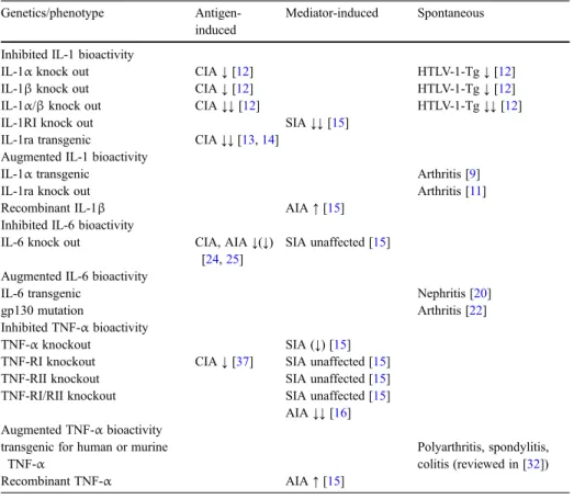
Table 1 Overview of models for immune-mediated diseases using transgenic or knock-out animals for IL-1, IL-6 and TNF-α
Genetics/phenotype Antigen-induced
Mediator-induced Spontaneous Inhibited IL-1 bioactivity
IL-1α knock out CIA↓ [12] HTLV-1-Tg↓ [12]
IL-1β knock out CIA↓ [12] HTLV-1-Tg↓ [12]
IL-1α/β knock out CIA↓↓ [12] HTLV-1-Tg↓↓ [12]
IL-1RI knock out SIA↓↓ [15]
IL-1ra transgenic CIA↓↓ [13,14] Augmented IL-1 bioactivity
IL-1α transgenic Arthritis [9]
IL-1ra knock out Arthritis [11]
Recombinant IL-1β AIA↑ [15]
Inhibited IL-6 bioactivity
IL-6 knock out CIA, AIA↓(↓) [24,25]
SIA unaffected [15] Augmented IL-6 bioactivity
IL-6 transgenic Nephritis [20]
gp130 mutation Arthritis [22]
Inhibited TNF-α bioactivity
TNF-α knockout SIA (↓) [15]
TNF-RI knockout CIA↓ [37] SIA unaffected [15]
TNF-RII knockout SIA unaffected [15]
TNF-RI/RII knockout SIA unaffected [15] AIA↓↓ [16] Augmented TNF-α bioactivity
transgenic for human or murine TNF-α
Polyarthritis, spondylitis, colitis (reviewed in [32])
Recombinant TNF-α AIA↑ [15]
Genetic modification of IL-1, IL-6, and TNF-α in animal models of immune-mediated inflammation
A large body of evidence demonstrating the importance of IL-1, IL-6, and TNF-α has been accumulated in the meantime in several overexpression or knock-out animal models (Table1). Most of the published data concern the development of arthritis, while the literature on genetically defined animal models for other target organs, such as colitis, skin inflammation, nephritis or CNS inflammation, and other important human inflammatory diseases, is clearly less extensive.
Cytokine effects on arthritis symptoms in mice show substantial variation and must be carefully interpreted, taking into account the specific background of different, always artificial, disease models. Spontaneous arthritis with 100% disease penetration occurs in IL-1 and TNF transgenic animals, and in some conditions also in animals with boosted IL-6 activity, thus providing evidence for arthritis induction by elevated cytokine expression itself, which mainly appears to be independent of defined co-conditions or environmental conditions.
Antigen-dependent arthritis models, such as collagen-induced arthritis (CIA), in which mice do not develop arthritis spontaneously, but only upon immunization with type II collagen (CII) in the presence of Freund’s adjuvant, are widely used for studies on the role of prophylactic or therapeutic administration of recombinant anti-cytokine proteins. This model includes all phases of immune reaction and inflammatory response.
The model of serum-transfer-induced arthritis is independent of antigen processing, presentation, and immunoreaction, but predominantly addresses the downstream processes of inflammation. Arthritis in this model is driven by injection of K/BxN serum from K/BxN T-cell receptor transgenic mice into naïve animals. Usually, C57Bl/6 mice are used here, which can be genetically modified for the interesting cytokine gene. Antibodies from the K/ BxN serum bind to different cartilage epitopes of C57Bl/6 mice and propagate arthritis. As an alternative, arthritis independent of antigen recognition and subsequent immunization can also be triggered by direct injection of CII antibodies.
Consequences of IL-1 modification in animal models
Human IL-1α transgenic C57BL/6N × B6C3F1 mice develop a spontaneous, macrophage-and neutrophil-dominated type of arthritis. The neutrophilia is apparently mediated by induced GM-CSF [9]. IL-1α transgenic mice with a mutation of Lys64 to Ala271 of the
human IL-1α amino acid sequence exhibit overproduction of membrane-associated IL-1 on macrophage and fibroblast surfaces. This results in a more severe phenotype than mice only transgenic for human IL-1α on the same genetic background, indicating that not only the quantity of IL-1α but also the sterical order or the association with other membrane components might be of functional relevance for IL-1 ligand/receptor interaction and disease development [10]. The importance of IL-1ra to control IL-1 activity was indicated in the spontaneous arthritis model of IL-1ra-deficient BAL/cA mice (which was not reproducible in C57BL/6J mice), so far suggesting influence of the IL-1/IL-1ra balance on arthritis development in certain conditions [11].
The pathogenic relevance of both IL-1α and IL-1β was further underlined when comparing induction of CIA in IL-1α−/−, IL-1β−/−, or IL-1α/β−/−animals with wild-type littermates on a DBA/1J background [12]. CII antibody production in CIA was still normal, but deficiency of IL-1α and IL-1β, and more so of both IL-1 molecules, ameliorated arthritis. In line with this, mice transgenic for IL-1ra were protected from CIA [13,14]. In the HTLV-I Tg model, in which BALB/c IL-1β−/−, IL-1α/β−/−mice crossed with HTLV-I transgenic mice showed a T-cell proliferative syndrome with arthritis, T-cell response and autoantibody production was clearly suppressed by IL-1α and IL-1β deficiency [12]. Comparing the results in CIA and HTLV-1-Tg led the authors to conclude that murine IL-1 is of importance for the T-cell-dependent processes of autoimmunity and arthritis [12].
Data from the serum-transfer model—which progresses independently of antibody formation—also supported a central role for IL-1-mediated processes in murine arthritis. In this study, mice were knocked out for IL-1R (type 1 equivalent), and also for IL-6, TNF-α and lymphotoxin, the p55 TNF-R1 and p75 TNF-R2 [15]. Interestingly, the most robust prevention of arthritis was observed in IL-1R knock-out animals, whereas, IL-6 and LT deficiency had no effect on the onset and course of serum-induced arthritis. TNF-α ligand knock-out mice had a variable and TNF-R knock-out mice a surprisingly unaffected disease course [15]. The equal importance of IL-1 and TNF-α in the induction and severity of
arthritis was suggested by the addition of TNF-α and IL-1β to CII antibodies with similar enhancement of antibody induced arthritis [15].
Other studies confirmed a pivotal role for IL-1 and TNF-α in models that are independent of antibody formation [16]. The inhibitory effect of IL-1R deficiency on the development of serum-transfer-induced arthritis could be circumvented by activating the downstream IL-1 pathway, e.g., by activating the myeloid differentiation factor MyD88 by LPS and TLR-4 signaling [17]. This suggests that other factors, such as products of gram-negative microbes binding to Toll-like receptors (TLR), may substitute for inhibited IL-1 effects.
To our knowledge, none of the IL-1 transgenic mice, or the IL-6 or TNF-α transgenic mouse models described so far evolved a spontaneous skin disease resembling that of psoriasis in human. Beta-1 integrin transgenic mice, however, over-express this molecule exclusively at the basal skin layer. They experience a sporadic psoriasis-like phenotype and display secondary up-regulation of IL-1α molecules in suprabasal skin layers upon activation of MAP kinases [18]. Inducible epidermal deletion of nuclear proteins c-Jun and Jun B also
leads to a psoriasis-like phenotype of the skin and also the joints [19]. Notably, and in contrast to the skin phenotype, the development of arthritic lesions required T and B cells and signaling through tumour necrosis factor receptor 1 (TNFR1) in this model.
Modulation of IL-6 activity in animal models
The contribution of IL-6 to the development of arthritis, when examined in IL-6 deficient or transgenic mice, appears more controversial than that of IL-1. IL-6 transgenic mice develop massive polyclonal plasmocytosis, which is in line with the role of IL-6 as a factor of B-cell differentiation [20]. IL-6 is known as an important growth factor for plasmocytoma cells. No malignant B-cell transformation occurs in this model, but in the presence of additional check point defects (such as mutation in thec-myc gene [21]) malignant transformation can be observed. No spontaneous arthritis occurred in the IL-6 transgenic mice mentioned so far,
although several autoimmune phenomena, such as autoantibody formation or autoimmune-mediated nephritis, were reported [20]. When the gp130 molecule is point-mutated to Y759F, leading to prolonged STAT-3 activation upon activation of the IL-6 receptor complex, late developing, spontaneous arthritis in C57BL/6 mice was induced [22].
An important clinical aspect of autoimmune diseases in juvenile individuals is a reduced longitudinal growth in chronic inflammation. As a correlate of this problem, NSE-h-IL-6 transgenic mice show significantly reduced weight gain and growth, which is explained by reduced insulin-like growth factor activity (IGF)-1 [23]. In this case, a special indication for IL-6 blockade in the treatment of children with inflammatory conditions may arise. The currently available mouse data suggest a relevant contribution of IL-6 in antigen-induced arthritis [24,25], but IL-6 is not required for arthritis development in TNF transgenic mice [24].
The role of IL-6 was also investigated in experimental autoimmune encephalomyelitis (EAE) induced by immunization with myelin oligodendrocyte glycoprotein (MOG), an animal model for a demyelinating disease, multiple sclerosis. In this case, the resistance to EAE in IL-6-deficient mice was associated with a deficiency of MOG-specific T cells [26]. Antagonising IL-6 levels by a monoclonal IL-6 antibody in the trinitrobenzenesulphonic (TNBS) acid-induced colitis model in Wistar rats to some extent normalized the impaired linear growth of these animals, although this was not completely explained by normalized food intake, but by anti-IL-6 treatment-associated normalization of insulin-like growth factor activity [27]. This observation further supports a direct role of IL-6 in growth regulation. In the dextrane sulphate-sodium-induced colitis model, IL-6−/− animals showed a reduced STAT-3 activation, indicating that the SH2 protein induced by IL-6 and STAT-3 tyrosine phosphorylation plays an essential role in the negative regulation of cytokine signaling [28].
Phenotype of animals transgenic or blocked for TNF-α
Different TNF-transgenic mouse strains have been developed on the C57BL/6CBA background with the CIA-resistant MHC haplotypes H-2kand H-2b. Stable human TNF-α overexpression has been achieved by cloning the human TNF-α gene with the 3′-modified β-globin UTR instead of the AU-rich element (ARE)-containing 3′UTR [29]. This untranslated gene region is crucial for the degradation of TNF transcripts that prevents TNF-α protein expression in resting cells. In activated cells, stability and translational efficiency of TNF-α mRNA is increased by mechanisms mediated by this gene sequence. Other models express a murine TNF-α transmembrane mutant either with the 3′-modified β-globin UTR [30] or an ARE-deleted mouse TNF-α gene, called TNFΔARE [31]. Human TNF-α can exclusively bind to the murine TNF-RI, while the murine isoform binds to both receptors. Different phenotypes of TNF-transgenic rodents have been developed. Overexpression of human TNF-α was predominantly associated with spontaneous polyarthritis, while murine TNF-α transgenic mice developed spondylitis, inflammatory bowel disease, or polyarthritis dependent on the particular genetic model [32]. The different modifications of human and murine TNF-α genes do not completely resolve some controversies about the functional consequences of activating the TNF-RI or TNF-RII, or a differential effect of the soluble or membrane-bound protein form [32].
Administration of anti-TNF-α antibodies was able to prevent arthritis in TNF-Tg mice [29]. The same was also true for antibody blockade of the IL-1RI, indicating that IL-1 is a potent downstream mediator of TNF-α in the pathogenesis of arthritis in these mice [33]. TNF-α serum concentrations were reduced in the IL-1R antagonist-treated TNF-α Tg mice. This underlines the well-known interrelationship regarding expression and function of IL-1 and TNF-α. TNF-Tg mice crossed with RAG-1 knock-out mice, which have no T cells or B cells, also develop erosive arthritis [34]. This circumstance indicates an immune-independent disease development. However, backcrossing of TNF-Tg onto an arthritis susceptible DBA/1 (H-2q) background promotes earlier arthritis development and suggests a link between raised levels of circulating TNF-α and immune-mediated processes [35]. Antigen-mediated B-cell dependent immune responses were not impaired in TNF-deficient, BAFF transgenic mice, although knocking out TNF led to a significant increase of B-cell lymphomas in the context of growth-factor-mediated B-cell stimulation [36].
Deletion of both TNF-R prevented disease in CII-antibody-induced arthritis of BALB/c mice [16], but had no effect on the development of arthritis in K/BxN mice. The latter, somewhat unexpected observation may be explained by the presence of other receptors of the TNF family, or up-regulation of downstream mediators in receptor-deficient mice that compensate for the TNF-R1/2 deficiency [15]. Also, TNF-α deficiency led to random
development or protection from serum-induced arthritis, which appeared to depend on environmental rather than epigenetic factors [15]. This observation reflects the situation in CIA, where p55 TNF-R1 deficiency reduced the incidence and severity of arthritis development [37].
Breakdown of self-tolerance by presentation of a foreign antigen in a specific arthritis-susceptible MHC background is a widely accepted hypothesis for the development of chronic inflammatory processes in humans. This led researchers to follow the generation of the T -cell receptor repertoire and antigen response in mice transgenic for susceptible and non-susceptible human MHC II molecules. It is noteworthy that stimulation of these animals with a potential antigen, human cartilage protein gp-39, led to a differential T-cell cytokine profile, with more TNF-α production in susceptible than in non-susceptible animals [38]. Importantly, however, chronic exposure of T cells to TNF-α impaired T-cell receptor signaling. Among other contributing factors, this observation may explain the frequently reported anergy of T cells in chronically inflamed tissues such as RA synovial membranes [39].
Summarizing the extensive literature on IL-1, IL-6, and TNF-α in various models in rodents, it appears appropriate to rank IL-1 equivalent or even superior to TNF-α, while IL-6 presents as a less attractive target molecule in immune-mediated inflammation.
Blockade of IL-1, IL-6, and TNF-α in human disease
As already mentioned in the introductory section, TNF-α blockade has made its way into clinical application, whereas, IL-1 and IL-6 blockade have not yet finished this process. What may be the reason for this?
All three molecules, IL-1α, IL-1β, and TNF-α are not the exclusive representatives of the IL-1 or TNF protein families. However, with respect to significant up-regulation in human disease and the enormous potential when modifying expression of one of these cytokines or
the respective receptors in rodent models of disease, it appears justified to suggest highest importance for IL-1, IL-6, and TNF-α. Some divergent results for the candidate cytokines in the rodent models were dependent on the genetic or environmental background. Differences were sometimes subtle, and the results were not always absolutely concordant.
Switching from experimental medicine to the perspective of a clinician, compounds for antagonizing IL-1, IL-6, and TNF activity and a large body of evidence in clinical application have become available. In the following section, we will focus on the effect of these drugs in prospective, randomized controlled trials (RCT).
IL-1 inhibition in human diseases
IL-1 antagonism in published RCT was accomplished by injecting or infusing recombinant IL-1ra in patients with sepsis [40–42], rheumatoid arthritis [43–45], and in graft vs host disease in patients undergoing allogeneic stem-cell transplantation [46]. Preliminary data also exist for stroke [47], osteoarthritis [48], and spondylitis [49,50]. Despite excellent data from animal models, and although activation of the IL-1 system has been described in several human conditions, clinical data have either proven ineffective or at least been controversial in terms of achievable results. The best evidence for effectiveness of blocking IL-1 currently exists for the erosive aspect of arthritic joints, while satisfactory clinical response to IL-1ra was rather infrequent even in RA trials. Also, addition of IL-1Ra to TNF blockade did not lead to increasing efficacy, but to accumulation of side effects [51]. Published clinical data on the effect of IL-1ra on colitis, psoriasis, nephritis, or encephalomyelitis are still missing. A few, but very convincing, case reports suggest a novel indication for IL-1ra in rare inherited auto-inflammatory disorders [52].
IL-6 blockade in human conditions
Randomized controlled trials for IL-6 inhibition have been published in the meantime for RA [53,54]. First trials were also done in Crohn’s disease [55] and in juvenile idiopathic arthritis (JIA) [56,57], in the latter indication with a special focus on the systemic onset subtype of JIA. This subtype is of particular interest, firstly because this entity is reported to be linked to a single nucleotide mutation in the IL-6 gene [58], and secondly because of the unfavourable outcome of systemic onset JIA upon TNF blockade. All the cited studies on IL-6 antagonism were performed with an IL-6R blocking, humanized antibody called tocilizumab (MRA). In line with the important pathogenetic role of IL-6 in this disease [59], MRA has also been successfully tested when shorter dose intervals than indicated in Table 2 were used in Castleman’s disease, a lymphoproliferative syndrome of non-malignant origin [60]. Affected patients suffer from lymphadenopathy, fever, general malaise, and wasting. Thus, increased plasma lipid concentrations were intended in these patients, but raised total and HDL cholesterol as well as triglycerides have also been reported in the RA trials. These parameters have to be kept under critical observation in ongoing studies for RA, a disease with an elevated risk per se for cardiovascular complications. As also suggested by the metabolic effects observed in the animal models, IL-6 blockade in patients also led to a significant
decrease of collagen cross-link products, as well as an increase of bone-formation-indicating parameters [54].
TNF blockade in human diseases
TNF-α blockade has proven efficacy in randomized controlled trials for several immune-mediated human diseases. Three TNF-binding recombinant protein constructs have entered the market since their first approval in 1998 in the US and EU: (1) Infliximab (cA2), a chimeric anti-TNF-α IgG1antibody; (2) etanercept, a human dimeric TNF-receptor type II-IgG1fusion protein (TNFR:Fc); and (3) adalimumab (D2E7), a human anti-TNF-α IgG1 antibody genetically engineered through phage display technology. At present, a fourth TNF-α binding protein, certolizumab (CDP870), a polyethylene-glycolated Fab’ fragment of anti-tumor necrosis factor is still in the pre-approval phase. Table2summarizes some key clinical trials and basic information on the available drugs.
Efficacy of TNF blockers in RA
Head-to-head comparison of the anti-TNF constructs in RA is missing, but with all the necessary caution to be taken, the null hypothesis of equivalence in terms of efficacy and safety when targeting TNF-α by different biologics has not been rejected, so far, for this indication. Significant improvement in signs and symptoms of RA (ACR20 response) was two to three times more likely than in placebo-treated control patient cohorts when adding infliximab or etanercept to methotrexate in active disease [61,62]. Efficacy of TNF-α was up
to five times higher than placebo when added to standard methotrexate and concomitant treatment in a later trial [63]. When recruiting severe cases of RA for these first trials on TNF
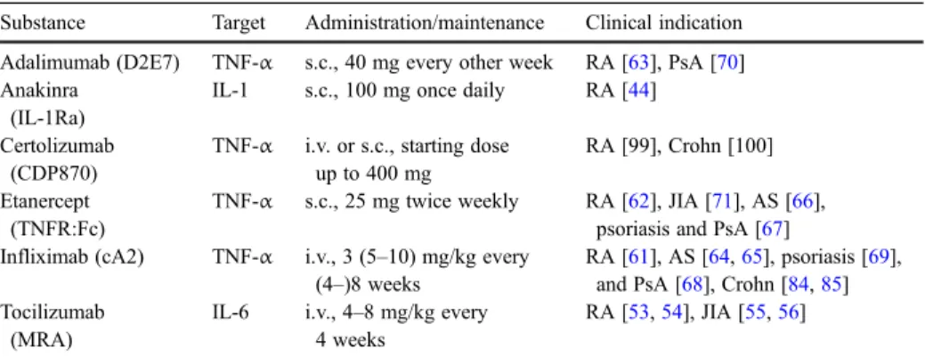
Table 2 Cytokine blocking recombinant proteins in clinical trials
Substance Target Administration/maintenance Clinical indication Adalimumab (D2E7) TNF-α s.c., 40 mg every other week RA [63], PsA [70] Anakinra
(IL-1Ra)
IL-1 s.c., 100 mg once daily RA [44] Certolizumab
(CDP870)
TNF-α i.v. or s.c., starting dose up to 400 mg
RA [99], Crohn [100] Etanercept
(TNFR:Fc)
TNF-α s.c., 25 mg twice weekly RA [62], JIA [71], AS [66], psoriasis and PsA [67] Infliximab (cA2) TNF-α i.v., 3 (5–10) mg/kg every
(4–)8 weeks
RA [61], AS [64,65], psoriasis [69], and PsA [68], Crohn [84,85] Tocilizumab
(MRA)
IL-6 i.v., 4–8 mg/kg every 4 weeks
RA [53,54], JIA [55,56]
AS Ankylosing spondylitis, i.v. intravenous infusion, JIA juvenile idiopathic arthritis, PsA psoriatic arthritis, RA rheumatoid arthritis, s.c. subcutaneous injection, SLE systemic lupus erythematosus
blockade, the new compound was added to standard medication including methotrexate. The response to different TNF blocking agents may differ in individual RA patients, and sequential attempts using more than one compound due to primary failure or secondary loss of efficacy of the first agent is justifiable. The reason for occasional individual differences in the response to TNF antagonists is difficult to identify: the induction of antibodies against the applied proteins was assumed to cause a loss of efficacy, but accumulated data from several clinical trials do not support a major contribution of antibody induction for this observation.
Efficacy of TNF blockers in other arthritides
Concomitant treatment with immunosuppressants was primarily considered to reduce antibody induction against the chimeric antibody infliximab, until application in AS proved efficacy in the absence of immunosuppressive co-medication [64]. Two studies with etanercept in AS patients were similarly performed without immunosuppressive co-medication, one with a two-to-three times higher probability of reaching the primary outcome endpoint than placebo [65], and the other with an even nine- to tenfold superiority over placebo [66]. All launched TNF-blocking agents have proven efficacy in psoriasis and psoriatic arthritis for both articular and skin manifestations independent of concomitant methotrexate treatment [67–70]. The likelihood of a disease flare was also reduced by one- to two-thirds of that observed in placebo-treated children with juvenile idiopathic arthritis [71], although the response in systemic onset JIA, as well as uveitis manifestation, appeared less favourable [72,73].
Safety of TNF blockers
It was the observation of infrequent but serious side effects in 2001, when more than 250,000 patients were already exposed to infliximab or etanercept, which transiently slowed the enthusiasm for TNF blockade. In this review, an increased risk of opportunistic infections and exacerbation of latent tuberculosis (http://www.rheumatology.org/publications/ hotline/0901tnf.asp, as well as increased mortality when treating patients with advanced cardiomyopathy within clinical trials became apparent [74,75]. Physicians have rapidly learned to ensure safety and protection from these complications by appropriate pre-treatment screening. At present, an acceptable short-term risk of non-opportunistic infections and allergic reactions to the compound still appear to be the price to pay for achieving superior efficacy by adding TNF blockade to conventional treatment [76]. The risk of lupus-like syndromes, one of the major concerns in previous development of TNF blocking agents, is now considered less important than previously expected [77, 78]. However, an increased risk of lymphoma due to TNF antagonism cannot yet be completely ruled out, although its appearance may be delayed (http://www.rheumatology.org/ publications/hotline/0303TNFL.asp).
TNF blockade in early or established RA?
However, accompanying the optimized efficacy, increased rates of non-opportunistic infections (pneumonia) were observed when TNF blockade was combined with standard treatment, including methotrexate, even in patients with early-stage RA [76]. This, together with the arguable superiority of TNF blockade over standard methotrexate treatment, starting with a head-to-head comparison in an RCT for early RA [79], and especially equivalence of the group with later switch to TNF blockade, leads us to the following conclusion. Before making definitive recommendations, we must wait for more evidence, perhaps from longer observation of ongoing studies, to better estimate the significance of additive TNF blockade for the early state of inflammatory conditions [80]. However, in advanced, active RA, the superiority of the combination of TNF-α blockade and conventional anti-inflammatory compounds has been more clearly shown [81].
TNF blockade in other immune mediated conditions
Different degrees of efficacy of the various TNF-blocking agents was suggested in the treatment of some entities. For example, disease activity in Crohn’s disease was ameliorated by infliximab treatment [82], and the chance of complete healing of the fistulas was three to five times higher than observed in the placebo control cohort [83], while RCT of other TNF blocking agents for this condition are still lacking. In this review, it was thus tempting to speculate that a specific interaction of the effective compound beyond reducing the amount of soluble TNF-α, e.g., interaction with membrane-bound TNF-α, might be of importance. Recent work suggests some differential effects on T-cell apoptosis, but the situation is still somewhat controversial [84–87]. Besides its effectiveness in Crohn’s disease, other types of
mucosa-associated pathology, e.g., active ulcerative colitis, could also be significantly ameliorated by infliximab treatment [88] as were cutaneous symptoms of Behçet’s disease
when treated with etanercept [89]. Despite some clinical casuistic evidence of successful treatment of other inflammatory conditions with TNF blocking agents [90], strong proof from RCTs of the safety and efficacy of TNF blocking agents is lacking. So far, evidence for a relevant action in systemic lupus erythematosus [78] is limited, and the data are negative for Sjoegren’s disease [91,92], or for maintenance therapy in Wegener`s granulomatosis [93].
Future perspectives
The potential of treating human immune-mediated diseases with recombinant proteins is still open. Looking back at the long but rapid journey from the discovery and validation of targets to the development of treatment options for IL-1, IL-6, and TNF-α may teach us a lot about future activities in the field. Critical knowledge has been obtained from animal models, but there may well be a considerable divergence between the preclinical expectations and the reality in the treatment of human disease.
Besides the scientific and medical aspects, financial implications have interfered with the introduction of expensive treatment into the medical systems of various countries. It has
become increasingly clear that economic evaluation of TNF blockade and of other recombinant proteins for the treatment of chronic immune-mediated conditions is of major importance. We need to consider the underlying condition, current disease activity, functional status, expected progression and treatment duration, and the individual dosing regimen. First data for RA and AS suggest that a financial cost acceptable to society is achievable [94,95]. Here, relevant, e.g., genetic, indicators for successful therapy [96] could be of major importance for avoiding unnecessary costs.
References
1. Dinarello CA (1997) Interleukin-1. Cytokine Growth Factor Rev 8:253–265 2. Dinarello CA (1996) Biologic basis for interleukin-1 in disease. Blood 87:2095–2147
3. Arend WP (2002) The balance between IL-1 and IL-1Ra in disease. Cytokine Growth Factor Rev 13:323–340
4. Ishihara K, Hirano T (2002) IL-6 in autoimmune disease and chronic inflammatory proliferative disease. Cytokine Growth Factor Rev 13:357–368
5. Jones SA (2005) Directing transition from innate to acquired immunity: defining a role for IL-6. J Immunol 175:3463–3468
6. Feldmann M, Maini RN (2001) Anti-TNF alpha therapy of rheumatoid arthritis: what have we learned? Annu Rev Immunol 19:163–196
7. Locksley RM, Killeen N, Lenardo MJ (2001) The TNF and TNF receptor superfamilies: integrating mammalian biology. Cell 104:487–501
8. Ibelgaufts H (1995) Dictionary of cytokines. Verlag Chemie, Weinheim
9. Niki Y, Yamada H, Seki S, Kikuchi T, Takaishi H, Toyama Y, Fujikawa K, Tada N (2001) Macrophage-and neutrophil-dominant arthritis in human IL-1 alpha transgenic mice. J Clin Invest 107:1127–1135 10. Niki Y, Yamada H, Kikuchi T, Toyama Y, Matsumoto H, Fujikawa K, Tada N (2004)
Membrane-associated IL-1 contributes to chronic synovitis and cartilage destruction in human IL-1 alpha transgenic mice. J Immunol 172:577–584
11. Horai R, Saijo S, Tanioka H, Nakae S, Sudo K, Okahara A, Ikuse T, Asano M, Iwakura Y (2000) Development of chronic inflammatory arthropathy resembling rheumatoid arthritis in interleukin 1 receptor antagonist-deficient mice. J Exp Med 191:313–320
12. Saijo S, Asano M, Horai R, Yamamoto H, Iwakura Y (2002) Suppression of autoimmune arthritis in interleukin-1-deficient mice in which T cell activation is impaired due to low levels of CD40 ligand and OX40 expression on T cells. Arthritis Rheum 46:533–544
13. Ma Y, Thornton S, Boivin GP, Hirsh D, Hirsch R, Hirsch E (1998) Altered susceptibility to collagen-induced arthritis in transgenic mice with aberrant expression of interleukin-1 receptor antagonist. Arthritis Rheum 41:1798–1805
14. Palmer G, Talabot-Ayer D, Szalay-Quinodoz I, Maret M, Arend WP, Gabay C (2003) Mice transgenic for intracellular interleukin-1 receptor antagonist type 1 are protected from collagen-induced arthritis. Eur J Immunol 33:434–440
15. Ji H, Pettit A, Ohmura K, Ortiz-Lopez A, Duchatelle V, Degott C, Gravallese E, Mathis D, Benoist C (2002) Critical roles for interleukin 1 and tumor necrosis factor alpha in antibody-induced arthritis. J Exp Med 196:77–85
16. Kagari T, Doi H, Shimozato T (2002) The importance of IL-1 beta and TNF-alpha, and the noninvolvement of IL-6, in the development of monoclonal antibody-induced arthritis. J Immunol 169:1459–1466
17. Choe JY, Crain B, Wu SR, Corr M (2003) Interleukin 1 receptor dependence of serum transferred arthritis can be circumvented by toll-like receptor 4 signaling. J Exp Med 197:537–542
18. Haase I, Hobbs RM, Romero MR, Broad S, Watt FM (2001) A role for mitogen-activated protein kinase activation by integrins in the pathogenesis of psoriasis. J Clin Invest 108:527–536
19. Zenz R, Eferl R, Kenner L, Florin L, Hummerich L, Mehic D, Scheuch H, Angel P, Tschachler PE, Wagner EF (2005) Psoriasis-like skin disease and arthritis caused by inducible epidermal deletion of Jun proteins. Nature 437:369–375
20. Suematsu S, Matsuda T, Aozasa K, Akira S, Nakano N, Ohno S, Miyazaki J, Yamamura K, Hirano T, Kishimoto T (1989) IgG1 plasmacytosis in interleukin 6 transgenic mice. Proc Natl Acad Sci USA 86:7547–7551
21. Hirano T (1991) Interleukin 6 (IL-6) and its receptor: their role in plasma cell neoplasias. Int J Cell Cloning 9:166–184
22. Ishihara K, Sawa S, Ikushima H, Hirota S, Atsumi T, Kamimura D, Park SJ, Murakami M, Kitamura Y, Iwakura Y, Hirano T (2004) The point mutation of tyrosine 759 of the IL-6 family cytokine receptor gp130 synergizes with HTLV-1 pX in promoting rheumatoid arthritis-like arthritis. Int Immunol 16:455–465
23. De Benedetti F, Alonzi T, Moretta A, Lazzaro D, Costa P, Poli V, Martini A, Ciliberto G, Fattori E (1997) Interleukin 6 causes growth impairment in transgenic mice through a decrease in insulin-like growth factor-I. A model for stunted growth in children with chronic inflammation. J Clin Invest 99:643–650 24. Alonzi T, Fattori E, Lazzaro D, Costa P, Probert L, Kollias G, De Benedetti F, Poli V, Ciliberto G (1998)
Interleukin 6 is required for the development of collagen-induced arthritis. J Exp Med 187:461–468 25. Ohshima S, Saeki Y, Mima T, Sasai M, Nishioka K, Nomura S, Kopf M, Katada Y, Tanaka T, Suemura
M, Kishimoto T (1998) Interleukin 6 plays a key role in the development of antigen-induced arthritis. Proc Natl Acad Sci USA 95:8222–8226
26. Samoilova EB, Horton JL, Hilliard B, Liu TS, Chen Y (1998) IL-6-deficient mice are resistant to experimental autoimmune encephalomyelitis: roles of IL-6 in the activation and differentiation of autoreactive T cells. J Immunol 161:6480–6486
27. Sawczenko A, Azooz O, Paraszczuk J, Idestrom M, Croft NM, Savage MO, Ballinger AB, Sanderson IR (2005) Intestinal inflammation-induced growth retardation acts through IL-6 in rats and depends on the −174 IL-6 G/C polymorphism in children. Proc Natl Acad Sci USA 102:13260–13265
28. Suzuki A, Hanada T, Mitsuyama K, Yoshida T, Kamizono S, Hoshino T, Kubo M, Yamashita A, Okabe M, Takeda K, Akira S, Matsumoto S, Toyonaga A, Sata M, Yoshimura A (2001) CIS3/SOCS3/ SSI3 plays a negative regulatory role in STAT3 activation and intestinal inflammation. J Exp Med 193:471–481
29. Keffer J, Probert L, Cazlaris H, Georgopoulos S, Kaslaris E, Kioussis D, Kollias G (1991) Transgenic mice expressing human tumour necrosis factor: a predictive genetic model of arthritis. EMBO J 10:4025–4031
30. Alexopoulou L, Pasparakis M, Kollias G (1997) A murine transmembrane tumor necrosis factor (TNF) transgene induces arthritis by cooperative p55/p75 TNF receptor signaling. Eur J Immunol 27:2588– 2592
31. Kontoyiannis D, Pasparakis M, Pizarro TT, Cominelli F, Kollias G (1999) Impaired on/off regulation of TNF biosynthesis in mice lacking TNF AU-rich elements: implications for joint and gut-associated immunopathologies. Immunity 10:387–398
32. Li P, Schwarz EM (2003) The TNF-α transgenic mouse model of inflammatory arthritis. Springer Semin Immunopathol 25:19–33
33. Probert L, Plows D, Kontogeorgos G, Kollias G (1995) The type I interleukin-1 receptor acts in series with tumor necrosis factor (TNF) to induce arthritis in TNF-transgenic mice. Eur J Immunol 25:1794–1797
34. Kollias G, Douni E, Kassiotis G, Kontoyiannis D (1999) On the role of tumor necrosis factor and receptors in models of multiorgan failure, rheumatoid arthritis, multiple sclerosis and inflammatory bowel disease. Immunol Rev 169:175–194
35. Butler DM, Malfait AM, Mason LJ, Warden PJ, Kollias G, Maini RN, Feldmann M, Brennan FM (1997) DBA/1 mice expressing the human TNF-alpha transgene develop a severe, erosive arthritis: characterization of the cytokine cascade and cellular composition. J Immunol 159:2867–2876 36. Batten M, Fletcher C, Ng LG, Groom J, Wheway J, Laabi Y, Xin X, Schneider P, Tschopp J, Mackay CR,
Mackay F (2004) TNF deficiency fails to protect BAFF transgenic mice against autoimmunity and reveals a predisposition to B cell lymphoma. J Immunol 172:812–822
37. Mori L, Iselin S, De Libero G, Lesslauer W (1996) Attenuation of collagen-induced arthritis in 55-kDa TNF receptor type 1 (TNFR1)-IgG1-treated and TNFR1-deficient mice. J Immunol 157:3178–3182 38. Cope AP, Patel SD, Hall F, Congia M, Hubers HA, Verheijden GF, Boots AM, Menon R, Trucco M,
Rijnders AW, Sonderstrup G (1999) T cell responses to a human cartilage autoantigen in the context of rheumatoid arthritis associated and non-associated HLA-DR4 alleles. Arthritis Rheum 42:1497–1507
39. Cope AP, Liblau RS, Yang XD, Congia M, Laudanna C, Schreiber RD, Probert L, Kollias G, McDevitt HO (1997) Chronic tumor necrosis factor alters T cell responses by attenuating T cell receptor signaling. J Exp Med 185:1573–1584
40. Fisher CJ Jr, Opal SM, Lowry SF, Sadoff JC, LaBrecque JF, Donovan HC, Lookabaugh JL, Lemke J, Pribble JP, Stromatt SC et al (1994) Role of interleukin-1 and the therapeutic potential of interleukin-1 receptor antagonist in sepsis. Circ Shock 44:1–8
41. Fisher CJ Jr, Slotman GJ, Opal SM, Pribble JP, Bone RC, Emmanuel G, Ng D, Bloedow DC, Catalano MA; IL-1RA Sepsis Syndrome Study Group (1994) Initial evaluation of human recombinant interleukin-1 receptor antagonist in the treatment of sepsis syndrome: a randomized, open-label, placebo-controlled multicenter trial. Crit Care Med 22:12–21
42. Vincent JL, Slotman G, Van Leeuwen PA, Shelly M, Nasraway S, Tenaillon A, Bander J, Friedman G (1999) IL-1ra administration does not improve cardiac function in patients with severe sepsis. J Crit Care 14:69–72
43. Campion GV, Lebsack ME, Lookabaugh J, Gordon G, Catalano M, The IL-1Ra Arthritis Study Group (1996) Dose-range and dose-frequency study of recombinant human interleukin-1 receptor antagonist in patients with rheumatoid arthritis. Arthritis Rheum 39:1092–1101
44. Bresnihan B, Alvaro-Gracia JM, Cobby M, Doherty M, Domljan Z, Emery P, Nuki G, Pavelka K, Rau R, Rozman B, Watt I, Williams B, Aitchison R, McCabe D, Musikic P (1998) Treatment of rheumatoid arthritis with recombinant human interleukin-1 receptor antagonist. Arthritis Rheum 41:2196–2204 45. Jiang Y, Genant HK, Watt I, Cobby M, Bresnihan B, Aitchison R, McCabe D (2000) A multicenter,
double-blind, dose-ranging, randomized, placebo-controlled study of recombinant human interleukin-1 receptor antagonist in patients with rheumatoid arthritis: radiologic progression and correlation of Genant and Larsen scores. Arthritis Rheum 43:1001–1009
46. Antin JH, Weisdorf D, Neuberg D, Nicklow R, Clouthier S, Lee SJ, Alyea E, McGarigle C, Blazar BR, Sonis S, Soiffer RJ, Ferrara JL (2002) Interleukin-1 blockade does not prevent acute graft-vs-host disease: results of a randomized, double-blind, placebo-controlled trial of interleukin-1 receptor antagonist in allogeneic bone marrow transplantation. Blood 100:3479–3482
47. Chevalier X, Giraudeau B, Conrozier T, Marliere J, Kiefer P, Goupille P (2005) Safety study of intraarticular injection of interleukin 1 receptor antagonist in patients with painful knee osteoarthritis: a multicenter study. J Rheumatol 32:1317–1323
48. Emsley HC, Smith CJ, Georgiou RF, Vail A, Hopkins SJ, Rothwell NJ, Tyrrell PJ; Acute Stroke Investigators (2005) Acute Stroke Investigators. A randomised phase II study of interleukin-1 receptor antagonist in acute stroke patients. J Neurol Neurosurg Psychiatry 76:1366–1372
49. Tan AL, Marzo-Ortega H, O’Connor P, Fraser A, Emery P, McGonagle D (2004) Efficacy of anakinra in active ankylosing spondylitis: a clinical and magnetic resonance imaging study. Ann Rheum Dis 63:1015–1041
50. Haibel H, Rudwaleit M, Listing J, Sieper J (2004) Open label trial of anakinra in active ankylosing spondylitis over 24 weeks. Ann Rheum Dis 64:296–298
51. Genovese MC, Cohen S, Moreland L, Lium D, Robbins S, Newmark R, Bekker P; 20000223 Study Group (2004) Combination therapy with etanercept and anakinra in the treatment of patients with rheumatoid arthritis who have been treated unsuccessfully with methotrexate. Arthritis Rheum 50:1412–1419
52. Seitz M, Kamgang RK, Simon HU, Villiger PM (2005) Therapeutic interleukin (IL) 1 blockade normalises increased IL1 beta and decreased tumour necrosis factor alpha and IL10 production in blood mononuclear cells of a patient with CINCA syndrome. Ann Rheum Dis 64:1802–1803
53. Choy EH, Isenberg DA, Garrood T, Farrow S, Ioannou Y, Bird H, Cheung N, Williams B, Hazleman B, Price R, Yoshizaki K, Nishimoto N, Kishimoto T, Panayi GS (2002) Therapeutic benefit of blocking interleukin-6 activity with an anti-interleukin-6 receptor monoclonal antibody in rheumatoid arthritis: a randomized, double-blind, placebo-controlled, dose-escalation trial. Arthritis Rheum 46:3143–3150 54. Nishimoto N, Yoshizaki K, Miyasaka N, Yamamoto K, Kawai S, Takeuchi T, Hashimoto J, Azuma J,
Kishimoto T (2004) Treatment of rheumatoid arthritis with humanized anti-interleukin-6 receptor antibody: a multicenter, double-blind, placebo-controlled trial. Arthritis Rheum 50:1761–1769 55. Ito H, Takazoe M, Fukuda Y, Hibi T, Kusugami K, Andoh A, Matsumoto T, Yamamura T, Azuma J,
Nishimoto N, Yoshizaki K, Shimoyama T, Kishimoto T (2004) A pilot randomized trial of a human anti-interleukin-6 receptor monoclonal antibody in active Crohn’s disease. Gastroenterology 126:989–996
56. Woo P, Wilkinson N, Prieur AM, Southwood T, Leone V, Livermore P, Wythe H, Thomson D, Kishimoto T (2005) Open label phase II trial of single, ascending doses of MRA in Caucasian children with severe systemic juvenile idiopathic arthritis: proof of principle of the efficacy of IL-6 receptor blockade in this type of arthritis and demonstration of prolonged clinical improvement. Arthritis Res Ther 7:R1281–1288 57. Yokota S, Miyamae T, Imagawa T, Iwata N, Katakura S, Mori M, Woo P, Nishimoto N, Yoshizaki K, Kishimoto T (2005) Therapeutic efficacy of humanized recombinant anti-interleukin-6 receptor antibody in children with systemic-onset juvenile idiopathic arthritis. Arthritis Rheum 52:818–825
58. Ogilvie EM, Fife MS, Thompson SD, Twine N, Tsoras M, Moroldo M, Fisher SA, Lewis CM, Prieur AM, Glass DN, Woo P (2003) The−174G allele of the interleukin-6 gene confers susceptibility to systemic arthritis in children: a multicenter study using simplex and multiplex juvenile idiopathic arthritis families. Arthritis Rheum 48:3202–3206
59. Mandler RN, Kerrigan DP, Smart J, Kuis W, Villiger P, Lotz M (1992) Castleman’s disease in POEMS syndrome with elevated interleukin-6. Cancer 69:2697–2703
60. Nishimoto N, Kanakura Y, Aozasa K, Johkoh T, Nakamura M, Nakano S, Nakano N, Ikeda Y, Sasaki T, Nishioka K, Hara M, Taguchi H, Kimura Y, Kato Y, Asaoku H, Kumagai S, Kodama F, Nakahara H, Hagihara K, Yoshizaki K, Kishimoto T (2005) Humanized anti-interleukin-6 receptor antibody treatment of multicentric Castleman disease. Blood 106:2627–2632
61. Maini R, St Clair EW, Breedveld F, Furst D, Kalden J, Weisman M, Smolen J, Emery P, Harriman G, Feldmann M, Lipsky P; ATTRACT Study Group (1999) Infliximab (chimeric anti-tumour necrosis factor alpha monoclonal antibody) versus placebo in rheumatoid arthritis patients receiving concomitant methotrexate: a randomised phase III trial. Lancet 354:1932–1939
62. Weinblatt ME, Kremer JM, Bankhurst AD, Bulpitt KJ, Fleischmann RM, Fox RI, Jackson CG, Lange M, Burge DJ (1999) A trial of etanercept, a recombinant tumor necrosis factor receptor:Fc fusion protein, in patients with rheumatoid arthritis receiving methotrexate. N Engl J Med 340:253–259
63. Weinblatt ME, Keystone EC, Furst DE, Moreland LW, Weisman MH, Birbara CA, Teoh LA, Fischkoff SA, Chartash EK (2003) Adalimumab, a fully human anti-tumor necrosis factor alpha monoclonal antibody, for the treatment of rheumatoid arthritis in patients taking concomitant methotrexate: the ARMADA trial. Arthritis Rheum 48:35–45
64. Braun J, Brandt J, Listing J, Zink A, Alten R, Golder W, Gromnica-Ihle E, Kellner H, Krause A, Schneider M, Sorensen H, Zeidler H, Thriene W, Sieper J (2002) Treatment of active ankylosing spondylitis with infliximab: a randomised controlled multicentre trial. Lancet 359:1187–1193 65. Gorman JD, Sack KE, Davis JC Jr (2002) Treatment of ankylosing spondylitis by inhibition of tumor
necrosis factor alpha. N Engl J Med 346:1349–1356
66. Brandt J, Khariouzov A, Listing J, Haibel H, Sorensen H, Grassnickel L, Rudwaleit M, Sieper J, Braun J (2003) Six-month results of a double-blind, placebo-controlled trial of etanercept treatment in patients with active ankylosing spondylitis. Arthritis Rheum 48:1667–1675
67. Mease PJ, Goffe BS, Metz J, VanderStoep A, Finck B, Burge DJ (2000) Etanercept in the treatment of psoriatic arthritis and psoriasis: a randomised trial. Lancet 356:385–390
68. Reich K, Nestle FO, Papp K, Ortonne JP, Evans R, Guzzo C, Li S, Dooley LT, Griffiths CE; EXPRESS study investigators (2005) Infliximab induction and maintenance therapy for moderate-to-severe psoriasis: a phase III, multicentre, double-blind trial. Lancet 366:1367–1374
69. Antoni CE, Kavanaugh A, Kirkham B, Tutuncu Z, Burmester GR, Schneider U, Furst DE, Molitor J, Keystone E, Gladman D, Manger B, Wassenberg S, Weier R, Wallace DJ, Weisman MH, Kalden JR, Smolen J (2005) Sustained benefits of infliximab therapy for dermatologic and articular manifestations of psoriatic arthritis: results from the infliximab multinational psoriatic arthritis controlled trial (IMPACT). Arthritis Rheum 52:1227–1236
70. Mease PJ, Gladman DD, Ritchlin CT, Ruderman EM, Steinfeld SD, Choy EH, Sharp JT, Ory PA, Perdok RJ, Weinberg MA; Adalimumab Effectiveness in Psoriatic Arthritis Trial Study Group (2005) Adalimumab for the treatment of patients with moderately to severely active psoriatic arthritis: results of a double-blind, randomized, placebo-controlled trial. Arthritis Rheum 52:3279–3289
71. Lovell DJ, Giannini EH, Reiff A, Cawkwell GD, Silverman ED, Nocton JJ, Stein LD, Gedalia A, Ilowite NT, Wallace CA, Whitmore J, Finck BK (2000) Etanercept in children with polyarticular juvenile rheumatoid arthritis. Pediatric Rheumatology Collaborative Study Group. N Engl J Med 342:763–769 72. Quartier P, Taupin P, Bourdeaut F, Lemelle I, Pillet P, Bost M, Sibilia J, Kone-Paut I, Gandon-Laloum S, LeBideau M, Bader-Meunier B, Mouy R, Debre M, Landais P, Prieur AM (2003) Efficacy of etanercept for the treatment of juvenile idiopathic arthritis according to the onset type. Arthritis Rheum 48:1093–1101
73. Schmeling H, Horneff G (2005) Etanercept and uveitis in patients with juvenile idiopathic arthritis. Rheumatology 44:1008–1011
74. Bozkurt B, Torre-Amione G, Warren MS, Whitmore J, Soran OZ, Feldman AM, Mann DL (2001) Results of targeted anti-tumor necrosis factor therapy with etanercept (ENBREL) in patients with advanced heart failure. Circulation 103:1044–1047
75. Chung ES, Packer M, Lo KH, Fasanmade AA, Willerson JT; Anti-TNF Therapy Against Congestive Heart Failure Investigators (2003) Randomized, double-blind, placebo-controlled, pilot trial of infliximab, a chimeric monoclonal antibody to tumor necrosis factor-alpha, in patients with moderate-to-severe heart failure: results of the anti-TNF Therapy Against Congestive Heart Failure (ATTACH) trial. Circulation 107:3133–3140
76. St Clair EW, van der Heijde DM, Smolen JS, Maini RN, Bathon JM, Emery P, Keystone E, Schiff M, Kalden JR, Wang B, Dewoody K, Weiss R, Baker D; Active-Controlled Study of Patients Receiving Infliximab for the Treatment of Rheumatoid Arthritis of Early Onset Study Group (2004) Combination of infliximab and methotrexate therapy for early rheumatoid arthritis: a randomized, controlled trial. Arthritis Rheum 50:3432–3443
77. De Rycke L, Baeten D, Kruithof E, Van den Bosch F, Veys EM, De Keyser F (2005) Infliximab, but not etanercept, induces IgM anti-double-stranded DNA autoantibodies as main antinuclear reactivity: biologic and clinical implications in autoimmune arthritis. Arthritis Rheum 52:2192–2201
78. Aringer M, Graninger WB, Steiner G, Smolen JS (2004) Safety and efficacy of tumor necrosis factor alpha blockade in systemic lupus erythematosus: an open-label study. Arthritis Rheum 50:3161–3169 79. Bathon JM, Martin RW, Fleischmann RM, Tesser JR, Schiff MH, Keystone EC, Genovese MC, Wasko
MC, Moreland LW, Weaver AL, Markenson J, Finck BK (2000) A comparison of etanercept and methotrexate in patients with early rheumatoid arthritis. N Engl J Med 343:1586–1593
80. Goekoop-Ruiterman YP, de Vries-Bouwstra JK, Allaart CF, van Zeben D, Kerstens PJ, Hazes JM, Zwinderman AH, Ronday HK, Han KH, Westedt ML, Gerards AH, van Groenendael JH, Lems WF, van Krugten MV, Breedveld FC, Dijkmans BA (2005) Clinical and radiographic outcomes of four different treatment strategies in patients with early rheumatoid arthritis (the BeSt study): a randomized, controlled trial. Arthritis Rheum 52:3381–3390
81. Klareskog L, van der Heijde D, de Jager JP, Gough A, Kalden J, Malaise M, Martin Mola E, Pavelka K, Sany J, Settas L, Wajdula J, Pedersen R, Fatenejad S, Sanda M; TEMPO (Trial of Etanercept and Methotrexate with Radiographic Patient Outcomes) Study Investigators (2004) Therapeutic effect of the combination of etanercept and methotrexate compared with each treatment alone in patients with rheumatoid arthritis: double-blind randomised controlled trial. Lancet 363:675–681
82. D’haens G, Van Deventer S, Van Hogezand R, Chalmers D, Kothe C, Baert F, Braakman T, Schaible T, Geboes K, Rutgeerts P (1999) Endoscopic and histological healing with infliximab anti-tumor necrosis factor antibodies in Crohn’s disease: a European multicenter trial. Gastroenterology 116:1029–1034 83. Present DH, Rutgeerts P, Targan S, Hanauer SB, Mayer L, van Hogezand RA, Podolsky DK, Sands BE,
Braakman T, DeWoody KL, Schaible TF, van Deventer SJ (1999) Infliximab for the treatment of fistulas in patients with Crohn’s disease. N Engl J Med 340:1398–1405
84. Van den Brande JM, Braat H, van den Brink GR, Versteeg HH, Bauer CA, Hoedemaeker I, van Montfrans C, Hommes DW, Peppelenbosch MP, van Deventer SJ (2003) Infliximab but not etanercept induces apoptosis in lamina propria T-lymphocytes from patients with Crohn’s disease. Gastroenterology 124:1774–1785
85. Mitoma H, Horiuchi T, Hatta N, Tsukamoto H, Harashima S, Kikuchi Y, Otsuka J, Okamura S, Fujita S, Harada M (2005) Infliximab induces potent anti-inflammatory responses by outside-to-inside signals through transmembrane TNF-alpha. Gastroenterology 128:376–392
86. Shen C, Assche GV, Colpaert S, Maerten P, Geboes K, Rutgeerts P, Ceuppens JL. Adalimumab induces apoptosis of human monocytes: a comparative study with infliximab and etanercept. Aliment Pharmacol Ther 21:251–258
87. Catrina AI, Trollmo C, af Klint E, Engstrom M, Lampa J, Hermansson Y, Klareskog L, Ulfgren AK (2005) Evidence that anti-tumor necrosis factor therapy with both etanercept and infliximab induces apoptosis in macrophages, but not lymphocytes, in rheumatoid arthritis joints: extended report. Arthritis Rheum 52:61–72
88. Rutgeerts P, Sandborn WJ, Feagan BG, Reinisch W, Olson A, Johanns J, Travers S, Rachmilewitz D, Hanauer SB, Lichtenstein GR, de Villiers WJ, Present D, Sands BE, Colombel JF (2005) Infliximab for induction and maintenance therapy for ulcerative colitis. N Engl J Med 353:2462–2476
89. Melikoglu M, Fresko I, Mat C, Ozyazgan Y, Gogus F, Yurdakul S, Hamuryudan V, Yazici H (2005) Short-term trial of etanercept in Behcet’s disease: a double blind, placebo controlled study. J Rheumatol 32:98–105
90. Aeberli D, Oertle S, Mauron H, Reichenbach S, Jordi B, Villiger PM (2002) Inhibition of the TNF-pathway: use of infliximab and etanercept as remission-inducing agents in cases of therapy-resistant chronic inflammatory disorders. Swiss Med Wkly 132:414–422
91. Mariette X, Ravaud P, Steinfeld S, Baron G, Goetz J, Hachulla E, Combe B, Puechal X, Pennec Y, Sauvezie B, Perdriger A, Hayem G, Janin A, Sibilia J (2004) Inefficacy of infliximab in primary Sjogren’s syndrome: results of the randomized, controlled Trial of Remicade in Primary Sjogren’s Syndrome (TRIPSS). Arthritis Rheum 50:1270–1276
92. Sankar V, Brennan MT, Kok MR, Leakan RA, Smith JA, Manny J, Baum BJ, Pillemer SR (2004) Etanercept in Sjogren’s syndrome: a twelve-week randomized, double-blind, placebo-controlled pilot clinical trial. Arthritis Rheum 50:2240–2245
93. Wegener’s Granulomatosis Etanercept Trial (WGET) Research Group (2005) Etanercept plus standard therapy for Wegener’s granulomatosis. N Engl J Med 352:351–361
94. Kobelt G, Andlin-Sobocki P, Brophy S, Jonsson L, Calin A, Braun J (2004) The burden of ankylosing spondylitis and the cost-effectiveness of treatment with infliximab (Remicade). Rheumatology 43:1158–1166
95. Kobelt G, Lindgren P, Singh A, Klareskog L (2005) Cost effectiveness of etanercept (Enbrel) in combination with methotrexate in the treatment of active rheumatoid arthritis based on the TEMPO trial. Ann Rheum Dis 64:1174–1179
96. Mugnier B, Balandraud N, Darque A, Roudier C, Roudier J, Reviron D (2003) Polymorphism at position −308 of the tumor necrosis factor alpha gene influences outcome of infliximab therapy in rheumatoid arthritis. Arthritis Rheum 48:1849–1852
97. Choy EH, Hazleman B, Smith M, Moss K, Lisi L, Scott DG, Patel J, Sopwith M, Isenberg DA (2002) Efficacy of a novel PEGylated humanized anti-TNF fragment (CDP870) in patients with rheumatoid arthritis: a phase II double-blinded, randomized, dose-escalating trial. Rheumatology 41:1133–1137 98. Schreiber S, Rutgeerts P, Fedorak RN, Khaliq-Kareemi M, Kamm MA, Boivin M, Bernstein CN, Staun
M, Thomsen OO, Innes A; CDP870 Crohn’s Disease Study Group (2005) A randomized, placebo-controlled trial of certolizumab pegol (CDP870) for treatment of Crohn’s disease. Gastroenterology 129:807–818