Endocrine, vol. 6, no. I, 11-19, February 1997 0969-71 lX/97/6:11-19/$10.25 9 1997 by Humana Press Inc. All rights of any nature whatsoever reserved.
Effects of Constant Infusion
with Insulin-Like Growth Factor-i (IGF-I)
to Immature Female Rats on Body Weight Gain,
Tissue Growth, and Sexual Function
Evidence that Such Treatment Does Not Affect Sexual Maturation or Fertility
Nadine M. Gruaz, ~ Violaine d'AIl~ves, 1 Yves Charnay, 2
Anna Skottner, ~ Sven Ekv~rn, 3 Linda Fryklund, 3 and Michel L. Auberf
~ Division of Biology of Growth and Reproduction, Department of Pediatrics, and Division of Neuropsychiatry; 2Department of Psychiatry, University of Geneva School of Medicine, 1211 Geneva 14, Switzerland;
3Pharmacia & Upjohn, Stockholm, Sweden
Plasma levels for insulin-like growth factor-I (IGF-I)
steadily increase in female rats between 20 and 40 d of
life, and this increase is intimately related to the well-
known growth spurt occurring at this age. Since spe-
cific actions of IGF-I related to sexual function have
been described at the ovarian and hypothalamic levels,
an endocrine role of rising circulating IGF-I levels
during sexual maturation cannot be excluded. There-
fore, the impact of adult-type plasma IGF-I levels dur-
ing the juvenile age, on body weight (BW) gain, growth
of several organs, sexual development, and fertility has
been evaluated. Female Sprague-Dawley rats were
infused with rhlGF-I (2 and 4 pg/g BW/d, using Alzet
minipumps), between 20 and 41 d of life. When infus-
ing 2 tag/g BW/d, plasma levels for IGF-I were increased
1.5- to 2-fold over controls at all ages studied. They
were further increased with the higher dosage, but only
after 35 d of age. Plasma levels for insulin-like growth
factor binding protein (IGFBP)-I to -3 were clearly
increased. BW gain was significantly increased, but
only with the higher dosage. Tail length was never
modified. In contrast, a growth acceleration for spleen,
kidneys, adrenals, and ovaries was observed with both
dosages. The ovarian weight of treated animals
represented approx 140% of control animals with
the 4 lag/g BW/d dosage. Histology of the enlarged
ovaries did not reveal any abnormalities. No meaning-
ful modification of the timing of vaginal opening was
observed, and fertility was not compromised by previ-
Received March 18, 1996; Revised September 15, 1996; Accepted September 30, 1996.
Author to whom all correspondence and reprint requests should be addressed: Dr. Michel L. Aubert, Clinique de P6diatrie, H6pital Cantonal Universitaire, 1211 Geneva 14, Switzerland. E-mail: [email protected]
ous rhlGF-I infusion during the 20-41 d age period. In
summary, early exposure to increased (adult-like)
plasma IGF-I levels did not modify BW gain or tail
length, but affected the development of spleen, kidneys,
adrenals, and ovaries. Exposure to supraphysiological
plasma IGF-I levels (>1200 ng/mL), accelerated BW
gain and increased the weight of all organs studied. No
signs of precocious sexual maturation were seen and
fertility was normal. In conclusion, prematurely
increased plasma IGF-I levels affected somatotropic
parameters, but not the onset of sexual function.
Key Words: Sexual maturation; puberty; insulin-like
growth factor-I (IGF-I); insulin-like growth factor bind-
ing proteins (IG F B Ps); gonadotropi n-releasi ng hormone
(GnRH); ovary; fertility; female rats.
11
Introduction
Plasma levels of insulin-like growth factor-I (IGF-I) steadily increase during the juvenile period in both male and female rats
(1,2),
and this increase is intimately related to the acceleration of growth at this age. Indeed, any experi- mental situation with reduced growth rate is linked to reduced IGF-I secretion(2-5).
In contrast, it is still unclear whether this acceleration of IGF-I secretion during the juvenile period can affect the course of sexual maturation. The authors have recently demonstrated that inhibition of growth hormone (GH) secretion by passive immunization of rat growth hormone-releasing factor (GRF), which mark- edly reduced IGF-I secretion, produced an important delay in growth, but no significant effects on the timing of sexual maturation and fertility(2).
Thus, ifa deficit in IGF-I secre- tion is of no consequence, it is not yet known whether adult-] 2 Treatment of Immature Female Rats with IGF-I/Gruaz et al. Endocrine type or supraphysiological plasma IGF-I levels imposed
during the juvenile age could affect ovarian development or more generally, the onset of sexual maturation. A stimu- latory effect of increased IGF-I can be postulated either at the hypothalamic or the gonadal level. Studies mostly per- formed in in vitro models have indicated that IGF-I can influence gonadotropin-releasing hormone (GnRH) release
(6, 7), possibly favoring the pubertal increase in GnRH release (6). However, the demonstration of such phenom- enon in in vivo situations is still lacking. A large body of knowledge demonstrating the existence of paracrine-type of actions of IGF-I in the ovary, with the involvement of insulin-like growth factor binding proteins (IGFBPs) is available (8,9), but evidence for an endocrine role of circu- lating IGF-I on ovarian development and function is scarce and often controversial (10-13).
It has been shown that treatment with IGF-I can promote growth in hypophysectomized rats (14-17), or in rat or mouse models with low or absent GH secretion (3). Using an appropriate dosage, IGF-I treatment in GH-deficient rats allowed to restore an almost normal growth rate, but unex- pectedly induced accelerated growth rate of several organs, possibly leading to organomegaly, particularly spleen, kid- neys, and adrenals (3). Transgenic mice with enhanced expression of the IGF-I gene also exhibit organomegaly
(18,19). Recently, treatment with rhlGF-I of severely growth- retarded patients with GH receptor defect (Laron-type dwarf- ism) has been introduced (20). Concerns have been expressed about adverse effects of exogenously administered IGF-I on gonadal growth and/or function as well as nonproportional growth of other organs. The authors, therefore, evaluated the impact of elevated IGF-I plasma concentration starting at 20 d of life on growth and initiation of sexual function in female rats. Constant infusion of IGF-I was used in order to prematurely raise plasma levels to normal adult levels, and by using a larger dosage, to supraphysiological levels. It has been demonstrated that ovarian growth can be influenced by circu- lating IGF-I since increased plasma IGF-I levels during the juvenile period produced a small but significant increase in ovarian weight at 41 d of life without modifying the timing of sexual maturation or fertility.
Results
A constant infusion of IGF-I was imposed between 20 and 41 d of life. Except for the few rats used for the fertility test, animals were sacrificed at the end of pump capacity while still under the influence of infused IGF-I. At sacri- fice, all pumps were found to have delivered the expected volume of IGF-I solution.
Effects of lGF-I Infusion
on Growth and the Somatotropic Axis Plasma IGF-! Concentration
Plasma IGF-I concentrations steadily increased between 20 and 41 d in untreated female rats, as illustrated with a full
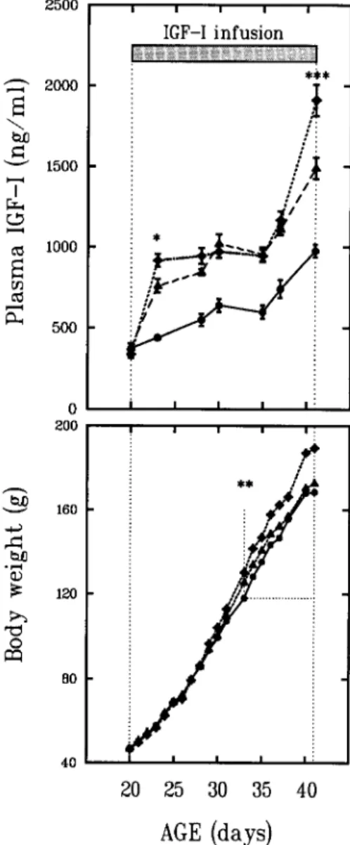
2500 2000 ~ 1500 I 1000 500 b/) bJD 0 CZ~ 0 200 1 6 0 120 8o 40 i i i i i IGF-I infusion I : ! : / ] / I | I I i m | u i r / : ** /
::,4,
20 25 30 35 40 AGE (days)Fig. 1. Pattern of changes between 20 and 41 d of life in body weight (BW) and plasma IGF-I concentration in Sprague-Dawley female rats either receiving a rhlGF-I infusion of 2 (A), or 4 ( t ) ~g/g BW/d using Alzet minipumps, starting at 20 d of life as indicated, or untreated (O). Mean + SE. No significant differ- ences in BW were observed between untreated rats and rats receiving 2/ag IGF-I/g BW/d; BW of rats infused with 4 ktg/g BW/d were different from controls starting at 33 d of life, as indicated. Plasma IGF concentrations of IGF-I-infused rats were highly significantly increased above control values. A significant difference in plasma IGF-I between rats receiving 2 and 4 gg/g BW/d, was observed at 23 and 41 d, as indicated. Rats from series #2:10 rats per point; other data presented in Table 1.
line in Fig. 1. Infusion with 2 ~tg IGF-I/g BW/d signifi- cantly increased plasma IGF-I levels at all ages, but the achieved absolute levels were lower at younger than older ages. In the IGF-I-infused rats presented in Fig. 1 and Table 1, plasma IGF-I increased from approx 700 ng/mL at 23 d to 1000 ng/mL at 35 d, as shown with filled triangles; there- after, a further increase occurred with plasma IGF-I levels reaching approx 1500 ng/mL at 41 d, consistent with the plasma levels achieved in the other series with the same
Vol. 6, No. 1 Treatment of Immature Female Rats with IGF-l/Gruaz et al 13
Table 1
Treatment of Immature Female Rats with a Constant Infusion of rhIGF-I Between Day of Life 20 and 41, as Illustrated in Fig. 1 (Series # 1)---Effects Seen at Sacrifice at Day of Life 41 on Growth and Somatotropic Parameters
IGF-I,/.tg/g/d Controls 2 4 AOV, p Growth parameters Body wt, g 168 + 4 173 + 4 189 + 4*** <0.005 Tail length, cm 14.9 + 0.2 14.6 + 0.2 14.9 + 0.2 NS Pituitary wt, mg 7.65 + 0.39 8.74 + 0.50 9.45 + 0.50* <0.05 mg/100g BW 4.49 + 0.18 4.95 + 0.19 4.88 + 0.22* NS Spleen wt, mg 706 + 38 1044 + 37*** 1428 + 115"** <0.001 mg/100 g BW 415 + 18 597 + 25*** 740 + 525*** <0.001 Kidney wt, g 1.59 + 0.04 1.92 + 0.05*** 2.31 + 0.06*** <0.001 g/100 g BW 0.93 + 0.01 1.09 + 0.02*** 1.20 + 0.02*** <0.001 Heart wt, mg 714 + 20 734 + 22 822 + 23** <0.005 mg/100 g BW 406 + 9 418 + 6 427 + 6* NS Adrenal wt, mg 41.4 + 1.4 47.7 + 2.6* 50.6 + 1.7"** <0.01 mg/100 g BW 24.3 + 0.6 26.8 + 1.0" 26.3 + 0.7* <0.05 Somatotropic axis Plasma IGF-I, ng/mL 976 + 40 1489 + 67*** 1913 + 96*** <0.001 Plasma GH, ng/mL 11.7 + 4.8 26.2 + 4.9 24.8 + 6.3 NS Pituitary GH, pg 126 + 15 174 + 17" 196 + 14"** <0.025 Plasma IGFBP-1 100 + 13 147 + 21 187 + 13"** <0.005 Plasma IGFBP-2 100 + 15 149 + 26* 177 + 19"* <0.05 Plasma IGFBP-3 100 + 11 160 + 20* 192 + 22** <0.005 Plasma IGFBP-4 100 + 7 83 + 7 86 + 7 NS Reproductive parameters Ovarian wt, mg 40.7 + 3.6 50.9 + 3.1" 57.3 + 3.6** <0.01 mg/100 g BW 23.8 + 1.9 29.0 + 1.6 29.8 + 1.7" <0.05 Uterine wt, mg 196 + 17 226 + 16 253 + 20 NS mg/100 g BW 115 + 9 128 + 7 132 + 10 NS Vaginal opening, d 38.6 + 0.8 37.2 + 0.8 38.2 + 0.9 NS
Mean + SE; 10 rats per group; significance, analysis of variance (AOV) withp values indicated for 2,27 degrees of freedom; plasma IGFBPs expressed in percent value of controls.
*p < 0.05, **p < 0.01, ***p < 0.001 with respect to untreated controls.
dosage at the same age (1404 + 41 ng/mL, Table 2). The pattern o f increased p l a s m a levels for I G F - I was similar for the higher dosage (4 p.g/g BW/d). There was a significant difference between the two dosages at 23 d, then no differ- ence between 25 and 35 d, both dosages generating similar adult p l a s m a levels (~1000 ng/mL). After 35 d, a marked increase in p l a s m a levels was seen for both dosages, and supraphysiological plasma levels were obtained with a clear difference at 41 d between the two dosages.
Pituitary GH Content and Plasma GH Levels
A dose-dependent, significant increase in pituitary G H content was seen in the series presented in Table 1. The s a m e trend, although not significant, was found in the other series. T h e r e w a s no d i f f e r e n c e in p l a s m a G H lev- els at sacrifice in control or I G F - I - t r e a t e d rats (Table 1), and e v i d e n c e f o r G H p e a k s w a s o b t a i n e d in the g r o u p o f rats r e c e i v i n g 4 ktg/g B W / d since three values a b o v e 50 n g / m L were measured. These elevated G H values speak
against a possible inhibition o f G H secretion b y increased circulating IGF-I.
Plasma IGFBPs Concentration
Analysis o f plasma I G F B P s in the series presented in Fig. 1 indicated that infusion o f I G F - I produced increases in I G F B P - 1 to -3 o f a s i m i l a r a m p l i t u d e , w h e r e a s no increase was seen for IGFBP-4. The increase in IGFBP-3
corresponded to 160 + 20% o f controls with the 2 ~g/g BW/d dosage, and 192 + 22 %, with the 4 p.g/g B W / d dosage at 41 d (Table 1).
Body Weight Gain and Tail Length
No significant effect on B W gain was observed with infusion o f 2 ~ IGF-I/g BW/d. With the higher dosage (4 p.g/ g BW/d), B W gain was significantly increased over con- trois starting at 33 d (p = 0.002), and r e m a i n e d so until 41 d (p < 0.001, Fig. 1, lower panel). In both series, no effects o f the I G F - I infusion were observed on tail length as measured at 41 d.
] 4 Treatment of Immature Female Rats with IGF-l/Gruaz et al. Endocrine
Table 2
Treatment of Immature Female Rats with a Constant Infusion
of rhIGF-I Between Day of Life 20 and 41 (Series #2)--Effects on Sexual Maturation and Fertility
Controls IGF-I, 2 pg/g BW/d Significance, p
Data at 41 d Plasma IGF-I, ng/mL 1017 + 25 1404 + 41 <0.001 Body wt, g 170 + 3 176 + 5 NS Pituitary wt, mg 7.47 + 0.42 8.02 + 0.49 NS mg/100 g BW 4.41 + 0.19 4.55 + 0.19 NS Spleen wt, mg 574 + 44 824 + 23 <0.001 rag/100 g BW 333 + 20 469 + 18 <0.001 Ovarian wt, mg 42.4 + 3.2 56.4 + 2 0.005 mg/100 g BW 24.9 + 1.7 32.0 + 1 0.006 Vaginal opening, d 36.2 _+ 0.4 37.8 + 0.6 0.031 Fertility Number of pups 13.2 + 0.8 12.5 + 0.8 NS Pup wt, g 7.00 + 0.07 7.06 + 0.10 NS
Mean + SE; 8 rats per group sacrificed at the end of pump capacity at 41 days of life, 6 rats per group used for the fertility test; assessment of day of vaginal opening done with all rats (14 rats per group).
Weight Gain of Nonreproductive Organs
In contrast to the absence of any large changes in BW gain, striking dose-dependent effects of infused IGF-I were seen on the growth of spleen, kidneys, heart, and adrenals (Tables 1 and 2). For spleen, kidney, and adrenals, this effect was also seen if organ weight was expressed per 100 g BW. The effects on the weight gain of the spleen were the greatest, followed by kidneys and adrenals, and were present at both dosages studied. The 3-wk IGF-I infusion at 4 gg/g BW/d produced a doubling of spleen weight. The increase in spleen weight was clearly present in each animal, therefore, representing an internal marker for the effect of IGF-I. Histological examination of enlarged spleen did not reveal any abnormalities, but rather an homoge- neous enlargement of this tissue.
Effects of lGF-I Infusion on the Reproductive Axis Weight Gain of Reproductive Organs
Treatment with IGF-I at the dose of 2 pg/g BW/d did not affect pituitary growth, but a significant increase was seen with the 4 pg/g BW/d dosage not present if pituitary weight is expressed in function of BW (Table 1). IGF-I infusion produced a significant, dose-dependent increase in ovarian weight (Tables 1 and 2). With the 4 ~g/g BW/d dosage, ovarian absolute weight of treated rats represented approx 140% of control weight and this increase was also seen if expressed in function of BW (Table 1).
Ovarian Morphology
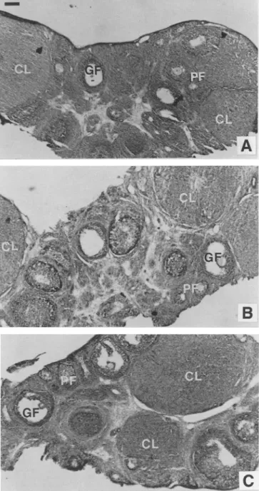
The histological aspect of ovarian tissues for control and IGF-I-treated rats for the two dosages, as seen at the light microscopic level, is illustrated in Fig. 2. No abnormalities were seen in the ovaries of the treated animals, and the analysis of the different follicles indicated a similar stage of development in the three sets of ovarian tissues examined.
The proportion of primary follicles, Graafian follicles, and corpora lutea assessed by inspection of a comparable num- ber of stained sections of ovaries originating from control and IGF-I-treated rats at both dosages was shown not to be different, as seen by a chi-square analysis (Table 3).
Uterine Weight
There was a moderate increase in uterine weight, but the analysis of variance did not indicate a dose-dependent increase.
Vaginal Opening and Fertility
One purpose of this study was to evaluate whether pre- maturely increased circulating IGF-I to adult levels would modify the timing of sexual maturation by an action at either the hypothalamic or gonadal level. In series # 1 that included both IGF-I dosages, there was no difference in the timing of vaginal opening (Table 1). In series #2, a small but sig- nificant delay of 1.6 d in vaginal opening was observed (Table 2). In two other series performed in similar condi- tions (2 gg IGF-I/g BW/d), vaginal opening took place at 36.5 + 1.0 d in treated animals vs 37.3 + 0.4 d in controls in one series and 38.5 + 1.0 d vs 37.3 + 0.4 d, in the other series (data not shown). Fertility was tested in half of the rats presented in Table 2. Alzet pumps were left in place, and mating occurred at 50 d of life. The outcome of mating and gestation was not different between control and IGF-I- treated rats. A vaginal plug was seen for all rats. Neither the number of pups, nor the weight of pups were different between control and IGF-I-treated rats (Table 2).
D i s c u s s i o n
The aim of this study was to evaluate the effects o f increased plasma IGF-I levels during the juvenile period of the female rat, defned as days of life 20-35 (21), on growth
Vol. 6, No. 1 Treatment of Immature Female Rats with IGF-I/Gruaz et al, 15
Fig. 2. Structure o fovarian tissues studied at the light microscope level in a control rat (A) and in representative rats treated with 2 (B), and 4 (C) gg IGF-I/g BW/d between 20 and 41 d. The ovarian slices were treated and stained according to the Masson- Goldner's trichrome method, as described in the Method section. PF, primary follicle; GF, Graafian follicle; CL, corpus luteum. Scale bar represents 200 pro.
and developmental aspects of sexual function. Since abnor- mally elevated plasma levels of IGF-I have been shown to increase the size of several organs
(3,18,19),
special atten- tion was given to possible effects oflGF-I on the growth of both reproductive and nonreproductive organs during development. It has recently been demonstrated that mark- edly reduced IGF-I plasma levels, secondary to the inhibi- tion of GH secretion by passive immunization against rat GRF starting at 15 d of life, drastically reduced growth rate,Table 3
Treatment of Immature Female Rats with a Constant Infusion of rhlGF-I Between Day of Life 20 and 4 1 -
Distribution of Primary Follicles, Graafian Follicles, and Corpora Lutea Expressed in Percentage of Observed Structures
IGF-I, pg/g/d Controls 2 4 Primary follicles 35.0 % 28.5 % 34.1% Graafian follicles 35.8 % 41.4 % 31.1% Corpora lutea 29.1% 30.1% 34.8 % Number of counted structures 120 133 161
No significant difference in distribution between treatment groups (chi-square test, a = 0.05).
but did not disturb the onset ofestrous cycles, thus indicat- ing that low plasma IGF-I levels do not prevent sexual maturation and fertility
(2).
The authors found it as impor- tant to address the question whether increased, rather than decreased, plasma IGF-I levels would affect sexual matu- ration, possibly producing precocious puberty. This issue is important for the pediatric endocrinologists who treat GH-resistent patients with IGF-I.We show that chronically elevated plasma IGF-I levels generated by constant infusion of IGF-I at two dosages pro- duced the expected differential acceleration of organ growth seen previously in studies performed with adult animals, with a significant increase in ovarian weight at 41 d of life. In contrast, the IGF-I infusion neither accelerated the timing of sexual maturation nor did it alter fertility later on.
The rationale of this study was to impose plasma IGF-I levels in young female rats comparable to those seen in sexually mature animals, thus raising these levels from approx 300 to 1000 ng/mL starting at 20 d of life. The authors found that the infusion of 2 gg/g BW/d generated lower plasma levels in younger animals, 600-700 ng/mL at 27 d of life, than later in older animals at 35 d of life, when plasma values clearly became above normal adult levels (> 1000 ng/mL). Increasing the dose to 4 gg IGF-I/g BW/d did not significantly increase circulating plasma IGF-I lev- els over those observed with the lower dosage during the 25-35 age range, pointing to some saturation mechanisms that could be related to the availability of IGFBPs at this young age. A dose-dependent difference in plasma IGF-I levels was only seen at 41 d. At this age, there was a good parallelism between achieved plasma IGF-I levels and the observed increase in circulating IGFBPs. Obviously, the achieved plasma IGF-I levels in infused animals depend upon several parameters: the IGF-I dosage used, a possible decrease in endogenous IGF-I secretion secondary to reduction in GH secretion, an IGF-I-stimulated increase in circulating IGFBPs, and possible changes of clearance rate as a result of other causes than IGFBPs concentration.
] 6 Treatment of Immature Female Rats with IGF-I/Gruaz et al. Endocrine The biological action of IGFs is closely modulated by
the presence of IGFBPs in the circulation
(5,22,23).
The induction of IGFBP-3, the most abundant circulating IGFBP that mainly conditions the half life for IGF-I, is hormonally regulated, GH being the most important regulator oflGFBP-3 synthesis(5).
However, IGF-I itself can also induce the production oflGFBP-3 in the rat(5,24).
IGF-I treatment o f hypophysectomized, or protein- deprived rats with low or absent GH can markedly increase IGFBPs(5,25).
The ability oflGF-I to trigger the synthesis of its own IGFBPs has also been demonstrated in vitro(26).
This study confirms that IGF-I treatment induces synthesis and release of large amounts of principally IGFBP-3, but also IGFBP-1 and -2.Various studies have demonstrated that IGF-I can inhibit GH secretion at the pituitary level, as seen with pituitary cells in culture
(27,28),
with permanent cell lines such as GH 3(29),
or with pituitary explants in culture(30,31).
The available published in vivo studies are more difficult to interpret since systemic administration of IGF-I would certainly act at the pituitary level to reduce GH release, so that any additional action at a higher level is less easily identified. Thus a central administration oflGF-I using the intracerebroventricular route is needed, not necessarily representing a peripheral action oflGF-I(32).
Tannenbaum et al. have shown that icv administration of IGF-I and -II could interact with the hypothalamic regulation of GH release, but apparently, IGF-I alone is unable to act cen- trally to inhibit GH secretion(33,34).
Thus, the feedback action of circulating IGF-I is most likely exerted at the level of pituitary somatotrophs.It was anticipated that the authors' IGF-I infusion para- digm would reduce GH secretion. Indeed, in a preliminary study, they observed a reduction of the pulsatile release of GH in some of the young rats receiving 2 lag IGF-I/g BW/d. In the present study the authors demonstrate increased pituitary GH content in IGF-I-treated rats consistent with a buildup phenomenon suggesting a decrease in GH release. However, with both dosages, normal to elevated plasma GH levels were measured at sacrifice, whereas plasma IGF-I levels were equal to or above 1400 ng/mL. This is negative evidence that circulating IGF-I at this concentra- tion abolish GH secretion. In a more recent study, an infu- sion of 8 lag IGF-I/g BW/d in 40-d-old rats generating plasma IGF-I levels of about 2000 ng/mL was shown to consistently inhibit pulsatile GH release as seen over a 24-h collection period (V. d'All6ves, N. M. Gruaz, A. Skottner, and M. L. Aubert, in preparation). Thus it appears that inhibition of plasma GH by IGF-I is a dose-related phe- nomenon and is probably present only with dosages above 4 lag/g BW/d.
No effect of infused IGF-I was seen on BW again in rats receiving 2 lag IGF-I/g BW/d roughly creating the physi- ological plasma levels of the adult female rat. Treatment with IGF-I has previously been shown to reestablish growth
in hypophysectomized rats
(3,14-16),
and there has been much debate whether such treatment could restore normal growth rate. It now appears that the choice of dosage is most important and Guler et al. have shown that 300 pg/d (or approx 2.5 p,g/g/d) was necessary to achieve this goal(16).
In mutant Dwarf rats, a dosage of 180 pg/d (1 p.g/g/d), only partially restored normal growth rate(3).
It is possible that simultaneous administration of GH and IGF-I might be more appropriate to promote growth(35),
probably by minimizing the lack of GH secretion because of the poten- tial inhibition imposed by exogenous IGF-I. In this study, the dose of 4 pg IGF-I/g BW/d generated supraphysio- logical plasma levels during the last 5 d of treatment and significantly increased BW gain during this period, con- firming that IGF-I can increase BW gain above the normal range. However, tail length was not affected in these conditions.IGF-I infusion during the juvenile age in the female rat produced a dose-dependent acceleration of growth of sev- eral organs as seen from increased weight, confirming ear- lier studies from one of us
(3),
and extending them to this younger age period. The most striking effect was on spleen weight with observed 1.5- to 2-fold increases. These increases are more important than corresponding BW increase as seen when organ weights are expressed per 100 g BW. This is in agreement with earlier studies per- formed either in GH-deficient rat models(3),
or transgenic mice overexpressing the IGF-I gene(18,19).
These effects of IGF-I infusion on spleen weight cannot be a result of a nonspecific inflammatory insult resulting from implanta- tion of the Alzet pump, since it was previously observed that when using a low IGF-I dosage (0.5 p,g/g BW/d) that did not produce any effects, spleen weight remained nor- mal despite the presence of the Alzet pump (data not shown). One interesting novel observation in this study was that IGF-I infusion produced acceleration of pituitary growth when using the 4 p,g/g BW/d dosage, indicating that IGF-I might affect growth of this organ, as alluded to in another study(15).
Kidney weight was increased at all ages studied and the achieved kidney weight in treated animals represented approx 145% that of control values with the 4 ~tg/g BW/d dosage. Histological examination of kidneys from control and IGF-I-treated rats performed with tissues removed at 40 d of life did not reveal any modifications of the renal structure (E. Girardin, personal communication). Heart weight was moderately increased and only in the high dose group.One important aim of this study was to evaluate the effect of increased plasma IGF-! levels on reproductive function. It has been shown that IGF-I treatment consistently pro- duced an increase in ovarian weight, as seen at 41 d of life. In a preliminary study the authors observed no increase in ovarian weight at 27 and 34 d when 2 pg IGF-I/g BW/d was infused, starting at 20 d. The increase in ovarian weight at 41 d was significant, but the achieved weight in IGF-I-
Vol. 6, No. 1 Treatment of Immature Female Rats with IGF-I/Gruaz et al ] 7
treated rats at this age did not exceed the weight generally seen for adult animals. Furthermore, the enlarged ovaries appeared completely normal histologically. In a toxicologi- cal study performed by two of the authors, adult female rats (n = 25-30) received daily sc injections of rhIGF-I (0.4 to 10 gg/g BW/d) for a period of 26 wk. No significant differ- ence in ovarian weight between controls (93 + 24 mg) and rats receiving 10 gg IGF-I/g BW/d (119 + 75 mg) was observed. Histological examination of the ovaries did not show any treatment-related pathological changes.
The concern that increased plasma IGF-I could advance sexual maturation was not verified. Indeed, prematurely increased plasma levels for IGF-I did not modify the pace of sexual maturation. Onset of estrous cyclicity as seen by vaginal opening was not accelerated in treated animals as seen with two IGF-I dosages. Provided that vaginal open- ing is checked only once daily, the small delay of 1.6 d observed in one series (series #2), is close to the normal variation seen for this parameter (p --- 0.031). It does not weaken the statement that no meaningful changes were observed. As said, in two other series performed in similar conditions (2 gg IGF-I/g BW/d), vaginal opening took place at the same time in IGF-I-treated and control animals. In a toxicological study in sexually mature female rats per- formed at Pharmacia in Sweden, the influence of increased IGF-I plasma levels upon reproductive function and fer- tility was assessed. Daily subcutaneous treatment with rhIGF-I at dose levels of 0.4--10 gg/g BW/d for 15 d before pairing and up to day 14 after mating did not cause any adverse effects on oestrous cycles, mating performance, or fertility (data not shown).
The possibility that circulating IGF-I produces more specific endocrine effects on ovarian function than promot- ing organ growth remains open. The relation between cir- culating GH and IGF-I on the ovarian function, and action of locally produced IGF-I is still unclear
(8, 9).
A possible role for circulating GH at the ovarian level could be through the local generation of IGF-I. Potentiating effects of GH and/or IGF-I at the ovarian level have been advocated(8, 9).
The existence of a well-defined intraovarian IGF system, involving IGF-I and II, specific receptor subtypes, and sev- eral populations of IGFBPs have been nicely documented
(8,13).
Apparently, the internal signaling system achieved by IGF-I that potentiates FSH action, is autonomous, specifically responding to gonadotropins, and might not depend upon other external influences such as GH or hepatic IGF-I. Nevertheless, some evidences for a trophic action of GH on the ovary have been documented(10-12).
There have been suggestions that the rising plasma lev- els of IGF-I could provide a stimulatory signal for the pubertal increase in GnRH release
(6).
In this situation, IGF-I would be able to act on GnRH neuron terminals in the median eminence, or possibly at perikarya if a transport mechanism through the blood-brain barrier is confirmed(36).
Hiney et al. (6) showed that IGF-I (but not insulin orIGF-II) can increase the in vitro release in GnRH from incubated median eminence obtained from 30-d-old female rats. The authors have shown that low IGF-I plasma con- centrations in GH-deprived rats do not modify the hypotha- lamic drive of sexual function in the rat
(2).
An action for IGF-I at the hypothalamic level cannot be excluded as dis- cussed in a recent report(7),
but appears unlikely.In conclusion, treatment of immature female rats with IGF-I significantly modified BW gain at the higher dosage tested and this effect could be related to the increase in IGFBPs that permits a longer half-life for IGF-I, thus prob- ably prolonging biological action. Significant biological action of IGF-I was seen in this study with the increase in weight of several organs, including ovarian weight. Despite the increased presence of plasma IGF-I, no major effects were seen on reproductive function. The normal outcome of gestation and delivery with rats treated during the juve- nile period, or in adult rats treated chronically with higher IGF-I dosages, indicates that excessive presence of circu- lating IGF-I is not deleterious to sexual function.
Materials and Methods
Hormones
Human IGF-I was produced by Pharmacia AB, Stock- holm, Sweden
(3),
and 5 or 10 mg/mL solutions were used to fill the Alzet minipumps.Animals
Sprague-Dawley female rats were purchased from IFFA- CREDO (69592 L'Arbresle, France), and were received at 10 d of life with 10 pups per dam. They were provided with water and standard rat pellets ad libitum unless indi- cated. Room temperature was maintained between 21 and 23~ and a 12/12 h light/dark cycle was used. Body weight (BW) was measured daily. All experiments were reviewed by the University of Geneva School of Medicine Ethical Committee for Animal Experimentation and approved by the State of Geneva Veterinary Office (autho- rization No. 31.1.28/459/II).
Experimental Protocols
Two series of rats were studied strictly using the same protocol. The principle was to impose adult levels of circu- lating IGF-I to immature rats throughout the entire juvenile period (20-41 d of life) and to study the effects of this treatment on somatic and reproductive parameters.
Series #1
This series included two IGF-I dosages (2 and 4 gg/g BW/d) for an infusion period between 20 and 41 d of life. Alzet pumps 2001 (200 gL capacity, 1.0 gL/h flow rate) were used. Pumps were filled with the 5 mg/mL IGF-I solution for the lower dosage and with the 10 mg/mL solution for the higher dosage. At 20 d, one pump was implanted; then at 27 d, the used pumps were removed and
l/3 Treatment of Immature Female Rats with IGF-I/Gruaz et al. Endocrine two new pumps were implanted; at 34 d, two new pumps
were implanted similarly. Control rats were also anesthe- tized and sham-operated on these days. Day of vaginal opening was assessed by daily vaginal inspection starting at 30 d of life. All rats were sacrificed at 41 d. In this series, a small volume of blood was collected from the tail tip of each rat at days of life 23, 28, 30, 35, and 37 to monitor plasma IGF-I concentration. Using the 5 mg/mL IGF-I solution to generate the 2 gg/g BW/d dosage, actual dosage of IGF-I based on measured BW and pump delivery was 2.58 gg/g BW/d at 20 d, 1.51 at 27 d before pump change, 2.80 at 28 d after inserting two pumps, 1.80 at 34 and 1.39 at 41 d, thus resulting in a mean dosage of 1.95 + 0.09 gg/ g BW/d. For the 4 gg/g BW/d dosage, for which the 10 gg/ mL IGF-I solution was used to fill the pumps, actual dosage was 5.16 at 20 d, 3.01 at 27, 5.56 at 28 d after the implan- tation of two pumps, 3.39 at 34 d, and 2.53 at 41 d, resulting in a mean dosage of 3.81 + 0.20 gg/g BW/d. Rats were sacrificed at 41 d (10 rats per group), trunk blood was col- lected in chilled heparinized tubes, pituitary, and ovaries were quickly dissected out, weighed and frozen, other organs such as kidneys and spleen were also dissected out but only weighed.
Series #2
Alzet pumps 200 L filled with the 5 mg/mL IGF-I solu- tion were used, resulting in a daily dose of 2 gg/g BW. As for the other series, one pump was implanted in the area of the neck under light ether anaesthesia at 20 d of life; then at 27 d, the used pumps were removed and two new pumps were implanted, always under light ether anaesthesia. At 34 d, two new pumps were implanted similarly. Control rats were also anesthetized and sham-operated on these days. Part of the rats were sacrificed at 41 d (8 rats per group), as described for series # 1. The remaining rats (6 rats per group) were not sacrificed, the pumps were left in situ,
and fertility was evaluated at 50 d of age, with six IGF-I- treated rats and five corresponding controls being left each with a normal male for 7 d and outcome of breeding, evaluated.
Hormone Determinations
Plasma and pituitary GH concentrations were analyzed by RIA as described earlier (37), using NIADDK-anti-rGH $5 and the RP-2 reference preparation. RIA for IGF-I was performed as described recently in detail (38) by using the antiserum UB 3-189 provided by Drs. L. Underwood and J. J. Van Wyk and a recombinant IGF-I preparation obtained from Pharmacia, Kabi Peptide Hormones, Stockholm, Swe- den. Serum extraction was performed according to the method described by Breier et al. (39). This extraction tech- nique has been validated as providing complete removal of IGFBPs. The antiserum (1:10,000 final dilution) was incubated for 90 min at 4~ with plasma specimens (dupli- cates), or standard amounts of biosynthetic IGF-I (tripli- cates) after which radioiodinated 125I-IGF-I (15,000 cpm) was added. Within and between assay variance was 8 and
17%, respectively. Minimal detectable concentration was 5 pg/tube.
Distribution of lGFBPs by Western Ligand Blot Analysis
The distribution of plasma IGFBPs was performed by the Western Ligand Blotting technique described by Hossenlopp et al. (40) as described in detail recently (2).
Briefly, plasma samples were diluted with buffer, boiled at 95~ for 5 min, cooled and immediately applied to a 11% SDS-polyacrylamide gel and run using a constant current (8 mA/gel) for about 20 h. The fractionated proteins were electroblotted on Immobilon membranes and after wash- ing, the membranes were incubated in a sealed plastic bag in presence of l25I-IGF-I at 4~ Radiolabeled IGFBPs were visualized by autoradiography and quantified using the program Image Quant, version 3.2. from Molecular Dynam- ics, on an Olivetti M 400-40 desk computer.
Analysis of Ovarian and Spleen Histology
Spleen and ovaries from rats treated with IGF-I and controls were fixed by immersion in 4% paraformaldehyde and stored at 4~ in the same fixative for several days. Tissue blocks were washed in 10% sucrose in phosphate buffer, pH 7.4, for up to 24 h, and cut in longitudinal plane using a cryostat. Fifteen ~rn sections were thaw-mounted on poly-L-lysine subbed glass slides and stained according to the procedure reported by Goldner (41). The sections finally mounted in Eukit were examined and photographed under Zeiss Axioskop microscope. The follicular matura- tion in control and treated animals was examined in stained sections through the ovaries (approx 12 sections/ovary). In order not to count a structure twice, number of follicle was estimated by counting oocytes that should appear only once when using 15 grn section; corpus lutea were counted only when at maximal profile. The distribution frequency of observed primary follicles, Graafian follicles and corpora lutea was assessed and compared between groups by a chi-square analysis.
Statistical Analysis
Analysis of variance (ANOVA) was first performed to evaluate the overall variation due to treatment, then indi- vidual variations were analyzed by the Duncan's range test, or unpaired t-test when appropriate.
Acknowledgments
The authors acknowledge the excellent technical assis- tance of Audrey Aebi, Anne Scherrer, Brigitte Delavy, and Brigitte Greggio. The skillful technical assistance of Jean- Jacques Goy in our animal quarter is gratefully acknowl- edged. We express our appreciation to Eric Girardin, Geneva, for investigating renal histology. This study was supported by grants from the National Research Science Foundation # 31-28'803-90 and 31-39729-93, and partly by Pharmacia AB, Peptide Hormones, Stockholm, Sweden.
Vol. 6, No. 1 Treatment of Immature Female Rats with IGF-I/Gruaz et al. 19
R e f e r e n c e s
1. Handelsman, D. J., Spaliviero, J. A., Scott, C. D., and Baxter, R. C. (1987). Endocrinology 120, 491-496.
2. Gruaz, N, M., Arsenijevic, Y., Wehrenberg, W. B., Sizo- nenko, P. C., and Aubert, M. L. (1994). Endocrinology 135, 509-519.
3. Skottner, A., Clark, R. G., Fryklund, L., and Robinson, I. C. A. F. (1989). Endocrinology 124, 251%2526.
4. Charlton, H. M., Clark, R. G., Robinson, I. C. A. F, Porter Goff, A. E., Cox, B. S., Bugnon, C., and Bloch, B. A. (1988). J. Endocrinol. 119, 51-58.
5. Clemmons, D. R., Thissen, J. P., Maes, M., Ketelslegers, J. M., and Underwood, L. E. (1989). Endocrinology 125, 2967-2972. 6. Hiney, J. K., Ojeda, S. R., and Dees, W. L. (1991). Neuroendo-
crinology 54, 420-423.
7. Bourguignon, J. P., G6rard, A., Alvarez-Gonzalez, M.-L., and Franchimont, P. (1993). Neuroendocrinology 58, 525-530. 8. Adashi, E. Y., Resnick, C. E., d'Ercole, A. J., Svoboda, M. E.,
and Van Wyk, J. (1985). Endocr. Rev. 6, 400-420. 9. Giudice, L. C. (1992). Endocr. Rev. 13, 641-669.
10. Davoren, J. B. and Hsueh, A. J. W. (1986). Endocrinology 118, 888-890.
11. Jia, X. C., Kalmijn, J., and Hsueh, A. J. W. (1986). Endocrinol- ogy 118, 1401-1409.
12. Hernandez, E. R., Roberts, C. T. Jr, LeRoith, D., and Adashi, E. Y. (1989). Endocrinology 125, 572-578.
13. Adashi, E. Y., Resnick, C. E., Hernandez, E. R., and Rosenfeld, R. G. (1990). Endocrinology 126, 1305-1307.
14. Schoenle, E., Zapf, J., Humbel, R. E., and Froesch, E. R. (1982). Nature 296, 252,253.
15. Hizuka, N., Takano, K., Shizume, K., Asakawa, K., Miyakawa, M., Tanaka, H., and Horikawa, R. (1986). Eur. J. Pkarmacol. 125, 143-146.
16. Gueler, H. P., Zapf, J., Scheiwiller, E., and Froesch, E. R. (1988). Proc. Natl. Acad. Sci. USA 85, 4889-4893.
17. Skottner, A., Clark, R. G., Robinson, I. C. A. F., and Fryklund, L. (1987). J. Endocrinol. 112, 123-132.
18. Mathews, L. S., Hammer, R. E., Behringer, R. R., D'Ercole, A. J., Bell, G. I., Brinster, R. L., and Palmiter, R. D. (1990). Endocrinology 123, 2827-2833.
19. Behringer, R. R., Lewin, T. M., Quaife, C. J., Palmiter, R. D., Brinster, R. L., and D'Ercole, A. J. (1990). Endocrinology 127, 1033-1040.
20. Rosenfeld, R. G., Rosenbloom, A. L., and Guevara-Aguirre, J. (1994). Endocr. Rev. 15, 36%390.
21. Ojeda, S. R., Urbanski, H. F., and Ahmed, C. E. (1986). Recent Prog. Horm. Res. 42, 385--440.
22. Baxter, R. C. and Martin, J. L. (1989). Prog. Growth Fact. Res. 1, 49-68.
23. Lamson, G., Giudice, L. C., and Rosenfeld, R. G. (1991). Growth Fact. 5, 1%28.
24. Zapf, J., Hauri, C., Waldvogel, M., Futo, E., Hhsler, H., Binz, K., Guler, H. P., Schmid, C., and Froesch, E. R. (1989). Proc. Natl. Acad. Sci. USA 86, 3813-3817.
25. Gargosky, S. E., Tapanainen, P., and Rosenfeld, R. G. (1994). Endocrinology 134, 2267-2276.
26. Bale, L. K. and Conover, C. A. (1992). Endocrinology 131, 608-614.
27. Brazeau, P., Guillemin, R., Ling, N., van Wyk, J., and Humbel, R. (1982). C. R. Acad. Sciences Paris. 295, 651-654. 28. Yamashita, S. and Melmed, S. (1986). Endocrinology 118,
176-182.
29. Yamashita, S. andMelmed, S. (1987).J. Clin. Invest. 79,449-452. 30. Goodyer, C. G., De St6phano, L., Wei Hsien Lai, Guyda, H. J.,
and Posner, B. I. (1984). Endocrinology 114, 1187-1195. 31. Goodyer, C. G., De St6phano, L., Guyda, H. J., and Posner,
B. I. (1984). Endocrinology 115, 1568--1576.
32. Berelowitz, M., Szabo, M., Frohman, L. A., Firestone, S., and Chu, L. (1991). Science 212, 1279-1281.
33. Tannenbaum, G. S., Guyda, H. J., and Posner, B. I. (1983). Science 220, 77-79.
34. Hazel, Z. and Tannenbaum, G. S. (1992). Endocrinology 131, 758-764.
35. Kupfer, S. R., Underwood, L. E., Baxter, R. C., and Clemmons, D. R. (1993). ,1. Clin. Invest. 91, 391-396.
36. Reinhardt, R. R. and Bondy, C. A. (1994). Endocrinology 135, 1753-1761.
37. Arsenijevic, Y., Rivest, R. W., Eshkol, A., Sizonenko, P. C., and Aubert, M. L. (1987). Endocrinology 121, 1487-1496. 38. Catzeflis, C., Pierroz, D. D., Rohner-Jeanrenaud, F., Rivier,
J. E., Sizonenko, P. C., and Aubert, M. L. (1993). Endocrinol- ogy 132, 224--234.
39. Breier, B. H., Gallaher, B. W., and Gluckman, P. D. (1991). 3". Endo- crinoL 128, 347-357.
40. Hossenlopp, P., Seurin, D., Segovia-Quinson, B., Hardouin, S., and Binoux, M. (1986). Anal. Biochem. 154, 138-143. 41. Goldner, J. (1938). Am. J. Pathol. 14, 237-243.