Publisher’s version / Version de l'éditeur:
Vous avez des questions? Nous pouvons vous aider. Pour communiquer directement avec un auteur, consultez la première page de la revue dans laquelle son article a été publié afin de trouver ses coordonnées. Si vous n’arrivez pas à les repérer, communiquez avec nous à PublicationsArchive-ArchivesPublications@nrc-cnrc.gc.ca.
Questions? Contact the NRC Publications Archive team at
PublicationsArchive-ArchivesPublications@nrc-cnrc.gc.ca. If you wish to email the authors directly, please see the first page of the publication for their contact information.
https://publications-cnrc.canada.ca/fra/droits
L’accès à ce site Web et l’utilisation de son contenu sont assujettis aux conditions présentées dans le site LISEZ CES CONDITIONS ATTENTIVEMENT AVANT D’UTILISER CE SITE WEB.
Proceedings of the National Academy of Sciences, 116, 37, pp. 18655-18663,
2019-09-27
READ THESE TERMS AND CONDITIONS CAREFULLY BEFORE USING THIS WEBSITE. https://nrc-publications.canada.ca/eng/copyright
NRC Publications Archive Record / Notice des Archives des publications du CNRC :
https://nrc-publications.canada.ca/eng/view/object/?id=dbe665e8-6c6d-4e7d-bf2f-36b112f7c0cb
https://publications-cnrc.canada.ca/fra/voir/objet/?id=dbe665e8-6c6d-4e7d-bf2f-36b112f7c0cb
NRC Publications Archive
Archives des publications du CNRC
This publication could be one of several versions: author’s original, accepted manuscript or the publisher’s version. / La version de cette publication peut être l’une des suivantes : la version prépublication de l’auteur, la version acceptée du manuscrit ou la version de l’éditeur.
For the publisher’s version, please access the DOI link below./ Pour consulter la version de l’éditeur, utilisez le lien DOI ci-dessous.
https://doi.org/10.1073/pnas.1907833116
Access and use of this website and the material on it are subject to the Terms and Conditions set forth at
A promising bioconjugate vaccine against hypervirulent Klebsiella
pneumoniae
Feldman, Mario F.; Mayer Bridwell, Anne E.; Scott, Nichollas E.; Vinogradov,
Evgeny; McKee, Samuel R.; Chavez, Sthefany M.; Twentyman, Joy;
A promising bioconjugate vaccine against
hypervirulent Klebsiella pneumoniae
Mario F. Feldmana,b, Anne E. Mayer Bridwella, Nichollas E. Scottc, Evgeny Vinogradovd, Samuel R. McKeea, Sthefany M. Chaveza, Joy Twentymane, Christina L. Stallingsa, David A. Rosena,e, and Christian M. Hardingb,1
aDepartment of Molecular Microbiology, Washington University School of Medicine, St. Louis, MO 63110;bVaxNewMo, St. Louis, MO 63108;cDepartment of Microbiology and Immunology, The Peter Doherty Institute for Infection and Immunity, University of Melbourne, Parkville, VIC 3010, Australia;dHuman Health Therapeutics, National Research Council Canada, Ottawa, ON K1A 0R6, Canada; andeDivision of Pediatric Infectious Diseases, Department of Pediatrics, Washington University School of Medicine, St. Louis, MO 63110
Edited by Dennis L. Kasper, Harvard Medical School, Boston, MA, and approved August 6, 2019 (received for review May 6, 2019)
HypervirulentKlebsiella pneumoniae (hvKp) is globally disseminating as a community-acquired pathogen causing life-threatening infec-tions in healthy individuals. The fact that a dose as little as 50 bacteria is lethal to mice illustrates the dramatic increase of virulence associ-ated with hvKp strains compared with classical K. pneumoniae (cKp) strains, which require lethal doses greater than 107bacteria. Until
recently, these virulent strains were mostly antibiotic-susceptible. However, multidrug-resistant (MDR) hvKp strains have been emerg-ing, spawning a new generation of hypervirulent“superbugs.” The mechanisms of hypervirulence are not fully defined, but overproduc-tion of capsular polysaccharide significantly impedes host clearance, resulting in increased pathogenicity of hvKp strains. While there are more than 80 serotypes ofK. pneumoniae, the K1 and K2 serotypes cause the vast majority of hypervirulent infections. Therefore, a glycoconjugate vaccine targeting these 2 serotypes could signif-icantly reduce hvKp infection. Conventionally, glycoconjugate vaccines are manufactured using intricate chemical methodologies to covalently attach purified polysaccharides to carrier proteins, which is widely considered to be technically challenging. Here we re-port on the recombinant production and analytical characterization of bioconjugate vaccines, enzymatically produced in glycoengineered Escherichia coli cells, against the 2 predominant hypervirulent K. pneumoniae serotypes, K1 and K2. The K. pneumoniae bioconju-gates are immunogenic and efficacious, protecting mice against le-thal infection from 2 hvKp strains, NTUH K-2044 and ATCC 43816. This preclinical study constitutes a key step toward preventing fur-ther global dissemination of hypervirulent MDR hvKp strains.
glycoconjugate
|
bioconjugation|
vaccine|
hypervirulent Klebsiella pneumoniaeK
lebsiella pneumoniae is an encapsulated, Gram-negative bacterium of the Enterobacteriaceae family recognized as an opportunistic pathogen causing nosocomial infections (1). K. pneumoniae is notorious mostly due to the emergence of carbapenem-resistant strains (2); however, the rise and global dissemination of a hypervirulent form of K. pneumoniae is alarming (3). While the majority of K. pneumoniae infections manifest in the hospital setting or in immunocompromised individuals (termed classical K. pneumoniae [cKp] infection), a subset of highly invasive, community-acquired K. pneumoniae infections, termed hyperviru-lent K. pneumoniae (hvKp) infections, are steadily increasing in frequency (3).First observed in the 1980s in Taiwan, hvKp infections are pyogenic and mainly present as hepatic abscesses that can be complicated by endophthalmitis, meningitis, osteomyelitis, and necrotizing fasciitis (4–7). One of the most notable bacterial phe-notypes associated with hvKp is the overproduction of the capsular polysaccharide (CPS) (8), which results in a hypermucoviscous phenotype. This phenotype can be demonstrated by a positive string test: a greater than 5 mm“string” between an inoculating loop and a plated bacterial colony (9). Overproduction of the CPS has been directly linked with increased resistance to host clearance
via impaired complement-mediated bacterial killing (10) and phagocytosis by neutrophils and macrophages (11).
More than 80 K. pneumoniae CPS serotypes have been iden-tified (12); however, only 2 serotypes, the K1 and K2 serotypes, are responsible for the vast majority of hvKp infections. In fact, K1 and K2 serotypes have been associated with ∼70% of all hvKp infections across many clinical institutions worldwide (8, 13–15). Additionally, while these infections have historically been susceptible to most antibiotic classes, there are now increasing reports emerging of hvKp strains acquiring multiple antibiotic-resistance determinants, rendering them refractory to most thera-peutic regimens (16, 17). Given the severity of disease associated with hvKp infections; their propensity for young, healthy hosts; the increasing rise of drug resistance in hvKp strains; and the observation that the majority of hvKp infections are caused by 2 serotypes, a bivalent glycoconjugate vaccine against the K1 and K2 serotypes would be an optimal prophylactic option.
Glycoconjugate vaccines, composed of a bacterial polysaccharide covalently attached to a carrier protein, are lifesaving prophylactic agents used to prevent colonization and disease by certain bacterial pathogens. Moreover, glycoconjugate vaccines elicit immunological
Significance
Klebsiella pneumoniae is considered a nosocomial pathogen, usually infecting immunocompromised patients. However, a pathotype ofK. pneumoniae, termed hypervirulent K. pneumoniae (hvKp), has emerged and is spreading throughout the community, causing severe, often fatal, disease in healthy individuals. Moreover, reports on multidrug-resistant hvKp isolates are increasing in fre-quency. It is imperative that strategies to combat hvKp begin im-mediately to prevent further dissemination of this new class of “superbugs.” Here, we show that bioconjugate vaccines targeting the capsule of hvKp can provide immunity and protection against extremely lethal hvKp strains. Further, we demonstrate that bioconjugation is a promising technology for rapid development of efficacious vaccines against emerging bacterial threats.
Author contributions: M.F.F., A.E.M.B., C.L.S., D.A.R., and C.M.H. designed research; A.E.M.B., N.E.S., E.V., S.R.M., S.M.C., J.T., D.A.R., and C.M.H. performed research; M.F.F., A.E.M.B., N.E.S., E.V., C.L.S., D.A.R., and C.M.H. analyzed data; and M.F.F., N.E.S., E.V., D.A.R., and C.M.H. wrote the paper.
Conflict of interest statement: M.F.F. and C.M.H. have a financial stake in VaxNewMo, a for-profit entity developing bioconjugate vaccines against Streptococcus pneumoniae and Klebsiella pneumoniae using patented technology derived from the data presented in this and other published manuscripts.
This article is a PNAS Direct Submission.
This open access article is distributed underCreative Commons Attribution-NonCommercial-NoDerivatives License 4.0 (CC BY-NC-ND).
1To whom correspondence may be addressed. Email: christian.harding@vaxnewmo.com. This article contains supporting information online atwww.pnas.org/lookup/suppl/doi:10. 1073/pnas.1907833116/-/DCSupplemental.
Published online August 27, 2019.
MIC
memory in all age groups, including infants and children, which is not the case for purely polysaccharide vaccines (18). Traditionally, glycoconjugate vaccines have been manufactured via chemical conjugation (19); however, this process requires the use of complex/multiple-step chemical protocols, making them labor-intensive, ultimately hindering the timely development of next-generation conjugate vaccines against emerging bacterial threats like hvKp (20). As an alternative, we and others have been developing methods to generate glycoconjugate vaccines by exploiting prokaryotic glycosylation systems in a process termed bioconjugation (21).
Bioconjugation relies on a conjugating enzyme, known as an oligosaccharyltransferase (OTase), to transfer polysaccharides from lipid-linked precursors to carrier proteins, all within the periplasm of Gram-negative bacterial expression systems such as Escherichia coli. Three conjugating enzymes have been utilized for bioconjugate vaccine development: PglB, PglL, and PglS (21– 23). Each OTase has unique properties enabling the transfer of different polysaccharide substrates to different carrier proteins. At least 2 bioconjugate vaccines are being tested in human clinical trials: Flexyn2a (24) and ExPEc4V (25), which target Shigella flexneri and extraintestinal E. coli, respectively. Many more bioconjugate vaccines are in different stages of preclinical development (26, 27). The overwhelming majority of these are being developed using PglB and, to a limited extent, PglL. However, both PglB and PglL are unable to conjugate polysac-charides containing glucose at the reducing end (the first sugar in the growing polysaccharide chain), such as most of the Strepto-coccus pneumoniae (28) and the K1 and K2 Klebsiella capsules (12). Recently, we identified a new class of conjugating enzyme, termed PglS, that is capable of transferring a diverse array of polysaccharides, including those that contain glucose as the re-ducing end sugar (23, 29). Importantly, more than 50% of all K. pneumoniae capsular serotypes are composed of polysaccharides with glucose at the reducing end, including both the K1 and K2 serotypes (12). Thus, PglB and PglL cannot be used to gen-erate a bioconjugate vaccine against hvKp.
Here we sought to develop a first-of-its-kind bioconjugate vaccine against hvKp infection. We have glycoengineered strains
of E. coli for the recombinant production of a bivalent K1/K2 K. pneumoniae bioconjugate vaccine. We present data on our glycoengineering approach and the analytical characterization of the resultant K1/K2 bioconjugate vaccines, and demonstrate the efficacy of the hvKp biconjugate vaccines in a murine model of infection.
Results
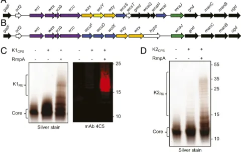
Glycoengineered Strains ofE. coli Require RmpA for Heterologous K1 and K2 CPS Expression.Prokaryotic glycoengineering exploits conserved polysaccharide synthesis and export pathways for the reprogrammable assembly and transfer of designer glycans in E. coli (SI Appendix, Fig. S1). The first step in glycoengineering bioconjugate vaccines against hvKp required building strains of E. coli for the heterologous expression of the K1 and K2 K. pneumoniae CPSs. As such, we cloned the K1 and K2 CPS loci from K. pneumoniae NTUH K-2044 (30) and K. pneumoniae 52.145 (31), respectively (Fig. 1 A and B). The cloned K1 CPS locus contained the genes from wzx to ugd. The cloned K2 CPS locus contained the genes from wcuF to ugd. The CPS regulatory genes wza, wzb, and wzc and export gene wzi are not required for heterologous CPS expression in E. coli (32) and were not in-cluded in the constructs. The galF and orf2 genes were also ex-cluded because E. coli carries its own copy of galF and the role of orf2 in K. pneumoniae CPS production is unclear. The plasmids containing the biosynthesis machinery for the K1 or K2 CPS were then introduced into E. coli CLM37, a reporter strain for heterologous polysaccharide expression and assembly (33). CLM37 cannot produce its natural O16 antigen due to a mu-tation in the WecA initiating glycosyltransferase. However, CLM37 expresses the WaaL O antigen ligase that transfers lipid-linked polysaccharides, like the K1 and K2 polysaccha-rides, to the outer core saccharide of LPS (34). After IPTG induction and overnight growth, LPS was extracted, separated by SDS/PAGE, and silver-stained from CLM37 cells expressing either the K1 or K2 CPS plasmids. As seen in Fig. 1 C and D, no appreciable O antigen polysaccharide was observed. We de-tected only the core saccharide, indicating that the K1 and K2 glycans were either not expressed or not transferred by the
galF orf2 wzi wza wzb wzc wzx wciY wzy wcsSwcsTgmd wcaG wcaHwcaI wcaJ gnd manC manB ugd
wcaJ gnd manC manB ugd
hypo wzx wzy
galF orf2 wzi wza wzb wzc wcuF wcuD wclX
K1CPS - + + RmpA - - + - + + - - + K2CPS - + + RmpA - - + - 25 - 15 - 10 - 35 - 55 Core K2RU Silver stain Core - 25 - 15 - 10 mAb 4C5 Silver stain K1RU
A
B
C
D
Fig. 1. RmpA enhances expression of the K1 and K2 glycans in E. coli CLM37. (A) The K. pneumoniae K1 CPS gene map and (B) the K. pneumoniae K2 CPS gene map. Black arrows indicate UDP-sugar biosynthesis genes, purple arrows indicate CPS regulatory/surface export genes, orange arrows indicate transport/ polymerase genes, blue arrows indicate glycosyltransferase genes, and green arrows indicate initiating glycosyltransferase genes. (C) Silver staining and Western blot analysis of LPS extracted from CLM37, CLM37 expressing the K1 locus, or CLM37 coexpressing the K1 locus and RmpA. (D) Silver staining of LPS extracted from CLM37, CLM37 expressing the K2 locus, or CLM37 coexpressing the K locus and RmpA.
WaaL ligase. Given that the WaaL has highly relaxed substrate specificity, we hypothesized that the absence of K1 and K2 polysaccharides was due to their poor expression. Pre-viously, Arakawa et al. demonstrated that the K2 CPS could be detected on the surface of E. coli JM109 when the complete K2 locus and the transcriptional activator RmpA were coex-pressed (35). Therefore, we cloned rmpA from K. pneumoniae NTUH K-2044 (NCBI accession no. BAH65944) into pACT3, a low-copy IPTG-inducible vector, and introduced this plasmid into CLM37 strains containing the K1 or K2 CPS-expressing
plasmids. When RmpA was coexpressed with either the K1 or K2 CPS expressing plasmids in CLM37, purified LPS contained observable O antigen polysaccharides (Fig. 1 C and D). Fur-ther, Western blot analysis using the monoclonal antibody 4C5, specific to the K1 CPS of K. pneumoniae (36), reacted with LPS purified from CLM37 coexpressing the K1 CPS locus and RmpA, indicating that the polysaccharide produced by this glycoengineered E. coli strain has a K1 structure. There are no commercially or publicly available antibodies to the K2 polysaccharide.
Fig. 2. Two-dimensional NMR analysis of K1- and K2-containing LPS extracted from CLM37. (A) The core structure of the K1 repeat unit. (B) The1H-13C HSQC spectrum of K. pneumoniae K1 polysaccharide produced in E. coli. Signals of the 2 variable O-acetylated fucoses are not shown because of low signal intensity. (C) NMR data for the K. pneumoniae K1 polysaccharide and the E. coli core (D2O, 25 °C, 600 MHz). Data for the main structure with O-acetylation (OAc) at Fucose A O-3. OAc 2.08/21.6 ppm. (D) The core structure of the K2 repeat unit. (E) The1H-13C HSQC spectrum of K. pneumoniae K2 polysaccharide produced in E. coli (D2O, 35 °C, 600 MHz). (F) NMR data for the K. pneumoniae K2 polysaccharide and the E. coli core (D2O, 35 °C, 600 MHz).
MIC
NMR Analysis of Glycoengineered K1 and K2 LPS and theK. pneumoniae NTUH K-2044 CPS.Next, we characterized the K1- and K2-containing LPSs purified from the glycoengineered strains of CLM37. The published structure of the K. pneumoniae NTUH K-2044 K1 CPS repeat unit has been reported as (→3)-β-D
-Glc-(1,4)-[2,3-(S)-pyruvate]-β-D-GlcA-(1,4)-α-L-Fuc-(1→) with varying degrees of
acetylation at the C2-OH and C3-OH of the fucose residue (37, 38). The NMR spectra and chemical shifts for the K1-containing LPS (Fig. 2 A–C) contained many signals from the core saccharide due to the short K1 polysaccharide chains, which can also be observed as low molecular weight polysaccharides on the silver-stained K1-containing LPS (Fig. 1C). Analysis of the 2D spectra (gCOSY, TOCSY, NOESY,1H-13C HSQC,1H-13C HMBC) allowed for the identification of the K1 repeating units containing the structure -3-β-Glc-4-β-GlcA-4-α-L-Fuc2/3OAc-, where fucose was present in
3 variants: nonacetylated or acetylated at positions 2 or 3. The published structure of the K1 CPS includes the same repeating unit as well as pyruvylation at the O-2, O-3 positions of the glucuronic acid (GlcA) residue. Although the spectra contained small signals attributable to pyruvate, there was no indication that it was linked to the GlcA residue or any other mono-saccharide of the repeating unit. Given the varying degrees of acetylation and pyruvylation between the previously published K1 structures (37, 38), we purified the capsular polysaccharide from K. pneumoniae NTUH K-2044 and reexamined it by NMR analysis. The NMR analysis (SI Appendix, Fig. S2) showed an identical structure to the K1-containing LPS without pyruvylation. Acetylation of the fucose was evident with O-acetylation at the O-2 (∼20%, A″) and the O-3 (∼40%, A′) of fucose, while the rest of fucose (A) was not acetylated. Small signals of terminal fucose A* were also visible. Last, signals of B4 and B5 were shifted between the native and deacylated K1 polysaccharide, likely due to different pH or salt formations by glucuronic acid. The data obtained here indicate that the polysaccharide structures produced by the K1 glycoengineered strain of E. coli and that of the K. pneumoniae K1 are nearly identical.
The K2-containing LPS was also extracted from glycoengineered E. coli CLM37 cells and analyzed by NMR to determine structure and linkage. As shown in Fig. 2 D–F, the K2 polysaccharide matched perfectly with the published structure of the native K2 polysaccharide (39).
Glycoengineering a K1 and K2 Bioconjugate Vaccine inE. coli. Pre-viously, we established a bioconjugation system for transferring poly-saccharides containing glucose at the reducing end to carrier proteins (23). The system employs the use of the oligosaccharyltransferase
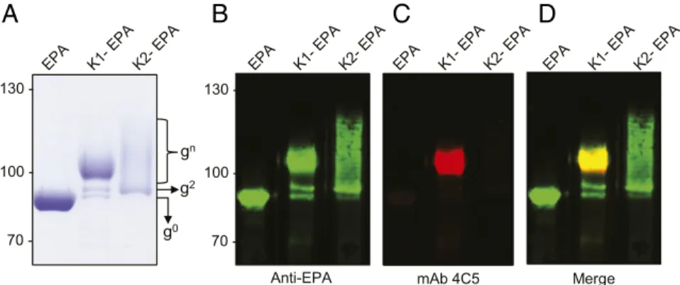
PglS from Acinetobacter baylyi ADP1 to transfer polysaccharides from a lipid-liked precursor to a genetically deactivated variant of exotoxin A protein from Pseudomonas aeruginosa (EPA) fused to a fragment of ComP, the natural acceptor protein of PglS. As such, we introduced either the K1 or the K2 CPS-expressing plasmid along with the transcriptional activator RmpA into a glycocompetent strain of E. coli, strain SDB1, expressing the PglS bioconjugation platform. SDB1 isΔwaaL derivative of CLM37, which prevents the use of the capsule for LPS synthesis. After induction and overnight growth, K1-EPA and K2-EPA glycopro-teins were purified using affinity, anionic exchange, and size-exclusion chromatography and examined by Coomassie staining and Western blotting. As seen in Fig. 3A, the unglycosylated EPA fusion protein migrates between the 75-kDa and 95-kDa markers (the theoretical molecular weight is 75,526.15 Da). The K1-EPA glycoprotein existed almost entirely as a semihomogenous glyco-form with a smeared electrophoretic mobility slightly larger than the 95-kDa marker. A similar electrophoretic mobility was also observed for the K1-containing LPS; however, more resolution can be seen due to the fact that the K1-containing LPS was sep-arated by using a 15% polyacrylamide gel whereas the K1-EPA glycoprotein was resolved on an 8% polyacrylamide gel. While this presentation for a bioconjugate vaccine is unusual, the smear-like banding is commonly observed for glycoconjugate vaccines pre-pared with chemical methodologies and is likely a product of nonstoichiometric levels of acetylation and pyruvylation of the K1 repeat unit. Western blotting using the 4C5 monoclonal anti-body specific to the K1 capsule confirmed that the K1-EPA bio-conjugate was indeed carrying K1 glycans (Fig. 3 B–D). The K2-EPA glycoprotein ran more similarly to conventional bioconjugate vaccines, exhibiting a modal, ladder distribution with each band corresponding to a glycoform with an additional K2 subunit.
Intact Mass Spectrometry of Bioconjugate Vaccines.Due to the lack of polyclonal or monoclonal antibodies specific to the K2 glycan, we next performed intact mass spectrometry analysis on the K2-EPA bioconjugate. This technique provides analytical data on the relative glycosylation ratios for bioconjugates existing in multiple different glycoforms as well as relative masses associated with each glycoform. As seen in Fig. 4A, the intact mass spectrum of the EPA bioconjugate showed a modal, ladder distribution of K2-EPA bioconjugates, each separated by∼662 Da, the mass of the K2 repeat unit (39). As many as 12 K2 repeat units were observed to be covalently attached to the EPA carrier protein by mass spectrometry; however, analysis of the Coomassie-stained purified glycoprotein preparation indicates that as many as 16 repeat units were transferred (Fig. 4A). Relative quantification of the different
g0 g2 gn 70 100 130 70 100 130
-Anti-EPA mAb 4C5 Merge
A
B
C
D
Fig. 3. K1-EPA and K2-EPA bioconjugate vaccines. (A) Coomassie-stained image of purified EPA, K1-EPA, and K2-EPA. Each lane was loaded with∼5 μg of glycoconjugate based on total protein. The unglycosylated EPA exists a single band. The K1-EPA exists as multiple glycoforms migrating in smear-like pattern. The K2-EPA bioconjugate migrates with a modal, ladder distribution. Each lane was loaded with∼0.5 μg of glycoconjugate based on total protein. (B–D) Western blot analysis probing for EPA and the K1 glycan. (B) Anti-EPA Western blot, (C) anti-K1 Western blot, and (D) merge image.
glycoforms showed that the most abundant glycoform contained 2 repeat units, with the next most abundant glycoform containing 7 K2 repeat units (Fig. 4B). Interestingly, when examined collec-tively, the K2-EPA glycoprotein seemed to be glycosylated with the K2 glycan in a semi–bell-curve distribution, indicating that PglS may prefer certain-sized lipid-linked polysaccharides as substrates. We also performed intact mass spectrometry on the K1-EPA bioconjugate. The EPA fusion protein was observed as a series of peaks compatible with different glycoforms containing the in-herently heterogenous K1 repeat units, which is nonstoichio-metrically modified with acetylation at the fucose residue and/or pyruvylated at the glucuronic acid residue (SI Appendix, Fig. S3).
K1 and K2 Bioconjugates Elicit Serotype-Specific IgG Responses.The K1-EPA and K2-EPA bioconjugates were then tested for their abilities to induce serotype-specific IgG responses. Four immu-nization groups, each containing 5 mice, were vaccinated with either a placebo (the unglycosylated EPA fusion protein), the K1-EPA bioconjugate, the K2-EPA bioconjugate, or a bivalent mixture of the K1- and K2-EPA bioconjugates. All vaccines were coformulated with an equal mixture of Imject Alum as an adjuvant
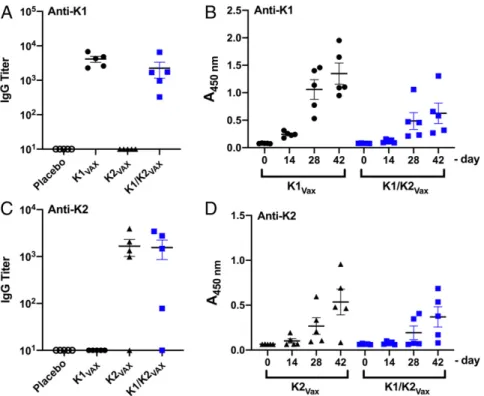
(50μL vaccine to 50 μL alum). The EPA-alone group, K1-EPA group (222 ng of K1 glycan), and K2-EPA group (195 ng of K2 glycan) all received 5μg of vaccine based on total protein quantity. The total polysaccharide content was measured using a modified anthrone-sulfuric assay (40). The bivalent vaccine was formulated by combining the K1-EPA and K2-EPA vaccine doses; thus, bivalent groups received a total of 10 μg of EPA, 222 ng of K1 glycan, and 195 ng of K2 glycan. Mice were vac-cinated 3 times s.c. on days 0, 14, and 28 and killed on day 42. Serum was collected throughout the course of the trial and used to characterize the IgG responses via ELISA with plates coated with either K. pneumoniae NTUH K-2044 (K1) or K. pneumoniae ATCC 43816 (K2) whole cells.
As seen in Fig. 5, mice vaccinated with K1-EPA bioconjugates had increased K1-specific IgG titers compared with mock-vaccinated mice. Mice receiving the bivalent K1-/K2-EPA vac-cine also had similar increases in K1-specific IgG titers compared with mock-vaccinated mice. The response was slightly lower than that of the single K1-EPA vaccinated group, but no statistically significant difference was observed. No K2-specific cross-reactivity was observed for K1-EPA vaccinated mice. In addition, mice
g
0g
2g
3g
4g
5g
6g
7g
8g
9g
10g
11g
12A
B
Fig. 4. Mass spectrometry analysis of K2-EPA. (A) Intact protein mass spectrometry analysis showing the MS1 mass spectra for K2-EPA. The EPA fusion protein has a theoretical mass of 79,526.15 Da and can be observed as the peak at 79,518.73 Da. The EPA fusion protein was also observed in multiple states of increasing mass corresponding to the K2 repeat unit, which has a mass of 662 Da. Varying glycoforms of K2-EPA were observed and are denoted by“gnumeric”, where“g” stands for glycoform and the “numeric” corresponds to the number of repeating CPS8 subunits. (B) Quantification of the relative abundance of each K2 glycoform.
MIC
vaccinated with the EPA biconjugate had increased K2-specific IgG titers compared with mock-vaccinated mice; how-ever, 1 mouse receiving the K2-EPA vaccine did not show in-creases in K2-specific IgG titers. Mice receiving the bivalent K1-/ K2-EPA vaccine also had similar increases in K2-specific IgG titers compared with mock-vaccinated mice. As was the case for the EPA group, 1 mouse did not show an increase in K2-specific IgG titers. No K1-K2-specific cross-reactivity was observed for mice vaccinated with the K2-EPA bioconjugate.
Mice sera were further examined for serotype-specific IgG kinetics over the course of the vaccination. As seen in Fig. 5, the kinetics of K1-specific total IgG responses increased over time, with K1-EPA vaccinated mice showing a more robust response compared with bivalent K1-EPA/K2-EPA vaccinated mice. The kinetics of total IgG responses specific to K2 were also mea-sured, and a similar trend was observed, with mice vaccinated with the K2-EPA bioconjugate showing the most robust response compared with bivalent vaccinated mice.
Next, mouse sera were pooled and examined by whole-cell ELISA for IgG subclass distributions specific to the K1 or K2 antigen. As seen in SI Appendix, Fig. S4, pooled K1-EPA vaccinated sera and pooled K1-/K2-EPA vaccinated sera dis-played an exclusively IgG1-specific response to the K1 antigen. The same trend was also observed for the pooled K2-EPA and pooled K1-/K2-EPA vaccinated sera when probing for specificity to the K2 antigen.
K1/K2 Bioconjugate Vaccines Protect Mice from hvKp Infection.The correlates of immunity that confer protection from classical or hypervirulent K. pneumoniae infection are not known. Therefore, we evaluated the protectiveness of the bivalent K1-/K2-EPA vaccine using a murine acute pulmonary infection model with 2 separate hypervirulent K. pneumoniae strains: NTUH K-2044 (K1) and ATCC 43816 (K2). Both strains are hyper-capsule producers (hypermucoviscous) and extremely virulent in mice.
In fact, the LD50for both strains has been shown to be less than 100 colony forming units (CFUs) in murine respiratory infection models (41). Two groups of mice (20 female BALB/c mice per group) were vaccinated with the carrier protein alone or with the bivalent K1-/K2-EPA bioconjugate vaccine. Briefly, mice were vaccinated on days 0, 14, and 28 using the same dosing and ad-juvant formulation as described above. After the vaccination regimen, the 2 groups (EPA placebo or bivalent bioconjugate vaccinated) were then challenged with either the K1 or K2 strain at doses close to the published LD50values. Specifically, 10 mice from each vaccination group were inoculated by aspiration with the hypervirulent K1 strain (NTUH K-2044) and 10 were in-oculated with the hypervirulent K2 strain (ATCC 43816) and subsequently monitored for survival and changes in weight for a 2-wk period (Fig. 6 A and B). As seen in Fig. 6A, 80% of placebo-vaccinated mice challenged with only 50 CFUs of the hypervirulent K1 strain (K. pneumoniae NTUH K-2044) succumbed to infec-tion, whereas only 20% of bioconjugate-vaccinated mice died (P= 0.0057 by log-rank test). While K. pneumoniae ATCC 43816 was not as virulent in this mouse model as K. pneumoniae NTUH K-2044, a dose of only 250 CFUs was able to kill 30% of the placebo-vaccinated mice (Fig. 6B), whereas no mice from the bioconjugate-vaccinated group challenged with ATCC 43816 died (P = 0.0669 by log-rank test). Additionally, placebo-vaccinated mice that did survive low-dose challenge had lower body weights than bioconjugate-vaccinated mice (SI Appendix, Fig. S5).
Given the success of the bioconjugate vaccine in preventing death from infection of hvKp at doses close to the LD50 values, we further challenged a separate group of placebo- and bioconjugate-vaccinated mice with hvKp strains at 100× the published LD50titers. For K. pneumoniae NTUH K-2044 (K1), all of the placebo-vaccinated mice rapidly died (Fig. 6C). The majority of bioconjugate-vaccinated mice also succumbed to this high-dose infection; however, the bioconjugate-vaccinated mouse Fig. 5. IgG responses to K1 and K2 bioconjugate vaccines. (A) Titers of K1-specific IgG antibodies in mice immunized with EPA, K1-EPA, K2-EPA, or a bivalent K-/EPA. (B) K1-specific IgG kinetics over the course of the immunization as measured by ELISA and quantified by absorbance at 450 nm. (C) Titers of K2-specific IgG antibodies in mice immunized with EPA, K1-EPA, K2-EPA, or a bivalent K-/K2-EPA. (D) K2-K2-specific IgG kinetics over the course of the immunization as measured by ELISA and quantified by absorbance at 450 nm.
group had a statistically significant increase in survival compared with the placebo-vaccinated group (P= 0.0250 by log-rank test). When mice were challenged with close to 100× the published LD50value of ATCC 43816 (K2), all placebo-vaccinated mice died by day 4, whereas 5 of the 10 mice survived the infection (P= 0.0038 by log-rank test; Fig. 6D). These data suggest that the biconjugate vaccine is efficacious in protecting some mice from K1 or K2 hvKp challenge, even at very high inocula.
Discussion
cKp infections are traditionally associated with nosocomial in-fections among hospitalized or immunocompromised patients, while hvKp can target healthy, immunocompetent hosts. Until recently, hvKp strains have been susceptible to common antibi-otic agents; however, many cases of carbapenem-resistance and even colistin-resistance phenotypes in hvKp isolates have been recently reported (42–45). Furthermore, hvKp strains generated via the acquisition of hypervirulence plasmids by MDR cKp are also emerging (16, 17). The extreme virulence of these strains coupled with a lack of antimicrobial treatment options is worri-some. As such, vaccines to prevent possible outbreaks of these hypervirulent, antimicrobial-resistant infections are urgently needed (46), and possibly prevent the spread of the hypervirulent plasmid. Here we report on the recombinant production and an-alytical characterization of rapidly produced bioconjugate vaccines against the 2 most common hypervirulent serotypes of K. pneu-moniae (K1 and K2), which account for more than 70% of the hvKp cases (15). Using a bioconjugation approach in glyco-engineered E. coli, we show that K1 and K2 bioconjugates are immunogenic and efficacious, protecting mice from lethal in-fection from 2 different hypervirulent strains of K. pneumonia.
For efficient bioconjugation of polysaccharides to proteins, usually only 3 components are needed: an oligosaccharyltransferase (also known as a conjugating enzyme), a target protein to be glycosylated, and a polysaccharide to be transferred. However, we found that a fourth factor, the RmpA transcriptional activator, was required to efficiently express K1 and K2 capsules in E. coli.
Multiple alleles of rmpA are associated with hypermucoviscous phenotypes (10, 47). K. pneumoniae isolates can carry as many as 3 different rmpA alleles. One is commonly found on the chromo-some (rmpAc), whereas 2 plasmid-encoded rmpA alleles (prmpA or prmpA2) are located on the virulence plasmid found in hypervir-ulent isolates of K. pneumoniae. In our study,prmpA functioned as transcriptional activator in glycoengineered E. coli cells; however, whether rmpAc or prmpA2 would have the same or possibly an additive effect on K1 and K2 CPS polysaccharide expression has not been determined.
A few vaccine strategies have been or are in development for K. pneumoniae. In fact, a 24-valent, purely capsular polysaccharide vaccine (Klebgen Bema) was developed in the 1980s by the Swiss Serum and Vaccine Institute and tested in human clinical trials (48). While the vaccine was shown to elicit increases in serotype-specific IgG responses in adult cohorts, as frequently happens with polysaccharide-only vaccines, total serotype-specific IgG levels dropped to near baseline levels 18 mo after vaccination for many capsular antigens (48). More recently, a glycoconjugate vaccine composed of 4 K. pneumoniae OPS serotypes conjugated to fla-gellin proteins from Pseudomonas aeruginosa was developed (49). The OPS vaccine was immunogenic, and mice passively trans-ferred with the OPS vaccine-induced antibodies were protected against systemic cKp infection. While these preclinical results are promising, OPS-based vaccines more appropriately target cKp strains, as molecular studies have shown that the capsular poly-saccharide of hypervirulent isolates can mask the OPS antigens (50). Also, it has not been shown that an OPS-based vaccine would be protective protect against hvKp isolates overproducing capsular polysaccharide. To the best of our knowledge, the K1-/K2-EPA bioconjugate presented here is the first case of a vaccine providing protection from extremely lethal hypervirulent isolates. While our in vivo work suggests efficacy against hvKp in the lung, additional studies need to assess efficacy against hvKp in other niches in-cluding the liver, bloodstream, and meninges.
Glycoconjugate vaccines elicit IgM-to-IgG class switching and im-munological memory (18). While this is common to all glycoconjugate Fig. 6. Survival of placebo- and bivalent bioconjugate-vaccinated mice after lethal challenge with hvKp. Groups of mice were vaccinated with either the placebo or the bivalent K1-/K2-EPA bioconjugate on days 0, 14, and 28. Anesthetized mice were aspirated with either the K. pneumoniae NTUH K-2044 or ATCC 43816 and monitored for survival for 14 d. For low-dose challenge studies, mice were infected with (A) 50 CFU of NTUH K-2044 or (B) 250 CFU of ATCC 43816. For high-dose challenge studies, mice were infected with (C) 4,700 CFU of NTUH K-2044 or (D) 4,300 CFU of ATCC 43816. Each graph represents data from a single experiment with n= 10 mice per group. Statistical analysis was performed via log-rank (Mantel–Cox) tests. MIC
vaccines, the distribution of IgG isotypes can be different for each antigenic stimulus, as well as differ based on the age of the cinated population. For instance, pneumococcal conjugate vac-cines elicit strong IgG1 responses in infants and young children (2–5 y), whereas the same vaccine elicits a predominantly IgG2 response in healthy adults (18–39 y) and geriatric patients (>50 y) (51). Using 6-wk-old BALB/c mice for our immunization model, we observed an exclusively IgG1 response for the both the K1 and K2 antigens. In fact, we were not able to observe any signals for IgG2a, IgG2b, or IgG3 subclasses by ELISA. IgG1 antibodies are known to efficiently activate the classical route of complement (52). Therefore, our data indicate that K1- and K2-specific IgG1 antibodies may be sufficient to provide protection to vacci-nated mice challenged with lethal doses of the hvKp isolates. Future experiments will be needed to establish if high levels IgG1 can be employed as an appropriate correlate of immunity and predict protection against hvKp.
While it is currently difficult to define which serotypes should be included in a capsular polysaccharide glycoconjugate vaccine targeting cKp infection or determine which populations are most at risk, the seroepidemiology of hvKp is much clearer. Specifi-cally, hvKp is endemic to certain parts of Asia, and 2 serotypes, K1 and K2, have emerged as the highly predominant disease-causing serotypes (53–55). Moreover, fewer than 10 serotypes have been reported to be associated with hypervirulent infections (K1, K2, K5, K16, K20, K54, K57, and KN1) (3). In addition to the K1 and K2 serotypes, the K5, K16, and K54 serotypes also contain glucose as the reducing end sugar (12), suggesting that they may also be appropriate polysaccharide substrates for the PglS bioconjugation platform. The remaining serotypes (K20, K57, and KN1) contain galactose at their reducing ends, which are
also compatible with the PglS bioconjugation platform. Therefore, PglS could be employed to develop a pan-hypervirulent bioconjugate vaccine against K. pneumoniae. Importantly, the use of the K1-/K2-EPA bioconjugate would also target K1 and K2 strains of K. pneu-moniae associated with classical, nosocomial infection. Thus, by expanding the serotype coverage, a broadly protecting glycoconjugate vaccine targeting both classical and hypervirulent pathotypes of K. pneumoniae could be developed rapidly by using our bioconjugation platform and employed to significantly reduce the burden of K. pneumoniae disease and possibly slow the rates of drug resistance and transmission of the hypervirulence plasmid.
The increasing incidence of community-acquired hvKp MDR strains poses a serious threat to global health. It is imperative that vaccine strategies to combat hvKp begin immediately, as the dissemination of the virulence plasmid into the cKp population could have devastating consequences. Our work demonstrates that bioconjugation is a promising approach to rapidly developing ef-ficacious antibacterial vaccines.
Materials and Methods
The bacterial strains, plasmids, and primers used in this study are listed inSI Appendix, SI Materials and Methods. A full description of all methods employed for this study is provided inSI Appendix, SI Materials and Meth-ods. All data are available in the main text or theSI Appendix.
ACKNOWLEDGMENTS. This work was supported by the National Institute of Allergy and Infectious Diseases Phase I Small Business Technology Transfer Grant R41AI136333-01 (to M.F.F.). In addition, this work was partially supported by the NIH Grant K08-AI127714 and the Children’s Discovery In-stitute of Washington University and St. Louis Children's Hospital funding (to D.A.R.).
1. S. S. Magill et al.; Emerging Infections Program Healthcare-Associated Infections and Antimicrobial Use Prevalence Survey Team, Multistate point-prevalence survey of health care-associated infections. N. Engl. J. Med. 370, 1198–1208 (2014). 2. Division of Healthcare Quality Promotion, National Center for Emerging and Zoonotic
Infectious Diseases, Centers for Disease Control and Prevention, Antibiotic Resistance Threats in the United States (Centers for Disease Control and Prevention, Atlanta, GA, 2013).
3. A. S. Shon, R. P. Bajwa, T. A. Russo, Hypervirulent (hypermucoviscous) Klebsiella pneumoniae: A new and dangerous breed. Virulence 4, 107–118 (2013).
4. D. L. Cheng, Y. C. Liu, M. Y. Yen, C. Y. Liu, R. S. Wang, Septic metastatic lesions of pyogenic liver abscess. Their association with Klebsiella pneumoniae bacteremia in diabetic patients. Arch. Intern. Med. 151, 1557–1559 (1991).
5. Y. C. Liu, D. L. Cheng, C. L. Lin, Klebsiella pneumoniae liver abscess associated with septic endophthalmitis. Arch. Intern. Med. 146, 1913–1916 (1986).
6. B. C. Prokesch et al., Primary osteomyelitis caused by hypervirulent Klebsiella pneumoniae. Lancet Infect. Dis. 16, e190–e195 (2016).
7. N. C. Cheng et al., Recent trend of necrotizing fasciitis in Taiwan: Focus on mono-microbial Klebsiella pneumoniae necrotizing fasciitis. Clin. Infect. Dis. 55, 930–939 (2012).
8. W. L. Yu et al., Comparison of prevalence of virulence factors for Klebsiella pneu-moniae liver abscesses between isolates with capsular K1/K2 and non-K1/ K2 serotypes. Diagn. Microbiol. Infect. Dis. 62, 1–6 (2008).
9. C. T. Fang, Y. P. Chuang, C. T. Shun, S. C. Chang, J. T. Wang, A novel virulence gene in Klebsiella pneumoniae strains causing primary liver abscess and septic metastatic complications. J. Exp. Med. 199, 697–705 (2004).
10. H. Y. Cheng et al., RmpA regulation of capsular polysaccharide biosynthesis in Klebsiella pneumoniae CG43. J. Bacteriol. 192, 3144–3158 (2010).
11. C. March et al., Role of bacterial surface structures on the interaction of Klebsiella pneumoniae with phagocytes. PLoS One 8, e56847 (2013).
12. Y. J. Pan et al., Genetic analysis of capsular polysaccharide synthesis gene clusters in 79 capsular types of Klebsiella spp. Sci. Rep. 5, 15573 (2015).
13. D. R. Chung et al.; Korean Study Group for Liver Abscess, Emerging invasive liver abscess caused by K1 serotype Klebsiella pneumoniae in Korea. J. Infect. 54, 578–583 (2007).
14. V. L. Yu et al.; International Klebseilla Study Group, Virulence characteristics of Klebsiella and clinical manifestations of K. pneumoniae bloodstream infections. Emerg. Infect. Dis. 13, 986–993 (2007).
15. C. M. Marr, T. A. Russo, Hypervirulent Klebsiella pneumoniae: A new public health threat. Expert Rev. Anti Infect. Ther. 17, 71–73 (2019).
16. J. F. Turton et al., Virulence genes in isolates of Klebsiella pneumoniae from the UK during 2016, including among carbapenemase gene-positive hypervirulent K1-ST23 and‘non-hypervirulent’ types ST147, ST15 and ST383. J. Med. Microbiol. 67, 118– 128 (2018).
17. D. Gu et al., A fatal outbreak of ST11 carbapenem-resistant hypervirulent Klebsiella pneumoniae in a Chinese hospital: A molecular epidemiological study. Lancet Infect. Dis. 18, 37–46 (2018).
18. E. De Gregorio, R. Rappuoli, From empiricism to rational design: A personal per-spective of the evolution of vaccine development. Nat. Rev. Immunol. 14, 505–514 (2014).
19. F. Berti, R. Adamo, Antimicrobial glycoconjugate vaccines: An overview of classic and modern approaches for protein modification. Chem. Soc. Rev. 47, 9015–9025 (2018). 20. C. E. Frasch, Preparation of bacterial polysaccharide-protein conjugates: Analytical
and manufacturing challenges. Vaccine 27, 6468–6470 (2009).
21. M. F. Feldman et al., Engineering N-linked protein glycosylation with diverse O an-tigen lipopolysaccharide structures in Escherichia coli. Proc. Natl. Acad. Sci. U.S.A. 102, 3016–3021 (2005).
22. A. Faridmoayer, M. A. Fentabil, D. C. Mills, J. S. Klassen, M. F. Feldman, Functional characterization of bacterial oligosaccharyltransferases involved in O-linked protein glycosylation. J. Bacteriol. 189, 8088–8098 (2007).
23. C. M. Harding et al., A platform for glycoengineering a polyvalent pneumococcal bioconjugate vaccine using E. coli as a host. Nat. Commun. 10, 891 (2019). 24. M. S. Riddle et al., Safety and immunogenicity of a candidate bioconjugate vaccine
against Shigella flexneri 2a administered to healthy adults: A single-blind, random-ized phase I study. Clin. Vaccine Immunol. 23, 908–917 (2016).
25. A. Huttner et al., Safety, immunogenicity, and preliminary clinical efficacy of a vaccine against extraintestinal pathogenic Escherichia coli in women with a history of re-current urinary tract infection: A randomised, single-blind, placebo-controlled phase 1b trial. Lancet Infect. Dis. 17, 528–537 (2017).
26. C. M. Harding, M. F. Feldman, Glycoengineering bioconjugate vaccines, therapeutics, and diagnostics in E. coli. Glycobiology 29, 519–529 (2019).
27. E. Kay, J. Cuccui, B. W. Wren, Recent advances in the production of recombinant glycoconjugate vaccines. NPJ Vaccines 4, 16 (2019).
28. K. A. Geno et al., Pneumococcal capsules and their types: Past, present, and future. Clin. Microbiol. Rev. 28, 871–899 (2015).
29. C. M. Harding et al., Acinetobacter strains carry two functional oligosaccharyl-transferases, one devoted exclusively to type IV pilin, and the other one dedicated to O-glycosylation of multiple proteins. Mol. Microbiol. 96, 1023–1041 (2015). 30. K. M. Wu et al., Genome sequencing and comparative analysis of Klebsiella pneumoniae
NTUH-K2044, a strain causing liver abscess and meningitis. J. Bacteriol. 191, 4492–4501 (2009).
31. L. M. Lery et al., Comparative analysis of Klebsiella pneumoniae genomes identifies a phospholipase D family protein as a novel virulence factor. BMC Biol. 12, 41 (2014). 32. N. L. Price et al., Glycoengineered outer membrane vesicles: A novel platform for
bacterial vaccines. Sci. Rep. 6, 24931 (2016).
33. D. Linton et al., Functional analysis of the Campylobacter jejuni N-linked protein glycosylation pathway. Mol. Microbiol. 55, 1695–1703 (2005).
34. M. F. Feldman et al., The activity of a putative polyisoprenol-linked sugar translocase (Wzx) involved in Escherichia coli O antigen assembly is independent of the chemical structure of the O repeat. J. Biol. Chem. 274, 35129–35138 (1999).
35. Y. Arakawa et al., Biosynthesis of Klebsiella K2 capsular polysaccharide in Escherichia coli HB101 requires the functions of rmpA and the chromosomal cps gene cluster of the virulent strain Klebsiella pneumoniae Chedid (O1:K2). Infect. Immun. 59, 2043– 2050 (1991).
36. E. Diago-Navarro et al., Antibody-based immunotherapy to Treat and prevent in-fection with hypervirulent Klebsiella pneumoniae. Clin. Vaccine Immunol. 24, e00456-16 (2017).
37. F. L. Yang et al., Structure and immunological characterization of the capsular polysaccharide of a pyrogenic liver abscess caused by Klebsiella pneumoniae: Acti-vation of macrophages through Toll-like receptor 4. J. Biol. Chem. 286, 21041–21051 (2011).
38. C. Erbing, L. Kenne, B. Lindberg, J. Lönngren, Structural studies of the capsular polysaccharide from Klebsiella Type 1. Carbohydr. Res. 50, 115–120 (1976). 39. M. M. Corsaro et al., 1H and 13C NMR characterization and secondary structure of the
K2 polysaccharide of Klebsiella pneumoniae strain 52145. Carbohydr. Res. 340, 2212– 2217 (2005).
40. C. Pan et al., Biosynthesis of conjugate vaccines using an O-linked glycosylation sys-tem. MBio 7, e00443-16 (2016).
41. R. A. Fodah et al., Correlation of Klebsiella pneumoniae comparative genetic analyses with virulence profiles in a murine respiratory disease model. PLoS One 9, e107394 (2014).
42. D. Shen et al., Emergence of a multidrug-resistant hypervirulent Klebsiella pneumo-niae sequence type 23 strain with a rare blaCTX-M-24-harboring virulence plasmid. Antimicrob. Agents Chemother. 63, e02273-18 (2019).
43. R. Zhang et al., Emergence of carbapenem-resistant serotype K1 hypervirulent Klebsiella pneumoniae strains in China. Antimicrob. Agents Chemother. 60, 709–711 (2015).
44. N. Dong, D. Lin, R. Zhang, E. W. Chan, S. Chen, Carriage of blaKPC-2 by a virulence plasmid in hypervirulent Klebsiella pneumoniae. J. Antimicrob. Chemother. 73, 3317– 3321 (2018).
45. Y. Feng, Y. Lu, Z. Yao, Z. Zong, Carbapenem-resistant hypervirulent Klebsiella pneumoniae of sequence type 36. Antimicrob. Agents Chemother. 62, e02644-17 (2018).
46. Boston Consulting Group, Vaccines to tackle drug resistant infections: An evaluation of R&D opportunities (Boston Consulting Group, Boston, MA, 2018).
47. C. R. Hsu, T. L. Lin, Y. C. Chen, H. C. Chou, J. T. Wang, The role of Klebsiella pneumoniae rmpA in capsular polysaccharide synthesis and virulence revisited. Microbiology 157, 3446–3457 (2011).
48. R. Edelman et al., Phase 1 trial of a 24-valent Klebsiella capsular polysaccharide vac-cine and an eight-valent Pseudomonas O-polysaccharide conjugate vacvac-cine adminis-tered simultaneously. Vaccine 12, 1288–1294 (1994).
49. N. Hegerle et al., Development of a broad spectrum glycoconjugate vaccine to pre-vent wound and disseminated infections with Klebsiella pneumoniae and Pseudo-monas aeruginosa. PLoS One 13, e0203143 (2018).
50. P. Williams, P. A. Lambert, M. R. Brown, Penetration of immunoglobulins through the Klebsiella capsule and their effect on cell-surface hydrophobicity. J. Med. Microbiol. 26, 29–35 (1988).
51. K. R. Lottenbach et al., Age-associated differences in immunoglobulin G1 (IgG1) and IgG2 subclass antibodies to pneumococcal polysaccharides following vaccination. In-fect. Immun. 67, 4935–4938 (1999).
52. C. I. Bindon, G. Hale, M. Brüggemann, H. Waldmann, Human monoclonal IgG isotypes differ in complement activating function at the level of C4 as well as C1q. J. Exp. Med. 168, 127–142 (1988).
53. C. R. Lee et al., Antimicrobial resistance of hypervirulent Klebsiella pneumoniae: Epidemiology, hypervirulence-associated determinants, and resistance mechanisms. Front. Cell. Infect. Microbiol. 7, 483 (2017).
54. C. Liu, J. Guo, Hypervirulent Klebsiella pneumoniae (hypermucoviscous and aero-bactin positive) infection over 6 years in the elderly in China: Antimicrobial resistance patterns, molecular epidemiology and risk factor. Ann. Clin. Microbiol. Antimicrob. 18, 4 (2019).
55. Y. Zhang et al., High prevalence of hypervirulent Klebsiella pneumoniae infection in China: Geographic distribution, clinical characteristics, and antimicrobial resistance. Antimicrob. Agents Chemother. 60, 6115–6120 (2016).
MIC