HAL Id: hal-02627312
https://hal.inrae.fr/hal-02627312
Submitted on 26 May 2020
HAL is a multi-disciplinary open access archive for the deposit and dissemination of sci-entific research documents, whether they are pub-lished or not. The documents may come from teaching and research institutions in France or abroad, or from public or private research centers.
L’archive ouverte pluridisciplinaire HAL, est destinée au dépôt et à la diffusion de documents scientifiques de niveau recherche, publiés ou non, émanant des établissements d’enseignement et de recherche français ou étrangers, des laboratoires publics ou privés.
Valley fever
Catherine Cêtre-Sossah, Aurélie Pedarrieu, Mikael Juremalm, Petrus Jansen
van Vuren, Alejandro Brun, Ahmed Bezeid Ould El Mamy, Jean-Michel
Héraud, Claudia Filippone, Jean-Pierre Ravalohery, Hassan Chaabihi, et al.
To cite this version:
Catherine Cêtre-Sossah, Aurélie Pedarrieu, Mikael Juremalm, Petrus Jansen van Vuren, Alejandro Brun, et al.. Development and validation of a pen side test for Rift Valley fever. PLoS Neglected Tropical Diseases, Public Library of Science, 2019, 13 (9), 16 p. �10.1371/journal.pntd.0007700�. �hal-02627312�
Development and validation of a pen side test
for Rift Valley fever
Catherine Cêtre-SossahID1,2*, Aure´lie Pe´darrieu1, Mikael Juremalm3, Petrus Jansen Van
VurenID4, Alejandro Brun5, Ahmed Bezeid Ould EL Mamy6, Jean-Michel He´raud7, Claudia Filippone7, Jean-Pierre Ravalohery7, Hassan Chaabihi
ID8, Emmanuel Albina1,9, Laure Dommergues10, Janusz Paweska4, Eric Cardinale1,2
1 ASTRE, Univ Montpellier, CIRAD, INRA, Montpellier, France, 2 CIRAD, UMR ASTRE, Sainte-Clotilde, La Re´union, France, 3 Boehringer Ingelheim Svanova, Virdingsalle´ , Uppsala, Sweden, 4 Centre for Emerging Zoonotic and Parasitic Diseases, National Institute for Communicable Diseases,
Sandringham-Johannesburg, South Africa, 5 INIA-CISA, Valdeolmos, Madrid, Spain, 6 ONARDEL, service de pathologies infectieuses, Nouakchott, Mauritania, 7 Virology Unit, Institut Pasteur de Madagascar, Antananarivo, Madagascar, 8 Agate bioservices, Bagard, France, 9 CIRAD, UMR ASTRE, Petit Bourg, Guadeloupe, France, 10 CoopADEM - GDS Mayotte, Coconi, Mayotte, France
*catherine.cetre-sossah@cirad.fr
Abstract
Background
Rift Valley fever (RVF) is one of the main vector borne zoonotic diseases that affects a wide range of ruminants and human beings in Africa and the Arabian Peninsula. A rapid and spe-cific test for RVF diagnosis at the site of a suspected outbreak is crucial for the implementa-tion of control measures.
Methodology/Principal findings
A first-line lateral flow immunochromatographic strip test (LFT) was developed for the detec-tion of the nucleoprotein (N) of the RVF virus (RVFV). Its diagnostic performance character-istics were evaluated using reference stocks isolates recovered from different hosts and in geographic regions mimicking clinical specimens and from known RVF negative serum samples. A high level of diagnostic accuracy (DSe (35/35), DSp (167/169)) was observed, including the absence of cross-reactivity with viruses belonging to different genera.
Conclusion/Significance
The fact no specialized reagents and laboratory equipment are needed, make this assay a valuable, first-line diagnostic tool in resource-poor diagnostic territories for on-site RVFV detection, however the staff require training.
Author summary
Rift Valley fever (RVF) is a viral disease that affects a wide range of animals and human beings in Africa and the Arabian Peninsula involving low case fatality rates. A rapid and
a1111111111 a1111111111 a1111111111 a1111111111 a1111111111 OPEN ACCESS
Citation: Cêtre-Sossah C, Pe´darrieu A, Juremalm M, Jansen Van Vuren P, Brun A, Ould EL Mamy AB, et al. (2019) Development and validation of a pen side test for Rift Valley fever. PLoS Negl Trop Dis 13(9): e0007700.https://doi.org/10.1371/ journal.pntd.0007700
Editor: Waleed Saleh Al-Salem, Saudi Ministry of
Health, SAUDI ARABIA
Received: November 2, 2018 Accepted: July 23, 2019 Published: September 11, 2019
Copyright:© 2019 Cêtre-Sossah et al. This is an open access article distributed under the terms of theCreative Commons Attribution License, which permits unrestricted use, distribution, and reproduction in any medium, provided the original author and source are credited.
Data Availability Statement: All relevant data are
within the manuscript and its Supporting Information files.
Funding: This study was funded by FEDER
INTERREG V TROI 2015-2020 and the Region Re´union under the DP One health Indian Ocean (www.onehealth-oi.org). The funders had no role in study design, data collection and analysis, decision to publish, or preparation of the manuscript.
Competing interests: The authors have declared
specific test for RVF diagnosis at the site of a suspected outbreak is crucial for the imple-mentation of control measures. Here, we report the development and the evaluation of the diagnostic performance characteristics of a pen-side test found to be a highly accurate and valuable first-line diagnostic tool for on-site RVF detection.
Introduction
Rift Valley fever (RVF) is an emerging mosquito-borne disease that affects a wide range of ani-mals and human beings in Africa and the Arabian Peninsula. It is caused by the mosquito-borne Rift Valley fever phlebovirus (RVFV) that belongs to the genusPhlebovirus in the family Phenuiviridae of the order Bunyavirales [1]. First identified in the Great Rift Valley in Kenya in 1930 and initially confined to the African continent, it subsequently spreads to Madagascar, the archipelago of Comoros, and the Arabian Peninsula [2]. There is a growing concern that RVFV will extend its current range due to the wide variety of mosquito species able to transmit to several mammal hosts. This includes species distributed in countries outside Africa and Arabian Peninsula where RVFV is not yet known to circulate despite the environmental fac-tors driving and favoring its circulation [3–6].
Recent outbreaks of RVF in Mayotte, Niger, Uganda and Sudan involving human deaths and characterized by mass abortion and high mortality rates of neonates in the ruminant pop-ulation raised international interests in improving diagnostic and vaccine control strategies [7–10].
When investigating disease outbreaks in animals, the earlier the clinical signs of disease are recognized by the farmer and the earlier the clinical diagnosis is confirmed by laboratory tests, along with rapid reporting to the relevant veterinary authorities, the better the disease will be controlled. Success partly relies on sending samples to a reference laboratory to test for the presence of RVFV, with high levels of viral particles in the serum during the acute phase of the disease.
The availability of a ‘pen-side’ diagnostic test would have the advantage of providing addi-tional support for the medical clinical judgment in the first instance and could reduce the time needed to confirm the test results in secondary cases of the disease. The use of rapid diag-nostic tests that can be conducted in the field, at the site of the outbreaks, where the infected human and animal populations are, will therefore facilitate earlier and more effective disease control. Conventional techniques for the diagnosis of RVF include virus isolation, detection of specific IgM or IgG antibodies, and detection of RVFV specific nucleic acids. Enzyme-linked immunosorbent assays (ELISA), based on whole virus antigens or the recombinant nucleocapsid protein N have been extensively validated for the serodiagnosis of RVF [11–12]. Conventional and real-time reverse transcriptase polymerase chain reaction (RT-PCR) assays are currently the most rapid and sensitive tests for the detection and quantification of RVFV during outbreaks [13–17]. Methods based on next generation sequencing (NGS) approaches [18] or colorimetry [19], TaqMan array cards [20] have been recently developed but most of these techniques are expensive and require dedicated trained personnel and equipped biosafety level 3 laboratories that are often not available in the areas where the dis-ease occurs. There is thus a need for a simpler, inexpensive and reliable pen-side test to facili-tate prompt and accurate field diagnosis. Lateral flow tests (LFT) also known as immuno-chromatographic strips are rapid, single-use, one-step test devices able to detect at point of care the presence of an analyte in a liquid sample, flowing along a membrane strip encased in a protective plastic frame. The result can be easily seen with the naked-eye (test line and
control line). Good examples have already been published for the detection of Ebola, rabies viruses and visceral leishmaniasis [21–23]. In this study, we evaluated the performances of a robust and rapid test for the detection of Rift Valley fever virus that should be a useful diag-nostic tool for RVF control as it will rapidly detect the first outbreak thereby limiting disease spread through appropriate surveillance in the framework of a disease management program in developing countries.
Materials and methods
Ethics statement
No endangered or protected species were involved in the surveys. Farmers in each zone gave their verbal consent to be included in the study. Consent for blood sampling on a herd was obtained from its owner verbally after information was provided in French (official language) or Shimaore, Malagasy (local languages). Animals sampled by qualified veterinarians were bled without suffering. The animal serum samples that originated from mainland France, Reunion Island, Tunisia and the Union of Comoros were collected during either a cross-sec-tional or a longitudinal survey as described previously [24–25]. Animal serum samples from Mayotte were collected under a national disease surveillance system SESAM with the approval of the Direction of Agriculture, Food and Forestry (DAAF) of Mayotte [26]. Animal serum samples from Madagascar were collected in collaboration with the Malagasy veterinary ser-vices [27].
RVFV N specific monoclonal antibodies used in the LFT
Plasmid, cloning and generation of recombinant baculoviruses. The N nucleoprotein
and the GN/Gc glycoprotein genes of the RVF ZH-548 strain isolated from a human infection
during the 1977 outbreak in Egypt [28] were used in this study. The coding sequence of the N recombinant nucleoprotein amplified using the primer N Rift-5’KpnI (5’- nnnnGGTAC-CATGGACAACTATCAAGAGC-3’) and the primer N Rift-3’BglII (5’-nnnnAGATCTT-TAGGCTGCTGTCTTGTAAGC-3’) was inserted in the KpnI-BglII restriction sites of the plasmid pΔPhC3T (Agate bioservices, Bagard, France) to obtain the transfer vector named pΔPhC3T-N. The vector contains the N coding sequence downstream of the AcMNPV baculo-virus p10 promoter, flanked by sequences for homologous recombination at the polyhedrin locus. The plasmid pΔPhC3T-N was used with a baculovirus DNA linearized at the polyhedrin locus (Agate bioservices, Bagard, France) for cotransfection in Sf9 cells to obtain the recombi-nant viral clone, Bac-N to produce the N nucleoprotein.
For the coproduction of N, GNand GCproteins (N- GN/GC) coding-sequences were
coex-pressed in a single baculovirus vector. When produced alone, the nucleoprotein N forms com-plex multimeric structures that were isolated from infected cell supernatants. However, when N and GN/GCwere coexpressed, virus like particles (VLPs) were generated and shown to
con-tain the correctly processed GN/GCprecursor [29], and to form spherical structures with
pro-jections protruding from the surface, which mimics RVF viral particles, with the GNand GC
proteins required for the stable morphology of the VLP structures. Hence, for the production of monoclonal antibodies, we decided, to coexpress N and GN/GCsequences, assuming that
together, these antigens would fold correctly. For this purpose, pTenTwin plasmid vector (Agate bioservices, Bagard, France) with tail-to-tail dual p10 promoters was used. The N sequence was amplified using the primer Nrif5’HindIII (5’- nnnnAAGCTTACCATGGA-CAACTATCAAGAGC-3’) and the primer Nrif3’BglII
(5’-nnnnAGATCTT-TAGGCTGCTGTCTTGTAAGC-3’), and inserted at the HindIII-KpnI sites under control of a first p10 promoter. The GN/GCsequence was amplified using GNGc5’BamHI
(5’-nnnnGGATCCACCATGGCAGGGATTGCAATG-3’) and GNGc3’NotI
(5’-nnnnGCGGCCGCTTATGAGGCCTTCTTAGTGG-3’) primers and inserted at the BamHI-NotI sites under the control on a second p10 promoter. The dual vector obtained, pTenTwin-N/ GNGc, was used for cotransfection with the BacTen baculovirus DNA [30] to generate a
recombinant viral clone, BacTen-N/ GNGc, coexpressing the N and GN/GCsequences at the
p10 locus.
Production and purification of the recombinant N and N-GN/GC. Insect cells have been
shown to be suitable for the production of RVFV proteins [30]. Bac-N and BacTen-N/GN/GC
recombinant baculoviruses were amplified to generate high-titer stocks in Sf9 cells grown in HyQ-SFX serum-free medium (GE Healthcare, France). For production, a total of 1.109Sf9 cells was infected at a MOI of 3, supernatants were collected at day 3 post-infection. The RVF N nucleoprotein and the N-GN/GCcoexpressed proteins were purified mainly as described by
Liu [29]. Briefly supernatants containing either recombinant baculovirus N or N-GN/GCwere
clarified by centrifugation for 30 minutes and precipitated by ultracentrifugation through a cushion of 20% sucrose. N was further purified by size exclusion chromatography, and N-GN/
GCcoexpressed proteins were submitted to a potassium tartrate-glycerol gradient. Cells,
super-natants and purified N-GN/GCwere analyzed by SDS-PAGE followed by Western blot. Briefly,
Western blot membranes were incubated either with RVF mouse anti-N (ID.vet, France) or anti-N/GN/GC(RVF polyclonal bovine hyper immune serum, Mayotte 2008) primary
antibod-ies, followed by either rabbit or bovine alkaline phosphatase-conjugated secondary antibodies and chemical detection.
Production and selection of monoclonal antibodies (Mabs). N-GN/GCantigens
copro-duced and copurified as described above were administered intraperitoneally to five OF1 mice at the dose of 100μg/mouse at days 0, 21 and 41. At day 63, a final boost was carried out three days before spleen cells were fused with SP2/0 myeloma cells at a ratio of 1:5 in the presence of polyethylene glycol 1500 (PEG, Sigma Aldrich, France). Hybridoma cells were selected in DMEM medium (Sigma Aldrich, France) containing the hypoxanthine-aminopterin-thymi-dine (HAT) selective medium (Sigma Aldrich, France), fetal bovine serum, 15% (Life Technol-ogies Gibco, France), hybridoma enhancing supplement macrophage-like origin (HES), 1% (Sigma Aldrich, France), L-glutamine, 200mM (Life Technologies, Gibco, France), penicillin, 10,000 units/streptomycin, 10 mg/ml (Life Technologies, Gibco, France). Stepwise the HAT medium was replaced by hypoxanthine-thymidine (HT) medium (Sigma Aldrich, France) fol-lowed by maintenance medium (Biotem, Le Rivier d’Apprieu, France). The hybridoma clone supernatants were screened and selected using three separate successive tests for their reactiv-ity against RVFV (i) a first screening test, an indirect ELISA with RVFV coated N-GN/GC
anti-gens at a concentration of 1μg/ml, and peroxidase-conjugated goat anti-mouse IgG/IgM detection antibody diluted 1:10000 (Jackson ImmunoResearch, USA), the positive cut-off value was an OD (Optical Density) > 0.300, and the OD value of the positive control > 1.2 (ii) an immunofluorescence assay (IFA) based on Vero cells infected with the RVFV Smithburn strain at a multiplicity of infection (MOI) of 0.25 pfu (particle forming unit) per cell with an anti-mouse FITC, diluted 1:1000 (Dako, France), as detection antibody. The presence of green fluorescence was considered as positive, Mab directed against peste des petits ruminants virus (PPRV) (gift from G. Libeau, CIRAD, France) as negative control and (iii) an ELISA based on the Bac-N nucleoprotein expressed by Sf9 insect cells with peroxidase-conjugated goat anti-mouse diluted 1:10000 (Jackson ImmunoResearch, USA), as detection antibody, the positive cut-off value being an OD value > 0.300, and the OD value of the positive control, Mab anti-N (ID.vet, Grabels, France) > 1.2.
Mabs selection and characterization. After fusion of splenocytes isolated from a mouse
SP2/0, seven hybridoma lines exhibiting high reactivity against the protein N of RVFV and belonging to the IgG subclass IgG1 or IgG2a following three rounds of cloning were selected and their suitability for RVFV antigen detection in the LFT was investigated.
Conjugation of Mab 8E10-4A4 to gold micro particles. The Mab 8E10-4A4 of IgG1
iso-type was selected and coupled with 40 nm colloidal gold particles. Briefly, the Mab was dia-lyzed in a 20mM Borat-Borax buffer at pH 7.3 and then mixed with an EDTA-NHS chelator prior to conjugation. Colloid gold particles (40nM) were mixed in UPH-water and activated by adjusting the pH to 7.1 with 100mM Na2CO3buffer. The Mab/chelate solution was slowly
added to the colloid gold solution under thorough mixing. The conjugation reaction was stopped by adding a blocking/stabilizer solution also under thorough mixing. The conjugate solution was then centrifuged at +8˚C, 14000 g for 20 minutes. The supernatant was removed and the pellet dissolved with a gold conjugation buffer, subsequently filtered through 0.22μm filter and stored at +4 ˚C until applied to the nitrocellulose membrane.
Adsorption of Mab 10H3-4E4-3D5 to nitrocellulose. The Mab 10H3-4E4-3D5 of IgG2a
isotype, dissolved in phosphate buffer 10 mM, NaCl 0.15 M, pH7.4 at a final concentration of 2.2 mg/ml was applied to the nitrocellulose membrane (Hi-Flow™ Plus HFC 13504 membrane, Millipore, USA) using Bio-Dot air-brush equipment (Bio-Dot, UK). Fifty microliters of the Mab solution were added per 30 cm of membrane. Rabbit anti-mouse antibodies (Dako, Den-mark) were applied (control band) at a concentration of 2.2 mg/ml parallel to the Mab line (test band). The membranes were dried at 37˚C for 45 min and stored in sealed foil sachets until use.
Adsorption of gold/Mab 8E10-4A4 conjugate antibody to filters. The Mab 8E10-4A4
gold conjugate of IgG1 isotype was applied to the freagent pads (Whatman 17 CHR, UK) using Bio-Dot air-brush equipment (Bio-Dot, UK), at a volume of 1μl/mm filter. The reagent pads were dried.
Design and lateral flow device test (LFT) procedure
The test strip was constructed on the principles of immunochromatography using colloidal-gold-labeled Mabs. We used the two Mabs generated against the N protein of RVFV
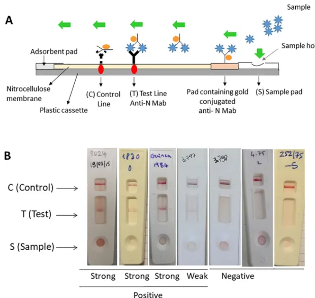
described above: the Mab 8E10-4A4 gold conjugate and the Mab 4E4-3D5. Mab 10H3-4E4-3D5 was immobilized onto a nitrocellulose membrane for the test line zone and rabbit anti-mouse antibodies (Dako, Denmark) were immobilized for the control line zone to capture unbound Mab. A reagent pad containing colloidal gold-labeled Mab 8E10-4A4 was located in front of the sample hole and overlaid onto the base of the nitrocellulose membrane, parallel to the control and the antibody bands, stuck to the membrane with adhesive cut into 0.8 cm wide strips, assembled in a device as described previously [31]. Aliquots (150μl) of either viral iso-lates mimicking clinical specimens, or viral supernatants diluted in DMEM, or serum samples were mixed with an equal volume of LFT sample buffer (0,1% Casein, 20 mM Borat-Borax buffer, 0,5% Tween 20) and the mixture was applied to the sample pad (S). This resulted in rehydration of the air-dried conjugated gold Mab and and their migration by capillary action along the membrane. If RVFV antigen was present in the sample then the RVFV-Mab-conju-gate complex was captured by the immobilized Mab on the membrane at the ‘T’ (test) line and resulted in their accumulation, which could be visualized as a red line to signify a positive result. Excess (or unbound) Mab-labelled gold particles continued to migrate along the device until being captured by the immobilized rabbit anti-mouse antibody and the formation of a red ‘C’ (control) line, to validate the test. The test (T) and control (C) lines were checked for the development of color after 10 minutes and again after 30 minutes as it might take longer time for weak positives to form a visual band scored subjectively from negative to strong.
Assessment of the diagnostic and analytical sensitivity ((DSe/ASe) and of
the diagnostic and analytical specificity (DSp/Asp)
According to the OIE guidelines [32], estimates of DSe (proportion of samples from known infected reference animals that test positive in an assay) and DSp (the proportion of samples from known uninfected reference animals that test negative in an assay) are the primary per-formance indicators established during validation of an assay. Analytical specificity (ASp) defined as the ability of the assay to distinguish the target analyte (e.g. a viral antigen) from non-target analytes, including matrix components and analytical sensitivity (ASe) defined as the estimated amount of analyte in a specified matrix that would produce a positive result at least a specified percent of the time are the first steps of the validation of an assay. The limit of detection (LOD) is a measure of the ASe. Although RVFV is considered as a single genotype and serotype, diagnostic sensitivity (DSe) was assessed with sera spiked with different RVF viral isolates from different geographical origins and collected over a period of 69 years to mimic clinical specimens (serum or fluids from aborted fetuses) (n = 25,Table 1A) and sera of the ongoing RVF outbreak occurring on Mayotte [10] (n = 10,Table 1A).
Arboviruses belonging to thePhlebovirus genus and viruses belonging to other viral genera
but producing the same clinical features in humans and/or ruminants (i.e. flaviviruses, alpha-viruses) (n = 9,Table 1B) were tested to assess diagnostic specificity (DSp), i.e. the proportion of samples from known uninfected reference animals that test negative in the assay, as they could be considered as the source of possible cross-reactions in diagnostic assays. In addition, animal serum samples known to be seronegative for RVF of different origins (mainland France, Tunisia, Reunion Island, Mayotte, Union of Comoros, and Madagascar) (n = 169,
Table 1B) were used for this evaluation.
To determine ASe, eight titrated suspensions from RVFV of different origins (Uganda, Madagascar, Mauritania and South Africa) adapted to cell culture were selected and diluted in RVF negative sheep sera to determine the limit of detection (LOD) of the LFT using 10-fold serial dilutions in RVFV negative cattle serum (Life Technologies, Gibco, France) mimicking clinical specimens. A volume of 150μL of each of the dilutions was tested. Samples were scored as true positives when detected positive with the real-time RT-PCR technique [16], which was considered as the gold standard in our study.
Results
Design of the lateral flow test (LFT)
Production and purification of the recombinant N and N-GN/GC. RVFV N or N-GN/
GCproteins coproduced in insect cells were analyzed by SDS-PAGE followed by Western Blot
incubated either with RVF mouse anti-N (Fig 1A) or anti-N/GN/GCantibodies (Fig 1B).
West-ern blot analysis using monoclonal antibody specific to RVFV N protein revealed a strong band of 26 KDa equivalent to the expected size of the N protein in the infected cell lysate, supernatant and in its purified form (Fig 1A, lane 1, 2 and 3 respectively). Western blot analy-sis using RVF polyclonal bovine hyper immune serum known to recognize N/GN/GCproteins
detected 3 bands at respectively 26 kDa (N protein),55 kDa (GNprotein) and 58kDa (GC
pro-tein) in the crude supernatant and as copurified proteins (Fig 1B, lane a and b). A double band at/above 45 kDa might correspond to products of GN/GCcleavage and maturation, and
possi-bly to partial and/or nonspecific processed forms.
Mab selection and characterization for use in the LFT. Based on the results of the three
separate tests previously described, two purified Mabs, Mab 10H3-4E4-3D5 as coating Mab and Mab 8E10-4A4 as the conjugate Mab were tested in combination at different
Table 1. Origin and results of the samples used to assess diagnostic sensitivity and specifity of the RVF LFT. Identification, year of isolation, origin of virus strains or
samples of sera tested in this study.
A. Sensitivity
Genus Strain/Species Year of isolation Country of origin Source LFT result (N˚ pos/total N˚)
Phlebovirus RVFV strain Smithburn 1944 Uganda mosquito Positive (1/1) Phlebovirus RVFV strain Lunyo 1955 Uganda mosquito Positive (1/1) Phlebovirus RVFV strain AN 1830 1956 South Africa sheep Positive (1/1) Phlebovirus RVFV strain KEN56/B2653/IB8 1963 Kenya bovine Positive (1/1) Phlebovirus RVFV strain 56/74 1974 South Africa cow Positive (1/1) Phlebovirus RVFV strain 252/75 1975 South Africa NA Positive (1/1) Phlebovirus RVFV strain ZH501 1977 Egypt human Positive (1/1) Phlebovirus RVFV strain VRL688/78 1978 Zimbabwe bovine Positive (1/1) Phlebovirus RVFV strain AR 20368 1981 South Africa mosquito Positive (1/1) Phlebovirus RVFV ARD 38388 1983 Burkina Faso mosquito Positive (1/1) Phlebovirus RVFV ARD 38661 1983 Senegal mosquito Positive (1/1) Phlebovirus RVFV 143/83 1983 Namibia human Positive (1/1) Phlebovirus RVFV ANK 6087 1984 Guinea bat Positive (1/1) Phlebovirus RVFV SPU45/85 1985 Zambia human Positive (1/1) Phlebovirus RVFV SPU204/855 1985 Angola human Positive (1/1) Phlebovirus RVFV An991 1991 Madagascar bovine Positive (1/1) Phlebovirus RVFV Tambul 1994 Egypt ovine Positive (1/1) Phlebovirus RVFV SPU12/98/2 1998 Somalia goat Positive (1/1) Phlebovirus RVFV strain AR21229 2000 Saudi Arabia mosquito Positive (1/1) Phlebovirus RVFV F057 2007 Kenya human Positive (1/1) Phlebovirus RVFV AL51 2009 Madagascar mosquito Positive (1/1) Phlebovirus RVFV HA 09–001 2008 Madagascar bovine Positive (1/1) Phlebovirus RVFV strain 26010 YK0 2010 Mauritania camel Positive (1/1) Phlebovirus RVFV strain MRU 2687 2013 Mauritania goat Positive (1/1) Phlebovirus RVFV strain SN 2 2013 Senegal goat Positive (1/1) Phlebovirus Z_LD_8023 2019 Mayotte, France ovine Positive (1/1) Phlebovirus Z_LD_8024 2019 Mayotte, France ovine Positive (1/1) Phlebovirus Z_LD_2700/5 2019 Mayotte, France bovine Positive (1/1) Phlebovirus Z_LD_8016 2019 Mayotte, France bovine Positive (1/1) Phlebovirus Z_LD_8107 2019 Mayotte, France bovine Positive (1/1) Phlebovirus Z_LD_8248 2019 Mayotte, France bovine Positive (1/1) Phlebovirus Z_LD_8627 2019 Mayotte, France bovine Positive (1/1) Phlebovirus Z_LD_8656 2019 Mayotte, France bovine Positive (1/1) Phlebovirus Z_LD_8657 2019 Mayotte, France bovine Positive (1/1) Phlebovirus Z_LD_Liver 160119 2019 Mayotte, France bovine Positive (1/1)
B. Specificity
Phlebovirus Arumowot virus 1963 Sudan Culex antennatus Negative (0/1) Phlebovirus Gabek forest virus 1981 Unknown Unknown Weak band (1/1) Phlebovirus Saint Floris virus 1981 Unknown Unknown Negative (0/1) Flavivirus Dengue virus, serotype 2 2012 Reunion Island human Negative (0/1) Orthobunyavirus Akabane virus 1978 Unknown Unknown Negative (0/1) Orthobunyavirus Shamonda virus 1972 Unknown Unknown Negative (0/1) Alphavirus Chikungunya virus Chik #4 2006 Reunion Island human Negative (0/1) Reovirus Bluetongue virus, serotype 2 2002 Corsica, France ovine Negative (0/1)
Table 1. (Continued) A. Sensitivity
Genus Strain/Species Year of isolation Country of origin Source LFT result (N˚ pos/total N˚)
Morbillivirus PPRV 75/1 1975 Nigeria goat Negative (0/1) NA Sera samples 2017 Mainland France goat/bovine Negative (1/31) NA Sera samples 2015 Union of Comoros goat/bovine Negative (0/20) NA Sera samples 2016 Reunion Island bovine Negative (0/30) NA Sera samples 2010 Tunisia goat/camel Negative (0/30) NA Sera samples 2011 Madagascar goat/bovine Negative (0/20) NA Sera samples 2016 Mayotte, France bovine Negative (0/19) NA Sera samples 2019 Mayotte, France bovine Negative (0/10) NA stands for Not Applicable, N˚ for Number and PPRV for Peste des Petits Ruminants Virus
https://doi.org/10.1371/journal.pntd.0007700.t001
Fig 1. Coproduction of RVFV N and GN/GCproteins in insect cells. Controls (uninfected and wild-type AcMNPV-infected cells)
gave no signal. A. SDS-PAGE/Western blot, anti-N detection. Lane 1: Sf9 crude cell lysates coproducing N and GN/GCproteins. Lane
2: Sf9 crude supernatant coproducing N and GN/GCproteins. Lane 3: purified N and GN/GCproteins. B. SDS-PAGE/Western blot,
anti-N/GN/GCdetection. Lane a: Sf9 crude supernatant coproducing N and GN/GCproteins. Lane b: copurified N/GN/GCproteins.
A double band at/above 45 kDa might correspond to products of GN/GCcleavage and maturation, and possibly to partial and/or
nonspecific processed forms.
concentrations based on experimental results and chosen for further sensitivity and specificity validation (Fig 2).
Design of in the LFT. In a positive sample, the RVFV-Mab-conjugate complex captured
by the immobilized Mab on the membrane corresponds to a solid red line at the ‘T’ (test) of the device. A red ‘C’ (control) line always appear and validate the test (Fig 3A). LFT strips showing negative, weak and positive results are shown inFig 3B.
Estimates of diagnostic sensitivity (DSe) and diagnostic specificity (DSp)
Diagnostic sensitivity of the LFT (DSe). A total of 25 isolated strains mimicking clinical
specimens of different geographical origins and 10 clinical samples originating from the ongo-ing outbreak of Mayotte (2019) [10] detected RVF positive by a Taqman RT-PCR technique
Fig 2. Specificity of the two Mabs 8E10-4A4 and 10H3-4E4-3D5 used in combination in the LFT for RVFV. Specific binding of
the Mabs to RVFV N protein was examined by (A) ELISA with RVFV copurified N/GN/GCproteins (ELISA N/GN/GC), ELISA with
coating of Bac-N nucleoprotein expressed by Sf9 insect cells (ELISA N). Readings are OD (Optical Density) values measured at 450 nm. Mabs were tested diluted 1:10 in culture medium and run in duplicates (mean +/- 3 SD). The positive cut-off value of the test is an OD value > 0.300, the positive control (mouse immunized with RVF N/GN/GCsampled at day 41, dilution 1:100) must have an
OD value > 1.200 and the negative control (non immunized OF1 mouse control) must have an OD value < 0.300. A tested sample is detected positive when the OD value > 0.300. (B) Immunofluorescence Assay (IFA) x 10, green fluorescence: positive sample, Mab directed against peste des petits ruminants virus (PPRV), negative control, no fluorescence.
which is considered as the current reference detection system [16] were also detected positive by the LFT (Table 1A) giving a DSe of 100% (CI 95%[90,1; 100]) (n = 35).
Diagnostic specificity (DSp) of the LFT. Diagnostic specificity (DSp) was assessed on
160 serum samples known to be negative for RVFV by seroneutralization test and cELISA but collected in tropical countries (Madagascar, Comoros, Mayotte) where RVF has been circulat-ing to detect possible cross reactions or in other areas where RVF introduction is considered at risk (Tunisia, mainland France, Reunion Island). Only one sample among 160 originating from mainland France was found positive with RVF LFT but negative with the gold standard reference test (n = 160,Table 1B). In addition, nine vector-borne viruses or viruses that pro-duce clinical signs similar to RVF, such as abortion or fever, were tested (n = 9,Table 1B) and were detected negative for RVF by LFT except for the Gabek forest virus that produced a weak band. Finally, the test gave a DSp of 98.81% (CI 95% [95.8; 99,7]) (n = 169) (Table 2).
Fig 3. RVF LFT strip test for the detection of RVF infection using the two selected Mabs. (A) Diagram of the rapid RVF LFT for
the detection of the RVF N protein, (B) Results of LFT strip. The serum sample or the viral suspension (150μl) was mixed with 150μl of sample buffer and applied to the S hole of the strips for migration. Results were recorded after 15 minutes. The test is valid when a red band is visible at the same level as the label C. S stands for Sample, C for Control and T for Test.
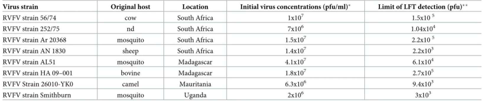
Determination of the limit of detection (LOD) of the LFT, a measure of ASe. Eight
titrated viral suspensions from RVFV originating from different countries (Uganda, Madagas-car, Mauritania and South Africa) (Table 3) were used spiked in RVF negative sera to detect the LOD, a measure of ASe. The lowest numbers of pfu detected with this assay were 2.3 x10³ pfu, 3x10³pfu and 9x10³pfu for the South African AN1830, the Smithburn and the Maurita-nian 26010-YK0 strains respectively.
Discussion
Recent outbreaks in several countries including Sudan, Uganda, Niger, Mali (2017–2018), Kenya and Mayotte (2018–2019) confirm the active circulation of the disease. Human and ani-mal movements in territories facing rural movements and civil wars are likely to facilitate RVFV spread and its extension outside its traditional boundaries towards northern Africa [33] and possibly to Europe. In European countries, currently free of RVFV, its introduction could cause severe outbreaks in naïve human and/or animal populations. As RVF is a vector borne disease, the threat is also increasing due to global warming leading to (i) an increase in mos-quito density and diversity, (ii) vector competence being acquired by new species [2,5,34]. The development of rapid diagnostic tools able to detect RVF infection is crucial for both human and animal health monitoring in endemic and at-risk areas for RVF. The aim of our study was (i) to develop and evaluate a prototype of a rapid and robust RVF LFT and (ii) to investigate its intrinsic parameters. As it is the case with viruses that are members of the
Bunyavirales order, the N nucleocapsid protein was shown to be a key immunogenic protein
that induces a strong immune response [35–36]. Antibodies against the nucleoprotein N are readily detected early after infection and in convalescent individuals, providing a robust basis for detection and diagnostic of the disease. ELISA diagnostic tests have been developed based
Table 2. Analytical specificity (ASp). Agreement between the RVF LFT results and the PCR results [16].
PCR results
Positive Negative Total
LFT results Positive 35 2 37
Negative 0 167 167
Total 35 169 204
https://doi.org/10.1371/journal.pntd.0007700.t002
Table 3. Analytical sensitivity (ASe) (estimated limit of detection (LOD)) based on 8 titrated RVF strains.
Virus strain Original host Location Initial virus concentrations (pfu/ml)� Limit of LFT detection (pfu)��
RVFV strain 56/74 cow South Africa 1x10⁷ 1.5x10⁵
RVFV strain 252/75 nd South Africa 7x10⁶ 1.04x10⁴
RVFV strain Ar 20368 mosquito South Africa 1.5x10⁷ 2.2x10⁵
RVFV strain AN 1830 sheep South Africa 1.4x10⁷ 2.2x10³ RVFV strain AL51 mosquito Madagascar 4.1x10⁷ 6.1x10⁴
RVFV strain HA 09–001 bovine Madagascar 1.8x10⁷ 2.7x10⁵
RVFV Strain 26010-YK0 camel Mauritania 6.3x10⁶ 9.4x10³
RVFV strain Smithburn mosquito Uganda 2x10⁶ 3x10³
�pfu/ml, particle forming units per ml, ��pfu deposited on the strip
nd, not determined
on this antigen and produced promising results [11,37]. This suggests the nucleoprotein N is a highly suitable target antigen for the detection of RVFV infection. In the present study, RVFV specific Mabs were produced against the N nucleoprotein and conjugated with colloidal gold to bind RVFV antigens captured by immobilized membrane to form a red band, which indi-cates the presence of RVFV antigens. The performance of the LFT we developed for detection of RVF infection was evaluated on sera spiked with known positive viral suspensions mimick-ing clinical specimens (n = 25) as well as viremic clinical samples of an ongomimick-ing outbreak (Mayotte 2019) (n = 10) and known RVF negative sera of different origins (Tunisia, Madagas-car, Mayotte, Reunion Island, Union of Comoros, mainland France). The RVF LFT was able to detect all 35 RVFV spiked or clinical samples from different geographical regions. Specific-ity was determined to be 98.81%, (CI 95% [95.8; 99,7]) based on (i) testing known negative ani-mal serum samples from different countries (n = 160) and (ii) vector-borne viruses or viruses giving clinical signs similar to RVF (n = 9). A weak positive band was detected when testing the Gabek forest virus (GFV). This could be explained by the high percentage of homology observed between the segments of RVFV and GFV illustrated by the closeness of the clade RVFV and GFV [38]. These preliminary data need to be strengthened with additional field serum samples originating from ongoing outbreaks around the world, even though the quanti-fication of the viral load present in serum field samples is hard to perform due to the very few laboratories able to handle advanced cell culture and molecular biology experiments located where the outbreaks occur. Comparison with other diagnostic techniques will help in that quantification. The limit of detection of this LFT was investigated using eight titrated suspen-sions of RVFV spiked in dilution in RVF negative sheep sera. The values varied among the iso-lates and ranked from 103to 105pfu. Previous ELISA based on RVF antigens were found to be less sensitive since they are able to detect 102.2to 103.2TCID50/reaction volume [37]. Viremia
becomes demonstrable in one-week-old lambs within 16 hours of peripheral infection and persists for the duration of the illness for at least 36 to 42 hours [39]. In older sheep, goats and cattle, viremia becomes demonstrable one to two days after infection and persists for seven days with a peak from day 2 to day 5. Observed viremia ranged from 105.6to 109of mouse median lethal doses (MIPLD50) per ml in naturally infected domestic animals up to 108.6in
humans [40]. Maximum viremia recorded were 1010.1MIPLD50/ml in lambs, 107.6in sheep
[41], 107.5in calves, and 105.6in goats [42]. Viremia titers in experimentally infected goats range from 104to 106.5log10RVF copies/ml [43–44]. Viremia of lower intensity and shorter
duration has been detected in other animal species that have been studied, but quantitative data are difficult to obtain. In adult African buffalo and ponies, viremia was recorded at 105.4TCID50/ml and 102.5MIPLD50/ml respectively [45–46].
The original aim of the LFT was to detect RVF infection in the field in serum samples in cases of suspected outbreaks. In that context, the viremia expected in infected animals is likely to be high with titers above 105pfu irrespective of the species of domestic animal tested (goats, sheep, cattle, camels). In that sense, the level of specificity obtained for the LFT would be high enough to test a field sample for a rapid first detection of initial outbreaks (which would still require confirmation of negative or positive results by a reference laboratory) or in the case of disease surveillance in the framework of a control programme when RVF has already been confirmed. The level of detection of the LFT could be improved either by experimenting with the addition of a secondary antibody on top of the conjugated Mab in the construction of the cassette or by testing several types of membranes. Alternative colloid gold conjugation meth-ods or other labels could also be evaluated to improve the sensitivity. Recovery of viral RNA from the same type of LFT membrane has been already proven for other viruses such as foot and mouth disease virus [47–48] and is another great outcome of this type of diagnosis, specifi-cally for viruses spreading in areas where access to diagnostic facilities is limited. The type of
samples that have been validated in this study is serum samples as it could be easily used in a field setting for early RVF detection. Comparison of whole blood versus serum, testing of long-term storage conditions are worth to be evaluated in future trials in order for this test to be field deployable to locations where this will ultimately be needed. The use of RVF LFT by trained personnel wearing the highest level of protective clothing and using appropriate equip-ment will provide rapid and objective support to veterinarians in their clinical judgequip-ment of the disease and also help dispatch field materials to national or international reference laboratories for confirmation. The sooner the disease is diagnosed, the sooner the appropriate measures can be taken. The use of this first-line LFT should accelerate the start of epidemiological inves-tigations in the case of RVF outbreaks. Furthermore, the difficulties and the high cost of send-ing infectious material from rural veterinary or district health facilities, where the disease often occurs, to the regional or national reference laboratory and the biosecurity risk it presents underline the need for a thermo-stable, non-infectious standard mode of transporting diag-nostic samples. The cost of the LFT itself and its shipment to regional facilities versus saving on expenses for standard sample collection and transportation is likely to vary from country to country and depends on the supply and demand as well as the competitive offers of the private companies able to produce it. Therefore the cost implications and the potential for this LFT to be easily affordable in low and limited resource settings needs to be taken into consideration in the long-term implementation of this LFT.
Conclusion
The performances of the RVF LFT are promising for field use, where the test could help to establish rapid preliminary diagnostic results particularly in suspected cases in the field, which would then have to be confirmed using WHO (World Health Organization) or OIE (World Organisation for Animal Health) recommended tests at central laboratories. The specificity and sensitivity of the evaluated test are lower than the ones of molecular-based techniques (LAMP, PCR) but are adequate for specific rapid initial detection of RVF outbreaks or disease surveillance in control programmes. However, there is still room for improvement in LFT per-formances by changing several parameters i.e. the sample buffer, the type of membrane or the addition of secondary labeled antibodies. This rapid and easy RVF LFT device does not require special laboratory equipment but does require trained staff wearing appropriate biosecure pro-tective clothing. Although the Lateral Flow Test (LFT) is easy to use for non-laboratorians out-side a biosafety containment reference laboratory normally used for RVF testing, it
nevertheless requires appropriate training to avoid any accidental infection during the removal and handling of a potentially infected sample of body fluid (serum being the ideal type of sam-ple in the case of RVF outbreak or fluid from aborted fetuses) for LFT testing. LFT can be per-formed in the field where epizootics occur. RVF LFT will be particularly valuable in remote areas or in territories where there are no diagnostic facilities but it is important to underline that staff needs to be trained to handle it safely under the highest possible biosecurity conditions.
Acknowledgments
The authors thank Dr Philippe Me´rot, DAAF Mayotte, the farmers of Mayotte, Dr Christian Schuler, Dr Lionel Dome´on, Dr Bertrand Bouyer, Dr Laure Bouyer, for collecting the animal samples, Chouanibou Youssouffi for entering the data in the database, Johny Hoarau, Abdou Achiraffi, Sitty Bahyat Chamassi and Chouanibou Youssouffi for their participation in the lab work.
Author Contributions
Conceptualization: Catherine Cêtre-Sossah, Aure´lie Pe´darrieu, Mikael Juremalm, Emmanuel Albina, Eric Cardinale.
Data curation: Catherine Cêtre-Sossah.
Formal analysis: Petrus Jansen Van Vuren, Alejandro Brun, Jean-Michel He´raud, Claudia
Filippone, Janusz Paweska.
Funding acquisition: Eric Cardinale.
Investigation: Mikael Juremalm, Petrus Jansen Van Vuren, Alejandro Brun.
Methodology: Catherine Cêtre-Sossah, Aure´lie Pe´darrieu, Mikael Juremalm, Petrus Jansen Van Vuren, Alejandro Brun, Jean-Pierre Ravalohery, Hassan Chaabihi, Emmanuel Albina, Janusz Paweska.
Resources: Petrus Jansen Van Vuren, Alejandro Brun, Ahmed Bezeid Ould EL Mamy,
Jean-Michel He´raud, Claudia Filippone, Jean-Pierre Ravalohery, Laure Dommergues, Janusz Paweska.
Supervision: Catherine Cêtre-Sossah.
Validation: Catherine Cêtre-Sossah, Aure´lie Pe´darrieu, Mikael Juremalm, Jean-Pierre Rava-lohery, Emmanuel Albina, Laure Dommergues, Janusz Paweska.
Writing – original draft: Catherine Cêtre-Sossah, Eric Cardinale.
Writing – review & editing: Catherine Cêtre-Sossah, Mikael Juremalm, Petrus Jansen Van Vuren, Alejandro Brun, Ahmed Bezeid Ould EL Mamy, Jean-Michel He´raud, Claudia Filip-pone, Emmanuel Albina, Laure Dommergues, Janusz Paweska, Eric Cardinale.
References
1. Maes P, Alkhovsky SV, Bào Y, Beer M, Birkhead M, Briese T, et al. Taxonomy of the family Arenaviri-dae and the order Bunyavirales: update 2018. Arch Virol. 2018; 163: 2295–2310.https://doi.org/10. 1007/s00705-018-3843-5PMID:29680923
2. Linthicum KJ, Britch SC, Anyamba A. Rift Valley fever: an emerging mosquito-borne disease. Annu Rev Entomol. 2016; 61: 395–415.https://doi.org/10.1146/annurev-ento-010715-023819PMID:26982443
3. Moutailler S, Krida G, Schaffner F, Vazeille M, Failloux AB. Potential vectors of Rift Valley fever virus in the Mediterranean region. Vector Borne Zoonotic Dis. 2008; 8: 749–753.https://doi.org/10.1089/vbz. 2008.0009PMID:18620510
4. Turell MJ, Wilson WC, Bennett KE. Potential for North American mosquitoes (Diptera: Culicidae) to transmit rift valley fever virus. J Med Entomol. 2010; 47: 884–889.https://doi.org/10.1603/me10007
PMID:20939385.
5. Brustolin M. Talavera S, Nuñez A, Santamarı´a C, Rivas R, Pujol N et al. Rift Valley fever virus and Euro-pean mosquitoes: vector competence of Culex pipiens and Stegomyia albopicta (= Aedes albopictus). Med Vet Entomol. 2017; 31: 365–372.https://doi.org/10.1111/mve.12254PMID:28782121
6. Tantely LM, Boyer S, Fontenille D. A review of mosquitoes associated with Rift Valley fever virus in Madagascar. Am J Trop Med Hyg. 2015; 92: 722–729.https://doi.org/10.4269/ajtmh.14-0421PMID:
25732680
7. Baudin M, Jumaa AM, Jomma HJE, Karsany MS, Bucht G et al. Association of Rift Valley fever virus infection with miscarriage in Sudanese women: a cross-sectional study. Lancet Glob Health. 2016; 4: e864–e871.https://doi.org/10.1016/S2214-109X(16)30176-0PMID:27692776
8. De St Maurice A. Nyakarahuka L, Purpura L, Ervin E, Tumusiime A, Balinandi S et al. Notes from the Field: Rift Valley Fever Response—Kabale District, Uganda, March 2016. MMWR Morb Mortal Wkly Rep. 2016; 65: 1200–1201.https://doi.org/10.15585/mmwr.mm6543a5PMID:27811840
9. Tambo E, Olalubi OA, Sacko M. Rift valley fever epidemic in Niger near border with Mali. Lancet Infect Dis. 2016; 16: 1319–1320.https://doi.org/10.1016/S1473-3099(16)30477-7PMID:27998581
10. Promed Archive Number: 20190222.6331808.
11. Fafetine JM, Tijhaar E, Paweska JT, Neves LC, Hendriks J, Swanepoel R et al. (2007) Cloning and expression of Rift Valley fever virus nucleocapsid (N) protein and evaluation of a N-protein based indi-rect ELISA for the detection of specific IgG and IgM antibodies in domestic ruminants. Vet Microb. 2007; 121: 29–38.https://doi.org/10.1016/j.vetmic.2006.11.008PMID:17187944
12. Paweska JT, Jansen van Vuren P, Swanepoel R. Validation of an indirect ELISA based on a recombi-nant nucleocapsid protein of Rift Valley fever virus for the detection of IgG antibody in humans. J Virol Methods. 2007; 146: 119–124.https://doi.org/10.1016/j.jviromet.2007.06.006PMID:17645952
13. Garcia S, Crance JM, Billecocq A, Peinnequin A, Jouan A, Bouloy M et al. Quantitative real-time PCR detection of Rift Valley fever virus and its application to evaluation of antiviral compounds. J Clin Micro-biol. 2001; 39: 4456–4461.https://doi.org/10.1128/JCM.39.12.4456-4461.2001PMID:11724861
14. Drosten C. Go¨ttig S, Schilling S, Asper M, Panning M, Schmitz H et al. Rapid detection and quantifica-tion of RNA of Ebola and Marburg viruses, Lassa virus, Crimean-Congo hemorrhagic fever virus, Rift Valley fever virus, dengue virus, and yellow fever virus by real-time reverse transcription-PCR. J Clin Microbiol. 2002; 40: 2323–2330.https://doi.org/10.1128/JCM.40.7.2323-2330.2002PMID:12089242. 15. Sall AA, Macondo EA, Sène OK, Diagne M, Sylla R, Mondo M et al. Use of reverse transcriptase PCR
in early diagnosis of Rift Valley fever. Clin Diagn Lab Immunol. 2002; 9: 713–715.https://doi.org/10. 1128/CDLI.9.3.713-715.2002PMID:11986283.
16. Bird BH, Bawiec DA, Ksiazek TG, Shoemaker TR, Nichol ST. Highly sensitive and broadly reactive quantitative reverse transcription-PCR assay for high-throughput detection of Rift Valley fever virus. J Clin Microbiol. 2007; 45: 3506–3513.https://doi.org/10.1128/JCM.00936-07PMID:17804663. 17. Wilson WC, Romito M, Jasperson DC, Weingartl H, Binepal YS, Maluleke MR, et al. Development of a
Rift Valley fever real-time RT-PCR assay that can detect all three genome segments J Vir Methods. 2013; 193: 426–431.
18. Brinkmann A. Ergu¨nay K, RadonićA, Kocak Tufan Z, Domingo C, Nitsche A, et al. Development and preliminary evaluation of a multiplexed amplification and next generation sequencing method for viral hemorrhagic fever diagnostics. PLoS Negl Trop Dis. 2017; 11 (11):e0006075.https://doi.org/10.1371/ journal.pntd.0006075PMID:29155823
19. Zaher MR, Ahmed HA, Hamada KEZ, Tammam RH. Colorimetric detection of unamplified Rift Valley Fever virus genetic material using unmodified gold nanoparticles. Appl Biochem Biotechnol. 2017. PMID:28918558
20. Liu J, Ochieng C, Wiersma S, Stro¨her U, Towner JS, Whitmer S et al. Development of a TaqMan array card for acute-febrile-illness and surveillance of emerging pathogens, including Ebola virus. J Clin Microbiol. 2016; 54: 49–58.https://doi.org/10.1128/JCM.02257-15PMID:26491176
21. Gao CH, Yang YT, Shi F, Wang JY, Steverding D, Wang X. Development of an Immunochromato-graphic test for diagnosis of visceral leishmaniasis based on detection of a circulating antigen. PLoS Negl Trop Dis. 2015; 9, e0003902.https://doi.org/10.1371/journal.pntd.0003902PMID:26125560
22. Cross RW, Boisen ML, Millett MM, Nelson DS, Oottamasathien D, Hartnett JN, et al. Analytical valida-tion of the ReEBOV antigen rapid test for point-of-care diagnosis of Ebola virus infecvalida-tion. J Infect Dis. 2016; 214 (suppl 3): S210–S217.https://doi.org/10.1093/infdis/jiw293PMID:27587634
23. Le´chenne M, Naïssengar K, Lepelletier A, Alfaroukh IO, Bourhy H, Zinsstag J, et al. Validation of a Rapid Rabies Diagnostic Tool for Field Surveillance in Developing Countries. PLoS Negl Trop Dis. 2016; 10(10):e0005010.https://doi.org/10.1371/journal.pntd.0005010PMID:27706156
24. Ayari-Fakhfakh E, Ghram A, Bouattour A, Larbi I, Gribaˆa-Dridi L, Kwiatek O, et al. First serological investigation of peste-des-petits-ruminants and Rift Valley fever in Tunisia. Vet J. 2011; 187: 402–404.
https://doi.org/10.1016/j.tvjl.2010.01.007PMID:20167519
25. Roger M, Beral M, Licciardi S, Soule´ M, Faharoudine A, et al. Evidence for circulation of the Rift Valley fever virus among livestock in the Union of Comoros. PLoS Negl Trop Dis. 2014; 8(7): e3045.https:// doi.org/10.1371/journal.pntd.0003045PMID:25078616
26. Me´tras R, Cavalerie L, Dommergues L, Me´rot P, Edmunds WJ, Keeling MJ, et al. The epidemiology of Rift Valley fever in Mayotte: Insights and perspectives from 11 years of data. PLoS Negl Trop Dis. 2016; 10: e0004783.https://doi.org/10.1371/journal.pntd.0004783PMID:27331402
27. Olive MM, Chevalier V, Grosbois V, Tran A, Andriamandimby SF, Durand B, et al. Integrated Analysis of Environment, Cattle and Human Serological Data: Risks and Mechanisms of Transmission of Rift Valley Fever in Madagascar. PLoS Negl Trop Dis. 2016; 10(7):e0004827.https://doi.org/10.1371/ journal.pntd.0004827PMID:27415438
28. El-Akkad AM. Rift Valley fever outbreak in Egypt. October—December 1977. J Egypt Public Health Assoc. 1978; 53: 123–128. PMID:572393.
29. Liu L, Celma CC, Roy P. Rift Valley fever virus structural proteins: expression, characterization and assembly of recombinant proteins. Virol J. 2008; 5: 82.https://doi.org/10.1186/1743-422X-5-82PMID:
18638365
30. Chaabihi H, Cêtre C, Berne A. A new vector for efficient generation of p10-single-late-promoter recom-binant baculoviruses. J Virol Methods. 1997; 63 (1–2): 1–7 PMID:9015270
31. Bru¨ning A, Bellamy K, Talbot D, Anderson J. A rapid chromatographic strip test for the pen-side diagno-sis of rinderpest virus. J Virol Methods. 1999; 81: 143–154. PMID:10488772.
32. OIE Terrestrial Manual OIE Chapter 1.1.5. Principles and methods of validation of diagnostic assays for infectious diseases. 2013: 1–17.
33. Bosworth A, Ghabbari T, Dowall S, Varghese A, Fares W, Hewson R, et al. Serologic evidence of expo-sure to Rift Valley fever virus detected in Tunisia. New Microbes New Infect. 2015; 9: 1–7.https://doi. org/10.1016/j.nmni.2015.10.010PMID:26740887
34. Lo´pez-Ve´ lez R, Molina R. Climate change in Spain and risk of infectious and parasitic diseases trans-mitted by arthropods and rodents. Rev. Esp. Salud Pu´blica. 2005; 79: 177–190. PMID:15913053
35. Swanepoel R, Struthers JK, Erasmus MJ, Shepherd SP, McGillivray GM, Erasmus BJ, et al. Compari-son of techniques for demonstrating antibodies to Rift Valley fever virus. J Hyg Camb. 1986; 97: 317– 329.https://doi.org/10.1017/s0022172400065414PMID:3537118.
36. Koser ML, McGettigan JP, Tan GS, Smith ME, Koprowski H, Dietzschold B, et al. Rabies virus nucleo-protein as a carrier for foreign antigens. Proc Natl Acad Sci USA. 2004; 101: 9405–9410.https://doi. org/10.1073/pnas.0403060101PMID:15197258
37. Jansen van Vuren P, Paweska JT. Laboratory safe detection of nucleocapsid protein of Rift Valley fever virus in human and animal specimens by a sandwich ELISA. J Virol Methods. 2009; 157, 15–24.
https://doi.org/10.1016/j.jviromet.2008.12.003PMID:19124041
38. Palacios G, Tesh RB, Savji N, Travassos da Rosa AP, Guzman H, Bussetti AV, et al. Characterization of the Sandfly fever Naples species complex and description of a new Karimabad species complex (genus Phlebovirus, family Bunyaviridae). J Gen Virol. 2014; 95: 292–300.https://doi.org/10.1099/vir.0. 056614-0PMID:24096318
39. Easterday BC. Rift Valley fever. Adv in Vet Science. 1965; 10: 65–127. PMID:5331042.
40. Peters CJ, Meegan JM. Rift Valley fever. In Beran G. (Ed.). CRC Handbook Series in Zoonosis. 1981. Sect B1, 403–20. CRC Press Inc., Boca Raton, Florida.
41. Easterday BC, Murphy LC, Bennett DG. Experimental RVF in calves, goats and pigs. Am J Vet Res. 1962; 23: 1224–1230.
42. Easterday BC, Murphy LC, Bennett DG. Experimental RVF in lambs and sheep. Am J Vet Res.1962; 23, 1231–1240.
43. Iman IZ, El-Karamany R, Kasem S. Studies on goats experimentally infected with Rift Valley fever virus. J Egypt Public Health Assoc. 1978; 53, 273–280. PMID:752709.
44. Nfon CK, Marszal P, Zhang S, Weingartl HM. Innate immune response to Rift Valley fever virus in goats. PLoS Negl Trop Dis. 2012; 6: e1623.https://doi.org/10.1371/journal.pntd.0001623PMID:
22545170
45. Davies FG, Karstad L. Experimental infection of the African buffalo with the virus of Rift Valley fever. Trop An Health and Prod. 1981, 13: 185–188. PMID:7344184.
46. Yedloutschnig RJ, Dardiri AH, Walker JS. The response of ponies to inoculation with Rift Valley fever virus. Contributions to Epidemiology and Biostatistics.1981; 3: 68–71.
47. Fowler VL, Bankowski BM, Armson B, Di Nardo A, Valdazo-Gonzalez B, Reid SM, et al. Recovery of viral RNA and infectious foot-and-mouth disease virus from positive lateral-flow devices. PLoS One 2014; 9: e109322.https://doi.org/10.1371/journal.pone.0109322PMID:25313787
48. Romey A. Relmy A, Gorna K, Laloy E, Zientara S, Blaise-Boisseau S, et al. Safe and cost-effective pro-tocol for shipment of samples from Foot-and-Mouth Disease suspected cases for laboratory diagnostic. Transbound Emerg Dis. 2018; 65, 197–204https://doi.org/10.1111/tbed.12648PMID:28387065