Publisher’s version / Version de l'éditeur:
Scientific Reports, 8, 2018-01-30
READ THESE TERMS AND CONDITIONS CAREFULLY BEFORE USING THIS WEBSITE. https://nrc-publications.canada.ca/eng/copyright
Vous avez des questions? Nous pouvons vous aider. Pour communiquer directement avec un auteur, consultez la
première page de la revue dans laquelle son article a été publié afin de trouver ses coordonnées. Si vous n’arrivez pas à les repérer, communiquez avec nous à PublicationsArchive-ArchivesPublications@nrc-cnrc.gc.ca.
Questions? Contact the NRC Publications Archive team at
PublicationsArchive-ArchivesPublications@nrc-cnrc.gc.ca. If you wish to email the authors directly, please see the first page of the publication for their contact information.
Archives des publications du CNRC
This publication could be one of several versions: author’s original, accepted manuscript or the publisher’s version. / La version de cette publication peut être l’une des suivantes : la version prépublication de l’auteur, la version acceptée du manuscrit ou la version de l’éditeur.
For the publisher’s version, please access the DOI link below./ Pour consulter la version de l’éditeur, utilisez le lien DOI ci-dessous.
https://doi.org/10.1038/s41598-018-19522-8
Access and use of this website and the material on it are subject to the Terms and Conditions set forth at
A novel human induced pluripotent stem cell blood-brain barrier model:
applicability to study antibody-triggered receptor-mediated
transcytosis
Ribecco-Lutkiewicz, Maria; Sodja, Caroline; Haukenfrers, Julie; Haqqani,
Arsalan S.; Ly, Dao; Zachar, Peter; Baumann, Ewa; Ball, Marguerite; Huang,
Jez; Rukhlova, Marina; Martina, Marzia; Liu, Qing; Stanimirovic, Danica;
Jezierski, Anna; Bani-Yaghoub, Mahmud
https://publications-cnrc.canada.ca/fra/droits
L’accès à ce site Web et l’utilisation de son contenu sont assujettis aux conditions présentées dans le site LISEZ CES CONDITIONS ATTENTIVEMENT AVANT D’UTILISER CE SITE WEB.
NRC Publications Record / Notice d'Archives des publications de CNRC:
https://nrc-publications.canada.ca/eng/view/object/?id=d891f0d8-7b5e-4fc6-b321-22e16d6b338a
https://publications-cnrc.canada.ca/fra/voir/objet/?id=d891f0d8-7b5e-4fc6-b321-22e16d6b338a
www.nature.com/scientificreports
A novel human induced pluripotent
stem cell blood-brain barrier model:
Applicability to study
antibody-triggered receptor-mediated
transcytosis
Maria Ribecco-Lutkiewicz, Caroline Sodja, Julie Haukenfrers, Arsalan S. Haqqani, Dao Ly,
Peter Zachar, Ewa Baumann, Marguerite Ball, Jez Huang, Marina Rukhlova, Marzia Martina,
Qing Liu, Danica Stanimirovic, Anna Jezierski & Mahmud Bani-Yaghoub
We have developed a renewable, scalable and transgene free human blood-brain barrier model, composed of brain endothelial cells (BECs), generated from human amniotic luid derived induced pluripotent stem cells (AF-iPSC), which can also give rise to syngeneic neural cells of the neurovascular unit. These AF-iPSC-derived BECs (i-BEC) exhibited high transendothelial electrical resistance (up to 1500 Ω cm2) inducible by astrocyte-derived molecular cues and retinoic acid treatment, polarized
expression of functional elux transporters and receptor mediated transcytosis triggered by antibodies against speciic receptors. In vitro human BBB models enable pre-clinical screening of central nervous system (CNS)-targeting drugs and are of particular importance for assessing species-speciic/selective transport mechanisms. This i-BEC human BBB model discriminates species-selective antibody- mediated transcytosis mechanisms, is predictive of in vivo CNS exposure of rodent cross-reactive antibodies and can be implemented into pre-clinical CNS drug discovery and development processes.
Central nervous system (CNS) drug development is hindered by high clinical attrition rates1,2. he complex
phys-iology of the human brain, the diiculty in achieving suicient drug concentrations in the brain3 and inadequate
animal models of human CNS pathology are key underlying causes. he development of translational and predic-tive models for assessing blood-brain barrier (BBB) delivery has become an important requirement in pre-clinical testing of CNS-targeting therapeutics.
he BBB is composed of specialized brain microvascular endothelial cells (BECs) that form a barrier between the bloodstream and the CNS4. his difusion barrier is formed by tight junctions between BECs, which result
in a high transendothelial electrical resistance (TEER). In addition to the physical paracellular barrier, the BBB endothelium is enriched with a battery of polarized elux transporters, that eliminate substrate-drugs from the brain, as well as specialized BBB inlux carriers that allow the selective, energy-dependent transport of essential nutrients such as amino acids, carbohydrates and small peptides into the brain5,6. he BBB is maintained and
reg-ulated by a complex crosstalk between BECs and cells of the neurovascular unit (pericytes, astrocytes, microglia and neurons), which work in concert to ensure proper brain homeostasis and function7.
he BBB also hinders the delivery of many potentially important diagnostic and therapeutic agents to the brain. Very few synthetic molecules (highly lipophilic or hydrophobic molecules with a molecular mass below 400–500 Da) and biologics delivered intravenously, can cross the BBB suiciently to produce a pharmacological efect8. In a study evaluating more than 7 000 drug compounds, only 5% could cross the BBB and produce a
phar-macological response in the CNS9,10.
In vitro BBB models have been developed to aid in the pre-clinical evaluation and selection of prospective BBB-permeant drugs and are widely implemented in the biopharmaceutical industry. Most in vitro BBB models
Human Health Therapeutics Portfolio, National Research Council of Canada, Ottawa, ON, K A R , Canada. Mahmud Bani-Yaghoub is deceased. Correspondence and requests for materials should be addressed to A.J. (email: anna.jezierski@nrc.ca)
Received: 19 May 2017 Accepted: 27 December 2017 Published: xx xx xxxx
are constructed using primary BECs isolated from animal brain tissues (reviewed in11,12); however, recent
dis-coveries of significant species differences in the abundance and function of key BBB transporters13–18 have
highlighted the need for the development of human BBB models. Such human BBB models aim to improve translational predictability and ultimately increase the clinical success of CNS pipelines. To date, human BEC sources for BBB models have been derived either from primary biopsied brain tissue13,14 or immortalized cell
lines15–18. Although both models have contributed valuable insights into the cellular and molecular biology of this
specialized endothelium, they have limitations as models for drug screening and transport evaluation. Primary BECs are limited in terms of availability of human tissues, scalability and rapid loss of BEC phenotype in cul-ture19; immortalized BECs are readily scalable but oten sufer from suboptimal barrier properties in culture such
as low baseline TEER values and discontinuous tight junction protein expression18. Recently, stem cell sources
have demonstrated a substantial advantage over other BEC sources for BBB modeling given their human origin, stability, scalability, self-renewal and potential to generate syngeneic cellular components of the neurovascular unit20–22. In vitro BBB models have been developed from human adult stem cells, speciically human endothelial
progenitor cells23 and human hematopoietic stem/progenitor cells24 as well as from human embryonic stem cells
and induced pluripotent stem cells (iPSCs)25,26 and were shown to possess many BBB-properties such as high
TEER, expression of BEC-speciic transporters and in vitro-in vivo predictability of transport for a subset of synthetic compounds24,26. Despite this signiicant progress, stem-cell derived BBB models require cell surface
marker enrichment and/or co-diferentiation and puriication steps to yield a pure population of specialized brain endothelial cells (BECs)23,24,26.
Here we describe an improved direct monolayer diferentiation protocol for the generation of induced BECs (i-BEC), as well as syngeneic neurons and astrocytes, from amniotic luid-derived induced pluripotent stem cells (AF-iPSCs). he i-BECs exhibit a BBB-speciic gene/protein expression proile, high inducible TEER values and functional polarized BBB transport. he i-BECs are used to assemble an in vitro BBB model which demonstrates correlative in vitro-in vivo receptor mediated transcytosis using species cross-reactive BBB-crossing antibodies. his is the irst stem cell derived human BBB model that is extensively characterized for receptor-mediated trans-port and its utility in evaluating antibody-based BBB carriers.
Results
Generation of iPSCs from amniotic luid cells.
Induced pluripotent stem cells (iPSCs) were generated from human amniotic luid derived cells (AF-iPSCs) using non-integrating episomal vectors in feeder-free con-ditions. AF cells were reprogrammed using the oriP/EBNA1 episomal vectors encoding OCT4, SOX2, c-Myc, KLF4, NANOG and LIN2827. Figure 1A illustrates the reprogramming kinetics of iPSC generation. iPSC-likecolonies began to appear approximately 6 days post-transfection and iPSC colonies were mechanically picked and passaged at day 18. his quick emergence of iPSC-like colonies, coupled with a relatively high reprogram-ming eiciency (0.05%) for episomal vectors in our cells, is consistent with previous studies highlighting the increased reprogramming eiciency of fetal-derived AF cells in generating iPSC lines28–30. he iPSC colonies
were distinguishable by their characteristic human embryonic stem-cell morphology, compact colonies with high-nucleus-to-cytoplasm ratio (Fig. 1B) and alkaline phosphatase activity (Fig. 1C). he established iPSC col-onies maintained a normal karyotype in culture (Supplementary Fig. 1A) and showed distinct OCT4, SOX2, NANOG and KLF4 nuclear staining (Fig. 1D) with several fold-increase in protein expression over parental AF cells (Fig. 1E). In contrast to the parental AF cells, the OCT4 and NANOG promoters were hypomethylated in the AF-iPSCs and PCR analysis demonstrated the absence of the vector and transgene sequences in the estab-lished AF-iPSCs conirming the endogenous expression of reprogramming genes (Supplementary Fig. 1B,C). Compared to the parental AF cells, the AF-iPSCs expressed human embryonic stem cell-speciic surface antigens (SSEA) SSEA3 and SSEA4 (Fig. 1F) as well as TRA-1-81 (Fig. 1G), which is detected primarily at later stages of reprogramming31. he concomitant expression of the cluster of diferentiation 30 (CD30), a speciic pluripotency
marker shown to distinguish fully reprogrammed iPSCs from other partially reprogrammed derivatives32, with
OCT4 and TRA-1-81 was also observed (Fig. 1G).
Diferentiation of AF-iPSCs into neurons and astrocytes.
he neural ectodermal lineage diferentia-tion of AF-iPSCs was induced using Stemdif Neural Inducdiferentia-tion medium. AF-derived induced neural progenitor (i-NP) cells began to emerge as a morphologically homogenous conluent monolayer negative for OCT4 staining and positive for neural progenitor markers SOX2, NESTIN and PAX6 (Fig. 2A,B). hese i-NPs formed neural rosettes and neurospheres in culture and under speciic induction conditions (see Methods), these neural rosettes and neurospheres could be diferentiated towards syngeneic astrocytes (Fig. 2C) and neurons (Fig. 2D). he purity of the neuronal and astrocytic diferentiation is shown in Supplementary Fig. 2. Following 4 weeks in neuronal induction conditions, mature neurons (i-Ns) appeared with characteristic neuronal morphology of deined cell bodies and neurite extensions accompanied by the expression of diferentiated neuronal markers βIII-TUBULIN, NCAM, NeuN, glutamate transporters VGLUT1, VGLUT2 and synaptic proteins SYNAPTOTAGMIN and SYNAPTOPHYSIN (Fig. 2E). To demonstrate that the i-Ns express the repertoire of voltage-gated ion chan-nels characteristic of active neurons, the i-Ns were recorded using whole-cell patch-clamp technique in current clamp mode. he i-Ns had an average resting membrane potential (Vm rest) of −84.21 ± 3.92 mV and an averaged input resistance (Rin) of 2454.53 ± 470.21 MΩ. he high value of the Rin is in accordance with previous reports on iPSC-derived neuronal electrophysiological recordings33. Following current injections, the i-Ns exhibited asustained series of action potentials. hese action potentials had an average amplitude of 77.82 ± 8.37 mV and a duration at half amplitude of 2.64 ± 0.38 msec. hey also had a mix of fast ater-hyperpolarizing and slow ater-hyperpolarizing currents (Fig. 2F). he addition of 1 µM tetrodotoxin (TTX), a potent sodium channel blocker, inhibited the generation of action potentials (Fig. 2G) conirming that the i-Ns were functional neurons.
www.nature.com/scientificreports/
Differentiation of AF-iPSCs into brain endothelial cells.
We specifically set out to differentiate AF-iPSCs into brain endothelial cells (i-BEC) following a two-step diferentiation protocol adapted and mod-iied from Lippmann et al.26 Osmolality of iPSC culture medium can inluence iPSC proliferation, survival anddiferentiation. An optimal osmolality to maintain iPSC pluripotency in culture was found to be approximately 340 mOsm/kg34 (osmolality of mTeSR1 medium) which is relatively higher compared to other mammalian cell
Figure 1. Generation and characterization of induced pluripotent stem cells (iPSCs) derived from amniotic luid cells (AFCs). (A) Schematic of AF-derived iPSC generation time course. iPSC-like colonies began to emerge at day 8 post-transfection and iPSC colonies were picked and sub-cultured at day 18. (B) Phase contrast image of parental AFCs (top panel) and the established AF-derived iPSC colony (bottom panel) which shows a typical embryonic stem cell-like morphology in feeder-free conditions. (C) Alkaline phosphatase staining of established iPSC colonies shows positive alkaline phosphatase activity in mTeSR1 maintenance conditions. (D) Immunoluorescence staining demonstrating the expression of pluripotency-associated markers OCT4, NANOG, SOX2, and KLF4 in iPSCs compared to parental AFCs. (E) Representative cropped western blot comparing the expression of OCT4, NANOG, SOX2 and KLF4 in AFCs, iPSCs and control NT2 cells. Fold-change increase in expression levels of OCT4, SOX2 and NANOG in iPSCs relative to AFCs is shown (mean ± SEM). (F) Immunoluorescence staining of human embryonic stem cell-speciic surface antigens SSEA3 and SSEA4 in iPSCs compared to AFCs. (G) Conirmation of fully reprogrammed state of iPSCs by the concomitant expression of TRA-1-81, OCT4 and CD30. Nuclei counterstained with Hoechst. Scale bar, 20 µm and 40 µm (top panel 1 G).
Figure 2. Diferentiation of AF-iPSCs into induced neural progenitors NPs) and functional neurons (i-Ns). (A) Immunoluorescence staining of i-NPs negative for OCT4 staining and positive for neural progenitor markers SOX2, NESTIN and PAX6. (B) Representative cropped western blot conirming increase in expression of PAX6, SOX2 and NESTIN in i-NPs compared to AFCs. Human neural progenitor cells (hNP1) and NT2 cells were used as positive controls. Under speciic neural induction conditions (see Methods), the i-NPs gave rise to syngeneic (C) GFAP positive astrocytes and (D) MAP2 positive neurons. (E) Phase contrast images of representative morphology of iPSCs (top panel) in maintenance culture and i-Ns following neuronal diferentiation. he mature neurons acquired characteristic neuronal morphology with deined cell bodies and branching neurite extensions (lower panel). Immunoluorescence staining for several neuronal markers including βIII-TUBULIN, NCAM, NeuN, VGLUT1, VGLUT2, SYNAPTOTAGMIN and SYNAPTOPHYSIN expressed in mature i-Ns. (F) Electrophysiological properties of i-Ns was assessed using typical voltage responses to a series of current pulses (bottom traces) applied at resting membrane potential (Vm rest) of -80 mV. Action potentials were evoked by depolarizing current pluses. Top right inset (dotted square) shows magniication of fast and slow ater-hyperpolarization (Ihap) of an evoked action potential. (G) Voltage responses to a 0.03 nA current step in absence (black trace) and presence of 1 µM tetrodotoxin (TTX; red trace), a potent sodium blocker which inhibits spiking activity. All recordings were obtained using the whole-cell patch-clamp technique in current clamp mode. Representative traces from individual neurons are shown (n = 8). Nuclei counterstained with Hoechst. Scale bar, 20 µm.
www.nature.com/scientificreports/
lines that range from 260 to 320 mOsm/kg35. Furthermore, manipulating the osmolality of the iPSC culture
medium can also induce diferentiation of the iPSCs towards speciic germ layer progenitor cells36. We found that
culturing the AF-iPSCs in low-osmolality KnockOut DMEM/F12 medium (~276 mOsm/kg, KOEB) caused the iPSCs to increase in size and assume a homogenous cobblestone-like morphology as early as 2–3 days in culture (Fig. 3A and Supplementary Fig. 3A). Once the cells formed a uniform monolayer of endothelial-like cells, around 5–7 days in KOEB, the medium was switched to endothelial diferentiation medium composed of human serum free endothelial medium (EM) supplemented with 1% platelet-poor plasma derived serum (PDS) and 20 ng/ml bFGF. Following 10 days in EM, the cells acquired a typical cobblestone morphology characteristic of diferenti-ated endothelial cells (Fig. 3A and Supplementary Fig. 3A). To monitor the diferentiation process, we examined the temporal expressions of BBB glucose transporter 1 (GLUT1) by immunoluorescence and low cytometry (Fig. 3B,C). In contrast to previously published protocols26, we found that the cells expressed low levels of GLUT1
as early as 3 days in KOEB with a 4-fold increase in GLUT1 mean luorescence intensity (MFI) observed following 10 days in EM culture (Fig. 3B–D). his degree of homogeneity, at the onset of pre-diferentiation (Supplementary Fig. 3A), enabled direct monolayer endothelial cell diferentiation and maturation to progress within the same well without the requirement for any co-diferentiation or additional puriication/enrichment steps. Although GLUT1 is also expressed by epithelial cells (but not neurons and astrocytes), the observed progression of GLUT1
Figure 3. Diferentiation of AF-iPSCs into induced brain endothelial cells (i-BECs). (A) Schematic of AF- iPSC BEC directed diferentiation process depicting the transitional stages of i-BEC monolayer diferentiation. (B) Representative immunoluorescence depicting temporal GLUT1 expression during pre-diferentiation and endothelial diferentiation and speciication in KOEB and EM medium, respectively. Days (d) in each diferentiation medium are shown. (C) Flow cytometry histograms depicting the increase in mean luorescence intensity of GLUT1 expression during diferent stages of diferentiation (3D KOEB, blue; 3D EM, green and 10D EM, red). Grey histogram depicts unstained cells. (D) Quantitation of mean luorescence intensity (arbitrary units, AU) of GLUT1 expression is shown during KOEB 3D and EM 10D diferentiation timeframe (mean ± SD, n = 3). Results are representative of at least two independent diferentiations. (E) Cropped gel electrophoresis of RT-PCR products for transcripts encoding Wnt-β-catenin signaling intermediates involved in BBB speciication such as: FZD4, FZD6, FZD7, FST and STRA6 in i-BECs and HBMECs. (F) Ubiquitous expression of SOX17, a Wnt-β-catenin downstream signaling target, in i-BECs and HBMECs. Nuclei counterstained with Hoechst. Scale bar, 20 µm.
expression in diferentiating cultures with the ubiquitous positive staining of the i-BECs for Ulex Europaeus Lectin 1 (ULEX) (Supplementary Fig. 3B), which selectively binds to the l-fucose glycoprotein residues on human brain endothelial cells37, conirms endothelial cell speciication.
Since the canonical Wnt-β-Catenin signaling pathway has been implicated in BBB speciication, microvascu-lar angiogenesis and barrier formation properties of brain endothelial cells26,38, we examined transcript
expres-sion of Wnt receptors Frizzled4 (FZD4), Frizzled6 (FZD6) and Frizzled7 (FZD7) and Wnt-downstream target genes Follistatin (FST) and STRA6, which have been shown to be upregulated during the course of BBB diferen-tiation26. Speciically, FZD6 and STRA6 expression has been shown to be enriched in adult brain endothelial cells
compared with lung and liver endothelial cells38 and FZD7 expression has been shown to control paracellular
permeability and junction organization39. hese Wnt receptors and target genes were all highly expressed in the
i-BEC in contrast to primary human brain microvascular endothelial cells (HBMEC), which did not express FDZ7 and STRA6 (Fig. 3E). SOX17, a downstream transcriptional target of Wnt-β-Catenin, which is robustly expressed in endothelial cells of intracerebral arteries40,41, was also expressed in i-BEC cells (Fig. 3F).
To ascertain whether the i-BECs mimic the transcriptome of primary HBMECs, we performed a comparative gene microarray analysis using a 434-gene array containing BBB- and endothelium-speciic genes. We found similar gene expression patterns between both endothelial cell types, with approximately 93% of the genes in the BBB microarray being expressed by the i-BECs (Fig. 4A). Of note, transcripts upregulated in i-BECs (com-pared to primary HBMEC) include several SLC transporters, ABC transporters as well as transcripts of secreted IGF1-binding proteins. Moderate down-regulation of cytoskeletal, ECM and adherens junction genes were also observed with the most down-regulated transcript being Caveolin-1. he diferentially expressed genes are shown in Fig. 4B and the full individual gene intensity microarray data set is included in Supplementary Table 4. he expression of a subset of these genes was further validated by RT-PCR (Supplementary Fig. 4) and/or immuno-luorescence microscopy (Fig. 4C and Supplementary Fig. 5). Similar to primary HBMEC, i-BECs expressed tran-scripts and/or proteins characteristic of endothelial cells including von Willbrand factor (vWf) and PECAM-1 (CD31), as well as those speciic for brain endothelial cells including tight junction proteins Claudin-5, Occludin, Zonula Occludens-1 (ZO-1), solute carrier (SLC) transporters enriched at the BBB such as the glucose transporter Glut-1 (SLC2A1) and large neutral amino acid transporter-1 (SLC7A5). ATP-binding cassette (ABC) transporters that mediate elux activity at the BBB including p-glycoprotein (P-gp, ABCB1), breast cancer resistant protein (ABCG2) as well as members of the multidrug resistance protein (MRP) family ABCC1(MRP1), ABCC2 (MRP2), ABCC3 (MRP3), ABCC4 (MRP4) and ABCC5 (MRP5) are also strongly expressed in i-BECs. Although we did not observe diferences in Claudin-5 gene expression levels, immunostaining for Claudin-5 showed continuous intercellular contacts in i-BECs compared to discontinuous intercellular borders with gaps in the HBMEC cul-tures (Supplementary Fig. 6).
Functional BBB characterization of i-BECs.
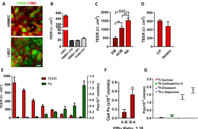
he i-BECs were evaluated in a two-compartment in vitro Transwell BBB transport assay. Following 10 days in EM culture, the i-BECs were dissociated and seeded at a density of 5.0 × 10⁵ cells/cm2 onto gelatin coated 0.9 cm2 Transwell polyethylene terephthalate (PET) perme-able inserts (1 µm pore size). he establishment of a conluent i-BEC monolayer on the inserts was monitored by a live staining technique using CFDA (a cellular viability dye) and CellMask Orange (CMO), a membrane stain that labels the plasma membrane (Fig. 5A and Supplementary Fig. 7). As shown in Fig. 5A, CFDA/CMO staining showed that the i-BECs formed continuous membrane contacts in conluent cultures, in contrast to the intercellular gaps observed in HBMECs. Transendothelial electrical resistance (TEER), a measurement indica-tive of barrier impermeability or “tightness” to paracellular difusion, showed average i-BEC values of 500 Ωcm2 (ranging between 300–800 Ωcm2 in maintenance culture) 48 hr ater seeding onto the inserts (Fig. 5B,E). In com-parison, TEER measurements of two commercially available primary HBMECs (HBMEC (from Cell Systems (CS) and AngioProteomie (AP)) and an immortalized HBMEC cell line (hCMEC/D3) ranged from 20–30 Ωcm2 (Fig. 5B). TEER measurements following culture of i-BECs in human astrocyte conditioned medium (ACM) increased over 2-fold (1090 ± 112 Ωcm2) compared to EM maintenance conditions (503 ± 94 Ωcm2) (Fig. 5C) and culture in basal ACM medium containing 1% FBS (779 ± 94 Ωcm2), indicating a speciic ‘tightening’ response to astrocytic cues. Retinoic acid (RA) treatment, which has been shown to enhance BBB properties and TEER in other iPSC-derived BECs25,42, also signiicantly increased TEER values in the i-BECs (1528.0 ± 132 Ωcm2; Fig. 5C). Given the homogeneity of the diferentiation protocol, the i-BECs were seeded directly onto gelatin coated inserts without any requirement to stimulate further maturation and puriication by sub-culturing onto collagen/ibronectin coated inserts, as described for other human iPSC-derived BEC protocols26. No signiicantchanges in TEER were observed following seeding of i-BECs onto Collagen IV/Fibronectin coated inserts, further supporting the purity of the i-BEC culture (Fig. 5D).
i-BEC paracellular permeability (Pe) for Sucrose, a benchmark of passive transport in in vitro models, inversely correlated with TEER values (Fig. 5E) and was similar (Pe = 2.8 × 10⁻⁵ cm/min) to that observed in other human iPSC-derived models (Pe = 3.4 × 10⁻⁵ cm/min)26 and substantially lower than that in the hCMEC/D3 line (Pe = 1.65 × 10⁻³ cm/min)18 and other animal BBB models43,44. Sucrose Pe did not change with increasing TEER beyond a threshold of 400 Ωcm2 (Fig. 5E).
he functional polarization of the transporter activity in maintenance medium was assessed using the model substrate for the elux transporter P-gp, Cyclosporine A (CsA). he Pe values indicated restricted transport of CsA with A-B Pe = 0.15 ± 0.05 × 10⁻³ cm/min and B-A Pe = 0.53 ± 0.13 × 10⁻³ cm/min with B-A/A-B elux ratio of 3.38 (Fig. 5F). Diazepam, a lipophilic small molecule with high BBB penetration (Pe = 1.36 ± 0.16 × 10⁻³ cm/ min) and H- l –Dopamine, a substrate for carrier mediated inlux via the organic cation transporter SLC7A5 (LAT-1) (Pe = 1.78 ± 0.17 × 10⁻³ cm/min) had the highest in vitro permeation (Fig. 5G). This illustrates a 12-fold dynamic range of permeability coeicient among compounds and transporter ligands of various classes (Cyclosporine A/H- l -Dopamine).
www.nature.com/scientificreports/
Receptor mediated transcytosis in i-BECs.
he focus of this study was the functional characteriza-tion of receptor mediated transcytosis (RMT) in the i-BECs, particularly for antibodies raised against speciic RMT receptors. RMT uses the vesicular traicking machinery of BECs to deliver a range of macromolecules to the brain initiated by ligand binding, ligand/receptor internalization, endosomal sorting and subsequent ligand release on the abluminal endothelial surface and receptor recycling back to the luminal membrane45–48. Hence, weexplored the utility of the i-BEC BBB model for evaluating BBB-crossing antibodies that can be used as molecular Trojan horses for otherwise non-permeable therapeutics.
he i-BECs express described RMT-mediating receptors, including Low-Density Lipoprotein Receptor (LDLR), Transferrin Receptor (TfR), Insulin (INSR) and Insulin-like Growth Factor Receptors (IGFIR and IGFRIIR), Low-Density Lipoprotein Receptor-related Protein 1 (LRP1), Receptor for Advanced Glycation End Products (AGER) and Leptin Receptor (LEPR) as well as the receptor for BBB-crossing antibody FC5 (TMEM30A, iso-form 1 and 2) (Fig. 6A,B). he endocytosis triggered by ligand binding to RMT receptors was evaluated using uptake of acetylated low-density lipoprotein (Ac-LDL; ligand to LDLR), as well as antibodies FC5-Fc (binding to
Figure 4. BBB and endothelial speciic gene expression in i-BECs. (A) A comparative gene microarray analysis comparing the individual gene intensity signals (arbitrary units) of 434- BBB and endothelium-speciic genes between HBMEC (y-axis) and i-BECs (x-axis). Scatter plot demonstrates a similar expression proile between both cell types, speciically in ABC transporters (green squares), tight junctions (purple triangles) and SLC transporters (red diamonds) transcripts (Pearson Correlation, r2 = 0.86, p = 0.001). (B) Table compiling a list of diferentially expressed transcripts in i-BECs when compared to HBMECs. Statistical signiicant was determined using Student’s T-Test, (*p ≤ 0.05) and a 1.35 fold change in ratio was used to generate the gene list. (C) Representative immunoluorescence staining of i-BECs for vWF, Claudin-5, CD31 (PECAM-1), Occludin, ZO-1, GLUT1 and P-gp. HBMECs were used as a positive control. All characterizations were carried out at day 21 of EM diferentiation. Nuclei counterstained with Hoechst. Scale bar, 20 µm.
TMEM30A) and IGF1R5-Fc (binding to IGF1R). A strong cellular uptake of all three ligands was clearly evident in the i-BECs (Fig. 6C), indicative of functional receptor-mediated endocytosis.
Directional transendothelial transcytosis using a panel of rat-human species cross-reactive camelid single-domain antibodies, (VHHs; 15 kDa) or their fusions with Fc (80 kDa), was compared in the i-BEC and rat immortalized brain endothelial cells (SV-ARBEC49) BBB models. he antibodies included a negative control
anti-body raised against C. diicile toxin A (A20.1; 16 kDa, no mammalian target), FC5, selected by biopanning for its ability to transmigrate the BBB and IGF1R5, raised against an ectodomain of the human IGF1R. An antibody (IgG) speciic for rat TfR (Ox26), was also used as a control. he rate of transcytosis of the antibodies (measured by the apparent permeability coeicient, PAPP) was quantiied using highly sensitive multiplexed nanoLC-SRM, as previously described50–52. he rat-human cross-reactive antibodies, FC5 and IGF1R5, in V
HH and Fc-fusion formats, showed similar PAPP in both human and rat BBB models, reproducing increased transcytosis rates in bi-valent FC5-Fc format, as previously described50 (Fig. 6D). In contrast, rat TfR-speciic Ox26 antibody,
demon-strated facilitated transcytosis in the rat SV-ARBEC BBB model, and no crossing in the human i-BEC BBB model (Fig. 6D). he PAPP of the control, non-binding A20.1 antibody, was 2-fold lower in the i-BEC than the SV-ARBEC model, which correlates with the higher TEER values for i-BEC over that observed in SV-ARBEC model (50– 70 Ωcm2)49 (Fig. 6D).
Figure 5. Functional characterization of the i-BECs. (A) Live staining of i-BEC and HBMEC cultures with CFDA (cellular viability) and CellMask Orange (plasma membrane) to visually assess the maintenance of continuous tight junctions. Intracellular gaps, indicative of poor BBB integrity, are easily visualized as seen in HBMEC cultures (asterisk, let panel) compared to the tight barrier integrity observed in i-BEC cultures (right panel). (B) Transendothelial electrical resistance (TEER, Ωcm2) of conluent i-BECs monolayer cultures on gelatin coated Transwell inserts. Higher TEER was observed in the i-BECs compared to HBMECs (CS and AP) as well as immortalized hCMEC/D3 (mean ± SD). (C) Enhanced TEER in i-BECs following 24 hr co-culture in astrocyte conditioned medium (ACM) and treatment with 10 µM all-trans retinoic acid (RA) compared to EM maintenance culture (mean ± SD, One-Way ANOVA **p < 0.01, ****p < 0.0001). (D) Comparison of TEER between i-BECS seeded onto gelatin and collagen/ibronectin (C/F) coated Transwell inserts (mean ± SD). (E) Comparison between TEER values (let y-axis) and 14C-Sucrose permeability coeicient (P
e, right y-axis) in i-BECs. Values are mean ± SD for each compound measured in 11 independent inserts from at least 8 biological replicates with varying TEER levels acquired during protocol optimization. he means relect variability from diferentiation to diferentiation during protocol optimization steps. (F) Permeability values of Cyclosporine A (CsA) from apical to basolateral (A-B) and basolateral to apical (B-A) compartments. Elux ratio (B-A/A-B) for CsA shown as 3.38. (mean ± SD, Student’s T-test **p < 0.01). (G) Functional i-BEC transporter activity was assessed by measuring apical to basolateral permeability (Pe) using radiolabeled small molecules: 14C-Sucrose, 3H-Cyclosporine A, 3H-Diazepam and 3H- l-Dopamine. Values are mean ± SD for each compound measured in at least 3 inserts. Results are representative of at least two independent diferentiations. Nuclei counterstained with Hoechst. Scale bar, 20 µm.
www.nature.com/scientificreports/
he PAPP values of the IGF1R5 and FC5, as single-domain antibodies and as fusions with human Fc domain observed in the i-BEC model, correlated with the apparent CSF exposure [EXPAPP] derived from the simultaneous pharmacokinetic measurements of the antibodies in rat CSF and serum [CSF AUC/Serum AUC]. he results show a signiicant correlation (Pearson correlation, r2 = 0.96, p = 0.04) between the in vitro P
APP and EXPAPP[CSF] for these antibodies (Fig. 6E), suggesting that the i-BEC model could be used for ranking/predicting in vivo rodent exposure of species cross-reactive BBB antibody carriers and potentially de-risking those that are uniquely speciic for human antigens.
Discussion
We have generated iPSCs, from ethically sourced human fetal AF cells, that are transgene-free and derived/main-tained in a feeder-free and chemically deined culture system. hese AF-iPSCs demonstrate high plasticity and represent a renewable, scalable and reproducible cell source for the derivation of i-BECs as well as other brain cells including neurons and astrocytes. Diferentiated i-BECs exhibit a molecular and functional phenotype of HBMECs in vivo such as high and inducible TEER, polarization of transporter activity and species-speciic properties suitable for use in ‘translational’ screening of early CNS targeting pipelines, especially those targeting receptor-mediated transcytosis.
In an improvement over existing human stem cell/iPSC BEC diferentiation protocols22–24,26, i-BECs were
derived from AF-iPSCs using an eicient and simpliied monolayer diferentiation protocol without the require-ment for cell surface marker enrichrequire-ment or co-diferentiation and puriication steps to yield a pure population of BECs. Modulating the osmolality of the pre-diferentiation medium, within a range of 270–290 mOsm/kg, increased the homogeneity of the cultures resulting in a higher eiciency of BEC diferentiation. Previously pub-lished protocols, which employ unconditioned medium (UM) composed of DMEM/Ham’s F12 medium, with an osmolality range of 290–300 mOsm/kg, induced simultaneous co-diferentiation of neural and endothelial line-ages26. his osmolality range for iPSC cultures has been shown to induce mesodermal as well as ectodermal germ
Figure 6. Functional receptor mediated transcytosis in i-BECs. (A) RT-PCR detection of receptor mediated transport (RMT) receptor transcripts including LDLR, TfR, INSR, IGF1R, IGFIIR, LRP1, AGER, LEPR and TMEM30A, isoform 1and 2 expressed in i-BECs compared to HBMECs. Cropped gels shown. (B) Immunoluorescence staining of i-BECs conirming expression of IGF1R, LRP1 and TfR. Scale bar, 20 µm. (C) Receptor mediated endocytosis of acetylated low-density lipoprotein (Ac-LDL), FC5-Fc and IGF1R5-Fc. Plasma membrane stained with Wheat-germ agglutinin (WGA). Scale bar, 20 µm (let panel) and 10 µm (right panel). Nuclei counterstained with Hoechst. (D) he in vitro apparent permeability coeicient (PAPP) was assessed for FC5-IgG (FC5), FC5-Fc, IGF1R5 and Ox26 as well as negative control A20.1 antibodies in human i-BEC and rat SV-ARBEC BBB models. (E) Correlation of PAPP values obtained in the i-BEC BBB model with the apparent CNS exposure (EXPAPP) for FC5-IgG, FC5-Fc, IGF1R5-Fc and control (A20.1-Fc, IgG) antibodies. EXPAPP is derived from simultaneous pharmacokinetic measurements of antibodies in CSF (serial sampling) and in the serum (AUCCSF/AUCserum) in the rat. he results show a signiicant correlation (Pearson correlation, r2 = 0.96, p = 0.04) among in vitro P
APP and in vivo EXPAPP (CNS) for this set of antibodies. Data are mean ± SD of repeated measurements from 3 to 10 in vitro BBB model assemblies and from 3 to 6 animals in each group.
layer progenitors from adherent cultures36. In our protocol, decreasing the osmolality of the pre-diferentiation
culture biased the cultures to preferentially diferentiate towards BEC. he cells began to acquire homogenous endothelial-like cobblestone morphology with the majority of cells expressing low levels of BBB transporter GLUT1 as early as 2–3 days in KOEB. During the diferentiation time course, the MFI of GLUT1 expression increased during the maturation in EM. By comparison, the majority of cells at 3–4 days in UM were neural-like with endothelial-like cells and GLUT1 expression increasing by 6 days in UM26. Given the homogeneity of the
diferentiation in KOEB, no additional sub-culture onto ibronectin/collagen matrix was required to achieve a pure, conluent monolayer of i-BECs. TEER values, averaging 500 Ωcm2, were achieved in i-BEC monolayer maintenance cultures in the absence of conditioned medium or hydrocortisone/RA treatments used to increase TEER in other human BBB models42,53. his high baseline TEER values translate into a low sucrose permeability
(2.8 × 10⁻⁵ cm/min) similar to that observed in other human iPSC-derived BBB models (3.4 × 10⁻⁵ cm/min)26
and much lower than that observed in hCMEC/D3 (1.65 × 10⁻³ cm/min)18. TEER values increased substantially
in the presence of human astrocyte conditioned medium (up to 1000 Ωcm2) and RA (up to 1500 Ωcm2). Recent reports have demonstrated a synergistic efect of RA and NVU co-culture models of pericytes, astrocytes and neurons resulting in increased TEER values ranging from 2500–5000 Ωcm220,22,42,54. In fact, human iPSC-derived
EC co-cultured in the presence of syngeneic pericytes, astrocytes and neurons generated culturally induced-BECs (ciBEC) with BBB speciic properties22. Although we have shown that TEER is inducible in the i-BEC model, the
potential synergies among co-culture with cells from the NVU and chemical (RA) induction remains to be fully examined.
Conirming the idelity of diferentiation, a comparative gene microarray analysis between i-BEC and pri-mary HBMECs (using a 434-gene array containing BBB- and endothelium-speciic genes) showed a statisti-cally signiicant overlap between the two cell types with 93% of the transcripts being similarly expressed. he expression of selected BBB transporters from SLC and ABC families has been conirmed in i-BECs by PCR and functional studies including a polarized transport of the P-gp substrate Cyclosporine A. Transcripts upregulated in i-BECs (compared to primary HBMECs) include several SLC transporters (SLC2A14, SLC38A2, SLC37A1, SLC2A1 and SLC17A3), ABC transporter (ABCC2) as well as transcripts of secreted IGF1-binding proteins (IGFBP5 and IGFBP3). Observed down-regulation of cytoskeletal, ECM and adherens junction genes could be attributed to the diferences in culture protocols and ECM coating used to culture the i-BEC (Matrigel/gelatin) versus the HBMECs (attachment factor). he most down-regulated transcript in i-BECs was Caveolin-1 (Cav-1), the principal component of caveolae, endocytic vesicles that provide a route for Cav-1 dependent endocyto-sis and potentially transcytoendocyto-sis. Caveolae are downregulated in mature BECs6. Evidence suggests that BBB
dys-function, following injury or disease, is accompanied by increased levels of Cav-1 expression and increase BBB permeability55,56. For example, BECs derived from patients with Huntington’s Disease expressed higher levels of
Cav-1 compared to BECs from healthy donors, suggesting an impaired transcytotic barrier57. Similarly, increased
bulk-transcytosis of circulatory albumin across the BBB has been observed in mfsda2 knock-out animals which exhibit up-regulation of caveolae in BECs58. Since the down-regulation (6-fold) of Cav-1 in i-BEC compared
to HBMECs was accompanied by >25-fold increase in TEER, lowering of Cav-1 may be a good biomarker of barrier maturation in cultured BECs. Although we did not observe diferences in Claudin-5 gene expression levels between i-BEC and HBMECs, the organization of intercellular contacts observed by immunostaining for Claudin-5 showed uniform and continuous staining in i-BEC cultures and punctate contacts with intercellular gaps in the HBMEC cultures. hese observations are consistent with published reports showing a strong correla-tion between intercellular Claudin-5 junccorrela-tional continuity and barrier integrity, which corresponds with the low TEER and paracellular tightness of primary and immortalized HBMECs20,42.
Recent studies using LC/MS/MS-based proteomic analyses on BBB tissues showed signiicant species difer-ences in the expression of BBB transporters59–62, necessitating human in vitro BBB models for accurate
bioavail-ability predictions of various synthetic compounds. In addition, the human BBB and neuropil cellular platforms are of particular importance for evaluating an emerging pipeline of antibodies against CNS targets, since they are usually raised against species-speciic epitopes. To enable access of therapeutic antibodies to their targets in the CNS, they can be re-engineered into bi-speciic antibodies, in which the second ‘arm’ functions as the BBB deliv-ery carrier that initiates a receptor-mediated transcytosis (RMT) of the antibody across the BBB. RMT-targeting antibodies such as TfR and Insulin receptor antibodies are species-selective, necessitating the use of ‘surrogate’ molecules in pre-clinical studies. herefore, human BBB models are an important tool for evaluating BBB carriers and their conjugates with therapeutic payloads that are selective for human receptors. Furthermore, the spe-ciic RMT receptor abundance, which can impact the capacity of a delivery system, can be signiicantly diferent among species. For example, TfR is 3-fold more abundant in rat compared to human brain vessels62–64,
under-scoring the importance of human models that replicate in vivo species diferences. Lastly, the human BBB model is of immense value for human speciic antibodies which would otherwise give ‘false-negative’ results in the rat model alone.
To explore the utility of the i-BEC model in the evaluation of RMT, we compared a panel of BBB-crossing anti-bodies, including FC5 (which binds TMEM30A), IGF1R5 (which binds IGF1R) and Ox26 (which binds TfR) in rat SV-ARBEC and human i-BEC models. FC5 and IGF1R5 have been selected for rodent-human cross-reactivity while Ox26 was selective for rat TfR. Whereas both FC5 and IGF1R5 (as either single-domain antibodies or in fusion with Fc domain) showed similar PAPP values in both models, OX26 transcytosis was only observed in the rat BBB model. In both models, Fc fusion of FC5 had higher PAPP values compared to monomeric FC5, despite being a substantially larger molecule (i.e., 75 kDa vs 15 kDa, respectively), as previously described50. As shown
in this example, coupling evaluation of RMT-targeting antibodies in rodent and human BBB models can be an important strategy for identifying species cross-reactive and similarly eicient antibody variants that could be used in pre-clinical in vivo brain distribution and pharmacokinetic studies. Using the set of species cross-reactive
www.nature.com/scientificreports/
BBB-crossing re-engineered antibodies50,65 we conirmed a signiicant correlation between their transport (P
APP values) in the i-BEC model and their in vivo CSF exposure measured from serial pharmacokinetic studies.
Herein, we described the development and characterization of a human BBB model in vitro (i-BEC) diferenti-ated from iPSCs derived from amniotic luid cells using a novel, simpliied and eicient method. Collectively, the data shown that i-BECs are a valuable tool for investigating drug classes that target human-enriched or speciic BEC transporters, receptors and signaling pathways. he described iBEC diferentiation strategy can be extended towards generating patient and disease speciic BBB models to examine BBB dysfunction with implications for therapeutics and drug delivery66,67. Although in vitro BBB culture models are a ‘reductionist’ approach to
eval-uating the ability of compounds to cross the BBB barrier and cannot truly recapitulate the complex anatomical and functional relationships of the NVU or in vivo pharmacokinetics, the emergence of human iPSC-derived BBB models represent an important advancement in the pre-clinical evaluation of CNS therapeutics towards improving clinical translation.
Methods
Generation of amniotic luid-derived induced pluripotent stem cells.
Amniotic luid (AF) cells at 26 weeks of gestation (AFC) were obtained from he Ottawa Hospital (Ottawa, Ontario, Canada) following routine amniocentesis. Informed consent was obtained from each woman to use the amniotic luid for research purposes and all the methods were carried out in accordance with relevant guidelines and regulations as approved by the Ottawa Hospital Research Ethics Board. All experimental protocols using AF cells were performed fol-lowing the guidelines established and approved by the National Research Council Canada Research Ethics Board. he AF cells were cultured in Dulbecco’s Modiied Eagle Medium (DMEM; Life Technologies) supple-mented with 20% inactivated fetal bovine serum (FBS; Wisent). To induce reprogramming, 0.5 × 10⁶ AFCs were nucleofected with 1.3 µg of pEP4 E02S EN2K (Addgene-20925), pCEP4 M2L (Addgene-20926) and pEP4 E02S ET2K (Addgene-20927) episomal vectors using the Nucleofector I Device (Amaxa) and plated into a 6-well plate coated with Matrigel hESC qualiied matrix (Corning), prepared as per manufacturer’s instructions, containing pre-warmed DMEM + 20% FBS supplemented with 10 µM Y27632 (ROCK Inhibitor, dissolved in DMSO as a 10 mM stock as per manufacturer’s instructions, Stem Cell Technologies). Two days later, the media was switched to mTeSR1 (Stem Cell Technologies) and replaced daily. Once the iPSC colonies were established, the cells were passaged at 80% conluency at a 1:12 ratio approximately every 5 days using ReLeSR (Stem Cell Technologies) onto Matrigel coated plates. he pluripotency and reprogramming eiciency of the established iPSC colonies was conirmed using Alkaline Phosphatase staining (AP, Invitrogen) and StainAlive Dylight 488 Mouse anti-human TRA1-81 Antibody (Stemgent), as per manufacturer’s instructions. Karyotype-G-banding on established iPSCs was carried out at WiCell Research Institute (WiCell Cytogenetics Lab, Wisconsin). All experiments were carried out between passage number 10–40.Diferentiation of iPSCs into neural progenitor cells.
To generate iPSC derived induced neural pro-genitor cells (i-NPs), iPSCs were dissociated using ReLeSR (Stem Cell Technologies) and passaged at a 1:6 ratio onto Matrigel coated plates in Stemdif Neural Induction Medium (Stem Cell Technologies) and changed daily, as per manufacturers protocols. he cells were dissociated using Accutase (Stem Cell Technologies) according to manufactures instructions. Ater 3 weeks in culture, the i-NPs were replated and maintained in Stemdif Neural Progenitor medium (Stem Cell Technologies) replenished daily.Neuronal differentiation of iPSC.
To induce neuronal differentiation, i-NPs were dissociated using Accutase (Stem Cell Technologies), resuspended in Neural Progenitor Maintenance BulletKit Medium (NPMM; Lonza) and plated on uncoated 10 cm petri dishes (Fisher Scientiic) for neurosphere formation or on Matrigel (Corning) coated plates for adherent cultures. he neurospheres were passaged by being dissociated into small aggregates using Accutase and re-plating onto Matrigel or 10 µg/ml coated poly- l-lysine (PLL, Sigma) coated plates in DMEM/F12 (Life Technologies) supplemented with B27 (Life Technologies) to induce neuronal dif-ferentiation. he medium was replaced every other day and mature neurons were observed following 4 weeks of diferentiation in B27 containing medium.Astrocyte diferentiation of iPSC.
To induce astrocyte diferentiation, i-NPs maintained in StemDif Neural Progenitor medium (Stem Cell Technologies) were dissociated using Accutase (Stem Cell Technologies) and plated at a 1:6 ratio in poly-l-ornithine PLO (20 µg/ml, Sigma) and laminin (10 µg/ml, Sigma) coated plates in Neural Progenitor medium for one day. he following day, the medium was changed to astrocyte induc-tion medium composed of DMEM/F12 with Glutamax (Stem Cell Technologies), 5% FBS, 20 ng/ml EGF (Life Technologies) which was replaced every other day for four days. At day 5, the medium was changed to complete astrocyte induction medium supplemented with 10 ng/ml CNTF (Sigma) and the medium was changed every other day. From day 8 to 14, the cells were cultured in DMEM/F12 supplemented with Glutamax, 1% FBS and 10 ng/ml CNTF and the medium was replaced every other day.Endothelial cell diferentiation of iPSCs.
Endothelial cell diferentiation protocol was adapted from the two-step diferentiation protocol by Lippmann et al.26 In the initial pre-diferentiation step, once the iPSCsreached 60–70% conluency the medium was switched from mTeSR1 to low osmolality KOEB medium composed of KnockOut DMEM/F12 medium (~276 mOsm/kg, Gibco) supplemented with 20% KnockOut serum replace-ment, 1 × Glutamax, 1 × non-essential amino acids and 55 µM β-mercaptoethanol (all from Life Technologies) for 5–7 days. During this time frame, major morphological changes were observed as the cells became bigger and began to assume a cobblestone-like morphology. Once the cells formed a uniform monolayer of endothelial-like cells, the medium was switched to endothelial diferentiation medium (EM) composed of human serum free endothelial medium (Life Technologies) supplemented with 1% platelet-poor plasma derived serum (PDS,
Alfa-Aesar) and 20 ng/ml bFGF for 9–10 days. While in EM culture, the cells acquired a typical cobblestone morphology characteristic of diferentiated endothelial cells (Supplementary Fig. 4). Ater 9–10 days in EM, the cells were dissociated with Accutase (Stem Cell Technologies) at 37 °C for 10–15 min. Once dissociated, the cells were iltered through a 40 µm sieve to eliminate residual basement membrane and endothelial cell clusters and re-suspended in EM containing 10 µM ROCK Inhibitor. Singularized i-BECs were plated onto 0.5% gelatin coated Transwell inserts as described under the “Preparation of transwell inserts and TEER measurements” section. All-trans 10 µM RA (reconstituted in DMSO at a stock concentration of 10 mM, Sigma) was added following seed-ing of i-BECs onto inserts where indicated. A mixture of collagen IV (80 µg/ml, Sigma) and ibronectin (20 µg/ ml, Sigma) coated on Transwell inserts was used where indicated. All i-BEC characterizations were performed at the end of the diferentiation process (21 days from iPSCs) following passage onto gelatin coated inserts and coverslips, where appropriate.
Cell culture.
Human primary brain microvascular endothelial cells (HBMECs; Cell Systems) were main-tained in CSC medium (Cell Systems) on attachment factor (Cell Systems) pre-coated tissue culture plates and passaged 1:4 using Trypsin-EDTA (Invitrogen). Human primary HBMECs (AngioProteomie) were main-tained in EGM-2MV Bulletkit medium (Lonza) on 0.5% gelatin coated tissue culture plates and passaged at a 1:4 split ratio using Trypsin-EDTA. All HBMEC studies were carried out within 3–10 passages. Unless other-wise speciied (Fig. 5C), HBMEC from Cell Systems were used as comparative positive controls in the experi-ments. Immortalized human brain capillary endothelial cells (hCMEC/D3; generous git from Dr. Pierre-Olivier Couraud, Institute Cochin, Paris, France), were maintained in EBM-2 basal medium (Lonza) supplemented with 2% heat-inactivated FBS (Hyclone) on rat tail collagen I (VWR) coated tissue culture plates18. he cells were splitusing Reagent Pack (Lonza) at a 1:3 ratio and cell passages 20–30 were used for these studies. Immortalized adult rat brain microvascular endothelial cells, SV-ARBEC, were established by SV-40 transfection of primary rat brain micorovascular endothelial cells, isolated from 24–30 days old Sprague–Dawley rats, as previously described49.
Cells were grown in M199 based feeding media containing: 0.25% Peptone, 0.9% d-glucose, BME Amino Acids, BME Vitamins and 10% FBS. Cells were routinely split 1:20 every week onto rat-tail collagen type I (VWR, 60 µg/ ml) coated plates. SV-ARBEC cell passages 79–86 were used for these studies. NT2/D1 (NT2) embryonal carci-noma (EC) cells (ATCC) were maintained in HG-DMEM (Invitrogen) and 10%FBS (Hyclone) replenished every 2 days and passaged at a 1:3 split ratio using Trypsin-EDTA, as previously described68. Human neural progenitor
cells (hNP, Aruna Biomedical) were cultured on Matrigel (Corning) coated plates maintained in complete neural progenitor medium as supplied in the hNP1 neural progenitor expansion kit (Aruna) and were used between passage 6–9. All primary cells were cultured according to the supplier’s recommendations and within speciic passages number limits.
Astrocyte conditioned media.
Human primary astrocytes (NHA, Lonza) were grown to 80% conluency in Astrocyte growth medium (AGM, Lonza). he cells were washed 2 × with HBSS and incubated in 10 ml AGM supplemented with 1% FBS per T75 lask for 72 hr. he astrocyte conditioned medium (ACM) was collected, pooled, ilter sterilized and aliquoted for storage at -20 °C. In the transport studies, 1 ml of ACM was added to 1 ml of EM in the bottom compartment of the companion plates 48 hr before measuring TEER. For control exper-iments, 1 ml of AGM medium supplemented with 1% FBS (ACM basal medium) was added to 1 ml of EM in the bottom compartment in parallel.Immunocytochemistry.
Cells were grown in 12 well plates on 15 mm round coverslips coated either with Matrigel (iPSC, i-NP, i-BECs) or PLO/laminin (Neurons and Astrocytes) in the respective growth media. For most antigens, the cells were ixed using Genoix (DNA Genotek) whereas 4% PFA (Sigma) was used for certain nuclear antigens, namely PAX6 and SOX17. In the latter case, the cells were permeabilized with 0.2% Triton X-100 (Sigma) in PBS (without Ca2+/Mg2+) for 20 min, washed and blocked using DAKO Protein Block Serum Free (Agilent) for 20 min at room temperature. Primary antibodies were prepared using the DAKO Antibody Diluent (Agilent), according to the dilutions described in Supplementary Table 1 and coverslips were incubated for 1hr at room temperature in a humidiied chamber. Coverslips were then washed three times for a minimum of 5 min with PBS (without Ca2+/Mg2+) and incubated with secondary antibodies diluted 1:500 in antibody diluent at room temperature for 1hr in the dark. Secondary only PBS controls were performed in parallel for all staining experiments. he coverslips were then washed three times for 5 min with PBS and mounted using DAKO luores-cent mounting medium (Agilent) spiked with 5 µg/ml of Hoechst 33258 (Sigma) to counterstain nuclei. Images were captured using the Axiovert 200 M Microscope (Zeiss). Cells on coverslips were imaged using a 20 × /0.5 Plan Neoluar objective and live cells were imaged using 20 × /0.4 LD Achroplan Korr(DICII) objective. All sec-ondary only control immunoluorescence images are shown in Supplementary Fig. 8.RNA extraction and RT-PCR.
Total RNA was extracted from cells, using TriReagent RT (Molecular Research Centre), as per manufacturer’s instructions. The RNA was treated with DNase (Turbo DNA-Free Kit, Ambion) to remove any residual DNA contamination and the RNA concentration was quantiied using a NanoDrop ND-100 (hermo Scientiic). cDNA was synthesized using 5–10 µg of RNA using Superscript II Reverse Transcriptase (Life Technologies) and AncT primers (Life Technologies) and puriied using the QiaQuick PCR puriication kit (Qiagen). he Oligreen Assay (Molecular Probes) was used to measure the concentration of cDNA samples. For each RT-PCR reaction, 10 ng of cDNA, 10 pmol/µl primer sets speciic for the genes of interest (Supplementary Table 2) and iQ Supermix (BioRad) were used and the PCR reaction was carried out in a PTC-200 DNA Engine hermal Cycler (MJ Research) by using the following parameters: Initial denaturation step at 94 °C for 3 min, followed by 30 cycles of a denaturation step at 94 °C for 20 sec, annealing at 60 °C for 20 secwww.nature.com/scientificreports/
and extension at 72 °C for 20 sec, and a inal extension step at 72 °C for 3 min. RT-PCR reactions were run on 2% agarose gel and images detected using a Fluochem 8900 imager (Alpha Innotech).
Protein extraction and Western blot analysis.
Cells were washed with PBS (without Ca2+/Mg2+) and lysed directly in the tissue culture plate on ice or from cell pellets, in ice-cold lysis bufer (25 mM Tris-HCL, pH 7.6, 150 mM NaCl, 1% Triton-X, 1% Na.Deoxycholate) containing protease inhibitor cocktail (Roche Diagnostics). Cell lysates were incubated on ice for 30 min and clariied by centrifugation at 21,000 × g at 4 °C for 15 min. Total protein concentrations were determined, using the Bio-Rad Protein Assay (Bio-Rad). he super-natant aliquots containing equal amounts of total protein (40 µg) were diluted 1:2 with Laemmli sample bufer (Bio-Rad) and run on a 10–12% SDS-PAGE at 100 V for 1.5 hr and transferred to a nitrocellulose membrane (Amersham), using a wet transfer apparatus (Bio-Rad) at 80 V for 1 hr at room temperature. Membranes were washed once with TBST (TBS containing 0.05% Tween-20, Sigma) and blocked with 5% skim milk in TBST for 2 hr at room temperature. he membrane was incubated with primary antibodies (see the Supplementary Table 1 for details) overnight at 4 °C with gentle rocking. β-ACTIN (ACTB) (Bio-Rad, 1:5000) was used as a loading con-trol. Membranes were washed three times for 10 min per wash in TBST. All secondary antibodies, anti-rabbit and anti-mouse IgG-HRP conjugates (Bio-Rad, 1:5000), were diluted in 5% skim milk and the membranes were incu-bated for 1 hr at room temperature. Membranes were washed three times for 10 min per wash in TBST and con-sequently detected by Clarity chemiluminescence reagent (BioRad). he ECL signal was analyzed by FluorChem 8900 (Alpha Innotech). Rainbow markers (Amersham) were used to determine the protein sizes.Flow Cytometry.
i-BECs were harvested using Accutase (Invitrogen) for 6 min and ixed using 3% para-formaldehyde (PFA) for 20 min. Cells were blocked with 3% BSA in PBS for 30 min at room temperature and incubated with Human Glut1 Alexa Fluor 700-conjugated Antibody (R&D Systems) at a dilution of 5 µl/1.0 × 106 cells in 800 µl of 1% BSA in PBS and 2 mM EDTA for 1 hr. Cells were washed three times with 1% BSA in PBS and re-suspended in 500 µl 1% BSA in PBS and analyzed on a BD LSRFortessa (BD Biosciences) low cytometer. Unstained cells were used as a control for autoluorescence and FSC and SSC gating was used to locate cells, exclude doublets and debris and dead cells. Flow cytometry data were analyzed using the FACS Diva sotware (Becton, Dickinson and Company). Mean luorescence intensity (MFI) was acquired to measure the shit in luo-rescence intensity of GLUT1 during the diferentiation process.Preparation of transwell inserts and TEER measurements.
Transwell inserts (0.9 cm2 cell growth area with 1 µm pore size, BD-Falcon, Cat. 353103) with polyethylene terephthalate (PET) membranes were coated with 0.5% gelatin, placed into 12-well companion plates and incubated overnight at 37 °C. he gelatin solution (Sigma) was removed the following day and 5 × 10⁵ i-BECs (at day 10 in EM) were seeded in each tran-swell insert in 1 ml complete endothelial medium (EM) composed of human endothelial-serum free medium (Life Technologies), supplemented with 1% Platelet-poor plasma derived serum (Alfa-Aesar), 20 ng/ml bFGF (Life Technologies)) and 10 µM Y27362 (ROCK Inhibitor). he inserts were incubated overnight at 37 °C in 5% CO2 and the following day, the complete EM medium (without ROCK Inhibitor) was replaced only in the apical cell layer of the transwell insert. For the transport studies, the apical volume is 1 ml and the basolateral volume is 2 ml. Transendothelial electrical resistance (TEER) measurements were carried 16–24 hr following media change using a CellZscope apparatus (Nanoanalytics) with 1 cm diameter electrodes and standard spectrum settings: frequency 1 Hz-100 kHz, points per decade 9 and logarithmic spacing. TEER values for the empty inserts were subtracted from the TEER values for the inserts with cells to obtain inal TEER values in Ωcm2. Following TEER measurements, cellular viability and BBB integrity were assessed as quality control measures using BBBView, a method where cells on inserts were live stained in Live Cell Imaging Bufer (Invitrogen) containing 2.5 µg/ ml of CFDA for 30 min at 37 °C to assess cellular viability. he cells were then placed on ice for 5 min and 5 µg/ ml of Cell Mask Orange dye (Invitrogen) was added for 5 min to label the plasma membrane. he cells were washed twice with cold PBS (without Ca2+/Mg2+) and imaged using a 20 × HMC objective on an Axiovert 200 M Microscope (Zeiss).SV-ARBEC BBB model.
Immortalized adult rat brain microvascular endothelial cells, SV-ARBECs, were established by SV-40 transfection of primary rat brain microvascular endothelial cells, isolated from 24–30 day old Sprague-Dawley rats, as previously described49. he cells were grown in M199 based medium (Wisent) androutinely passaged at a split ratio of 1:20 every week. For transport studies, the SV-ARBEC cells were seeded at 80,000 cells/membrane on rat-tail collagen I (VWR)-coated 0.9 cm2 Falcon cell inserts, 1 µm pore size (same as for the iBEC cells), in 1 ml SV-ARBEC feeding medium without phenol red (Invitrogen). he SV-ARBEC BBB model characterization and transport experiments were performed as previously described49,51.
Permeability transport assay.
A 2 × input solution of 1 µCi/ml (0.575 µM) of 14C -Sucrose (Perkin Elmer), and 3 µCi/ml (0.389 µM) of 3H-Cyclosporine A (Perkin Elmer), 3 µCi/ml (3.75 µM) of 3H -Diazepam (ARC) and 3 µCi/ml (5.00 µM) 3H- l -H-Dopamine (ARC) was prepared in transport bufer (5 mM MgCl2 and 10 mM HEPES in HBSS, pH 7.4) and warmed to 37 °C. All radiolabeled compounds were dissolved in ethanol as per manufacturer’s instructions and three blank inserts were used in each experiment. he 12-well transwell inserts (BD-Falcon) containing a conluent monolayer of i-BECs were dipped sequentially for three consecutive washes 5–10 min in wells containing 2 ml pre-warmed HBSS to remove any residual medium. he inserts were then placed into companion plates containing 2 ml of pre-warmed transport bufer, equilibrated to 37 °C in an incubator for 5–10 min and then 500 µl of the media was carefully removed from the top (apical) chamber of each insert and replaced with 500 µl of 2 × input solution. he inserts were incubated at 37 °C with gentle rota-tion using the 311DS Labnet (Labnet Internarota-tional Inc.) incubator containing orbital shaking platform set at 20 rpms for an hour and 100 µl of transport bufer was collected from the bottom (basal) wells at 15, 30, 45 and
60 min intervals for permeability analysis. Following each collection, 100 µl pre-warmed transport bufer were added back to the bottom wells and the plates were returned to the incubator. he sample collection was also carried out in 3 inserts without cells at 3, 7, 10, 15, 20, 30, 45 and 60 min and clearance slopes were calculated from the linear portion of the curves (0–30 min). Samples were collected in 24-well microbeta sample plates (Perkin Elmer). Duplicate 2 × inal concentration input solution was also collected by adding 10 µl of each respective input solution to each well and 90 µl transport bufer. For quantitation, 400 µl high aqueous capacity scintillation luid was added to each sample well, the plate covered with a microbeta plate seal and contents mixed well by gently shaking the plate until solution was homogeneous and clear. he amount of radioactivity per sample was counted in a scintillation counter (Perkin Elmer), using normalized protocol: 14C and 3H chan-nel, 2 min per well, disintegration per minute (dpm). he permeability coeicient (Pe) calculations are described in Supplementary Methods.
Quantiication of antibodies in transport assay, CSF and serum samples using multiple
reac-tion monitoring (MRM).
All pure antibodies and proteins from in vitro transport samples or body lu-ids samples were reduced, alkylated and trypsin digested using a previously described protocol50,51. For isotopicdilution-based quantiication, isotopically heavy versions of the peptides were synthesized from a commercial source (New England Peptide LLC, Gardner, MA) that contained heavy C-terminus K (+8 Da). To develop the SRM assay for proteins, each protein was irst analyzed by nanoLC-MS/MS using data-dependent acquisition to identify all ionizable peptides. For each peptide, 3 to 5 of the most intense fragment ions were chosen. An initial MRM assay was developed to monitor these fragments at attomole amounts of the digest (about 100–300 amol). Fragments that showed reproducible intensity ratios at low amounts (i.e., had Pearson r2 ≥ 0.95 compared to higher amounts) were considered stable and were chosen for the inal MRM assay (see Supplementary Table 3). he apparent permeability coeicient (PAPP) and apparent CNS exposure (EXPAPP) values were calculated, as described previously50,51.