REVIEW ARTICLE
OPEN
Application of a risk-management framework for integration
of stromal tumor-in
filtrating lymphocytes in clinical trials
Jan Hude
ček
1, Leonie Voorwerk
2, Maartje van Seijen
3, Iris Nederlof
2, Michiel de Maaker
3, Jose van den Berg
4,
Koen K. van de Vijver
5, Karolina Sikorska
6, Sylvia Adams
7, Sandra Demaria
8,9, Giuseppe Viale
10,11, Torsten O. Nielsen
12,
Sunil S. Badve
13, Stefan Michiels
14,15, William Fraser Symmans
16, Christos Sotiriou
17, David L. Rimm
18,19, Stephen M. Hewitt
20,
Carsten Denkert
21,22, Sibylle Loibl
23, Sherene Loi
24, John M. S. Bartlett
25,26,27, Giancarlo Pruneri
28,29, Deborah A. Dillon
30,
Maggie C. U. Cheang
31, Andrew Tutt
32, Jacqueline A. Hall
33, Zuzana Kos
34, Roberto Salgado
24,35✉, Marleen Kok
2,36,
Hugo M. Horlings
3,4and International Immuno-Oncology Biomarker Working Group*
Stromal tumor-in
filtrating lymphocytes (sTILs) are a potential predictive biomarker for immunotherapy response in metastatic
triple-negative breast cancer (TNBC). To incorporate sTILs into clinical trials and diagnostics, reliable assessment is essential. In this
review, we propose a new concept, namely the implementation of a risk-management framework that enables the use of sTILs as a
strati
fication factor in clinical trials. We present the design of a biomarker risk-mitigation workflow that can be applied to any
biomarker incorporation in clinical trials. We demonstrate the implementation of this concept using sTILs as an integral biomarker
in a single-center phase II immunotherapy trial for metastatic TNBC (TONIC trial, NCT02499367), using this work
flow to mitigate risks
of suboptimal inclusion of sTILs in this specific trial. In this review, we demonstrate that a web-based scoring platform can mitigate
potential risk factors when including sTILs in clinical trials, and we argue that this framework can be applied for any future
biomarker-driven clinical trial setting.
npj Breast Cancer (2020) 6:15 ; https://doi.org/10.1038/s41523-020-0155-1
INTRODUCTION
Clinical trials in cancer research are increasingly incorporating
biomarkers, for example, as an inclusion criterion or for strati
fica-tion of patients to control for confounding factors. Practical
challenges, such as interobserver variation in the assessment of
biomarkers during the execution of the trial, are often overlooked.
If not handled appropriately, these challenges can limit the
effectiveness and ability to complete the biomarker and drug
development process. According to Hall et al.
1, the risks inherent
to biomarker integration can be divided into risks to patients,
operational risks, and direct risks to biomarker development. A
practical risk-management framework developed by a National
Cancer Institute (NCI), National Cancer Research Institute (NCRI),
and European Organization for Research and Treatment of Cancer
(EORTC) Working Group
1was proposed to manage the risks
inherent to biomarker integration into clinical trials.
Stromal tumor-in
filtrating lymphocytes (sTILs) have been
strongly associated with prognosis in early-stage triple-negative
breast cancer (TNBC) and HER2-positive breast cancer. In addition,
sTILs are predictive for neo-adjuvant chemotherapy response in
early breast cancer
2,3. Furthermore, sTILs correlate with outcome
after immune checkpoint blockade in metastatic TNBC
4–6. The
readout of sTILs, however, can be challenging impeding its
effective use as a biomarker and its usage in the clinic
7. The
International Immuno-Oncology Biomarker Working Group
(here-after called the TIL Working Group) has provided guidelines for
the scoring of sTILs in breast cancer
8, and the St. Gallen Breast
Cancer Conference of 2019 endorsed sTILs being routinely
1
Department of Research IT, The Netherlands Cancer Institute, Amsterdam, The Netherlands.2
Division of Tumor Biology and Immunology, The Netherlands Cancer Institute, Amsterdam, The Netherlands.3Division of Molecular Pathology, The Netherlands Cancer Institute, Amsterdam, The Netherlands.4Department of Pathology, The Netherlands Cancer Institute, Amsterdam, The Netherlands.5
Department of Pathology, Ghent University Hospital, Ghent, Belgium.6
Department of Biometrics, The Netherlands Cancer Institute, Amsterdam, The Netherlands.7Department of Medicine, Perlmutter Cancer Center, New York University School of Medicine, New York, NY, USA.8Department of Radiation Oncology, Weill Cornell Medicine, New York, NY, USA.9
Department of Pathology and Laboratory Medicine, Weill Cornell Medicine, New York, NY, USA.10
International Breast Cancer Study Group Central Pathology Office, Department of Pathology and Laboratory Medicine, IEO European Institute of Oncology IRCCS, Milan, Italy.11
University of Milan, Milan, Italy. 12
Department of Pathology and Laboratory Medicine, Genetic Pathology Evaluation Centre, University of British Columbia, Vancouver, BC, Canada. 13
Department of Pathology and Laboratory Medicine, Indiana University Simon Cancer Center, Indianapolis, IN, USA.14
Service de Biostatistique et d’Epidémiologie, Gustave Roussy, CESP, Université-Paris Sud, Université Paris-Saclay, Villejuif, France.15
CESP, Fac. de médecine - Univ. Paris-Sud, Fac. de médecine - UVSQ, INSERM, Université Paris-Saclay, Villejuif, France.16
Department of Pathology, M.D. Anderson Cancer Center, Houston, TX, USA.17
Breast Cancer Translational Research Laboratory, Institut Jules Bordet, U-CRC, Université Libre de Bruxelles, Brussels, Belgium.18Department of Pathology, Yale School of Medicine, New Haven, CT, USA.19Department of Medicine, Yale University School of Medicine, New Haven, CT, USA.20
Laboratory of Pathology, Center for Cancer Research, National Cancer Institute, Bethesda, MD, USA.21
Institute of Pathology, Charité Universitätsmedizin Berlin, Berlin, Germany.22Institute of Pathology, Philipps-University Marburg, Marburg, Germany.23German Breast Group, Neu-Isenburg, Germany.24Division of Research and Clinical Medicine, Peter MacCallum Cancer Centre, University of Melbourne, Melbourne, VIC, Australia.25
Ontario Institute for Cancer Research, Toronto, ON, Canada.26
IGMM, Edinburgh, UK.27
Edinburgh Cancer Research Centre, Western General Hospital, Edinburgh, UK.28
Department of Pathology and Laboratory Medicine, IRCCS Fondazion - Instituto Nazionale Tumori, Milan, Italy.29
School of Medicine, University of Milan, Milan, Italy.30Department of Pathology, Brigham and Women’s Hospital, Harvard Medical School, Boston, MA, USA.31
Clinical Trials and Statistics Unit, The Institute of Cancer Research, Surrey, UK.32
Breast Cancer Now Toby Robins Research Centre, The Institute of Cancer Research, London, UK.33
Research and Development, Vivactiv Ltd, Chesham, Buckinghamshire, UK.34
Department of Pathology and Laboratory Medicine, University of Ottawa, Ottawa, ON, Canada.35
Department of Pathology, GZA-ZNA Ziekenhuizen, Antwerp, Belgium.36
Department of Medical Oncology, The Netherlands Cancer Institute, Amsterdam, The Netherlands. *A list of authors and their affiliations appears at the end of the paper. ✉email: roberto@salgado.be
1234567
characterized
in
TNBC
and
reported
according
to
these
guidelines
8.
RISKS ASSOCIATED WITH INTEGRATION OF BIOMARKERS IN
CLINICAL TRIALS
In contemporary clinical research there is an increasing trend
toward the use of biomarker results obtained in daily practice to
select patients for inclusion in clinical trials. Although biomarker
research is more and more prominent in clinical trials, most
biomarkers will not make into the clinic
9. Therefore, continuous
monitoring of the predefined risks and the solutions can improve
the quality of the biomarker, which can be applied in a clinical trial
setting, as well as in daily practice. The recommendations of the
TIL Working Group
8,10for appropriate scoring, and the
risk-management framework of the NCI, NCRI, and EORTC Working
Groups
1will help to effectively and ef
ficiently improve the
incorporation of biomarkers in clinical trials in
first instance.
Several risks are associated with biomarker development and
integration of biomarkers in clinical trials. Roughly, risks can be
divided into three categories: risks to patient safety, operational
risks, and risks to biomarker development. Not all risks are
applicable to all clinical trials and upon designing a
biomarker-incorporating clinical trial, risks should be de
fined and mitigation
approaches formulated. It is highly recommended that during a
clinical trial, risks are not only pre-identi
fied but are also
continuously monitored to prevent stagnation in biomarker
development
1. For example, incorporating biomarkers in a large
multi-center international clinical trial involves different risks than
a small single-center trial. In the
first case, there might be different
legislation regarding data confidentiality, and inter-laboratory
variability can be an issue. When incorporating a biomarker as
inclusion criterion or stratification factor in clinical trials, rapid
turnaround times are needed and the highest level of quality is
necessary for correct interpretation of the results. In the next steps
of biomarker development, high-quality results are needed to
ensure implementation in daily clinical practice.
USE OF DIGITAL PATHOLOGY IN CLINICAL TRIALS AND
DEVELOPMENT OF A NOVEL WEB APPLICATION
In larger trials, usually phase II
–III, central pathology review (CPR)
plays an important role in the reliable assessment of biomarker
scoring. However, logistical issues, such as the sending of tumor
blocks or slides, can be time consuming, costly for the pathology
laboratory, and error prone with signi
ficant consequences for
patient inclusion if the wrong material is sent to the central lab.
Digital sharing of histology slides and patient data simplifies
logistics for CPR
11. Besides digital sharing and scoring of slides,
digital image analysis and machine learning approaches are
emerging in clinical research
12,13. The use of digital pathology or
digital evaluation of histology slides most prominently mitigates
risks associated with operational processes. It can reduce the
number of missing samples, since the sharing of material is
simpli
fied; it enables rapid turnaround times; reduces manual
errors; and can streamline local versus central assessment of
biomarker.
For clinicians and researchers to use digital pathology,
applications and websites should be user-friendly and intuitive.
As an example, a web-based tool called Slide Score (
www.
slidescore.com
) was developed as a cross-platform web
applica-tion to facilitate the scoring of whole slide images and tissue
microarray (TMA) cores. Application programming interface (API)
was implemented that allowed programmatic administration of
studies, uploading slides, fetching results, and retrieving pixel data
for regions of images. This API enabled automating creation of
new studies from internal database system for managing
biobanking workflows. Additionally, a plugin was developed for
QuPath
14—open-source image analysis software—which uses this
API to run image analysis algorithms on slides stored on the Slide
Score platform avoiding the need to download the slides. This
web-based platform was used in high-impact projects
6,15, for
example, for the digital scoring of biomarkers in the
first stage of
the TONIC trial
6, and the estimation of the immune in
filtrate of
tumors of melanoma patients used for single-cell sequencing
15.
Furthermore, the web-based platform is currently used for several
other types of research, such as interrater variability studies,
retrospective TMA, and whole slide scoring and prospective
biomarker scoring.
DESIGN OF A WORKFLOW TO MITIGATE RISKS ASSOCIATED
WITH BIOMARKER DEVELOPMENT: AN EXAMPLE
We identi
fied seven distinct risks with the risk-management
framework published by Hall et al.
1as possibly interfering with the
quality and integration of prospective sTILs scores in a clinical trial,
and designed our work
flow accordingly (Table
1
). These risks are
speci
fic for this trial, but some of them are applicable also to other
trials. They span all three categories mentioned above
1and
included (1) poor-quality biopsies, (2) possible loss of data
con
fidentiality, (3) interrater variability, (4) poor sample quality,
(5) poor scoring quality, (6) delay in patient registration, and (7)
manual errors (Table
1
). We then de
fined solutions to mitigate
these risks and integrated these solutions in a work
flow that can
be applied across clinical trials and across biomarkers (Fig.
1
). The
work
flow can be modified according to local guidelines, research
questions, and clinical trial designs. We used the following
workflow to obtain timely and reliable sTILs scores (summary in
Supplementary Fig. 1).
After obtaining informed consent of a patient, three biopsies of
one metastatic lesion (lymph node, skin, liver, or other) were
obtained in this trial. Previous research has shown that three 14 G
core needle biopsies should be suf
ficient for accurate breast
cancer diagnosis
16. A hematoxylin and eosin (H&E)-stained slide of
one biopsy was then evaluated, to ensure that the biopsy
contained enough tumor cells (more than 100 cells) for further
analysis (risk 1). Next, a high-resolution digital scan was obtained
and automatically pseudonymized with study-speci
fic identifiers
(risk 2) before uploading to Slide Score. Display of the original
labels was masked to ensure con
fidentiality of all data within Slide
Score (Supplementary Fig. 2b). Pathologists and administrators
had to login with their username and password to access the
slides and were able to add a two-factor authentication
application. Four well-trained breast pathologists, based in three
different institutes and in two different countries, were noti
fied via
email to score each slide using existing sTIL scoring guidelines of
the TIL Working Group
8,10to reduce interrater variability (risk 3).
sTILs are scored as the percentage of lymphocytes in the total
stromal area (in close proximity of the tumor cells). Interrater
variability can lead to bias in the results, when assessment of a
biomarker is skewed towards either the lower or higher ranges.
When there was a disagreement (using a 5% cut-off) a
concordance-score was agreed upon (Supplementary Fig. 1).
Low-quality, inaccurate collection or processing of samples can
result in low sample availability and introduce batch effects or bias
in the results (risk 4) and lead to non-consistent scores (risk 5).
High quality of samples was ensured by standardization of our
work
flow in which all steps were performed in the same manner
for every biopsy (Supplementary Fig. 1). Oversight of the entire
work
flow by one person, referred to as the central manager, is
essential for timely identi
fication of technical errors. The central
manager tracked the timing of the biopsies, noti
fied the
pathologists immediately after the scan was uploaded and sent
reminders if necessary, kept track of the scores and timing, and
noted the score in the patient record for trial office notification.
We predefined acceptable timeframes for obtaining the scores of
2
1234567
the reviewers and tracked these during the study progress (risk 6;
Supplementary Fig. 1). Pathologists were noti
fied via email the
next working day when the slide was not scored yet to minimize
the waiting period to start treatment (risk 6). Finally, using Slide
Score, we reduced the risks of typos and other manual errors by
collecting all slides within one online study group (collection of
slides) and a customized scoring form was built to standardize
scores and obtain structured data (risk 7).
IMPLEMENTATION OF WORKFLOW IN THE TONIC TRIAL
The TONIC trial (NCT02499367)
6is a phase II, non-comparative
randomized multi-cohort single-center trial (full title: Adaptive
phase II randomized non-comparative Trial Of Nivolumab after
Induction treatment in TNBC patients), designed to assess the
efficacy of induction of an anti-cancer immune response by
low-dose chemotherapy or irradiation to increase response to
anti-PD-1 in patients with metastatic TNBC. In the
first part of the trial
6,
patients with metastatic TNBC were randomized to nivolumab (1)
without induction or two-week low-dose induction, with (2)
irradiation (3 × 8 Gy), (3) cyclophosphamide, (4) cisplatin, or (5)
doxorubicin, all followed by nivolumab (anti-PD-1; 3 mg/kg). Based
on a Simon’s two-stage design
17and prespecified pick-the-winner
criteria, only the doxorubicin cohort was allowed to continue in
the second part of the trial
6. In the second part of the TONIC trial,
patients were randomized between anti-PD-1 monotherapy
(control group) and two cycles of low-dose doxorubicin (15 mg
flat dose, weekly), followed by anti-PD-1 (Supplementary Fig. 2a).
Randomization was strati
fied for sTILs. Stratification is done by
dividing patients in two categories, namely sTIL
high(equal or
exceeding 5%) and sTIL
low(lower than 5%). The cut-off was
determined based on data obtained in the
first part of the TONIC
trial, in which we observed that sTILs were predictive of response
to anti-PD-1, both continuous and when a cut-off of 5% was used
6.
These data confirmed the predictive value of sTILs of at least 5% in
another trial, which tested the ef
ficacy of anti-PD-1 in patients
with metastatic TNBC
4. The full protocol, including four
amend-ments, and the informed consent form were approved by the
medical-ethical committee of The Netherlands Cancer Institute. All
patients provided written informed consent before enrollment.
The trial was registered on 17 August 2015. The 47 patients of the
second part of the trial were randomized between March 2018
Table
1.
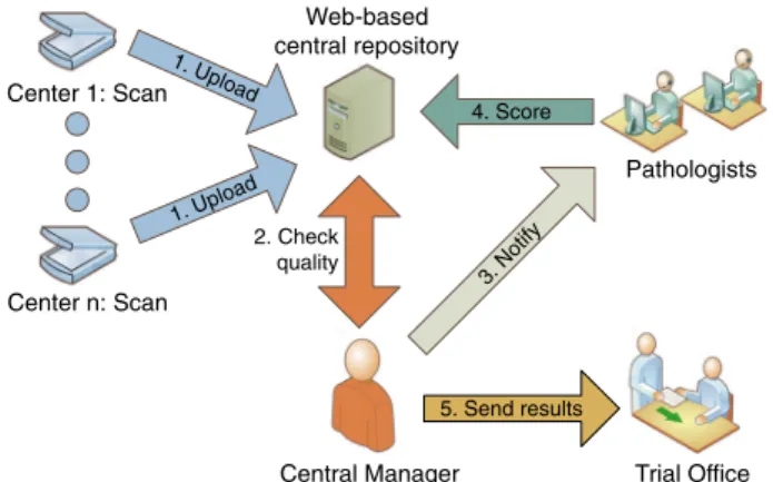
Risks with poss ible high impact identi fi ed in a p hase II immun otherapy trial 6based on the perspe ctive s o f Hall et al. 1wi th our approach to m itigation o f that risk. T ype of risk Risk Descript ion of risk Mitigation appro ach 1. Risks to patie nts No stroma or tumor cells in b iopsy Discom fo rt and risk s ass ociated wi th a samp ling inter v ention Take multip le biopsie s from o n e lesion at the same time (a minim um of thre e biopsi es p e r lesion 9) and chec k amou nt of tumo r cells bef ore analy sis of sTIL s and inclu sion in the trial 2. Risks to patie nts L oss of data con fi den tiality P atient samp les sent to multip le instituti ons and revie wers P seudo n y m ization should b e appl ied , hide slid e labe ls , impl emen t strict acce ss control , e n sure no metada ta is linked to a slide 3. Risks to biomar ker dev elopme nt Inter-labora tor y v ariability and interob ser v e r v aria bility Diff erent metho dologies us ed to score slides , interrat er v ariab ility Use of intern ational guid elines fo r scor ing and trai ning , use con sensus scor e of fo u r exp er t patho logists from thre e in stitutes 4. Operat ional risks Failure of sam ple collection, proce ssing , and quali ty Missing or poo r-quality sam ples resul ting in poor consensu s scoring Standa rdized tissue proces sing , wor kfl ow mana geme nt 5. Operat ional risks Ina dequate imag e qualit y o r n o abil ity to access imag e Missing , poo r, or inaccurate scor ing T rack the scor es of all path ologists and noti fy when score s are incon sistent 6. Operat ional risks/risks to patients L ong turnaround time Dela y in pati ent rand omiza tion and treatm ent Time line tracking incorpo rated in work fl ow 7. Operat ional risks Data mana gement failure Errors in collecting manua l score s, typ os , data con v ersion issues Structured dig ital score s from pathologist to the analyst Web-based central repository Center n: Scan Center 1: Scan 1. Upload 1. Upload Central Manager 3. Notify Pathologists Trial Office 5. Send results 4. Score 2. Check qualityFig. 1 Organization of a workflow for reliable and timely
biomarker scoring in a general single-center or multi-center trial.
Personnel at individual centers scan the slides after processing by
the local pathology department. Digital slides are uploaded to a
central web-based repository, such as Slide Score. A study-speci
fic
identi
fier is assigned to each sample. The central manager is notified
by the system when new slides are available and requests
pathologists to review it. When a consensus score is obtained, the
trial of
fice is notified for randomization of the patient.
and July 2019. Full eligibility criteria and trial procedures have
been described previously
6.
In the second part of the TONIC trial, we could implement our
work
flow with a focus on accurate and reproducible sTIL scores
within a reasonable timeframe after a biopsy was taken (72 h). For
all 47 patients included in the trial, reliable sTIL scores were
obtained with 45 biopsies scored within the 72-h timeframe
(Supplementary Fig. 3). During the course of the study, the server
of Slide Score was available 99.9% of the time. Five biopsies had to
be re-evaluated due to a discrepancy in the categorical scores,
when not all pathologists agreed on the appropriate category of
the sTIL score (lower than 5% versus higher or equal to 5%). In
three of these cases the score of one pathologist was higher (5 or
10%) than the score of the other two or three pathologists (0–3%).
The average sTIL score was obtained and the pathologist causing
the disagreement was noti
fied. In the fourth and fifth case, two
pathologists scored 5 and 10%, whereas the other pathologists
scored 1%. All four pathologists were noti
fied of the disagreement
and a consensus score of 5% was obtained. We observed an
intraclass correlation coef
ficient of 0.94 (95% confidence interval
(CI): 0.91
–0.97) for sTILs as a continuous variable. Interrater
agreement for the categorical variable used in the strati
fication
(sTILs <5% or
≥5%) was 0.86 (multirater Fleiss’ κ
18; 95% CI: 0.73
–1;
Supplementary Fig. 2c). In the anti-PD-1 monotherapy cohort, we
observed that 13 out of 23 patients (56.5 %) had sTILs below 5%,
as compared to 15 out of 24 patients in the doxorubicin cohort
(62.5 %; Fisher
’s exact test p value 0.77). The distribution of the
sTIL scores is depicted in Supplementary Fig. 2d. These data
indicate effective stratification based on the cut-off of 5%, but a
slightly uneven distribution in the higher ranges of sTIL scores
(10% or higher) inherent to the use of our cut-off. We observed a
median time from biopsy until the scanning of the H&E slide of
30 h (range 24–98 h) and a median time from the biopsy until at
least three scores were obtained of 43 h (range 27
–106 h). In total,
the median time from biopsy until registration in the patient
records was 49 h (range 41
–106 h; Supplementary Fig. 2e), with
96% of biopsies scored within 72 h. Two biopsies were not scored
within the 72-h time limit, due to additional processing of one
sample and one delay in registration time due to the absence of
the central manager (Supplementary Figs 1 and 3).
ADVANTAGES AND LIMITATIONS OF A WEB-BASED
RISK-MITIGATION WORKFLOW
Our proposed solutions involved standardization of our work
flow,
obtaining digital images and the use of a web-based tool such as
Slide Score for the managing and scoring of digital images.
Anticipating the incorporation of digital images in routine
diagnostics, our workflow shows that it is feasible for a pathologist
to score digital images with high reliability. Moreover, a
web-based tool can facilitate the process of coordinated uploading of
digital images, pseudonymizing slides, and regulate access to
studies and proper data management. Web-based platforms are
therefore of high interest in biomarker research and can help with
automation that can be transferred to clinical practice in the
future.
In this study, we obtained sTIL scores within 72 h after a biopsy
was taken, which is a reasonable timeframe for clinicians to start
randomization of patients to treatment arms in a clinical trial. We
observed an excellent interrater agreement score between our
panel of four expert pathologists. In an accompanying paper
7we
demonstrate using data from three RING studies of the TIL
Working Group that the concordance achieved using a
risk-management approach as detailed in this study is substantially
higher than observed outside this risk-management perspective
as observed in the three RING studies and in other published
studies
19,20. However, our sample size is small and the four
pathologists in the current study were trained and experienced in
the scoring of sTILs in breast cancer. Also, the biopsies used in this
study were checked for containing suf
ficient tumor cells (≥100
cells) before the slide was scored for sTILs, which could have
further improved our results. In the future, it is to be expected that
computational work
flows will further improve the scoring of
sTILs
13. Although we obtained reliable and timely results in 96% of
cases, the presence of a central manager is crucial. In one case
there was a delay in registration time due to the absence of the
central manager. The manual intervention of quality checks,
processing of the slides, and data cannot be circumvented in our
work
flow.
Stratification in this study was performed using sTILs as a binary
variable (lower than 5% versus higher or equal to 5%).
Consequently, we observed an uneven distribution in continuous
sTILs scores between the cohorts (Supplementary Fig. 2d). This
was mainly due to more patients with sTILs scores above 10% in
the anti-PD-1 monotherapy cohort. Inherent to the use of a binary
cut-off for strati
fication, the median of the continuous
measure-ment might still differ between cohorts. Alternatively, multiple
categories for the same variable can be used in strati
fication.
However, this approach generates more strata, with lower number
of patients in each stratum, possibly leading to an imbalance in
distribution
21,22. Moreover, at the time of writing of this paper no
cut-offs for sTILs are established and/or properly validated for
predictive purposes.
During the trial, we continuously monitored whether our
strategy was still feasible within the set timeframe by means of
regular evaluation by the pathologists and the study coordinators.
This led to rapid adjustment of the workflow if needed, ensuring
the quality of the sTIL scores. For example, pathologists could
easily login remotely and score a digital H&E outside the hospital
ensuring that sTILs were still scored within 72 h after biopsy.
Ongoing evaluation during the clinical trial is of critical importance
for risk mitigation in biomarker research
1.
FUTURE APPLICATIONS OF THE WORKFLOW
Our strategy can serve as a template for risk management and
mitigation of all identi
fied risks in future clinical trials
incorporat-ing biomarkers for inclusion, enrichment, or strati
fication. By no
means will risks identi
fied in this study be similar for all clinical
trials. Each trial will have its own risks that need to be mitigated,
although there will be similarities between the risks across clinical
trials. De
fining the risks that come with biomarker development
will help tested biomarkers eventually make their way to the clinic.
However, one may even argue that a similar risk-management
strategy can be applied in daily practice. In the BELLINI trial
(NCT03815890), two cycles of neo-adjuvant anti-PD-1 are
adminis-tered in patients with early-stage TNBC or luminal B breast cancer.
All patients are required to have at least 5% sTILs in the
pretreatment biopsy and patients are thereafter stratified in three
sTIL categories. Our work
flow will be used to ensure timely and
reliable sTIL scores for the right patient selection. By using our
work
flow, scoring of sTILs is highly standardized, allowing also
smaller centers with less extensive experience in sTILs scoring to
participate in a clinical trial.
CONCLUSIONS
In contemporary clinical research there is an increasing trend
toward the use of biomarker results obtained in daily practice to
select patients for inclusion in clinical trials. Therefore, continuous
monitoring of the prede
fined risks and the solutions can improve
the quality of the biomarker, as can be applied in a clinical trial
setting, as well as in daily practice. The recommendations of the
TIL
Working
Group
8,10for
appropriate
scoring,
the
risk-management framework of the NCI, NCRI, and EORTC Working
Groups
1, as well as our proposed strategies to reduce risks will
4
help to effectively and ef
ficiently improve the incorporation of
biomarkers in clinical trials in
first instance, herewith illustrated
using sTILs as a paradigm of this development.
DATA AVAILABILITY
The data that support thefindings of this study are available from the corresponding author upon reasonable request.
Received: 16 July 2019; Accepted: 18 February 2020;
REFERENCES
1. Hall, J. A., Salgado, R., Lively, T., Sweep, F. & Schuh, A. A risk-management approach for effective integration of biomarkers in clinical trials: perspectives of an NCI, NCRI, and EORTC working group. Lancet Oncol. 15, e184–193 (2014). 2. Loi, S. et al. Tumor-infiltrating lymphocytes and prognosis: a pooled individual
patient analysis of early-stage triple-negative breast cancers. J. Clin. Oncol. 37, 559–569 (2019).
3. Denkert, C. et al. Tumour-infiltrating lymphocytes and prognosis in different subtypes of breast cancer: a pooled analysis of 3771 patients treated with neoadjuvant therapy. Lancet Oncol. 19, 40–50 (2018).
4. Loi, S. et al. LBA13—relationship between tumor infiltrating lymphocyte (TIL) levels and response to pembrolizumab (pembro) in metastatic triple-negative breast cancer (mTNBC): results from KEYNOTE-086. Ann. Oncol. 28(Suppl_5), v605–49,https://doi.org/10.1093/annonc/mdx440(2017).
5. Emens, L. A. et al. Long-term clinical outcomes and biomarker analyses of ate-zolizumab therapy for patients with metastatic triple-negative breast cancer: a phase 1 study. JAMA Oncol.https://doi.org/10.1001/jamaoncol.2018.4224(2018). 6. Voorwerk, L. et al. Immune induction strategies in metastatic triple-negative breast cancer to enhance the sensitivity to PD-1 blockade: the TONIC trial. Nat. Med.https://doi.org/10.1038/s41591-019-0432-4(2019).
7. Kos, Z. et al. Pitfalls in assessing stromal tumor infiltrating lymphocytes (sTILs) in breast cancer. npj Breast Cancer. https://doi.org/10.1038/s41523-020-0156-0
(2020).
8. Salgado, R. et al. The evaluation of tumor-infiltrating lymphocytes (TILs) in breast cancer: recommendations by an International TILs Working Group 2014. Ann. Oncol. 26, 259–271 (2015).
9. Hayes, D. F. et al. Breaking a vicious cycle. Sci. Transl. Med. 5, 196cm196 (2013). 10. Hendry, S. et al. Assessing tumor-infiltrating lymphocytes in solid tumors: a practical review for pathologists and proposal for a standardized method From the International Immunooncology Biomarkers Working Group: Part 1: assessing the host immune response, TILs in invasive breast carcinoma and ductal carci-noma in situ, metastatic tumor deposits and areas for further research. Adv. Anat. Pathol. 24, 235–251 (2017).
11. Mroz, P., Parwani, A. V. & Kulesza, P. Central pathology review for phase III clinical trials: the enabling effect of virtual microscopy. Arch. Pathol. Lab. Med. 137, 492–495 (2013).
12. Pell, R. et al. The use of digital pathology and image analysis in clinical trials. J. Pathol. 5, 81–90 (2019).
13. Amgad, M. et al. Report on computational assessment of tumor infiltrating lymphocytes from the international immuno-oncology biomarker working group. npj Breast Cancer.https://doi.org/10.1038/s41523-020-0154-2(2020).
14. Bankhead, P. et al. QuPath: Open source software for digital pathology image analysis. Sci. Rep. 7, 16878 (2017).
15. Li, H. et al. Dysfunctional CD8 T cells form a proliferative, dynamically regulated compartment within human melanoma. Cell 176, 775–789.e718 (2019). 16. Sauer, G. et al. Ultrasound-guided large-core needle biopsies of breast lesions:
analysis of 962 cases to determine the number of samples for reliable tumour classification. Br. J. Cancer 92, 231–235 (2005).
17. Simon, R. Optimal two-stage designs for phase II clinical trials. Control. Clin. Trials 10, 1–10 (1989).
18. Fleiss, J. L. Statistical Methods for Rates and Proportions 2nd edn (1981). 19. Tramm, T. et al. Standardized assessment of tumor-infiltrating lymphocytes in
breast cancer: an evaluation of inter-observer agreement between pathologists. Acta Oncol. 57, 90–94 (2018).
20. Dieci, V. et al. Association of tumor-infiltrating lymphocytes with distant disease-free survival in the ShortHER randomized adjuvant trial for patients with early HER2+ breast cancer. Ann. Oncol. 30, 418–423 (2019).
21. Therneau, T. M. How many stratification factors are “too many” to use in a randomization plan? Control. Clin. Trials 14, 98–108 (1993).
22. Silcocks, P. How many strata in an RCT? Aflexible approach. Br. J. Cancer 106, 1259–1261 (2012).
ACKNOWLEDGEMENTS
The Department of Pathology of the Netherlands Cancer Institute is thanked for the support of this study and ensuring the rapid turnaround times. The Breast Cancer Research Foundation and Bristol-Myers-Squibb (BMS) are thanked for financial support. We also thank the BMS-International Immuno-Oncology Network (BMS/II-ON) and the Dutch Cancer Society (NKI2015-7710) for funding the clinical trial costs and this feasibility study (NKI2016-10510). S.L., R.S., and M.K. are supported by a grant from the Breast Cancer Research Foundation (BCRF, NY, US). The following is a list of current members of the International Immuno-Oncology Working Group (TILs Working Group). A member is defined as a person willing to be involved, informed and be part of the activities of the TILs Working Group. The authors alone are responsible for the views expressed in the work of the TIL Working Group and they do not necessarily represent the decisions, policy, or views of their employer.
AUTHOR CONTRIBUTIONS
J.H. developed Slide Score and wrote the manuscript with L.V., R.S., M.K. and H.M.H. L. V. coordinated study procedures and performed data-analyses. M.v.S., I.N., S.A., S.D., G.V., T.O.N., S.S.B., S.M., W.F.S., C.S., D.L.R., S.H., C.D., S.L., Sh.L., J.M.S.B., G.P., D.A.D., M.C. U.C., A.T., J.A.H. and Z.K. gave critical input. M.d.M. provided logistical support with the sample processing. J.v.d.B., K.K.v.d.V., R.S. and H.M.H. scored the slides. K.S. performed the statistical analysis. M.K. is the principal investigator of the TONIC trial. J.H., L.V., R.S., M.K. and H.M.H. designed this feasibility study. All authors edited and approved the manuscript.
COMPETING INTERESTS
J.H. is the owner of Slide Score. B.V. L.V., M.v.S., I.N., M.d.M., J.v.d.B., K.K.v.d.V., K.S., S.A., S.D., G.V., S.S.B., S.M., W.F.S., C.S., S.M.H., C.D., S.L., G.P., M.C.U.C., Z.K., and H.M.H. have nothing to disclose. T.O.N. has consulted for Nanostring and received compensation and has intellectual property rights/ownership interests from Bioclassifier LLC, not related to the subject material under consideration and received funding support from the Canadian Cancer Society. D.L.R. reports research funding from AstraZeneca, Cepheid, Navigate BioPharma, NextCure, Lilly, and Ultivue; instrument support from Ventana, Akoya/PerkinElmer, and NanoString; advisory board of Amgen, AstraZeneca, Cell Signaling Technology, Cepheid, Daiichi Sankyo, GSK, Konica/Minolta, Merck, NanoString, PerkinElmer, Ventana, and Ultivue; consultancy for Biocept; honorarium and travel support from BMS; royalties from Rarecyte and is a founder and equity holder of PixelGear. Sh.L. receives research funding to her institution from Novartis, Bristol Meyers Squibb, Merck, Roche-Genentech, Puma Biotechnology, Pfizer, and Eli Lilly, acted as consultant (not compensated) to Seattle Genetics, Pfizer, Novartis, BMS, Merck, AstraZeneca, and Roche-Genentech and acted as consultant (paid to her institution) to Aduro Biotech. J.M.S.B. reports research funding from ThermoFisher, Genoptix, Agendia, NanoString Technologies, Stratifyer GmbH, and Biotheranostics and advisory roles for Insight Genetics, BioNTech AG, Biotheranostics, Pfizer, RNA Diagnostics, and OncoXchange. J.M.S.B. reports the following patents: Methods and Devices for Predicting Anthracycline Treatment Efficacy, US utility (January 2017; 15/ 325,472; EPO– 15822898.1; Canada – not yet assigned), Systems, Devices and Methods for Constructing and Using a Biomarker, US utility (January 2017; 15/ 328,108; EPO– 15824751.0; Canada – not yet assigned), Histone gene module predicts anthracycline benefit (October 2016; PCT/CA2016/000247), 95‐Gene Signature of Residual Risk Following Endocrine Treatment (December 2016; PCT/ CA2016/000304), Immune Gene Signature Predicts Anthracycline Benefit (December 2016; PCT/CA2016/000305). D.A.D. is on the advisory board and consults for Oncology Analytics Inc., and has consulted for and received travel funds from Novartis for work unrelated to the current manuscript. A.T. reports benefits from ICR’s Inventors Scheme associated with patents for one of PARP inhibitors in BRCA1/2-associated cancers. A.T. also reports Honoraria from Pfizer, Vertex, Prime Oncology, and Artios, honoraria and stock in InBioMotion, honoraria andfinancial support for research from AstraZeneca, Medivation, Myriad Genetics, and Merck Serono. J.A.H. is the director and owner of Vivactiv Ltd. R.S. reports research funding from Roche, Puma, and Merck; advisory board and consultancy for BMS; travel funding from Roche, Merck, and AstraZeneca, outside the scope of this work. M.K. reports funding to the institute from BMS, Roche and an advisory role for BMS, outside the submitted work.
ADDITIONAL INFORMATION
Supplementary information is available for this paper athttps://doi.org/10.1038/ s41523-020-0155-1.
Correspondence and requests for materials should be addressed to R.S. Reprints and permission information is available at http://www.nature.com/ reprints
Publisher’s note Springer Nature remains neutral with regard to jurisdictional claims in published maps and institutional affiliations.
Open Access This article is licensed under a Creative Commons Attribution 4.0 International License, which permits use, sharing, adaptation, distribution and reproduction in any medium or format, as long as you give appropriate credit to the original author(s) and the source, provide a link to the Creative Commons license, and indicate if changes were made. The images or other third party material in this article are included in the article’s Creative Commons license, unless indicated otherwise in a credit line to the material. If material is not included in the article’s Creative Commons license and your intended use is not permitted by statutory regulation or exceeds the permitted use, you will need to obtain permission directly from the copyright holder. To view a copy of this license, visithttp://creativecommons. org/licenses/by/4.0/.
© The Author(s) 2020
INTERNATIONAL IMMUNO-ONCOLOGY BIOMARKER WORKING GROUP
Aini Hyytiäinen
37, Akira I. Hida
38, Alastair Thompson
39, Alex Lefevre
40, Alexander J. Lazar
41, Allen Gown
42, Amy Lo
43, Anna Sapino
44,
Anant Madabhushi
45,46, Andre Moreira
47, Andrea Richardson
48, Andrea Vingiani
49, Andrew H. Beck
50, Andrew M. Bellizzi
51,
Angel Guerrero
52, Anita Grigoriadis
53,54, Anna Ehinger
55, Ana Garrido-Castro
56, Anne Vincent-Salomon
57, Anne-Vibeke Laenkholm
58,
Ashish Sharma
59, Ashley Cimino-Mathews
60, Ashok Srinivasan
61, Balazs Acs
62, Baljit Singh
63, Benjamin Calhoun
64, Benjamin
Haibe-Kans
65, Benjamin Solomon
66, Bibhusal Thapa
67, Brad H. Nelson
68, Brandon D. Gallas
69, Carlos Castaneda
70,71, Carmen
Ballesteros-Merino
72, Carmen Criscitiello
73, Carolien Boeckx
74, Cecile Colpaert
75, Cecily Quinn
76, Chakra S. Chennubhotla
77, Charles Swanton
78,
Cinzia Solinas
79, Crispin Hiley
78, Damien Drubay
80,81, Daniel Bethmann
82, David A. Moore
83,84, Denis Larsimont
85,
Dhanusha Sabanathan
86, Dieter Peeters
87, Dimitrios Zardavas
88, Doris Hö
flmayer
89, Douglas B. Johnson
90, E. Aubrey Thompson
91,
Edi Brogi
92, Edith Perez
93, Ehab A. ElGabry
94, Elisabeth Specht Stovgaard
95, Elizabeth F. Blackley
66, Elvire Roblin
96,97,
Emily Reisenbichler
18, Enrique Bellolio
98,99, Eva Balslev
95, Ewa Chmielik
100, Fabien Gaire
101, Fabrice Andre
102, Fang-I Lu
103,
Farid Azmoudeh-Ardalan
104, Federico Rojo
105,106, Tina Gruosso
107, Francesco Ciompi
108, Franklin Peale
109, Fred R. Hirsch
110,
Frederick Klauschen
21, Frédérique Penault-Llorca
111, Gabriela Acosta Haab
112, Gelareh Farshid
113, Gert van den Eynden
114,
Giuseppe Curigliano
115,116, Giuseppe Floris
117,118, Glenn Broeckx
119, Gonzalez-Ericsson
120, Harmut Koeppen
43, Harry R. Haynes
121,
Heather McArthur
122, Heikki Joensuu
123, Helena Olofsson
124, Huang-Chun Lien
125, I-Chun Chen
126,127, Ian Cree
128, Isabel Frahm
129,
Iva Brcic
130, Jack Chan
131, James Ziai
43, Jane Brock
132, Jelle Wesseling
133, Jennifer Giltnane
43, Jennifer K. Kerner
134, Jeppe Thagaard
135,136,
Jeremy P. Braybrooke
137,138, Jeroen A. W. M. van der Laak
108, Jerome Lemonnier
139, Jiping Zha
140, Joana Ribeiro
141, Jochen K. Lennerz
142,
Jodi M. Carter
143, Joel Saltz
144, Johan Hartman
145, Johannes Hainfellner
146, John Le Quesne
147, Jonathon W. Juco
148, Jorge
Reis-Filho
92,149, Joselyn Sanchez
150, Joseph Sparano
151, Joël Cucherousset
152, Juan Carlos Araya
98, Julien Adam
153, Justin M. Balko
154,
Kai Saeger
155, Kalliopi Siziopikou
156, Karen Willard-Gallo
157, Karsten Weber
23, Katherine L. Pogue-Geile
158, Keith E. Steele
140,
Kenneth Emancipator
148, Khalid AbdulJabbar
159, Khalid El Bairi
160, Kim R. M. Blenman
161, Kimberly H. Allison
162, Konstanty Korski
163,
Lajos Pusztai
161, Laura Comerma
164,106, Laurence Buisseret
157, Lee A. D. Cooper
165, Leming Shi
166, Loes F. S. Kooreman
167,
Luciana Molinero
109, M. Valeria Estrada
168, Magali Lacroix-Triki
169, Maise Al Bakir
78, Manu M. Sebastian
170, Marc van de Vijver
171,
Marcelo Luiz Balancin
172,173, Maria Vittoria Dieci
174, Marie-Christine Mathieu
175, Marlon C. Rebelatto
140, Martine Piccart
176,
Matthew G. Hanna
92, Matthew P. Goetz
93, Matthias Preusser
146, Mehrnoush Khojasteh
177, Melinda E. Sanders
178,
Meredith M. Regan
179,180, Michael Barnes
181, Michael Christie
182, Michael Misialek
183, Michail Ignatiadis
184, Mieke van Bockstal
185,
Miluska Castillo
71, Mohamed Amgad
186, Nadia Harbeck
187, Nadine Tung
188, Nele Laudus
189, Nicolas Sirtaine
190, Nicole Burchardi
191,
Nils Ternes
14, Nina Radosevic-Robin
192, Oleg Gluz
193, Oliver Grimm
101, Paolo Nuciforo
194, Paul Jank
195, Paula Gonzalez-Ericsson
196,
Pawan Kirtani
197, Petar Jelinic
148, Peter H. Watson
198, Peter Savas
24,199, Prudence A. Francis
200,201, Prudence A. Russell
202,
Rajendra Singh
203, Rim S. Kim
204, Robert H. Pierce
205, Robert Hills
206, Roberto Leon-Ferre
93, Roland de Wind
190, Ruohong Shui
207,
Sabine De Clercq
208, Sam Leung
209, Sami Tabbarah
210, Sandra C. Souza
211, Sandra O
’Toole
212, Sandra Swain
213, Sarah Dudgeon
214,
Scooter Willis
215, Scott Ely
216, Seong-Rim Kim
217, Shahinaz Bedri
218, Sheeba Irshad
219,220, Shi-Wei Liu
221, Shom Goel
200,201,
Shona Hendry
222, Simonetta Bianchi
223, So
fia Bragança
224, Soonmyung Paik
61, Stephan Wienert
225, Stephen B. Fox
222,
Stephen J. Luen
24, Stephen Naber
226, Stuart J. Schnitt
30,227, Luz F. Sua
228, Sunil R. Lakhani
229, Susan Fineberg
230, Teresa Soler
231,
Thomas Gevaert
232, Timothy D
’Alfonso
233, Tom John
234, Tomohagu Sugie
235, Uday Kurkure
236, Veerle Bossuyt
237, Venkata Manem
65,
Vincente Peg Cámara
238, Weida Tong
239, Weijie Chen
69, Wentao Yang
207, William T. Tran
210, Yihong Wang
240, Yinyin Yuan
159,
Yves Allory
241, Zaheed Husain
242and Zsuzsanna Bago-Horvath
243 37Department of Oral and Maxillofacial Diseases, Helsinki, Finland.38
Department of Pathology, Matsuyama Shimin Hospital, Matsuyama, Japan.39
Surgical Oncology, Baylor College of Medicine, Houston, TX, USA.40
Roche Diagnostics, Mechelen, Belgium.41
Departments of Pathology, Genomic Medicine, Dermatology, and Translational Molecular Pathology, The University of Texas MD Anderson Cancer Center, Houston, TX, USA.42
PhenoPath Laboratories, Seattle, WA, USA.43
Research Pathology, Genentech Inc., South San Francisco, CA, USA.44
University of Turin/Candiolo Cancer Institute - FPO, IRCCS, Candiolo, Italy.45
Case Western Reserve University, Cleveland, OH, USA.46
Louis Stokes Cleveland Veterans Health Administration Medical Center, Cleveland, OH, USA.47
Pulmonary Pathology, New York University Center for Biospecimen Research and Development, New York University, New York, NY, USA.48
Department of Pathology, Johns Hopkins Hospital, Baltimore, MD, USA.49
Department of Pathology, Istituto Europeo di Oncologia, University of Milan, Milan, Italy.50PathAI, Inc, Cambridge, MA, USA.51Department of Pathology, University of Iowa Hospitals and Clinics, Iowa City, IA, USA.52Department of Oncology, IVO, Valencia, Spain.53
Cancer Bioinformatics Lab, Cancer Centre at Guy’s Hospital, London, UK.54
School of Life Sciences and Medicine, King’s College London, London, UK.55 Lund University, Skane University Hospital, Department of Clinical Sciences Lund, Oncology and Pathology, Lund, Sweden.56Dana Farber Cancer Institute, Boston, MA, USA.57Institut Curie, Paris Sciences Lettres Université, Inserm U934, Department of Pathology, Paris, France.58
Department of Surgical Pathology, Zealand University Hospital, Køge, Denmark. 59
Department of Biomedical Informatics, Emory University, GA, USA.60Departments of Pathology and Oncology, The Johns Hopkins Hospital, Baltimore, MD, USA.61National Surgical Adjuvant Breast and Bowel Project Operations Center/NRG Oncology, Pittsburgh, PA, USA.62
Department of Pathology, Karolinska Institutet, Karolinska, Sweden.
6
63
Department of Pathology, New York University Langone Medical Centre, New York, NY, USA.64
Department of Pathology and Laboratory Medicine, UNC School of Medicine, Chapel Hill, NC, USA.65Bioinformatics and Computational Genomics Laboratory, Princess Margaret Cancer Center, Toronto, ON, Canada.66Department of Medical Oncology, Peter MacCallum Cancer Centre, Melbourne, VIC, Australia.67
Department of Medicine, University of Melbourne, Parkville, VIC, Australia.68
Trev & Joyce Deeley Research Centre, British Columbia Cancer Agency, Victoria, BC, Canada.69Division of Imaging, Diagnostics, and Software Reliability (DIDSR), Office of Science and Engineering Laboratories (OSEL), Center for Devices and Radiological Health (CDRH), Rockville, MD, USA.70
Department of Research, Instituto Nacional de Enfermedades Neoplásicas, Lima, Peru.71
Department of Research, Instituto Nacional de Enfermedades Neoplásicas, Lima 15038, Peru.72
Providence Cancer Research Center, Portland, Oregon, USA.73
Department of Medical Oncology, Istituto Europeo di Oncologia, Milan, Italy.74
Roche Diagnostics, Mechelen, Belgium.75
Department of Pathology, AZ Turnhout, Turnhout, Belgium.76
Department of Pathology, St Vincent’s University Hospital and University College Dublin, Dublin, Ireland.77
Department of Computational and Systems Biology, University of Pittsburgh, Pittsburgh, PA, USA. 78
Cancer Research UK Lung Cancer Centre of Excellence, University College London Cancer Institute, University College London, London, UK.79
Azienda AUSL, Regional Hospital of Aosta, Aosta, Italy.80
Université Paris-Sud, Institut National de la Santé et de la Recherche Médicale, Villejuif, France.81
Gustave Roussy, Universite Paris-Saclay, Villejuif, France. 82
University Hospital Halle (Saale), Institute of Pathology, Halle, (Saale), Germany.83
Department of Pathology, UCL Cancer Institute, UCL, London, UK.84
University College Hospitals NHS Trust, London, UK.85
Department of Pathology, Jules Bordet Institute, Brussels, Belgium.86
Department of Clinical Medicine, Macquarie University, Sydney, Australia. 87
HistoGeneX NV, Antwerp, Belgium and AZ Sint-Maarten Hospital, Mechelen, Belgium.88Oncology Clinical Development, Bristol-Myers Squibb, Princeton, NJ, USA.89Institut für Pathologie, UK Hamburg, Germany.90
Department of Medicine, Vanderbilt University Medical Centre, Nashville, TN, USA.91
Department of Cancer CV, Jacksonville, FL, USA. 92
Department of Pathology, Memorial Sloan Kettering Cancer Center, New York, NY, USA.93
Department of Oncology, Mayo Clinic, Rochester, MN, USA.94
Roche, Tucson, AZ, USA. 95
Department of Pathology, Herlev and Gentofte Hospital, Gentofte, Denmark.96
Université Paris-Saclay, Univ. Paris-Sud, Villejuif, France.97
Service de biostatistique et d’épidémiologie, Gustave Roussy, Villejuif, France.98
Department of Pathology, Universidad de La Frontera, Temuco, Chile.99
Departamento de Anatomía Patológica, Universidad de La Frontera, Temuco, Chile.100
Tumor Pathology Department, Maria Sklodowska-Curie Memorial Cancer Center, Gliwice, Poland.101
Pathology and Tissue Analytics, Roche, Neuherberg, Germany.102
Department of Medical Oncology, Gustave Roussy, Villejuif, France.103
Sunnybrook Health Sciences Centre, Toronto, ON, Canada.104
Tehran University of Medical Sciences, Tehran, Iran.105
Pathology Department, Instituto de Investigación Sanitaria Fundación Jiménez Díaz (IIS-FJD), Madrid, Spain.106
GEICAM-Spanish Breast Cancer Research Group, Madrid, Spain.107
Translational Research, Montreal, Canada.108
Computational Pathology Group, Department of Pathology, Radboud University Medical Center, Nijmegen, The Netherlands.109Oncology Biomarker Development, Genentech-Roche, Neuherberg, Germany.110Division of Medical Oncology, Department of Medicine, University of Colorado Anschutz Medical Campus, Aurora, CO, USA.111
Centre de Lutte Contre le cancer - Centre Jean Perrin, Clermont-Ferrand, France.112
Department of Pathology, Hospital de Oncología Maria Curie, Buenos Aires, Argentina.113Directorate of Surgical Pathology, SA Pathology, Adelaide, Australia.114Department of Pathology, GZA-ZNA Ziekenhuizen, Wilrijk, Belgium.115
University of Milano, Instituto Europeo di Oncologia, IRCCS, Milano, Italy.116
Division of Early Drug Development for Innovative Therapy, IEO, European Institute of Oncology IRCCS, Milan, Italy.117
Department of Imaging and Pathology, Laboratory of TranslCal Cell & Tissue Research, Leuven, Belgium.118 KU Leuven-University Hospitals Leuven, Department of Pathology, Leuven, Belgium.119
Department of Pathology, University Hospital of Antwerp, Antwerp, Belgium.120
Paula I, Breast Cancer Research Program, Vanderbilt University Medical Center, Nashville, TN, USA.121
Translational Health Sciences, Department of Cellular Pathology, North Bristol NHS Trust, University of Bristol, Bristol, UK.122
Medical Oncology, Department of Medicine, Cedars-Sinai Medical Center, Los Angeles, CA, USA.123
Helsinki University Central Hospital, Helsinki, Finland.124
Department of Clinical Pathology, Akademiska University Hospital, Uppsala, Sweden.125
Department of Pathology, National Taiwan University Hospital, Taipei, Taiwan. 126
Department of Oncology, National Taiwan University Cancer Center, Taipei, Taiwan.127
Graduate Institute of Oncology, College of Medicine, National Taiwan University, Taipei, Taiwan.128
International Agency for Research on Cancer (IARC), World Health Organization, Lyon, France.129
Department of Pathology, Sanatorio Mater Dei, Buenos Aires, Argentina. 130Institute of Pathology, Medical University of Graz, Graz, Austria. 131Department of Oncology, National Cancer Centre Singapore, Singapore, Singapore. 132
Department of Pathology, Brigham and Women’s Hospital, Boston, MA, USA.133
Department of Pathology, Netherlands Cancer Institute, Amsterdam, The Netherlands. 134
PathAI Inc, Cambridge, MA, USA.135
Visiopharm A/S, Hørsholm, Denmark.136
DTU Compute, Department of Applied Mathematics, Technical University of Denmark, Lyngby, Denmark.137Nuffield Department of Population Health, University of Oxford, Oxford, UK.138
Department of Medical Oncology, University Hospitals Bristol NHS Foundation Trust, Bristol, UK.139
R&D UNICANCER, Paris, France.140
Translational Sciences, MedImmune, Gaithersberg, MD, USA.141
Breast Unit, Champalimaud Clinical Centre, Lisboa, Portugal. 142
Department of Pathology, Massachusetts General Hospital, Boston, MA, USA.143
Department of Laboratory Medicine and Pathology, Mayo Clinic, Rochester, MN, USA. 144
Biomedical Informatics Department, Stony Brook University, Stony Brook, NY, USA.145
Department of Oncology and Pathology, Karolinska Institutet and University Hospital, Solna, Sweden.146
Department of Medicine, Clinical Division of Oncology, Comprehensive Cancer Centre Vienna, Medical University of Vienna, Vienna, Austria.147
Leicester Cancer Research Centre, University of Leicester, Leicester, and MRC Toxicology Unit, University of Cambridge, Cambridge, UK.148
Merck & Co., Inc, Kenilworth, USA.149
Human Oncology and Pathogenesis Program, Memorial Sloan Kettering Cancer Center, New York, NY, USA.150Department of Research, Instituto Nacional de Enfermedades Neoplasicas, Lima 15038, Peru.151
Department of Medicine, Department of Obstetrics and Gynecology and Women’s Health, Albert Einstein Medical Center, Bronx, USA.152
GHI Le Raincy-Montfermeil, Chelles, Île-de-France, France.153Department of Pathology, Gustave Roussy, Grand Paris, France.154Departments of Medicine and Cancer Biology, Vanderbilt University Medical Centre, Nashville, TN, USA.155
VMscope GmbH, Berlin, Germany.156
Department of Pathology, Breast Pathology Section, Northwestern University, Chicago, IL, USA.157
Molecular Immunology Unit, Institut Jules Bordet, Université Libre de Bruxelles, Brussels, Belgium.158
NSABP/NRG Oncology, Pittsburgh, PA, USA.159
Division of Molecular Pathology, Centre for Evolution and Cancer, The Institute of Cancer Research, London, UK.160
Cancer Biomarkers Working Group, Faculty of Medicine and Pharmacy, Université Mohamed Premier, Oujda, Morocco.161
Yale Cancer Center Genetics, Genomics and Epigenetics Program, Yale School of Medicine, New Haven, CT, USA. 162 Pathology Department, Stanford University Medical Centre, Stanford, CA, USA.163
Pathology and Tissue Analytics, Roche Innovation Centre Munich, Penzberg, Germany.164 Pathology Department, Hospital del Mar, Parc de Salut Mar, Barcelona, Spain.165
Department of Pathology, Northwestern University Feinberg School of Medicine, Chicago, IL, USA.166 Center for Pharmacogenomics and Fudan-Zhangjiang, Center for Clinical Genomics School of Life Sciences and Shanghai Cancer Center, Fudan University, Fudan, China.167GROW -School for Oncology and Developmental Biology, Maastricht University Medical Centre and Department of Pathology, Maastricht University Medical Centre, Maastricht, The Netherlands.168Biorepository and Tissue Technology Shared Resources, University of California San Diego, San Diego, CA, USA.169Department of Pathology, Gustave Roussy, Villejuif, France.170
Departments of Epigenetics and Molecular Carcinogenesis, The University of Texas MD Anderson Cancer Center, Houston, TX, USA.171
Department of Pathology, Academic Medical Center, Amsterdam, The Netherlands.172
Department of Pathology, University of São Paulo, São Paulo, Brazil.173
Hospital das Clínicas, Sao Paulo, Brasil.174
Department of Surgery, Oncology and Gastroenterology, University of Padova, Padua, Italy.175
Department of Medical Biology and Pathology, Gustave Roussy Cancer Campus, Villejuif, France.176
Institut Jules Bordet, Universite Libre de Bruxelles, Brussels, Belgium.177
Roche Tissue Diagnostics, Digital Pathology, Santa Clara, CA, USA. 178
Department of Pathology, Microbiology and Immunology, Vanderbilt University Medical Centre, Nashville, TN, USA.179
Division of Biostatistics, Dana-Farber Cancer Institute, Boston, MA, USA.180
Harvard Medical School, Boston, MA, USA.181
Roche Diagnostics Information Solutions, Belmont, CA, USA.182
Department of Anatomical Pathology, Royal Melbourne Hospital, Parkville, VIC, Australia.183Vernon Cancer Center, Newton-Wellesley Hospital, Newton, MA, USA.184Department of Medical Oncology, Institut Jules Bordet, Université Libre de Bruxelles, Brussels, Belgium.185
Service de pathologique, Cliniques universitaires Saint-Luc, Bruxelles, Belgique.186
Department of Biomedical Informatics, Emory University School of Medicine, Atlanta, GA, USA.187Breast Center, Dept. OB&GYN and CCC (LMU), University of Munich, Munich, Germany.188Division of Hematology-Oncology, Beth Israel Deaconess Medical Center, Boston, MA, USA.189
University of Leuven, Leuven, Belgium.190
Department of Pathology, Institut Jules Bordet, Université Libre de Bruxelles, Brussels, Belgium.191German Breast Group, Neu-Isenburg, Germany.192Department of Surgical Pathology and Biopathology, Jean Perrin Comprehensive Cancer Centre, Clermont-Ferrand, France.193
Johanniter GmbH - Evangelisches Krankenhaus Bethesda Mönchengladbach, West German Study Group, Mönchengladbach, Germany. 194
Molecular Oncology Group, Vall d’Hebron Institute of Oncology, Barcelona, Spain.195
Department of Pathology, University of Marburg, Marburg, Germany.196
Breast Cancer Program, Vanderbilt-Ingram Cancer Center, Vanderbilt University Medical Center, Nashville, TN, USA.197
Department of Histopathology, Manipal Hospitals Dwarka, New Delhi, India.198
Department of Pathology and Laboratory Medicine, University of British Columbia, Vancouver, BC, Canada.199
The Sir Peter MacCallum Department of Oncology, University of Melbourne, Melbourne, VIC, Australia.200
Peter MacCallum Cancer Centre, Melbourne, VIC, Australia.201
Sir Peter MacCallum Department of Oncology, University of Melbourne, Melbourne, VIC, Australia.202
Department of Anatomical Pathology, St Vincent’s Hospital Melbourne, Fitzroy, VIC, Australia.203
Icahn School of Medicine at Mt. Sinai, New York, NY, USA.204NRG Oncology/NSABP, Pittsburgh, PA, USA.205Cancer Immunotherapy Trials Network, Central Laboratory and Program in Immunology, Fred Hutchinson Cancer Research Center, Seattle, WA, USA.206
Clinical Trial Service Unit & Epidemiological Studies Unit, University of Oxford, Oxford, UK.207
Department of Pathology, Fudan University Cancer Center, Shanghai, China.208Department of Pathology, GZA-ZNA Hospitals, Antwerp, Belgium.209University of British Columbia, Vancouver, BC, Canada. 210
Department of Radiation Oncology, Odette Cancer Centre, Sunnybrook Research Institute, Toronto, ON, Canada.211
Merck Oncology, Kenilworth, NJ, USA.212 The Cancer Research Program, Garvan Institute of Medical Research, Darlinghurst, NSW, Australia.213
Georgetown University Medical Center, Washington, DC, USA.214
FDA/CDRH/OSEL/
7
Division of Imaging, Diagnostics, and Software Reliability, Silver Spring, MD, USA.215
Department of Molecular and Experimental Medicine, Avera Cancer Institute, Sioux Falls, SD, USA.216Translational Medicine, Bristol-Myers Squibb, Princeton, NJ, USA.217National Surgical Adjuvant Breast and Bowel Project Operations Center/NRG Oncology, Pittsburgh, PA, USA.218
Anatomic Pathology, Boston, MA, USA.219
Guy’s Hospital, London, UK.220
King’s College London, London, UK.221
Peking University First Hospital Breast Disease Center, Beijing, China.222Department of Pathology, Peter MacCallum Cancer Centre, Melbourne, VIC, Australia.223Dipartimento di Scienze della Salute (DSS), Firenze, Italy.224Department of Oncology, Champalimaud Clinical Centre, Lisbon, Portugal.225
Charité - Universitätsmedizin Berlin, corporate member of Freie Universität Berlin, Humboldt-Universität zu Berlin, and Berlin Institute of Health, Institute of Pathology, Berlin, Germany.226
Department of Pathology and Laboratory Medicine, Tufts Medical Center, Boston, MA, USA. 227
Dana-Farber Cancer Institute, Boston, MA, USA.228
Department of Pathology, Fundación Valle del Lili, Cali, Valle del Cauca, Colombia.229
The University of Queensland Centre for Clinical Research and Pathology Queensland, Brisbane, QLD, Australia.230
Department of Pathology, Montefiore Medical Center and the Albert Einstein College of Medicine, Bronx, NY, USA.231Department of Pathology, University Hospital of Bellvitge, Oncobell, IDIBELL, L’Hospitalet del Llobregat, Barcelona 08908 Catalonia, Spain.232
Department of Development and Regeneration, Laboratory of Experimental Urology, KU Leuven, Leuven, Belgium.233
Department of Pathology, Memorial Sloan Kettering Cancer Center, New York, NY, USA.234
Department of Medical Oncology, Austin Health, Heidelberg, VIC, Australia.235
Department of Surgery, Kansai Medical School, Hirakata, Japan.236
Roche Tissue Diagnostics, Digital Pathology, Santa Clara, CA, USA.237
Department of Pathology, Massachusetts General Hospital, Boston, MA, USA.238
Pathology Department, H.U. Vall d’Hebron, Barcelona, Spain.239Division of Bioinformatics and Biostatistics, US Food and Drug Administration, Silver Spring, MD, USA.240Department of Pathology and Laboratory Medicine, Rhode Island Hospital and Lifespan Medical Center, Providence, RI, USA.241
Université Paris-Est, Créteil, France.242
Praava Health, Dhaka, Bangladesh.243
Department of Pathology, Medical University of Vienna, Vienna, Austria.