International Journal of
http://www.arjournals.org/index.php/ijpm/index
Effect of phenolic extracts of Algerian medicinal plants on the bacterial
growth and adherence of Staphylococcus aureus and Bacillus
pathogens responsible of food
Belkacem Imane
*Corresponding author:
Belkacem Imane
1Laboratory of Microbiology and vegetal Biology, Faculty of Natural Sciences and Life, University of Mostaganem, ALGERIA. Received: 08 February 2017 Accepted: 09 May 2017 Published: 02 September 2017
A b s t r a c t
Artemisia, one of the larger genera in the family Asteraceae, comprises from 200 to more than 500 taxa at the specific or subspecific level. Artemisia herba
Africa and certain parts of Asia and Middle East. It is one of
Algerian folk medicine. The antioxidant and free radical scavenging activities of extracted materials were tested together with their antibacterial effects against bacterial strains causing food po
phenolic and flavonoid content (161, 64 mg/g and 16,83mg/g, respectively) in T. capitatus L which represent a phenolic content of 131, 48 mg/g and 14, 96 content. The res
antioxidant (DPPH, FRAP assays) and antibacterial activities, which ethnopharmacological use and A. herba alba represent the highest values (IC and OD 1, 13 at 700nm). Our results show the interest of A. herba
L, among other medicinal plants, in search of new chemo
and planktonic growth of food spoilage pathogens. Further studies are most interesting molecules responsible for these activities. It is from Artemisia herba alba Asso, exert strong antioxidant polyphenol contents. These findings demons
valuable source of Natural antioxidants. Keywords:
Introduction
Foodborne illness is a major problem with enormous associated costs. Foodborne pathogens occur widely in nature and it is difficult to prevent them from entering raw foods. Staphylococcus aureus is a Gram-positive bacterium and is considered one of the most prevalent causes of gastroenteritis worldwide [1], with milk and dairy products and meat products (particularly cooked and fermented) being some of the most frequently involved Bacillus cereus belongs to a genus of Gram-positive, endospore forming bacteria, which is involved in foodborne outbreaks with variable incidence from country to country [2].
primary and secondary metabolite constituents have a long history of use in old and modern medicines and in certain systems of traditional medicines, and are considered the sources of important medications such as vincristine, codeine, quinine, morphine, digoxin, atropine and others1. In Algeria Artemesia herba
DOI:10.5138/09750185.2018
This article is distributed under the terms of the
and redistribution provided that the original author and source are credited.
International Journal of Phytomedicine 9 (2017) 4
http://www.arjournals.org/index.php/ijpm/index
Original Research Article
Effect of phenolic extracts of Algerian medicinal plants on the bacterial
adherence of Staphylococcus aureus and Bacillus
pathogens responsible of food poisoning.
Belkacem Imane*
1, Rebai Ouafa
1, Djibaoui Rachid
1A b s t r a c t
Artemisia, one of the larger genera in the family Asteraceae, comprises from 200 to more than 500 taxa at the specific or subspecific level. Artemisia herba-alba Asso (Shih) is grown in Africa and certain parts of Asia and Middle East. It is one of the most widely used Algerian folk medicine. The antioxidant and free radical scavenging activities of extracted materials were tested together with their antibacterial effects against bacterial strains causing food poisoning. In summary, A. herba
phenolic and flavonoid content (161, 64 mg/g and 16,83mg/g, respectively) in T. capitatus L which represent a phenolic content of 131, 48 mg/g and 14, 96 content. The results shows also that the methanolic extracts of the two plants antioxidant (DPPH, FRAP assays) and antibacterial activities, which ethnopharmacological use and A. herba alba represent the highest values (IC
nd OD 1, 13 at 700nm). Our results show the interest of A. herba
L, among other medicinal plants, in search of new chemo-preventive agents and planktonic growth of food spoilage pathogens. Further studies are
most interesting molecules responsible for these activities. It is concluded that organic extracts from Artemisia herba alba Asso, exert strong antioxidant activities which are related to their polyphenol contents. These findings demonstrate the remarkable potential of these plants as valuable source of Natural antioxidants.
Keywords: food poisoning, S. aueus, B. cereus, A. herba alba, T. capitatus, Antioxydant.
roblem with enormous associated occur widely in nature and it is difficult Staphylococcus aureus is positive bacterium and is considered one of the most gastroenteritis worldwide [1], with milk and products (particularly cooked and fermented) being some of the most frequently involved foods. positive, endospore- nvolved in foodborne outbreaks with variable incidence from country to country [2]. Medicinal plants primary and secondary metabolite constituents have a long history old and modern medicines and in certain systems of the sources of important medications such as vincristine, codeine, quinine, morphine, atropine and others1. In Algeria Artemesia herba-alba, and
thyme (T. capitatus) are largely used in traditional treatment of digestive diseases and culinary but not very well studied scientifically. Artemesia herba-alba and thyme (T. capitatus) are aromatic plants naturally grown in mountain areas of Algeria. A. herba-alba is used for treatment of gastric
diarrhea, abdominal pain and for healing external wounds. It is also used as remedy for gastritis and inflammation of the gastrointestinal tract [3]. The thyme has
considered as an anthelmintic, antispasmodic, carminative, emmenagogue, expectorant, rubefactien
tonic. The plant has been used as a folk
arteriosclerosis, colic, bronchitis, coughs and diarrhea [4]. On the other hand, phenolic compounds, in addition to their antimicrobial properties, are known to have antioxidant ability
role as free radical scavengers. Their antioxidant
on the number and arrangement of the hydroxyl groups and the extent of structure conjugation [5]. A reagent with a triple function as flavoring, antioxidant (preventing the chemical spoilage of food) and antimicrobial (against pathogenic and spoilage
This article is distributed under the terms of the Creative Commons Attribution License, which permits unrestricted use n provided that the original author and source are credited.
479-489
Effect of phenolic extracts of Algerian medicinal plants on the bacterial
adherence of Staphylococcus aureus and Bacillus cereus
poisoning.
Artemisia, one of the larger genera in the family Asteraceae, comprises from 200 to more than alba Asso (Shih) is grown in North the most widely used plants in the Algerian folk medicine. The antioxidant and free radical scavenging activities of the methanolic extracted materials were tested together with their antibacterial effects against isolated isoning. In summary, A. herba-alba Asso represent a good phenolic and flavonoid content (161, 64 mg/g and 16,83mg/g, respectively) in comparison with T. capitatus L which represent a phenolic content of 131, 48 mg/g and 14, 96 of flavonoid ults shows also that the methanolic extracts of the two plants possess a strong antioxidant (DPPH, FRAP assays) and antibacterial activities, which supports their ethnopharmacological use and A. herba alba represent the highest values (IC 50: 2, 35 mg/ml nd OD 1, 13 at 700nm). Our results show the interest of A. herba-alba Asso and T. capitatus preventive agents against biofilm and planktonic growth of food spoilage pathogens. Further studies are envisaged to target the concluded that organic extracts activities which are related to their remarkable potential of these plants as
food poisoning, S. aueus, B. cereus, A. herba alba, T. capitatus, Antioxydant.
used in traditional treatment of culinary but not very well studied alba and thyme (T. capitatus) are grown in mountain areas of Algeria. A. alba is used for treatment of gastric disturbances, such as n and for healing external wounds. It is also as remedy for gastritis and inflammation of the The thyme has traditionally been considered as an anthelmintic, antispasmodic, carminative, expectorant, rubefactient, sedative, stimulant, and tonic. The plant has been used as a folk medicine against asthma, arteriosclerosis, colic, bronchitis, coughs and diarrhea [4]. On the other hand, phenolic compounds, in addition to their antimicrobial ave antioxidant ability because of their role as free radical scavengers. Their antioxidant potential depends on the number and arrangement of the hydroxyl groups and the structure conjugation [5]. A reagent with a triple function (preventing the chemical spoilage of food) and antimicrobial (against pathogenic and spoilage
ISSN: 0975-0185
PAGE |
480
|
microorganisms) would be of interest to the food industry, because it would enable the total amount of additives used in foods to be reduced.
Materials and Methods
Isolation of Bacillus cereus strains from different Food
Sources
Several Samples from local commercial supermarket and home-made foods were collected. The used foods are mainly: fresh and raw ground meats, poultry, fish, dairy products and some cooked dishes. All food samples were transported in sterile plastic boxes. The isolation of Bacillus species was performed according to the conventional procedure by serial dilution in sterile phosphate buffered saline. 10 g of food sample was added in 90 mL of phosphate buffered saline; the different solutions were heated at 85 C° for 10 min. 100µl from the appropriate dilutions were surface plated on LB agar, and on Mannitol egg-yolk polymyxin B (MYP) agar plates [6]. The islation of S. aureus was done by the method described previously using Baird parker agar, after a 10-fold serial dilution, a volume from each dilution was spread onto plate containing Baird Parker (BP) agar with 20% egg yolk telluride emulsion by drop plating technique. Plates were incubated aerobically at 37°C for 24 - 48 h. [7,8]. The characterization of the isolates was performed by studying colonies morphology, Gram stain, licithinase, hemolysis on blood agar, coagulase production and biochemical tests, using API 20 E, API Staph system and Bergey’s Manual of Systematic Bacteriology. [9].
Screening of Bacillus cereus and Staphylococcus
aureus virulence
The hemolytic activity was assessed on blood agar medium (The isolates were spotted on blooded nutritive agar medium. [10]. After incubation at 33°C for 18 h. The strains were classified as α (partial), β (total), or non-hemolytic. The production of phospholipase-C was measured by adding egg yolk emulsion up to 5% (v/v) to nutritive agar. Each isolate was spotted on the medium and the plates were incubated at 30°C. Opaque zones around the colonies, caused by hydrolysis of lecithin indicated lecithinase production. [11]. Caseinase activity was identified according to the method of Gudmudsdo [12] on milk agar medium. The isolated bacteria were streaked on the appropriate medium for 24 h at 37°C. A transparent zone around the colonies indicated caseinase activity. The ability to hydrolyse starch was tested by inoculating on the starch agar medium; the zones were detected by adding lugol to the plate’s surfaces. [10].
Coagulase test tube
Confirmation of the genus, Staphylococcus was done by Gram staining and various biochemical tests, while the species, S. aureus was confirmed by Coagulase test as described by Monica [13].
Original CRA (CRAori) test
S. aureus strains were cultivated on BHIA (Difco) with 0.08 % (w/v) Congo red (Sigma- Aldrich, Germany) supplemented with 5% (w/v) sucrose. The strains were inoculated in streaks and incubated at 35 C under aerobic conditions for 24 and 48 h. The staphylococci bio film producer strains formed black colonies, while the non-bio film producer strains formed red colonies. The stains were incubated at 35 °C under microaerophilic conditions and were evaluated after 24 and 48 h [14].
Plant material
The plant materials were studied following its large use in Algerian culinary and its potential medicinal uses. The plant used in this study consisted by Thymus capitatus, collected during spring season from the region of Safsaf (Mostaganem, Algeria), Artemisisa herba alba (Asso) from the region of Saida, Algeria. The identification of the different plant studied was done at Microbiology and vegetal biology Laboratory at Mostaganem University. The plants were washed with distilled water and dried at room temperature under shade. The dried aerial parts were powdered by a blender and stored away from light for further studies.
Extraction procedure
70% methanol (v/v), was used to extract phenolic compounds. 10% w/v of powdered plant material was extracted by 100 ml of solvent. The extraction of both plants A. herba alba and T. capitatus was done under reflux using methanol as solvent. The extraction was performed during 30 min. Centrifugation was done at 3000 rpm/ 20min, and vacuum filtration was used to separate the liquid extract. The filtrates were collected and the solvent was evaporated at 40°C using a rotary vacuum evaporator.
Total phenolic and Flavonoid content
Phenolic content of the obtained extracts was determined by the method of Singleton and Rossi, [15]. Gallic acid concentrations were prepared as standard in methanol. To determine the flavonoid content (TFC), the aluminum chloride colorimetric assay was used as described by Liu et al. [16]. Quercetin was used for the calibration curve. The results of flavonoids content were expressed as mg quercetin equivalents (QE) per g. All the samples were analyzed in triplicate.
PAGE |
481
|
The antioxidant activity of the phenolic compounds used as antimicrobials was determined according to two procedures: 1) the scavenging of DPPH radical, 2) the FRAP method. The scavenging of DPPH radical was carried out as described by Shimada et al., [17]. The FRAP method (procedure 2) was conducted according to the procedure of Oyaizu [18]. All data were expressed from three assay methods.
Antimicrobial activity of plant extracts
Agar-well diffusion assay: The antibacterial activity of the different plant extracts was evaluated against five isolates of food spoilage Bacillus cereus and five of Staphylococcus aureus. The turbidity of the bacterial suspensions was adjusted to an equivalent to 0.5 McFarland. The agar-well diffusion assay was performed according to the recommended method of Valgas et al. [19]. A standardized bacterial inoculum was uniformly surface spread on a Mueller-Hinton agar. Next, 80 μL of each plant extract dissolved in distilled water (10mg /ml) was added into the wells of 6 mm in diameter, the plates were incubated at 30°C for 24h and the measure of the diameter of inhibition zones was done. Well containing the solvent and water only were used as controls. All tests were done in triplicate.
Effect of plant extracts on the adherence and plank
tonic growth of B. cereus and S. aureus
This test was used to detect the ability of bacteria to adhere to smooth surface (glass tubes), and to evaluate the effect of A. herba alba and T. capitatus extracts on the adherence and planktonic growth of the tested strains. The test was done by cultivation the tested bacteria in glass tubes containing (2ml) of Trypticase soy broth (TSB) with 200µl of the different concentrations of plant extracts (10µL-200µL) obtained after dilution of a solution stock (2mg/ml) of each plant extract. The control tube contains (2ml) of TSB without extracts. All the tubes were incubated for 24 h at 37°C. After incubation, the absorbance of the bacterial suspensions was read at 600 nm. After that, the contents of tubes were removed and tubes were stained by adding (2 ml) of 0.1% crystal violet for 30 minutes, then the effect of plant extracts on adhering bacteria was seen. Slime production was visible through a
film that occurred on tube walls. After 30 minutes, the dye was removed and the wells were washed thoroughly, 2 ml of 95% ethanol was added and incubated for 15 minutes. The reaction mixture was read at 595 nm. [20].
Statistical analysis
All the experiments were carried out in triplicate. The data were analysed by ANOVA. The IC50 values were calculated from linear regression analysis. Correlations between variables were established by excel.
Results
Isolation and identification of the isolated
Bacillus
cereus
and
S. aureus
strains
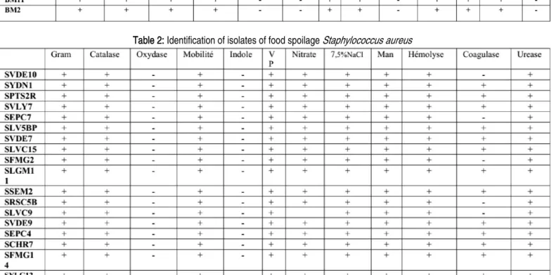
24 isolates of B. cereus and 18 of S. aureus were selected after screening of their ability of resistance to penicillin G and other antibiotics (data not shown). 5 isolates of each genus showing high resistance to the different antibiotics were selected for this study. The colonies of isolated B. cereus bacteria were big waxy white or gray, surrounded by an opacity zone. Unable to catabolize mannitol when cultured on MYP agar. The isolates were gram positive rods. Motile, and grow positively in 7% NaCl but could not grow at 50°C. (Table 1). While, the colonies of S. aureus isolates were black with an opacity zone, all Staphylococcus aureus isolates were found able to ferment mannitol sugar and positive to coagulase, and 12 strains (66,66%) of S. aureus were able to form biofilm when were cultivated on CRA agar. The isolates were gram positive cocci not motile and were able to grow in 7, 5 % NaCl (Chapman agar). The presence of S. aureus in these samples was determined by plating on BP agar, followed by confirmation via biochemical tests. (Table 2). The identification of the above species was confirmed by API 20 E and API Staph system as recommended by bergy’s manual. All
Bacillus cereus and S. aureus stains were beta-hemolytic producing phospholipase C, amylase and caseinase. The obtained results indicate that the studied Bacillus cereus and S. aureus
PAGE |
482
|
Table 1: Identification of isolates of food spoilage Bacillus cereus
Table 2: Identification of isolates of food spoilage Staphylococcus aureus
Total phenolic and Flavonoid content
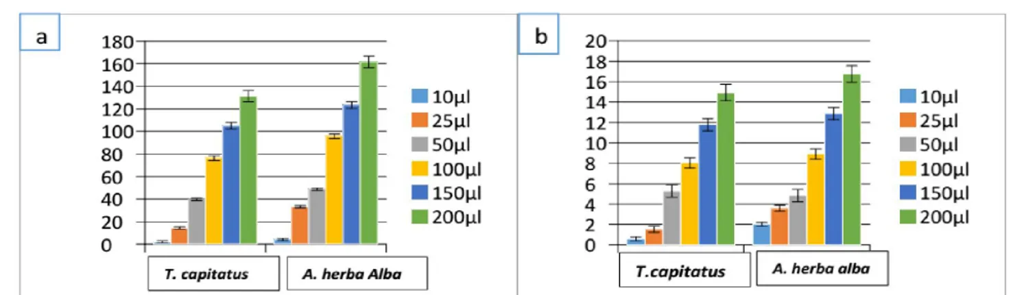
The results in figure (1) show the extraction yield of total phenols and flavonoid from the different plant extracts. This yield increase
with the increase of the concentration of the extracts used in this study and A. herba alba represents the highest content of polyphenols and flavonoids (161, 64 mg/g and 16,83mg /g) followed by T. capitatus (131, 48 mg/g and 14,96).
PAGE |
483
|
Figure 1.Total phenolic content (a) and flavonoid content (b) of T. capitatus and A. herba alba’ extracts.
Measurement of radical-scavenging activities
To evaluate the antioxidant activity, two methods were used DPPH and FRAP. For the first method and in comparison with ascorbic acid (AA) as positive control, it was found that methanolic extract of
A. herba alba represented the highest DPPH scavenging activity (91, 65% +0,001), this value was significantly different from T. capitatus extracts which represent 80,13%+ 0,027. (Figure. 2).These values were lower than those obtained by ascorbic acid (positive control) which represent 91, 79%. IC 50 values obtained
for the investigated plant extracts, methanolic extract of A. herba
alba was found to be 2, 35 mg/ml while T. capitatus extracts was 13,62 mg/ml and ascorbic acid represent a value of 12,21 mg/ml. The results indicate that the scavenging ability of methanolic extract of A. herba alba (Asso) on DPPH radical was strong. The results of FRAP method confirmed those obtained by DPPH assay and the extract of A. herba alba (Asso) represented the highest values of DO (0, 16-1, 13) at 700 nm (Figure 3). These results were correlated with the results of phenolic contents.
Figure 2.Scavenging ability of different plant extracts.
PAGE |
484
|
Antimicrobial activity of plant extracts (Agar-well
diffusion assay)
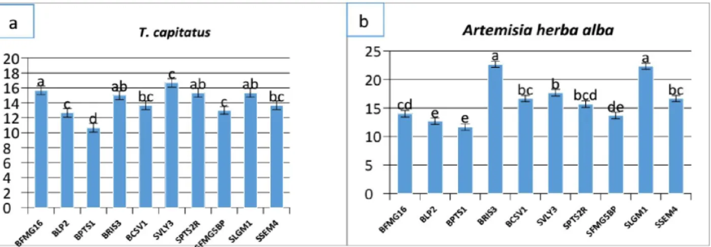
The results of antibacterial activity against strains of Bacillus cereus
and S. aureus are illustrated in figure 4. The inhibition zones of
bacterial strains obtained by the A. herba alba extract were in the range of 10,33+0,05 to 12,66+0,05 ± 1 mm, which was significantly different from those obtained by a T. capitatus extracts with 7+0,05 to 9+0,05, respectively. It can be observed that the extracts studied possessed an inhibitory effect on all tested strains, depending on plant extracts and amounts of phenolic compounds.
Figure 4. Antimicrobial activity of T. capitatus (a) and Artemesia herba alba(b) extracts against isolates of Bacillus cereus and Staphylococcus aureus.
Effect of plant extracts on the adherence and planktonic
growth of
B. cereus
and
S. aureus
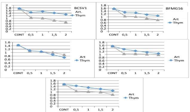
According to the obtained results we found that both methanolic extracts Thymus capitatus and Artemesia herba-alba inhibited bacterial growth of different isolates of the two species studied. The results presented in the figure (5 and 6) showed that in the case of low concentrations (0.5 mg / ml) of the tested extracts, a small decrease in bacterial growth of Bacillus cereus and Staphylococcus aureus strains (OD 1.12 to 1.09) and (OD of 1.54 to 0.75), respectively, was observed by the Thymus capitatus extract. About Artemisia herba- alba the OD values obtained are 1.21 to 1.12 for Bacillus cereus, and 1.09 to 0.75 for Staphylococcus aureus. A significant reduction in bacterial biomass formed by the two strains studied was observed in the presence of methanol extracts of
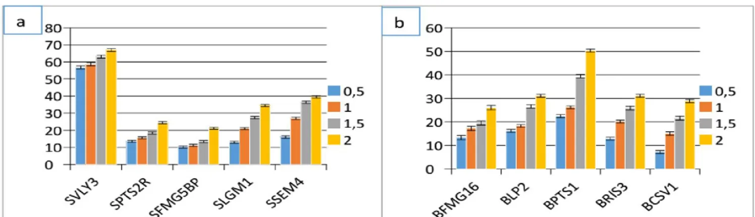
Thymus capitatus and Artemisia herba alba at a concentration of 2 mg/ ml. From the results obtained in the inhibition of bacterial growth of Staphylococcus aureus and B. cereus by the two plant extracts, we found that these inhibition differ from strain to another. The results presented in the figures 7 and 8 have shown that biofilm formation decreases with the increase of the concentration of methanolic extracts of Thymus capitatus and Artemesia herba -alba, except for BFMG16, BPTS1, SVLY3 and SSEM4 strains that the bacterial biofilm grew in the presence of plant extracts studied, which explains that these strains are resistant to our extracts tested. The antibiofilm activity of Artemisia herba-alba extract was
higher on both bacterial species than the extract of Thymus capitatus. We tested by the method of crystal violet the capacity of ten (10) different strains of our cohort belonging to Bacillus cereus and Staphylococcus aureus to form in vitro bacterial bio films in the presence of extracts from Thymus capitatus and Artemisia herba-alba. For inhibiting biofilm formation most of the tested bacterial strains was lower in the case of the concentration (0.5 mg / ml) and for Bacillus cereus strains, the percentage inhibition obtained for the extract Thymus capitatus was 7.19 up to 22.48% for the
Artemisia herba-alba was 23.41 to 40.10%. The percentage of biofilm formation inhibition obtained with the extract of the plant
Thymus capitatus against the growth of Staphylococcus aureus is 10, 20% to 56.75% for the extract of Artemisia herba-alba the values obtained are 1.13% up to 34.59%. At high concentrations (2 mg/ ml), the percent of inhibition of biofilm formation was raised to extract Thymus capitatus was 26, 02% up to 50.33%, while the
Artemisia herba-alba represented a percentage of inhibition of 38.82% to 59.42% vis-a- vis the various Bacillus cereus isolates. On Staphylococcus aureus strains were less sensitive to different concentrations of both methanolic extracts tested. The percentage inhibition obtained with the extract of Thymus capitatus was 21.12% to 67.07%, and 13.59% up to 47.53% were obtained by the extract of Artemisia herba-alba. All these results showed that the percentage of inhibition of biofilm formation by both extracts of the test plants, increases with increasing the concentration of the methanol extract of each plant extract of Artemisia herba-alba was more effective at inhibiting biofilm formation than Thymus capitatus.
PAGE |
485
|
Figure 5.Inhibition of planktonic growth of Bacillus cereus by methanolic extract of Artemisia herba-alba and Thymus capitatus.
PAGE |
486
|
Figure 7. % Inhibition of mature bio films of Staphylococcus aureus(a) and Bacillus cereus(b) by the methanolic extract of Thymus capitatus.
Figure 8. % Inhibition of mature bio films of Staphylococcus aureus(a) and Bacillus cereus(b) by the methanolic extract of Artemisia herba-alba.
Discussion
Spices are important vectors for various microorganisms implicating possible health problems for consumers as well as quality and shelf-life problems for foods. Studies on the microbiological quality of spices demonstrated profiles of micro-organisms, including total heterotrophs, Bacillus cereus, Clostridium perfringens, Staphylococcus aureus, Escherichia coli,
Salmonella and Shigella [21, 22]. Meat and milk products are found to be among the most frequently involved matrices during outbreaks of food poisoning [23, 24]. 13 staph enterotoxins foods that have been frequently incriminated in staphylococcal intoxication include meat and meat products, poultry and egg products, milk and dairy products, bakery products, particularly cream filled pastries and cakes salads, , and sandwich fillings [25, 26]. B. cereus is an important underestimated foodborne pathogen that is ubiquitous in the environment [27]. The contamination of milk and other dairy products with B. cereus is a common problem due to its effect on the quality of the products and the potential health hazards of the presence of toxigenic strains [28]. The pathogenicity of foodborne S. aureus is associated with the ability of some strains to produce enterotoxins [23]. The resistance to antimicrobials, particularly to b-lactam antibiotics, has also raised high concern as
an emerging problem in the food environment [29]. According to the results of the present study, most B. cereus group strains produced extracellular enzymes, such as protease, lipase, lecithinase, gelatinase and amylase. In Italy, Cosentino et al. [30] reported that nearly all strains with their origin in dairy products origin showed a strong enzymatic activity by hydrolyzing casein, gelatin, starch, and olive oil. In addition, Molva et al. [31] noted that most of B. cereus
and B. thuringiensis strains from cheese samples were found to produce lecithinase, gelatinase, lipase and protease. In another survey, De Jonghe et al. [32] investigated the production of the spoilage enzymes that adversely affect milk quality among the Bacillus species. Bacillus strains were strongly proteolytic and lipolytic, but lecithinase activity seems to be the less abundant trait Pathogenic staphylococci are commonly identified by their ability to produce coagulase, and thus clot blood [33]. Following Ross et al. [35], a great number of phenolic compounds can be obtained by methanol extraction such as phenolic acids, flavanons, flavanols, anthocyanins, catechins, and procyanidins. The quantification of the total phenolic content in the extracts showed that the aqueous mixtures at 70% solvent and in comparison with pure solvents, were more effective to extract the phenolic compounds. [36]. In fact, recent studies showed that many flavonoids and related polyphenols contribute significantly to the total antioxidant activity
PAGE |
487
|
of many medicinal plants [37]. Thus, our data support conclusions of others who attributed antioxidant activities to the presence of phenolic compounds in thyme [38, 39]. Recent studies showed that natural antioxidants such as polyphenols are often added to foods to stabilize them and prevent off-flavour development and considerable interest for their potential role as functional foods or nutraceuticals [40]. In fact, it has been found that antioxidant molecules such as polyphenols, flavonoids, and tannins reduce and discolour DPPH due to their hydrogen donating ability [41]. These results may be due to hydroxyl groups existing in the chemical structure of phenolic compounds from T. capitatus extracts that can provide the necessary component as a radical scavenger [42, 43, 44]. Thymol is a natural phenolic monoterpene, known for its antioxidant [45] and anti- inflammatory [46] properties. It also has a significant effect against several pathogens such as bacteria [47]. Phenolic compounds were reported to be very strong antioxidants [48]. The correlation between antioxidant activity and total phenol contents has been largely studied in different food products. [49]. It has been reported that amount of total phenolic compounds in Aseteraceae varieties is higher than in other families. Indeed, amounts of total phenolic compounds in extracts of plants we tested were higher compared to what was reported in other plants [34]. Interestingly, the amounts of total phenolics and flavonoids in
A. herba-alba extract were higher compared to what was found elsewhere [50]. Al Mustafa and Al Thunibat [51] found a phenolic amount in methanolic extracts from A. herba-alba shoots fourfold lower that what we measured. These differences may be due to variability of phenolic metabolism in different A. herba-alba organs, but also to differences in climate conditions (hot temperature, high solar exposure, dryness, short growing season) between Central Tunisia and other countries. It was reported that Artemisia herba-alba has a weaker antibacterial activity than the related species of Artemisia [52, 53]. Antibacterial activities of flavonoids were described previously [54]. Various plant products, particularly spices and extracts of various herbal plants were widely utilized as natural antimicrobials and antioxidants [55]. The demand of these herbal products is increasing because of their antimicrobial activity against many human as well as animal pathogens [56]. In clinical and foodborne pathogenesis bio film associated infection is known
as a trigger to chronic diseases, food spoilage. Even dairy and refrigerated food spoilage was also created by the bacterial bio film. [57, 58]. Developed studies are demonstrating that there is a biological rationale between quorum sensing and bio film which work on a coordinate manner leading to spoilage. [59]. Adhesion and bio film formation are also important virulence factors in S. aureus since they promote the colonization of food environments [60]. In fact, bio films formed on food processing surfaces enhance the tolerance to disinfectants, thereby increasing the risk of cross-contamination of food. Of note, bio film formation by S. aureus can be enhanced by some processing conditions used in the food industry, such as a suboptimal growth temperature or the combined presence of salt and glucose [61].
Conclusion
The contamination level of food contact surfaces with S. aureus
suggests that the handling of livestock as well as cleaning and disinfection of food industry facilities must be improved. In this respect, it must be pointed out that all isolates showed the potential ability to form bio films, which may allow them to adhere to different food contact surfaces. These bio films could be a potential source of food contamination, likely due to an unsatisfactory application of disinfectants against bio film-associated cells. In addition, food industry surfaces seemed to be a reservoir for other food pathogenic bacteria as well as for some food spoilage microorganisms coexisting with S. aureus. In turn, the presence of
S. aureus in mixed-species bio films could also enhance the colonization and persistence of this bacterium in the food environment. These results therefore point toward the need to improve hygiene conditions during the production of food.
Acknowledgments
Authors would like to express gratitude to the members of the laboratory of Microbiology and vegetal Biology at the University of Mostaganem, ALGERIA.
References
[1]. Seo KS, Bohach GA. Staphylococcus aureus. InFood Microbiology: Fundamentals and Frontiers, Third Edition American Society of Microbiology.2007;493-518.
[2]. Griffiths MW, Schraft H. Bacillus cereus
food poisoning. In D. O. Cliver, & H. P. Riemann (Eds.), Foodborne diseases (2nd Ed.). (261e270) London: Academic Press. 2002.
[3]. Feuerstein I, Muller D, Hobert K, Danin A, Segal R. The constituents of essential oilsfrom Artemisia herba alba
population of Israel and Sinai. Phytochemistry. 1986; 25:2343-2347. [4]. Akrout A, El Jani H, Amouri S, Neffati
M. Screening of antiradical and antibacterial activities of essential oils of Artemisia campestris L, Artemisia herba alba Asso and Thymus capitatus
Hoff. Et Link. Recent Research in Science and Technology. 2010; 2(1): 29-39.
[5]. Rice-Evans CA, Miller NJ, Paganga G. Structure antioxidant activity relationships of flavonoids and phenolic acids. Free Radical Biology and Medicine.1996; 20: 933-956.
[6]. Van der Veen S, Abee T. Mixed species bio films of Listeria
PAGE |
488
|
monocytogenes and Lactobacillus plantarum show enhanced resistance to benzalkonium chloride and peracetic acid. Int. J. Food Microbiol. 2011; 144:421–431.
[7]. FDA Bacteriological Analytical Manual:
Staphylococcus aureus, In: AOAC International, 8th rev. ed., Gaithersburg, MD. 1998; 12.01-12.05 [8]. Yousef AE, Carlstrom C.
Staphylococcus aureus. In: Food Microbiology: A Laboratory Manual, A Wiley- Interscience publication. 2003. [9]. Cappucino JG, Sherman N.
Microbiology A Laboratory Manuel Pearson Education (Singapore) Indian Branch. New Delhi. 2004.
[10]. Collins CH, Lyne PM, Grange JM. Collins and Lyne’s microbiological methods. 7thed. London: Arnold. 2001. [11]. Guttman DM, Ellar DJ. Phenotypic and
genotypic comparisons of 23 strains from the Bacillus cereus complex for a selection of khown and putative B. thuringesis virulence factors. FFMS Microbial LeH. 2000; 118(1): 7-13. [12]. Gudmundsdo BK. Comparison of
extracellular proteases produced by
Aeromonas salmonicida strains isolated from various fish species. Applied Bacterioly. 1996; 80: 105–113. [13]. Monica C. Medical Laboratory manual
for Tropical countries. ELBS. 1991; 60– 63.
[14]. Cotter JJ, gara JP, Mack D, Casey E. Oxygen-mediated regulation of biofilm development is controlled by the alternative sigma factor σβ in
Staphylococcus epidermidis. Appl Environ Microbiol. 2009; 75(1):261–4. [15]. Singleton VL, Rossi JA. Colorimetry of
total phenolics with phosphomolybdic– phosphotungstic acid reagents. American Journal of Enology and Viticulture. 1965; 16:144–158.
[16]. Liu X, Zhao M, Wang J, Yang B Jiang Y. Antioxidant activity of methanolic extract of emblica fruit (Phyllanthus emblica L.) from six regions in China. Journal of Food Composition and Analysis. 2008; 21: 219–228.
[17]. Shimada K, Fujikawa K, Yahara K, Nakamura T. Antioxidative properties of xanthan on the autoxidation of
soybean oil in cyclodextrin emulsion. Journal of Agricultural and Food Chemistry. 1992; 40: 945-948. [18]. Oyaizu M. Studies on products of
browning reaction. The Japanese Journal of Nutrition and Dietetics. 1986; 44(6): 307-15.
[19]. Valgas C, Souza SMD, Smânia EFA, and A. Smânia Jr: Screening methods to determine antibacterial activity of natural products. Brazilian Journal of Microbiology. 2007; 38:369–380. [20]. Christensen GD, Purisi JT, Bisno AL,
Sineson WA and Beachey EH. Characterization of clinically significant strains of coagulase-negative Staphylococci. J. Clin. Microbiol. 1983; 18: 258-264.
[21]. Powers EM, Lawyer R, Masuoka Y. Microbiology of processed spices. J. Milk Food Technol. 1975; 38: 683–687. [22]. Banerjee M, Sarkar PK. Growth and enterotoxin production by sporeforming bacterial pathogens from spices. Food Control. 2003; 15(6): 491-496.
[23]. Le Loir Y, Baron F, Gautier M.
Staphylococcus aureus and food poisoning. Genet Mol Res. 2003; 2 (1): 63-76
(www.funpecrp.com.br/gmr/year2003/v ol1- 2/sim0009_full_text.htm accessed on 18 May 2011).
[24]. Zschöck M, Botzelr D, Blöcher S, Sommerhäuser J, Hamann HP. Detection of genes for enterotoxins (ent) and toxic shock syndrome toxin-1 (tst) in mammary isolates of
Staphylococcus aureus by polymerase chain reaction. Int Dairy J. 2000; 10: 569-574.
[25]. Tamarapu, S, McKillip JL, Drake M. Development of a multiplex polymerase chain reaction assay for detection and differentiation of Staphylococcus aureus in dairy products. J. Food Prot. 2001; 64: 664–668.
[26]. Wieneke AA, Roberts D, Gilbert RJ. Staphylococcal food poisoning in the United Kingdom, 1969–1990 Epidemiol. Infect. 1993; 110: 519–531. [27]. Ceuppens S, Boon N, Uyttendaele M.
Diversity of Bacillus cereus group strains is reflected in their broad range of pathogenicity and diverse ecological
lifestyles. FEMS Microbiol Ecol. 2013; 84: 433-50
[28]. Yobouet BA, Kouame-Sina SM, Dadi ´ e A, Makita K, Grace D, ´ Dje KM, et al. Contamination of raw milk with ` Bacillus cereus from farm to retail in Abidjan, Coted ’Ivoire and possible health implications. Dairy Sci Technol. 2014; 94: 51-60.
[29]. Lee JH. Methicillin (oxacillin)-resistant
Staphylococcus aureus strains isolated from major food animals and their potential transmission to humans. Appl. Environ. Microbiol. 2003; 69:6489 – 6494.
[30]. Cosentino S, Mulargia AF, Pisano B, Tuveri P, Palmas F. Incidence and biochemical characteristics of Bacillus flora in Sardinian dairy products. Int J Food Microbiol. 1997; 38:235e8. [31]. Molva C, Sudagidan M, Okuklu B.
Extracellular enzyme production and enterotoxigenic gene profiles of Bacillus cereus and Bacillus thuringiensis strains isolated from cheese in Turkey. Food Control. 2009; 20:829-34.
[32]. De Jonghe V, Coorevits A, De Block J, Coillie EV, Grijspeerdt K, Herman L, et al. Toxinogenic and spoilage potential of aerobic spore-formers isolated from raw milk. Int J Food Microbiol. 2010; 136:318-25.
[33]. Kloos WE, Musselwhite MS. Distribution and persistence of Staphylococcus and Micrococcus species and other aerobic bacteria on human skin. Appl. Microbiol. 1975; 30:381–385.
[34]. Kaur C, Kapoor HC. Antioxidant activity and total phenolic content of some Asian vegetables. Int. J. Food. Sci. Technol. 2002; 37: 53–161.
[35]. Ross KA, Beta T, Arntfield SD. A comparative study on the phenolic acid identified and quantified in dry beans using HPLC as affected by different extraction and hydrolysis methods. Food Chemistry. 2009; 113:336–344. [36]. Rødtjer A, Skibsted LH, Andersen ML.
Antioxidative and prooxidative effect of extracts made from cherry liqueur pomace. Food Chemistry. 2006; 99: 6-14.
PAGE |
489
|
[37]. Bourgou S, Ksouri R, Bellila A, Skandrani I, Falleh H, Marzouk B. Phenolic composition and biological activities of Tunisian Nigella sativa L. shoots and roots. Comptes Rendus Biologie. 2008; 331: 48–55.
[38]. Bounatirou S, Smiti S, Miguel MG, Faleiro L, Rejeb MN, Neffati M, Costa MM, Figueiredo A, Barroso JG, Pedro LG. Chemical composition antioxidant and antibacterial activities of the essential oils isolated from Tunisian Thymus capitatus Hoff et Link. Food Chemistry. 2007; 105:146–155. [39]. Miguel G, Simoes M, Figueiredo AC,
Barroso JG, Pedro LG, Carvalho L. Composition and antioxidant activities of the essential oils of Thymus caespititius, Thymus camphoratus and Thymus mastichina. Food Chemistry. 2004; 86:183–188.
[40]. Espin JC, Garcia-Conesa, MT, Tomas-Barberan FA. Nutraceuricals: Facts and fiction. Phytochemistry. 2007; 68, 2986–3008.
[41]. Kumaran A, Karunakaran RJ. In vitro antioxidant activities of methanol extracts of five Phyllanthus species from India. Food Science and Technology. 2007; 40:344–352. [42]. Das NP, Pereira TA. Effects of
flavonoids on thermal autooxidation of Palm oil: structure- activity relationship. J. Am. Oil Chem. Soc.1990; 67: 255-258.
[43]. Matkowski A. Plant in vitro culture for the production of antioxidants - A review. Biotechnol. Adv. 2008; 26: 548-560.
[44]. Shimoi K, Masuda S, Shen B, Furugori B, Kinae N. Radioprotective effect of antioxidant plant flavonoids in mice. Mutat. Res. 1996; 350: 153-161. [45]. Braga PC, Sasso MD, Culici M,
Galastri L, Marceca MT, Guffanti EE. Antioxidant potential of thymol
determined by chemiluminescence inhibition in human neutrophilis and cell free systems. Pharmacology. 2006; 76: 61–68.
[46]. Braga PC, Dal Sasso M, Culici M, Bianchi T, Bordoni L, Marabini L. Anti- inflammatory activity of thymol: inhibitory effect on the release of human neutrophil elastase. Pharmacology. 2006; 77: 130–136. [47]. Khanuja SPS, Srivastava S, Shasney
AK, Darokar MP, Kumar TRS, Agrawal KK, Ahmed A, Patra,NK, Sinha P, Dhawan S, Saikia D, Kumar S. Formulation comprising thymol useful in the treatment of drug resistant bacterial infections. US Patent. 2004; 6: 795–824.
[48]. Pietta PG. Flavonoids as antioxidants. J. Nat. Products. 2000; 63: 1035-1042. [49]. Kiselova Y, Ivanova D, Chervenkov T,
Gerova D, Galunska B, Yankova T. Correlation between the in vitro antioxidant activity and polyphenol content of aqueous extracts from bulgarian herbs. Phytotherapy Research. 2006; 20(11):961- 965. [50]. Djeridane A, Yousfi M, Nadjemi B,
Boutassouna D, Stocker P, Vidal N. Antioxidant activity of some Algerian medicinal plants extracts containing phenolic compounds. Food Chem. 2006; 97:654–660.
[51]. Al Mustafa AH, Al Thunibat OY. Antioxidant activity of some Jordanianplants used tradionally for treatment of diabetes. Pak. J. Biol. Sci. 2008; 11, 351–358
[52]. Yashphe J, Segal R, Breuer A, Erdreich-Naftali G. Antibacterial activity of Artemisia herba alba. J. Pharm. Sci. 1979; 68(7): 924-925.
[53]. Kaur S, Shinna GK. Antibacterial activity of volatile oils and their important constitutents from some indigenous plants. Indian J. Phys. Nat.
Sci. 1982; 15: 43-47.
[54]. Middleton E Jr, Kandaswami C, Theoharides TC. The effects of plant flavonoids on mammalian cells: implications for inflammation, heart disease, and cancer. Pharmacol. Rev. 2000; 52: 673-751.
[55]. George FOA, Ephraim RN, Obasa SO, Blankole MO. Antimicrobial properties of some plant extracts on organisms associated with fish spoilage. AJMR. 2009; 6 (2):12-17.
[56]. Gur S, Balik DT and Gur N. Antimicrobial activities and some fatty acids of Turmeric, ginger root and Linseed used in the treatment of infectious diseases. World Journal of Agricultural Sciences. 2006; 2(4): 439-442.
[57]. Teh KH, Flint S, Palmer J, Andrewes P, Bremer P, Lindsay D. . Biofilm an unrecognised source of spoilage enzymes in dairy products? International Dairy Journal. 2014; 34: 32–40.
[58]. Mizan MFR, Jahid IK, Ha SD. Microbial bio films in seafood: a food-hygiene challenge. Food Microbiology. 2015; 49:41–55.
[59]. Bai AJ, Vittal RR. Quorum sensing inhibitory and anti-biofilm activity of essential oils and their in vivo efficacy in food systems. Food Biotechnology. 2014; 28:269–292.
[60]. Brooks JD, Flint SH. Bio films in the food industry: problems and potential solutions. Int. J. Food Sci. Technol. 2008; 43:2163–2176.
[61]. Rode TM, Langsrud S, Holck A, Møretrø T. Different patterns of biofilm formation in Staphylococcus aureus under food-related stress conditions. Int. J. Food Microbiol. 2007; 116:372– 383.