HAL Id: hal-03013735
https://hal.archives-ouvertes.fr/hal-03013735
Submitted on 24 Nov 2020HAL is a multi-disciplinary open access archive for the deposit and dissemination of sci-entific research documents, whether they are pub-lished or not. The documents may come from teaching and research institutions in France or abroad, or from public or private research centers.
L’archive ouverte pluridisciplinaire HAL, est destinée au dépôt et à la diffusion de documents scientifiques de niveau recherche, publiés ou non, émanant des établissements d’enseignement et de recherche français ou étrangers, des laboratoires publics ou privés.
Heterochromatin replication goes hand in hand with
telomere protection
Aaron Mendez-Bermudez, M.J. Giraud-Panis, Jing Ye, Eric Gilson
To cite this version:
Aaron Mendez-Bermudez, M.J. Giraud-Panis, Jing Ye, Eric Gilson. Heterochromatin replication goes hand in hand with telomere protection. Nature Structural and Molecular Biology, Nature Publishing Group, 2020, 27 (4), pp.313-318. �10.1038/s41594-020-0400-1�. �hal-03013735�
Heterochromatin replication goes hand in hand with telomere protection
Aaron Mendez-Bermudez1,2, Marie-Josèphe Giraud-Panis1, Jing Ye2* and Eric Gilson1,2,3*
1Côte-d’Azur University, Institute for Research on Cancer and Aging, Nice (IRCAN), CNRS UMR
7284 - INSERM U1081, France.
2International Research Laboratory “Hematology,Cancer and Aging”, Shanghai Ruijin Hospital
affiliated to Shanghai Jiaotong University, CNRS, Inserm, Côte-d’Azur University, Shanghai, China.
3CHU Nice, Department of Genetics, France
• Correspondence: [email protected] (E.G.), [email protected] (J.Y.)
Abstract
Telomeres arose from the need to stabilize natural chromosome ends, resulting in terminal chromatin structures with specific protective functions. Their constituent proteins also execute global functions within heterochromatin, where they mediate late replication and facilitate fork progression. Emerging insights into the mechanisms governing heterochromatin replication suggest concerted actions between telomeres and heterochromatin during development and aging. They also suggest a common evolutionary origin of these two chromosome regions during eukaryogenesis.
Introduction
During evolution, chromosomal DNA molecules have adopted two types of geometry: linear for all eukaryotes and circular for most prokaryotes, with the exception of some bacteria and bacteriophages. Chromosome linearity imposes two molecular problems: first, the inherent difficulty of replicating chromosomal termini and second, the potential deleterious activation of the DNA damage response (DDR) due to the presence of DNA ends. Our growing knowledge of the comparative biology of telomeres between organisms reveals a wide variety of specialized nucleoprotein organizations that function to compensate for replicative erosion (e.g. telomerase) and to protect from DDR processes (e.g. shelterin) 1. In many organisms, telomeric DNA forms a 3’ overhang that can invade internal homologous duplex DNA sequences to form a protective terminal chromatin loop (t-loop).
In this Perspective, we discuss a wealth of recent studies showing that some of the specialized telomere protective proteins also regulate heterochromatin replication. These findings reveal unexpected genome-wide concerted actions between telomeres and heterochromatin, and invite us to consider the hypothesis of a common evolutionary origin between telomeres and heterochromatin.
Heterochromatin and telomeres, old acquaintances
If the original definition of heterochromatin is a form of chromatin that remains condensed throughout the cell cycle 2, a more molecular definition is preferred today that considers heterochromatin as regions enriched in specific post-translational histone modifications,
including H3K9me3 and H3K27me3 3. A general property of heterochromatin is its ability to
repress the expression of genes located in its vicinity in a variegated manner 4.
The first functional indication of the heterochromatic character of telomeres was the demonstration of variegated expression of genes inserted in their proximity in budding yeast,
a phenomenon named TPE for Telomere Position Effect 5. In fact, constitutive
heterochromatin in budding yeast is localized within the telomeric regions, and is generated by the formation of a long array of Rap1 proteins on the telomeric DNA that serves as a
platform for the loading and spreading of the Silent Information Regulator (SIR) complex 6.
Since its initial discovery, TPE was considered an ancient mechanism of gene expression
regulation 5 and opened the way to the idea that the telomeric state can remotely influence
gene expression 7. However, telomere protection does not necessarily rely on
heterochromatin formation, since telomeres that have lost their heterochromatic character
are nonetheless propagated in budding yeast 8 or in mammalian cells 9 without any apparent
effect on their stability. Similarly, telomeres of many human cell types do not exhibit
characteristic heterochromatin marks 9,10. Nevertheless, in some circumstances, the
formation of terminal heterochromatin may serve as a protective mechanism, as in fission
yeast strains that lack telomerase 11 {Jain, 2010 #4609} and in Drosophila 12.
In agreement with the intimate links between telomere and heterochromatin, several general
heterochromatin factors are involved in telomere protection 3. In the following sections, we
consider reciprocal relationships of specialized telomere protective factors and specific regulators of heterochromatin replication genome-wide that have recently been reported.
Heterochromatic domains are generally replicated late during S-phase. Such a timed process is of paramount importance for the regulation of transcriptional programs and for
chromosome maintenance during the cell cycle, as well as differentiation and development 13.
In fission yeast, Taz1 is a key shelterin subunit that specifically binds telomeric DNA through a
C-terminal Telobox domain (Figure 1a) 14. Taz1 was found to delay replication firing of roughly
half of S. pombe late origins by associating with short stretches of nearby telomeric DNA repeats 15. An independent study reported that Taz1 also associates with a subset of late origins to assemble heterochromatin by recruiting Ccq1, another shelterin subunit that
interacts with the histone H3K9 methyltransferase complex CLRC 16. Thus, Taz1 functions with
other shelterin subunits to couple telomere protection, heterochromatin assembly, late replication firing and gene silencing at several internal regions of fission yeast chromosomes (Figure 1a).
Another protein initially identified as a telomere-specific factor, Rif1, was subsequently shown to be a general regulator of late replication timing. In budding yeast, Rif1 was discovered as a
Rap1 interacting factor that negatively regulates TPE and telomere elongation 17. These roles
of Rif1 are dependent on its interaction with Protein Phosphatase 1 (PP1), which inhibits the recruitment and activation of Tel1 (ATM, ataxia-telangiectasia mutated, in mammals), a kinase crucial for telomerase recruitment to short telomeres 18. In fission yeast, Rif1 also binds telomeres indirectly through the shelterin subunit Taz1, and contributes to telomere length regulation 19. In mammals, Rif1 does not appear to be directly recruited to telomeres, but rather exerts its telomere maintenance functions by controlling subtelomeric
heterochromatin and the expression of genes involved in telomere length regulation 20.
Overall, it appears that Rif1 is widely conserved through eukaryotic evolution connecting telomere length regulation and heterochromatin formation (Figure 1a).
Rif1 also exhibits extratelomeric functions by scheduling late replication origin firing through the recruitment of PP1, which dephosphorylates and inhibits the Dbf4-dependent cdc7 kinase (DDK), a crucial factor for origin firing that phosphorylates the replicative minichromosome maintenance complex (MCM) DNA helicase. PP1 also dephosphorylates components of the MCM complex, thus counteracting the effects of DDK. This conserved function of Rif1 was documented in fission yeast 21; budding yeast 22,23,24, Drosophila 25 and vertebrates 26,27,28,29.
In addition to its local role as PP1 recruiter to late origins, Rif1 controls and limits the number of late replicating domains as well as their spatial organization (Figure 1b) 27,30,31,32. In budding and fission yeasts, only a subset of the Rif1-controlled origins are generated by Rif1
recruitment through Rap1 and Taz1 respectively 15,33, implying that Rif1 can control the
structure of late replication domains through pathways independent from the binding to
telomeric factors. Among the known properties of Rif1, multimerization 34, as well as binding
to particular DNA conformations 35,36 or heterochromatin factors 20 {Dan, 2014 #4249} may
play important roles in its chromatin domain-organizing role. The crosstalk between telomeres and heterochromatin has important implications, since the telomere changes that occur during development and aging can impact heterochromatin replication genome-wide. For instance, in fission yeast, mutations that over-elongate telomeres lead to accelerated replication at internal late origins due to the sequestration of PP1 at telomeres in a Taz1-Rif1
dependent manner 37. In a similar way, the telomeres of budding yeast concentrate most of
the cellular Rif1 protein explaining why the Rif1 action is largely restricted to subtelomeric
origins in this organism 38. There are also examples where telomere and heterochromatin
behave in a concerted manner. For instance, replication stress in fission yeast triggers a vast chromosome spatial rearrangement leading to the telomeric association of the subset of late origins controlled by Taz1, a process that requires the two shelterin subunits Rap1 and Ccq1
39. During Drosophila development, Rif1 expression is regulated to allow late replication to
emerge at the mid-blastula transition by delaying replication at heterochromatin 25. Finally, in
mouse embryonic stem cells, Rif1 modulates the expression of the subtelomeric gene
Zscan4 involved in telomere length elongation 20.
Telomere proteins facilitate fork progression through hard-to-replicate regions
A characteristic, shared feature of telomeres and heterochromatin is their ability to hinder replication fork progression, leading to frequent paused or stalled forks that reduce
replication speed and trigger topological stress 40. The obstacles that block fork progression in
these regions are only partially understood, but include DNA secondary structures such as G quadruplexes (G4), condensed and looped chromatin, transcription of non-coding RNA forming R-loops, attachment to the nuclear matrix and envelope and, for telomeres, t-loops. In both prokaryotes and eukaryotes, excessive topological constraints at stalled forks promote their reversal, which leads to the conversion of a typical replication fork (three-way junction) into a four-way junction (resembling a Holiday junction) by the annealing of the two newly synthesized strands, forming the regressed arm, and the re-annealing of the parental strands
41 (Figure 2a). This process is believed to decrease supercoiling constraints and to transiently
protect the forks to allow more time to relieve the replication block, repair the DNA lesions
and resume fork progression 42.
Amazingly, the telomeric protective factors shown to be involved in the control of late replication timing (TRF2, Taz1 and Rif1) are also required to facilitate fork progression through hard-to-replicate regions at telomeres and at heterochromatin. The first indication came from studies of the fission yeast Taz1, which was shown to block the progression of replication forks through a stretch of telomeric DNA sequences artificially inserted inside a yeast chromosome
43. The mammalian Taz1 orthologs, TRF1 and TRF2, were also shown to have replicative
functions. Specifically, TRF1 prevents telomere replication fork stalling and ATR activation by recruiting the Bloom helicase (BLM), most likely to remove secondary structures formed by the G-rich strand of telomeres 44. For its part, TRF2 is involved in the progression of the
replisome through telomeric chromatin by relieving torsional stress 45 and by recruiting the
BUB1-BUB3 complex, a component of the spindle assembly checkpoint that acts in synergy with TRF1 and BLM, suggesting a coordination between TRF1 and TRF2 to cope with replication stress at telomeres 46.
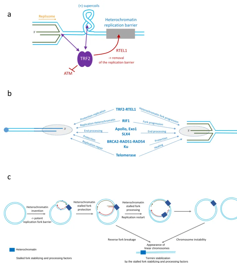
The role of TRF2 in fork progression is not limited to telomeres. Indeed, in human cancer cells, TRF2 also binds and protects the pericentromeric heterochromatin DNA sequences from DDR
activation (Figure 2a) 40,47. One of the mechanisms by which TRF2 facilitates pericentromeric
replication is through recruitment of the helicase RTEL1, which relieves heterochromatin
replication blockade 40. The effects of TRF2-RTEL1 on heterochromatin replication are most
likely linked to G4-like conformations, as they are modulated by G4 ligands and since RTEL1 is known to unwind G4 structures efficiently. These G4-related structures may constitute one of the barriers impeding replication fork progression at pericentromeric heterochromatin. TRF2 recruitment at replicating pericentromeres does not depend upon its telomeric sequence-specific DNA binding but on its capacity to bind several structural features of topologically constrained reversed stalled forks, particularly four-way DNA junctions and positive supercoils (Figure 2a) 40,48,49.
Like TRF2 and Taz1, Rif1 also controls replication fork progression, as shown in both yeast 33
and mammals, where its interaction with PP1 dephosphorylates the DNA2-WRN complex and
Was the ancestral telomere born out of a regressed replication fork?
An intriguing implication of the fork reversal process is the formation of a DNA terminus at the regressed arm that has to be protected during replication, a situation bearing striking similarities with the need to protect telomeric DNA from unwanted DDR. Indeed, regressed forks and telomeres exhibit numerous similarities (Figure 2b and Figure 3), and regressed
forks share several mechanisms of formation with t-loops 49,53,54,55. Moreover, regressed fork
and telomeres are protected by common factors 33,40,50,51,52,56,57,58,59,60. Not only are formation
and protection similar between telomeres and regressed forks, but also the way that their DNA termini are processed via a controlled 5’ resection reaction involving the nucleases Apollo
61,62 and Exo1 63. It is also noteworthy that SLX4, which acts as a scaffold to coordinate the
recruitment of various structure-specific endonucleases involved in the processing of stalled
forks, interacts with telomeres via TRF2 64. Finally, telomeric DSBs and regressed forks can be
healed by telomerase in yeast and mammalian cells 65,66.
Together, these findings suggest that the mechanisms that were implemented during early eukaryogenesis to protect regressed forks and telomeres were intertwined and possibly of common origin (Figure 3). We propose that, during eukaryogenesis, the evolution of condensed heterochromatin-like structures led to massive replicative and topological stresses that required robust mechanisms to be put in place to allow the DNA to be faithfully replicated. In the light of our present knowledge of the common mechanisms protecting telomeres and regressed forks discussed above, these ancestral mechanisms might include proteins that recognize positive supercoils, such as the TRFH domain of the shelterin complex, and that promote four-way DNA junction formation, such as the basic domain of TRF2, to
sense and remodel topologically blocked forks. In this hypothesis, the origin of ancestral telomeres would stem from replication difficulties at the time of heterochromatin invention. In favor of the scenario that regressed forks can be a source of free chromosome ends, replicative stress in E. coli can generate cells with a heritable but unstable chromosome
carrying a broken end 67. Thus, the early evolution of factors protecting DNA double-strand
breaks (DSBs) of the regressed arm of regressed forks could have been employed as a “first aid” to protect the free ends of primitive linearized genomes.
A prior model of telomere invention during eukaryogenesis proposed that the invasion of ancestral circular genomes by group II introns would have created conditions that stabilize
DNA termini by allowing the formation of primitive t-loops 68. It is quite conceivable that group
II intron repeats would have also favored heterochromatin formation to limit their propagation as it is observed today for retrotransposons and consistent with the
demonstrated toxicity conferred by an experimental invasion of group II introns in E. coli 69.
Therefore, the group II intron model of telomere invention by t-loop formation is compatible with the one proposed here based on replication accident at ancestral heterochromatin.
Conclusions
A direct link between telomeres and heterochromatin replication genome-wide represents an important breakthrough in our understanding of the specific mechanisms of heterochromatin replication and how telomeres and heterochromatin act in concert to control chromosome maintenance and function. Moreover, such findings invite us to envisage a common evolutionary origin between telomeres and heterochromatin.
We believe that the genome-wide roles of key telomere protective proteins in heterochromatin replication (reviewed here) and in transcriptional regulation 70, reflect a
fundamental need to couple the telomeric state to cell fate. It is therefore quite conceivable that in the course of evolution, the original relationships between telomeres, heterochromatin replication and transcriptional regulation genome-wide have been preserved and diversified. Such mechanisms are expected to provide an explanation for the broad contributions of telomeres to development as well as normal and pathological ageing.
Acknowledgments
This work in the EG lab was supported by the cross-cutting Inserm program on aging (AGEMED), the ANR program TELOCHROM, the Sino-French PHC Cai Yuanpei program and the ‘‘Investments for the Future’’ LABEXSIGNALIFE (reference ANR-11-LABX-0028-01). We apologize to all authors whose publications have not been cited due to space limitation.
Figure 1. Telomere proteins and late replicating domains.
a) Schematic representation comparing the DNA-protein organization of telomeres, subtelomeres and heterochromatin (late replication) domains in Saccharomyces cerevisiae,
Schizosaccharomyces pombe, and mammals. Heterochromatin domains are boxed in grey
rectangles.
b) Higher-order chromatin structures controlled by Rif1. In S. cerevisiae, telomeres are clustered at the nuclear periphery, forming a heterochromatic late-replicating domain where the Rif1 proteins are enriched. In mammals, Rif1 determines the formation of late-replicating heterochromatin domains. The divergent arrows within the late replicating domains indicate the ability of Rif1 to assemble and limit the 3D organization of these domains in both yeast and mammalian nuclei.
Figure 2. Telomere proteins and heterochromatin replication elongation.
a) TRF2 is required to protect stalled forks formed in pericentromeric heterochromatin and to facilitate replication fork progression by recruiting the helicase RTEL1 and blunting ATM activation. TRF2 can bind stalled forks within heterochromatin through the positively supercoiled DNA that accumulates ahead of the stalled replication fork or the four-way DNA junctions of the reversed fork.
b) Similarities between the protective mechanisms at play at telomeres (left) and at reverse stalled forks (right). Common factors and their context-dependent functions are indicated.
Figure 3. Model of telomere evolution.
At the early stages of eukaryogenesis, we hypothesise that the appearance of heterochromatin-like structures within circular chromosomes triggered a high level of replicative and topological stress, leading to the formation of reversed stalled forks at a high rate and, consequently, the requirement for potent mechanisms of reversed fork protection and maturation. This, in turn, may have favoured stabilization of linearized chromosomes resulting either from stalled fork breakage or chromosome instability generated, for instance, by group II intron invasion, high levels of ultraviolet irradiation and oxidative stress, or frequent desiccation periods.
References
1. Giraud-Panis, M.J. et al. One identity or more for telomeres? Front Oncol 3, 48 (2013).
2. Heitz E. Das Heterochromatin der Moose. Jarhb. Wiss. Bot. 69, 762-818 (1928).
3. Nishibuchi, G. & Dejardin, J. The molecular basis of the organization of repetitive DNA-containing constitutive heterochromatin in mammals. Chromosome Res 25, 77-87 (2017).
4. Müller H.J. The remaking of chromosomes. The Collecting Net 13, 182-198 (1938).
5. Gottschling D.E., Aparicio O.M., Billington B.L. & Zakian, V.A. Position effect at
S. cerevisiae telomeres : reversible represssion of Pol II transcription. Cell 63,
751-762 (1990).
This paper reveals that gene expression can be repressed due to telomere proximity
6. Gasser, S.M. & Cockell, M.M. The molecular biology of the SIR proteins. Gene 279,
1-16 (2001).
7. Maillet, L. et al. Evidence for silencing compartments within the yeast nucleus: a role
for telomere proximity and Sir protein concentration in silencer-mediated repression.
Genes Dev 10, 1796-811 (1996).
8. Palladino F. et al. SIR3 and SIR4 proteins are required for the positioning and integrity
of yeast telomeres. Cell 75, 543-555 (1993).
9. Gauchier, M. et al. SETDB1-dependent heterochromatin stimulates alternative
lengthening of telomeres. Sci Adv 5, eaav3673 (2019).
10. Cubiles, M.D. et al. Epigenetic features of human telomeres. Nucleic Acids Res 46,
2347-2355 (2018).
11. Jain, D., Hebden, A.K., Nakamura, T.M., Miller, K.M. & Cooper, J.P. HAATI
survivors replace canonical telomeres with blocks of generic heterochromatin.
Nature 467, 223-7 (2010).
This paper provides evidence in fission yeast for the use of heterochromatic blocks to replace telomeres and to provide end protection in the absence of telomerase.
12. Fanti, L., Giovinazzo, G., Berloco, M. & Pimpinelli, S. The heterochromatin protein 1
prevents telomere fusions in Drosophila. Mol Cell 2, 527-38 (1998).
13. Zhao, P.A., Rivera-Mulia, J.C. & Gilbert, D.M. Replication Domains: Genome
Compartmentalization into Functional Replication Units. Adv Exp Med Biol 1042, 229-257 (2017).
14. Cooper J P., Nimmo E R., Allshire R C. & Cech, T.R. Regulation of telomere length
and function by a Myb-domain protein in fission yeast. Nature 385, 744-747 (1997).
15. Tazumi, A. et al. Telomere-binding protein Taz1 controls global replication timing
through its localization near late replication origins in fission yeast. Genes Dev 26, 2050-62 (2012).
This paper shows the Taz1 replication timing control of a subset of late origins. This is mediated by the direct binging of Taz1 to telomeric repeats found proximal to origins.
16. Zofall, M., Smith, D.R., Mizuguchi, T., Dhakshnamoorthy, J. & Grewal, S.I.S.
Taz1-Shelterin Promotes Facultative Heterochromatin Assembly at Chromosome-Internal Sites Containing Late Replication Origins. Mol Cell 62, 862-874 (2016). This work shows that Shelterin subunits assemble facultative heterochromatin at internal chromosomal sites containing late replication origins.
17. Hardy C.F.J., Balderes D. & Shore, D. Dissection of a carboxy-terminal region of the
yeast regulatory protein RAP1 with effects on both transcriptional activation and silencing. Mol. Cell. Biol. 12, 1209-1217 (1992).
18. Kedziora, S. et al. Rif1 acts through Protein Phosphatase 1 but independent of
replication timing to suppress telomere extension in budding yeast. Nucleic Acids Res 46, 3993-4003 (2018).
19. Miller, K.M., Ferreira, M.G. & Cooper, J.P. Taz1, Rap1 and Rif1 act both
interdependently and independently to maintain telomeres. EMBO J 24, 3128-35 (2005).
20. Dan, J. et al. Rif1 maintains telomere length homeostasis of ESCs by mediating
21. Hayano, M. et al. Rif1 is a global regulator of timing of replication origin firing in fission yeast. Genes Dev 26, 137-50 (2012).
This paper reveals that the global replication timing in S. pombe is controled by Rif1 in a Taz1 independent fashion.
22. Hiraga, S. et al. Rif1 controls DNA replication by directing Protein Phosphatase 1
to reverse Cdc7-mediated phosphorylation of the MCM complex. Genes Dev 28, 372-83 (2014).
This paper shows that the budding yeast Rif1 controls DNA replication genome-wide and that Rif1 exerts this control through PP1-mediated dephosphorylation of the MCM complex early in the cell cycle.
23. Mattarocci, S. et al. Rif1 controls DNA replication timing in yeast through the PP1
phosphatase Glc7. Cell Rep 7, 62-9 (2014).
This work links Rif1 to the negative regulation of replication origin firing through recruitment of the Glc7 phosphatase.
24. Dave, A., Cooley, C., Garg, M. & Bianchi, A. Protein phosphatase 1 recruitment
by Rif1 regulates DNA replication origin firing by counteracting DDK activity.
Cell Rep 7, 53-61 (2014).
This paper shows that Rif1, regulates replication timing in budding yeas through its interaction with PP1 phosphatases.
25. Seller, C.A. & O'Farrell, P.H. Rif1 prolongs the embryonic S phase at the
Drosophila mid-blastula transition. PLoS Biol 16, e2005687 (2018).
This study demonstrates the direct role played by Rif1 during development by delaying the replication of satellite sequences.
26. Cornacchia, D. et al. Mouse Rif1 is a key regulator of the replication-timing programme
in mammalian cells. EMBO J 31, 3678-90 (2012).
27. Yamazaki, S. et al. Rif1 regulates the replication timing domains on the human genome.
EMBO J 31, 3667-77 (2012).
28. Sukackaite, R. et al. Mouse Rif1 is a regulatory subunit of protein phosphatase 1 (PP1).
Sci Rep 7, 2119 (2017).
29. Alver, R.C., Chadha, G.S., Gillespie, P.J. & Blow, J.J. Reversal of DDK-Mediated
MCM Phosphorylation by Rif1-PP1 Regulates Replication Initiation and Replisome Stability Independently of ATR/Chk1. Cell Rep 18, 2508-2520 (2017).
30. Foti, R. et al. Nuclear Architecture Organized by Rif1 Underpins the
Replication-Timing Program. Mol Cell 61, 260-73 (2016).
This study identifies Rif1 as the molecular link between nuclear architecture and replication-timing establishment in mammals.
31. Toteva, T. et al. Establishment of expression-state boundaries by Rif1 and Taz1 in
fission yeast. Proc Natl Acad Sci U S A 114, 1093-1098 (2017).
32. Ogawa, S. et al. Shelterin promotes thethering of late replication origins to telomeres
for replication-timing control. EMBO J 37, e98997 (2018).
33. Hiraga, S.I. et al. Budding yeast Rif1 binds to replication origins and protects DNA at
blocked replication forks. EMBO Rep 20, e48152 (2019).
34. Moriyama, K., Yoshizawa-Sugata, N. & Masai, H. Oligomer formation and
G-quadruplex binding by purified murine Rif1 protein, a key organizer of higher-order chromatin architecture. J Biol Chem 293, 3607-3624 (2018).
35. Kanoh, Y. et al. Rif1 binds to G quadruplexes and suppresses replication over long
distances. Nat Struct Mol Biol 22, 889-97 (2015).
36. Masai, H. et al. Rif1 promotes association of G-quadruplex (G4) by its specific G4
37. Hasegawa, Y., Yamamoto, M., Miyamori, J. & Kanoh, J. Telomere DNA length-dependent regulation of DNA replication timing at internal late replication origins. Sci
Rep 9, 9946 (2019).
38. Hafner, L. et al. Rif1 Binding and Control of Chromosome-Internal DNA
Replication Origins Is Limited by Telomere Sequestration. Cell Rep 23, 983-992 (2018).
This work shows that the global replication dynamics in budding yeast are controlled by telomeric sequestration of Rif1 by Rap1.
39. Mizuguchi, T. et al. Shelterin components mediate genome reorganization in response
to replication stress. Proc Natl Acad Sci U S A 114, 5479-5484 (2017).
40. Mendez-Bermudez, A. et al. Genome-wide Control of Heterochromatin
Replication by the Telomere Capping Protein TRF2. Mol Cell 70, 449-461 e5 (2018).
This paper provides evidence for the involvment of TRF2 in pericentromereic heterochromaétin replication by the recruitment of the helicase RTEL1.
41. Postow, L. et al. Positive torsional strain causes the formation of a four-way junction at
replication forks. J Biol Chem 276, 2790-6 (2001).
42. Rickman, K. & Smogorzewska, A. Advances in understanding DNA processing and
protection at stalled replication forks. J Cell Biol 218, 1096-1107 (2019).
43. Miller, K.M., Rog, O. & Cooper, J.P. Semi-conservative DNA replication through
telomeres requires Taz1. Nature 440, 824-8 (2006).
44. Zimmermann, M., Kibe, T., Kabir, S. & de Lange, T. TRF1 negotiates TTAGGG
repeat-associated replication problems by recruiting the BLM helicase and the TPP1/POT1 repressor of ATR signaling. Genes Dev 28, 2477-91 (2014).
45. Ye, J. et al. TRF2 and apollo cooperate with topoisomerase 2alpha to protect human
telomeres from replicative damage. Cell 142, 230-42 (2010).
46. Li, F. et al. The BUB3-BUB1 Complex Promotes Telomere DNA Replication. Mol Cell
70, 395-407 e4 (2018).
47. Simonet, T. et al. The human TTAGGG repeat factors 1 and 2 bind to a subset of
interstitial telomeric sequences and satellite repeats. Cell Res 21, 1028-38 (2011).
48. Fouche, N. et al. The basic domain of TRF2 directs binding to DNA junctions
irrespective of the presence of TTAGGG repeats. J Biol Chem 281, 37486-95 (2006).
49. Poulet, A. et al. TRF2 promotes, remodels and protects telomeric Holliday junctions.
EMBO J 28, 641-51 (2009).
50. Alabert, C. et al. Nascent chromatin capture proteomics determines chromatin dynamics
during DNA replication and identifies unknown fork components. Nat Cell Biol 16, 281-93 (2014).
51. Garzon, J., Ursich, S., Lopes, M., Hiraga, S.I. & Donaldson, A.D. Human RIF1-Protein
Phosphatase 1 Prevents Degradation and Breakage of Nascent DNA on Replication Stalling. Cell Rep 27, 2558-2566 e4 (2019).
52. Mukherjee, C. et al. RIF1 promotes replication fork protection and efficient restart to
maintain genome stability. Nat Commun 10, 3287 (2019).
53. Fouche, N., Ozgur, S., Roy, D. & Griffith, J.D. Replication fork regression in repetitive
DNAs. Nucleic Acids Res 34, 6044-50 (2006).
54. Verdun, R.E. & Karlseder, J. The DNA damage machinery and homologous
recombination pathway act consecutively to protect human telomeres. Cell 127, 709-20 (2006).
55. Amiard, S. et al. A topological mechanism for TRF2-enhanced strand invasion. Nat
56. Sarek, G., Vannier, J.B., Panier, S., Petrini, J.H. & Boulton, S.J. TRF2 recruits RTEL1 to telomeres in S phase to promote t-loop unwinding. Mol Cell 57, 622-35 (2015).
57. Jaco, I. et al. Role of mammalian Rad54 in telomere length maintenance. Mol Cell Biol
23, 5572-80 (2003).
58. Badie, S. et al. BRCA2 acts as a RAD51 loader to facilitate telomere replication and
capping. Nat Struct Mol Biol 17, 1461-9 (2010).
59. Sfeir, A. & de Lange, T. Removal of shelterin reveals the telomere end-protection problem. Science 336, 593-7 (2012).
60. Teixeira-Silva, A. et al. The end-joining factor Ku acts in the end-resection of double
strand break-free arrested replication forks. Nat Commun 8, 1982 (2017).
61. Lam, Y.C. et al. SNMIB/Apollo protects leading-strand telomeres against
NHEJ-mediated repair. EMBO J 29, 2230-41.
62. Mason, J.M. et al. The SNM1B/APOLLO DNA nuclease functions in resolution of
replication stress and maintenance of common fragile site stability. Hum Mol Genet 22, 4901-13 (2013).
63. Aggarwal, M., Sommers, J.A., Morris, C. & Brosh, R.M., Jr. Delineation of WRN
helicase function with EXO1 in the replicational stress response. DNA Repair (Amst) 9, 765-76 (2010).
64. Guervilly, J.H. & Gaillard, P.H. SLX4: multitasking to maintain genome stability. Crit
Rev Biochem Mol Biol 53, 475-514 (2018).
65. Myung, K., Chen, C. & Kolodner, R.D. Multiple pathways cooperate in the suppression
of genome instability in Saccharomyces cerevisiae. Nature 411, 1073-6 (2001).
66. Margalef, P. et al. Stabilization of Reversed Replication Forks by Telomerase Drives
Telomere Catastrophe. Cell 172, 439-453 e14 (2018).
67. Sinha, A.K. et al. Broken replication forks trigger heritable DNA breaks in the terminus
of a circular chromosome. PLoS Genet 14, e1007256 (2018).
68. de Lange, T. A loopy view of telomere evolution. Front Genet 6, 321 (2015).
69. Lee, G. et al. Testing the retroelement invasion hypothesis for the emergence of the ancestral eukaryotic cell. Proc Natl Acad Sci U S A 115, 12465-12470 (2018).
70. Ye, J., Renault, V.M., Jamet, K. & Gilson, E. Transcriptional outcome of telomere signalling. Nat Rev Genet 15, 491-503 (2014).
3’ 5’
Subtelomeric heterochromatin -> Telomeric Position Effect
(TPE) HP1 HP1 HP1 HP1 Rap1 TRF1 TRF2 TIN2 Tpp1 Pot1 Rap1 TRF1 TRF2 TIN2 Tpp1 Rap 1 TRF 1 TRF 2 TIN 2 Tp p1 Rap1 TRF1 TRF2 TIN 2 Tp p 1 Rap1 TRF1 TRF2 TIN2 Tpp1 Rif1 Rif1 PP1 ORI DDK ?
late replication domain Heterochromatin TRF2
Figure 1
Saccharomyces cerevisiae Schizosaccharomyces pombe Mammalsb
a
Sir4 Sir3 Sir2 Rap1 Sir4 Sir3 Sir2 Rap1 Sir4 Sir3 Sir2 Sir4 Sir3 Sir2 Sir2 Sir spreading Subtelomeric heterochromatin -> Telomeric Position Effect(TPE)
Telomere
Telomerase
Rif1
PP1 Tel1/ATM
Late replication origins
3’ 5’ Sir4 Sir3 Rif1 PP1 ORI DDK ? Rif1 PP1 ORI DDK Rap1 Rif1 PP1 ORI DDK ? Taz1-independent late replication domain
Rap1 Ccq1 Tpz1 Poz1 Taz1 Subtelomeric heterochromatin -> Telomeric Position Effect
(TPE) Telomere Rif1 5’ Rif1 PP1 ORI DDK Swi6/HP1 Taz1 Taz1 Swi6/HP1 Swi6/HP1 Pot1 CLRC Rap1 Ccq1 Tpz1 Poz1 Taz1 Rif1 Swi6/HP1 CLRC Rap1 Ccq1 Poz1 Taz1 Tpz1 Interstitial heterochromatin late replication domain
mammals
S. cerevisiae
Early replicating region Late replicating region
Rif1 PP1 PP1 Rif1 Rif1 PP1 Rap1 Rif1 Rap1 Rif1 Rap1 ORI ORI PP1 Rif1
Figure 2
a
b
c
Apollo, Exo1 SLX4 BRCA2-RAD51-RAD54 Ku TRF2-RTEL1 3’ RIF1 Telomerase 3’ Protecti on-replication Heterochromatin fo
rk progressio n Replication-Heterochro
matin
Fork progression
End processing End processing
Protection Protection Healing Replication-r epair Heterochromatin
Stalled fork stabilizing and processing factors
Appearance of linear chromosomes x x x Heterochromatin invention -> potent replication fork barrier
Heterochromatin stalled fork protection Heterochromatin stalled fork processing Replication restart
Reverse fork breakage Chromosome instability
Termini stabilization
by the stalled fork stabilizing and processing factors 3’ Heterochromatin replication barrier (+) supercoils Replisome RTEL1 -> removal of the replication barrier
ATM
Figure 1. Telomeric-heterochromatic DNA connection.
a) Saccharomyces cerevisiae telomeres are coated with Rap1 proteins, serving as platform for the binding and spreading of the SIR complex to the subtelomeres. In addition, Rap1 is the binding site of Rif1, whose interaction with PP1 suppresses the firing of late origins at subtelomeres. In
Schizosaccharomyces pombe, Taz1 is a key shelterin protein, that not only protects telomeres but
also controls the firing of late origins through the recruitment of Rap1 and the Rif1-PP1 complex. Rif1-PP1 can also modulate late origins independently of Taz1-Rap1. In addition, the formation of subtelomeric heterochromatin is mediated by another shelterin protein, Ccq1, that together with the CLRC complex mediates the spreading of the heterochromatic Swi6/HP1 protein. Mammalian telomeres lack the common heterochromatic marks, however, the subtelomeric region is rich in H3K9me3, H3K20me3 and HP1. Rif1 is involved in the maintenance of subtelomeric
heterochromatin as well as in the control of late replication origin timing. Is still unknown how Rif1 binds late replicating regions independently of Rap1. Grey rectangles represent
heterochromatic DNA blocks.
b) Subnuclear organization of Rif1 domains. In S. cerevisiae, telomeres are cluster into few nuclear envelop foci where the Rif1 protein is mainly found. The high concentration of Rif1 in these regions create a late replication compartment. In mammals, Rif1 modulates nuclear architecture by regulating the boundaries between euchromatin and heterochromatin creating late replicating regions.
Figure 2. Similarities between telomeres and stalled forks
a) Several proteins involved in telomere protection, repair and replication are also part of the factors involved in the protection and processing of stalled replication forks generated in difficult-to-replicate regions of the genome.
b) TRF2 is required to protect stalled forks formed in pericentromeric heterochromatin and
facilitates the progression of the replication fork by recruiting the helicase RTEL1. TRF2 can bind heterochromatic DNA in several ways: through the presence of G4s, positive supercoil DNA accumulating ahead of stalled replication fork, and four-way DNA junctions.
c) Model of telomere formation. The appearance of heterochromatin-like structures or the generation of abnormal DNA structures (e.g. by high levels of ultraviolet irradiation, high
production of reactive oxygen species or frequent desiccation periods) led to huge replicative and topological stress. All this created major problems in the progression of replication forks giving rise to a high rate of reversed stalled forks and double strand breaks. The stabilization of the ancestral stalled forks may have behaved as the first mechanism of stabilization of ancestral telomeres.