HAL Id: hal-01913815
https://hal.archives-ouvertes.fr/hal-01913815
Submitted on 6 Nov 2018HAL is a multi-disciplinary open access archive for the deposit and dissemination of sci-entific research documents, whether they are pub-lished or not. The documents may come from teaching and research institutions in France or abroad, or from public or private research centers.
L’archive ouverte pluridisciplinaire HAL, est destinée au dépôt et à la diffusion de documents scientifiques de niveau recherche, publiés ou non, émanant des établissements d’enseignement et de recherche français ou étrangers, des laboratoires publics ou privés.
Macrophages of distinct origins contribute to tumor
development in the lung
Pierre-Louis Loyher, Pauline Hamon, Marie Laviron, Aïda
Meghraoui-Kheddar, Elena Goncalves, Zihou Deng, Sara Torstensson, Nadège
Bercovici, Camille Baudesson De Chanville, Béhazine Combadière, et al.
To cite this version:
Pierre-Louis Loyher, Pauline Hamon, Marie Laviron, Aïda Meghraoui-Kheddar, Elena Goncalves, et al.. Macrophages of distinct origins contribute to tumor development in the lung. Journal of Experi-mental Medicine, Rockefeller University Press, 2018, 215 (10), pp.2536 - 2553. �10.1084/jem.20180534�. �hal-01913815�
1
Macrophages
of
distinct
origins
contribute
to
tumor
development in the lung
Pierre-Louis Loyher1,4*, Pauline Hamon1*, Marie Laviron1, Aïda Meghraoui-Kheddar1, Elena Goncalves1, Zihou Deng4, Sara Torstensson1, Nadège Bercovici2, Camille Baudesson de Chanville1, Béhazine Combadière1, Frederic Geissmann4, Ariel Savina3, Christophe Combadière1 and Alexandre Boissonnas1.
1
Sorbonne Université, Institut universitaire de cancérologie (IUC), Inserm, CNRS,
Centre d’Immunologie et des Maladies Infectieuses CIMI, F-75013, Paris, France.
2
Inserm, U1016, Institut Cochin, CNRS UMR8104, Université Paris Descartes,
Sorbonne Paris Cité, Paris, France.
3
Institut Roche, 30, Cours de l'Ile Seguin, 92650 Boulogne-Billancourt Cedex,
France.
4
Immunology Program, Sloan Kettering Institute, Memorial Sloan Kettering Cancer
Center, New York, New York 10065, USA.
Corresponding author: alexandre.boissonnas@upmc.fr
*PL.L. and P.H. are co-first authors
Non-standard abbreviations: TAM: Tumor-associated macrophage Res-TAMs: Tissue-resident macrophages MoD-TAMs: Monocyte derived macrophages
PyMT-Chova: MMTV PyMT-P2A-mCherry-P2A-OVA mice OH-TAMs: 4-hydroxytamoxifen
AM: Alveolar macrophage IM: Interstitial macrophage Mo: Monocyte
CP: Cyclophosphamide
2 Abstract
Tissue-resident macrophages can self-maintain without contribution of adult hematopoiesis. Herein we show that tissue-resident interstitial macrophages (Res-TAMs) in mouse lungs contribute to the pool of tumor-associated macrophages (TAMs) together with CCR2-dependent recruited macrophages (MoD-TAMs). Res-TAMs largely correlated with tumor cell growth in vivo while MoD-TAMs accumulation was associated with enhanced tumor spreading. Both cell subsets were depleted after chemotherapy, but MoD-TAMs rapidly recovered and carried out phagocytosis-mediated tumor clearance. Interestingly, anti-VEGF treatment combined with chemotherapy inhibited both Res and Mod-TAM reconstitution without affecting monocyte infiltration and improved its efficacy. Our results reveal that the developmental origin of TAMs dictates their relative distribution, function and response to cancer therapies in lung tumors.
3 Introduction
The tumor-microenvironment (TME) can regulate malignant potential and contributes to tumor heterogeneity. Tumor-associated macrophages (TAMs) are the most abundant host cells within the TME (Qian and Pollard, 2010) and have been implicated in the promotion of invasiveness (Wyckoff et al., 2007), growth (Pollard, 2004), angiogenesis (Lewis et al., 2016), metastasis (Kitamura et al., 2015) and immunosuppression (Boissonnas et al., 2013; Broz et al., 2014). TAMs have been suggested to limit the efficacy of chemotherapeutic agents and to promote tumor relapse (Hughes et al., 2015), although they can in some cases be required for optimal therapy response (De Palma and Lewis, 2013).
It is considered that TAMs mainly arise from the differentiation of monocytic precursors (Cortez-Retamozo et al., 2012; Franklin et al., 2014). However, in many tissues, pools of resident macrophages have been identified; these originate from embryonic precursors and self-maintain independently of hematopoietic stem cells (Gomez Perdiguero et al., 2015). Distinct transcriptional programs initiated in embryonic, fetal or adult progenitors (Mass et al., 2016) and the exposure to specific tissue environments (Gosselin et al., 2014; Lavin et al., 2014) may explain the specialization and diversity of macrophages in healthy as well as neoplastic tissues. The lung environment is densely colonized by subsets of mononuclear phagocytic cells displaying various spatial organizations, functions and dependence for blood monocytes in their maintenance. Interstitial macrophages (IMs) represent a discrete population in the steady state lung largely outnumbered by alveolar macrophages (AMs) (Gibbings et al., 2017; Rodero et al., 2015). IMs and AMs express different surface markers which allow their identification and they have been described to arise from distinct developmental waves without interconverting (Guilliams et al., 2013; Tan and Krasnow, 2016).
So far, the contribution of these different resident macrophage subsets in the generation of lung TAMs has not been reported.
4 Herein, the TAM network in lung tumors was studied based on transgenic fluorescent reporter mice and fate mapping models that enable the discrimination of the lung mononuclear phagocyte subsets according to their origin and localization. We showed that the TAM compartment is intermingled by both yolk sac-derived interstitial and monocyte-derived recruited macrophages, differentially represented in the TME depending on the anatomical site of tumor development in the lung. Finally, we highlight their respective implication on lung tumor development and response to various anti-cancer therapies.
5 Results
Lung macrophage subsets differentially accumulate during tumor development
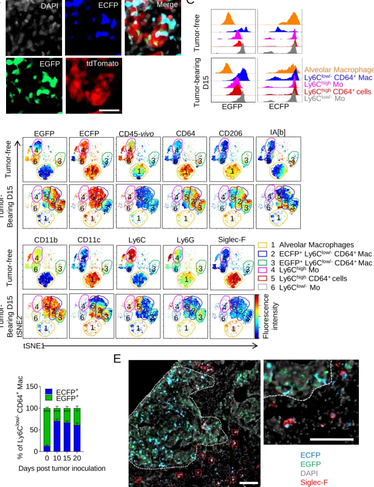
We studied the impact of tumor growth on the different subset of lung myeloid cells after inoculating TC-1 lung carcinoma cells, which induce multifocal tumor nodules (Ji et al., 1998; Lin et al., 1996). The tumor-associated myeloid signature was monitored along tumor evolution using flow cytometry phenotyping combined with a non-supervised viSNE analysis. The generated tSNE-plot was calculated with 12 parameters including cell anatomic distribution between the tissue parenchyma and the vasculature. This distinction is achievable using anti-CD45 antibody injected intravenously that allows a blood/tissue partitioning of cells (see dashed gates Figure 1A and S1). Ten clusters obtained using unsupervised analysis were subsequently assigned to a specific cell population according to expression level of each marker and previously described phenotypes (Gibbings et al., 2017; Misharin et al., 2013; Sabatel et al., 2017) (Figure S1A). Briefly, cluster 7 and cluster 8 were identified as classical Ly6Chigh and non-classical Ly6Clow/- monocytes (Mo) respectively, with CD11bhighSiglec-F-Ly6G-Fc-gamma receptor 1low (CD64low) expression profile. Cluster 2 included CD11blowCD11chighSiglec-Fhigh cells, representing alveolar macrophages (AMs), while cluster 1 included CD11bhighSiglec-F-Ly6G-CD64+ cells representing a distinct subset of lung macrophages named here Ly6Clow/-CD64+ Mac. These different macrophage subsets were clearly distinguished from cluster 3, identified as CD11b-I-A[b]+CD11c+CD103+Ly6C -CD64- cells and cluster 5 identified as CD11b+I-A[b]+CD11c+Ly6C-CD64- cells known as conventional dendritic cells cDC1 and cDC2 respectively. Cluster 6 and 9 are defined as CD11bhighLy6G+ cells (representing two subsets of neutrophils with differential expression of CD24). Cluster 10 represented CD11bhighSiglec-Fint cells identified as eosinophils (Figure 1A, S1A-B). In the absence of tumor, Ly6Chigh-Mo (cluster 7) and Ly6Clow/--Mo (cluster 8) resided almost exclusively within lung vessels whereas AMs (cluster 2) and Ly6Clow/-CD64+ Mac (cluster 1) were detected in the lung parenchyma only (Figure 1A and S1C). With tumor expansion, cluster 4 including Ly6Chigh-CD64+ cells appeared together with a progressive and
6 massive accumulation of Ly6Clow/-CD64+ Mac (cluster 1) (Figure 1A). Supervised analysis of Ly6C, CD64 expression and intravascular cell labeling on CD11b+Siglec-F-Ly6G- gated cells suggested that Ly6Chigh-Mo progressively upregulate CD64 and differentiate into Ly6C low/-CD64+ Mac upon tumor infiltration (Figure S1C-D). In contrast, Ly6Clow/--Mo did not apparently accumulate and they remained mostly intravascular (Figure 1 and S1). Blood/tissue partitioning of monocyte and macrophage subsets (Figure 1C-D) was next performed using supervised analysis and the observations made on tSNE plots were confirmed. While the Ly6Clow/-CD64+ Mac subset massively accumulated in the tumor parenchyma exclusively, the number of AMs per mg of tissue strongly diminished with tumor growth, leading to 50% reduction in their number per whole lungs after 20 days (Figure 1B). Overall, these observations suggest that monocyte-derived macrophages (MoD-Mac) and lung-resident macrophages might differentially contribute to the tumor microenvironment.
Macrophages have distinct origins within lung tumors
We previously demonstrated that the MacBlue x Cx3cr1EGFP/+ mouse can be used to discriminate monocyte and macrophage subsets in lungs according to their relative expression of the ECFP and EGFP fluorescent reporters (Rodero et al., 2015). Histological analysis of TC1tdTomato tumor-bearing mice along tumor expansion unveiled that tumor nodules were infiltrated by distinct cell subsets expressing EGFP and ECFP (Figure 2A, S2A). The fluorescent signatures of monocytes and macrophages in free and tumor-bearing MacBlue x Cx3cr1EGFP/+ mice were compared using tSNE algorithm and clusters were assigned as previously described (Figure S2B). Briefly, classical Ly6Chigh (cluster 4) and non-classical Ly6Clow/- (cluster 6) monocyte subsets both expressed high level of ECFP and respectively low and high levels of EGFP according to their relative expression of the
Cx3cr1 gene reporter. AMs (cluster 1) expressed high level of ECFP but no EGFP (Figure
S2C). Interestingly, the Ly6Clow/-CD64+ Mac subset was distributed between clusters 2 and 3 suggesting different cell origin in this subset. In tumor-free animals, cluster 3 was dominant and was mainly composed of EGFPhigh ECFPneg (named EGFP+ macrophages, representing
7 87±4.2% of the total Ly6Clow/-CD64+ Mac) (Figure S2D). We previously observed that this subset typically represents interstitial macrophages (IMs) located in the pleura, along blood vessels and nearby large airways of the lungs (Rodero et al., 2015). Following tumor inoculation, cluster 2 including Ly6Clow/-CD64+ Mac subset, expressing high level of ECFP and EGFP (named ECFP+ macrophages), accumulated along with cluster 3 but became dominant as soon as day 10 (70.4±9.8% of the Ly6Clow/-CD64+ Mac) (Figure S2).
The reduction of AMs was confirmed in the second tSNE signature (cluster 1) (Figure S2B). Co-labeling of MacBlue x Cx3cr1EGFP/+ mice with Siglec-F on histological sections showed that ECFP+Siglec-F+ AMs remained exclusively localized in the healthy alveolar space, outside tumor nodules (Figure S2E), suggesting that AMs are progressively eliminated during tumor expansion or that they completely change their phenotype. We thus hypothesized that tumor development leads to the accumulation of lung tumor-associated macrophages (TAMs) from distinct origins. To address this, the distribution of EGFP+ or ECFP+ cells was analyzed in tumor-bearing MacBlue x Cx3cr1EGFP/+ x Ccr2-/- mice. ECFP+ macrophages were substantially reduced in Ccr2-/- mice whereas EGFP+ macrophages and AMs were unaffected (Figure 2B). This suggests a monocytic origin of ECFP+ macrophages while EGFP+ macrophage accumulation is CCR2-independent. Macrophage distribution was next compared on histological lung sections of tumor-bearing MacBlue x Cx3cr1EGFP/+ (WT), MacBlue x Cx3cr1EGFP/+ x Ccr2-/- (Ccr2-/-), and C57Bl6 host parabiont with the MacBlue x
Cx3cr1EGFP/+ donor mouse. In pulmonary nodules of WT mice, the ratio of ECFP+/EGFP+ cells was 0.57±0.10 whereas the corresponding ratio was 0.14±0.08 in Ccr2-/- mice and 0.96±0.07 in host parabiont mice (Figure 2C). These results support that TAMs in lung tumors are composed of both ECFP+ monocyte-derived macrophages (ECFP+-TAMs) and a CCR2-independent local accumulation of EGFP+ resident interstitial macrophages (EGFP+ -TAMs).
These two fluorescent subsets were also present within lewis lung carcinoma (LLC) nodules with similar proportion. ECFP+/EGFP+ cells ratio within nodules was 0.65±0.03 on histological sections, among which ECFP+ TAMs represented 58±8.5% of total TAMs as depicted by
8 flow cytometry analysis (Figure 2D). We next evaluated the origin of TAMs in spontaneous pulmonary metastases using the PyMT-ChOVA breast cancer model. Within spontaneous pulmonary metastases of MacBlue x Cx3cr1EGFP/+ x PyMT-ChOVA mice the ECFP+/EGFP+ cells ratio was 0.54±0.14, while EGFP cells were absent in nodule of parabiont mice (Figure 2E). These results suggest that TAMs are of dual origins both during the growth of lung carcinoma cells and metastatic cells.
Lung interstitial macrophages of embryonic origin accumulate within tumors
To further confirm that lung interstitial macrophages contributed to the TAM compartment, we performed fate mapping experiments using Csf1rMeriCreMer; RosaLSL-tdTomato reporter mice pulsed with OH-TAM at E8.5 to label cells derived from erythro-myeloid progenitors (EMP)(Mass et al., 2017; Schulz et al., 2012). In this context, a small fraction of Ly6C low/-CD64+ Mac, and to some lesser extent Siglec-F+ alveolar macrophages were labeled (Figure 3A). In the presence of tumor, the embryonic-derived tdTomato+ Ly6Clow/-CD64+ Mac strongly expanded but not tdTomato+ Siglec-F+ AMs (Figure 3A, right panel) confirming the previous observation made in the MacBlue x Cx3cr1EGFP/+ model. Expression of Tnfrs11a during early EMP-derived macrophage differentiation allows more efficient and relatively specific lineage tracing of tissue-resident macrophages using the Tnfrs11aCre (Mass et al., 2016). Ly6C low/-CD64+ Mac and Siglec-F+ AMs were mostly YFP+ in the healthy lungs of Tnfrs11aCre; Rosa26LSL-YFP mice whereas less than 20% of each Mo subsets and neutrophils were labeled consistent with studies showing an embryonic origin of the former populations (Guilliams et al., 2013; Tan and Krasnow, 2016). Upon tumor development, only the proportion of YFP+ cells among the total Ly6Clow/-CD64+ Mac diminished in accordance with the appearance of a YFP- Ly6Clow/-CD64+ Mac (Figure 3B). YFP+ Ly6Clow/-CD64+ Mac, but not Siglec-F+ AMs, dramatically increased in absolute count confirming the expansion of the embryonic-derived interstitial subset with tumor growth (Figure 3B, right panel). These different fate mapping models further confirm that interstitial resident macrophages of embryonic origin contribute to the pool of TAMs in lung tumors together with monocyte-derived macrophages
9 Resident and MoD-TAMs harbor distinct phenotypes and distribution
Because of their different origin, we speculated that the distribution and phenotype of EGFP+ -and ECFP+-TAMs might be different. We previously demonstrated that, in tumor-free lungs of MacBlue x Cx3cr1EGFP/+, EGFP+ interstitial macrophages were mostly localized in the lung pleura and in the vicinity of large airways (Rodero et al., 2015). Accordingly, in tumor nodules located nearby the lung pleura, EGFP+ cells showed a gradient of distribution falling with increase distance from the pleura, while the ECFP+ cell distribution was equal (Figure 4A). In tumors that developed in the central alveolar space of the lung, EGFP+ cells represented 40.5±7.8% of total fluorescent cells while in tumors that developed near large airways, the ratio of EGFP+ cells was higher (65±8.6%) (Figure 4B). EGFP+ cells displayed a more stellar-like morphology compared to ECFP+ cells. EGFP+ cells were relatively sessile but interacted with each other and exhibited a highly protrusive activity across tumor cells (Figure S3A and video 1 and 2). The dynamics of ECFP+ cells was heterogeneous, likely reflecting the diversity of their composition, including monocytes or macrophages with higher displacement compared to EGFP+ cells as depicted by the relative track straightness distribution (Figure S3B).
Similarly to EGFP+ cells, YFP+ TAMs in Tnfrs11aCre; Rosa26LSL-YFP micewere more abundant in tumor nodules developing next to the pleura compared to nodules located in the alveolar space (Figure 4C). Along with tumor expansion (between day 15 and 20), accumulation of ECFP+ cells was observed at the tumor margin, whereas the proportion of EGFP+ cells remained higher in the tumor core (Figure S3C). This suggests that the relative composition of EGFP+-TAMs and ECFP+-TAMs in the TME is determined by the specific site of tumor development as well as the phase of tumor evolution. Based on phenotypic surface markers (CD206, IA[b], CD11c), we did not find any distinct expression between the two TAM subsets (Figure S2B) suggesting that both subsets are composed of M1/M2-like profiles. To further compare the two TAM subsets, we performed whole transcriptome microarray analysis on EGFP+ and ECFP+ TAMs sorted 20 days after TC-1 inoculation. Up to 604 differentially
10 expressed genes (either up or down, with a p-value <0.05 by Student’s t-test) were identified between the two TAM subsets (Figure S3D). The Ingenuity Knowledge Base identified their association with functional groups and the most relevant groups (with a cut-off value at p<0.01, given by the score from Fisher’s Exact Test) were listed (Figure S3E). These functional groups were involved in cellular signaling, cell morphology and trafficking, tissue remodeling associated to cancer development. We found a set of transcripts related to extracellular matrix and vasculature interactions that were differentially expressed between EGFP+-TAMs and ECFP+-TAMs. For instance, the transcripts Marco, Mmp8, F7, Tnfsf14 and
Thbs1 were found to be expressed at higher levels in ECFP+-TAMs compared to EGFP+ -TAMs (Figure 4D). The transcripts for Col14a1, Ccl2, Cxcl13 as well as Vcam1 and Plxna4 (involved in adhesion-dependent processes and angiogenesis, Gambardella et al., 2010; Tamagnone, 2012), were all up-regulated in EGFP+-TAMs compared to ECFP+-TAMs.
Col14a Ccl2, Cxcl13 transcripts were also higher in YFP+ TAMs in the Tnfrs11aCre; Rosa26LSL-YFP model, whereas YFP- TAMs expressed higher level of Mmp8. YFP+ and YFP -TAMs expressed similar levels of csf1r transcripts (Figure 4E). VCAM1 expression was confirmed at the protein level and defined a marker mostly restricted to the EGFP+-TAM subset and was expressed accordingly in YFP+ TAMs of Tnfrs11aCre; Rosa26LSL-YFP lungs (Figure 4F). Near the tumor vasculature, EGFP+ cells were more abundant than ECFP+ cells, displaying a typical perivascular-like morphology around the vessels (Figure 4G). We concluded that despite a similar surface marker expression profile, ECFP+-TAMs and EGFP+-TAMs are distinct subsets and we speculated they might be differentially involved in tumor growth.
Resident TAMs support tumor cell growth and MoD-cells are associated with tumor spreading in the lung
The relative contribution of the TAM subsets on tumor growth was next evaluated comparing tumor evolution in WT and CCR2-deficient mice. Tumor growth was similar in WT and Ccr2 -/-mice, as monitored by bioluminescence (Figure 5A). However, histological analysis showed
11 that nodule surface was smaller in CCR2-deficient mice compared to WT mice (Figure 5B). This discrepancy might be explained by a more disperse and lower density of tumor cells within pulmonary nodules of WT compared to Ccr2-/- (Figure 5C). Overall, these results confirm that even in the absence of monocyte-derived TAMs, tumor cells can efficiently grow
in vivo and suggest that resident TAMs are sufficient to support tumor cell expansion while
MoD-cells might contribute to tumor cell dissemination.
Transient anti-CSF1R treatment is known to target mature macrophages but does not block monocyte infiltration into tumors (Kitamura et al., 2017). Compared to other resident macrophages, AMs have been described to be uniquely dependent on GM-CSF (Guilliams et al., 2013; Schneider et al., 2014) and as a result should not be targeted by the treatment. Treatment of tumor-bearing WT mice with anti-CSF1R depleted ECFP+-TAMs and more profoundly EGFP+-TAMs but not monocytes and AMs (Figure 5D). Anti-CSF1R treatment does not allow to distinguish the relative contribution of monocytes, ECFP+-TAMs and EGFP+-TAMs on tumor growth. To investigate the contribution of resident TAMs only on tumor growth, we performed anti-CSF1R treatment on CCR2-deficient mice. This treatment strongly depleted the remaining EGFP+-TAMs in tumor nodules of CCR2-deficient mice as well and strongly reduced tumor burden (Figure 5D-E). Our results corroborate the role of interstitial lung macrophages as a trophic support for tumor cells while MoD-cells are associated with tumor remodeling and spreading.
Distinct sensitivity and recovery of Res-TAMs and MoD-TAMs after chemotherapy TAMs play major roles in the response to anti-cancer therapies (Mantovani and Allavena, 2015). We next addressed how the two TAM subsets respond to conventional chemotherapy. Cyclophosphamide (CP) is a classical alkylating agent with known myeloablative properties (Jacquelin et al., 2013). A single injection of CP led to a strong reduction in tumor burden, which relapsed 15 days after chemotherapy (Figure 6A). The number of circulating Ly6Chigh-Mo was reduced 2 days after chemotherapy but recovered with a significant overshoot between day 5 and day 10 post-therapy (Figure S4A).
12 Circulating Ly6Clow/--Mo displayed a delayed recovery compared to Ly6Chigh-Mo but the numbers of both monocyte subsets finally dropped at day 15 post CP, correlating with tumor relapse (Figure S4A). Intravascular CD45 staining was performed and the recovery of myeloid cells in the lungs was monitored (Figure 6B). Monocyte and macrophage subsets were also depleted in both vascular and parenchymal compartments of the lungs within 2-3 days (Figure 6C). Monocyte subsets transiently rebounded at day 5 after CP treatment and their accumulation was associated with macrophage recovery, peaking at day 10 (Figure 6C). Among macrophages, both EGFP+ and ECFP+ subsets were depleted by CP treatment but the massive recovery at day 10 was mainly constituted by ECFP+-TAMs (Figure 6D). ECFP+ MoD-cells accumulated in the vicinity of living tumor cells between 5 and 10 days post CP and participated in the clearance of the apoptotic debris (Figure 6E). The proportion of phagocytic cells among different subsets was quantified by flow cytometry between 10 and 15 days post CP (Figure 6F). ECFP+-TAMs represented the most abundant phagocytic subsets while EGFP+-TAMs poorly contributed to tumor clearance (Figure 6F). The numbers of monocytes and macrophages were lower in Ccr2-/- mice compared to WT mice 15 days after CP treatment (Figure S4B). This defect was associated with a reduced efficacy of chemotherapy (Figure S4C-D). We conclude that CP treatment targets both EGFP+-TAMs and ECFP+-TAMs but these subsets differentially recover and contribute to tumor elimination. Because one single dose of CP was not sufficient to completely eradicate the tumor and led to tumor relapse, we next aimed at improving therapy efficacy.
Anti-VEGF combination with CP reduces TAM recovery and enhances chemotherapy efficacy
The pro-angiogenic molecule VEGF has been implicated in vessel reconstruction and tumor relapse following chemotherapy (Hughes et al., 2015; Lewis et al., 2016). In addition, the combination of anti-VEGF with chemotherapy has shown greater efficacy than chemotherapy or targeted therapy alone in patients bearing non-small cell lung cancer and metastatic breast cancer (Cohen et al., 2007; Montero and Gluck, 2012). Because TAMs have been
13 shown to express VEGFR1 (FLT1) (Qian et al., 2015), we speculated that the combination of anti-VEGF with CP could directly target TAMs and improve therapeutic outcome. Tumor-bearing mice were treated or not with CP in combination or not with anti-VEGF (Figure 7A). We determined the impact of the combined therapy on the myeloid signature of the tumor microenvironment using the previous unsupervised viSNE analysis based on 12 parameters including intravascular CD45 staining (dashed gates) (Figure 1A, S1, and 7B). The myeloid signature of the vascular compartment was similar in each condition. Anti-VEGF treatment in combination with CP induced a striking reduction of the TAM signature (cluster 1) in comparison to single treatments. Interestingly, in the combined regimen, the tumor-infiltrating Ly6Chigh CD64+ cell subset (cluster 4) was increased compared to CP or anti-VEGF treatments alone (Figure 7B). We thus quantified the recovery of monocytes and macrophages between D5 and D10 after chemotherapy in mice treated with anti-VEGF or isotype control (Figure 7C). The combination of CP and anti-VEGF blocked TAM recovery between days 15 and 20, whereas neither the Ly6Chigh-Mo rebound nor the infiltration of Ly6Chigh CD64+ cells were affected, suggesting that TAM diminution was not a result of a reduction of monocyte infiltration. AM number remained unaffected between the two conditions (Figure 7C). The efficacy of the combined therapy was evaluated on advanced stages of tumor development (day 20 after tumor inoculation). Compared to both treatments alone, the combination resulted in prolonged mouse survival and normalization of the lung weight (Figure 7D).
Anti-VEGF targets Res-TAM and MoD-TAM accumulation
To further investigate the action of anti-VEGF on myeloid cells, we adoptively transferred bone-marrow monocytes in anti-VEGF or isotype-treated WT mice (Figure 8A). The proportion of recovered TAMs was significantly reduced at the expense of Ly6Chigh-Mo (Figure 8B) while the infiltration (measured by intravascular CD45 staining) of the latter was unchanged (Figure 8C), indicating that anti-VEGF did not block monocyte infiltration but rather reduced their differentiation into TAMs and/or TAM survival. FLT1 expression was
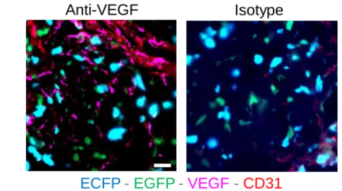
14 already detected on Ly6Chigh CD64+ cells, but the expression on Ly6Chigh and Ly6Clow/--Mo was low (Figure S5A). EGFP+-TAMs and ECFP+-TAMs harbored similar expression of FLT1 (Figure 8D). VEGF was found mainly along blood vessels but also in the tumor parenchyma, in proximity to TAMs without preferential co-localization with ECFP+ or EGFP+ cells (Figure S5B). Anti-VEGF treatment of tumor-bearing mice led to a significant reduction in the number of TAMs, but the proportions of EGFP+-TAMs and ECFP+-TAMs were similar demonstrating that both macrophage subsets are reduced by this treatment (Figure 8E). Our results support that anti-VEGF targets both monocyte-derived and resident TAM accumulation through a mechanism independent of their recruitment but rather affects their survival or proliferation.
15 Discussion
Embryonic-derived macrophages have recently been shown to contribute to the generation of TAMs in the pancreas and in the brain (Bowman et al., 2016; Zhu et al., 2017). These discoveries challenge the dogma on the origin of TAMs and raise the question whether this observation is applicable to other tissues such as the lungs, which are colonized by distinct macrophage subsets. AMs represent the main and typical resident macrophages of the lungs, maintaining immune homeostasis in the alveoli lumen (Trapnell and Whitsett, 2002). AMs acquire their unique signature and self-maintain via GM-CSF-dependent induction of PPAR-γ after birth (Guilliams et al., 2013; Schneider et al., 2014). Less is known about the functions and origin of IMs, but it has been suggested that they develop earlier than AMs in the embryo (Tan and Krasnow, 2016) and self-maintain independently of adult hematopoiesis (Gibbings et al., 2017; Rodero et al., 2015).
The implication of the chemokine receptor CCR2 in the recruitment of monocytes and on their subsequent differentiation into TAMs is well established in both primary and metastatic sites of various tumor types. This CCR2/CCL2 axis can contribute to an amplification loop of tumor progression (Franklin et al., 2014; Kitamura et al., 2015; Loyher et al., 2016). In these studies, the role of this axis on resident macrophages could not be excluded. However, lung IMs have been shown to expand independently of CCR2 and to display regulatory functions in the context of allergy (Sabatel et al., 2017).
Using the MacBlue x Cx3cr1EGFP/+ model, we unveiled the accumulation of an EGFP+ TAM subset that was unaffected by CCR2 deficiency and not reconstituted in parabiosis experiments, demonstrating that this subset originates from macrophages that were already present in the healthy lungs before tumor development. Preferential labeling of CD11b+ IMs was previously achieved using Csf1rMeriCreMer mice pulsed with OH-TAM at E8.5 (Schulz et al.,
2012). Using the same approaches to trace EMP-derived macrophages, we unveiled that embryonically seeded lung-resident IMs persist and proliferate to represent a large fraction of TAMs within pulmonary tumors and confirmed our hypothesis made using the MacBlue x
16 from AMs differentiation, it is unlikely that upon tumor parenchymal infiltration AMs lose the expression of Siglec-F and ECFP reporter while upregulating CD11b and EGFP. No progressive change in the expression of these surface markers that could support this hypothesis was observed in the AM population during tumor development. Thus, resident IMs are likely to represent a unique tissue-resident subset involved in the accumulation of EGFP+ TAMs. Loss of ECFP expression from monocyte-derived macrophages could lead to overestimation of EGFP+-TAMs, however monocyte derived cells maintained ECFP expression in parabiosis and transfer experiment, suggesting that this phenomenon barely occurs during this time frame and would only minimally perturb our quantification of EGFP+ -TAMs. Fate mapping studies led to similar observations and strengthen the fidelity of the MacBlue x Cx3cr1EGFP/+ system to study lung macrophages. Concomitantly, we observed an increase in the number of Ly6Chigh monocytes. Infiltrating Ly6Chigh-Mo seemed to up-regulate CD64 suggesting an intermediate toward the progressive differentiation into TAMs. We subsequently identified a distinct population of monocyte-derived TAMs arising from CCR2-dependent monocyte recruitment. At later time points, these TAMs became from far the most abundant population. The dual origin of macrophages was also observed in TAMs of LLC lung nodules and PyMT-ChOVA spontaneous pulmonary metastases suggesting that it might occur for any neoplastic tissue development in the lung.
Within lung tumor nodules, the relative distribution and abundance of EGFP+-TAMs compared to the recruited ones were in accordance with the localization of IMs prior to tumor development. We previously showed in the MacBlue x Cx3cr1EGFP/+ mice that interstitial EGFP+ macrophages are abundant in the pleura, airways and at the periphery of large blood vessels (Rodero et al., 2015). Lineage tracing of yolk sac-derived macrophages labeled mostly IMs that persisted in adults and localized in these same particular locations but scarce in the central lung parenchyma (Tan and Krasnow, 2016). The local environment can dictate macrophage phenotypes in vivo (Gosselin et al., 2014; Lavin et al., 2014; van de Laar et al., 2016). Despite a very close proximity between the two TAM subsets in tumor nodules, their transcriptomic profiles were distinct and were associated with different distribution depending
17 on the anatomic localization of the tumor, further arguing that origin poises macrophages for differing functions. No typical M1 or M2 profile could be attributed to EGFP+-TAMs or ECFP+ -TAMs, suggesting that this paradigm does not fully resolve the polarization process of TAMs. Nevertheless, the relative proportion and specific features of tissue-resident macrophages might contribute to the heterogeneity of different TME according to the anatomical site of tumor development. Further studies are needed to investigate whether it could serve as a prognostic factor of tumor growth and response to therapies.
Anti-CSF1R treatment depleted most of EGFP+-TAMs but ECFP+-TAMs were only partially targeted while Ly6Chigh monocytes and AMs were unaffected. Despite the lack of ECFP expression in the MacBlue mouse, it was previously shown that adult tissue-macrophages express CSF1R. The differential utilization of the truncated CSF1R promoter of the MacBlue binary transgene in macrophages was proposed to reflect different survival dependency on CSF1. Thus, cell expressing the ECFP reporter would be CSF1-independent in contrast to EGFP+ macrophages that require the upstream depleted region of the CSF1-regulated promoter region of the MacBlue transgene (Hawley et al., 2018; Sauter et al., 2014). This was supported by the reduced impact of anti-CSF1R treatment on ECFP+-TAMs compared to EGFP+-TAMs. Depletion of EGFP+-TAMs in Ccr2-/- mice led to drastic reduction in tumor growth, which links resident TAMs more directly to tumor trophic functions. ECFP+-TAMs displayed increased motility, in accordance to enriched cellular movement associated pathways and turned up to accumulate at the tumor margin. In this regard, Mmp8 and
Tnfsf14 enrichment (implicated in airway remodeling) (Doherty et al., 2011) could argue for a
licensing of monocyte-derived TAMs for remodeling of the surrounding environment and modification of the tumor architecture. Indeed, recruitment of MoD-cells was associated with reduced tumor cell density, higher spreading and increased invasion of pulmonary nodules. We could not fully differentiate the relative contribution of tumor-infiltrating ECFP+ monocytes versus ECFP+-TAMs. Monocyte-dependent cytotoxic activity could be suspected as observed after CP-induced monocyte rebound. Thus ECFP+-cells represent a heterogeneous population balancing between tumor destruction and remodeling, favoring spreading and
18 invasiveness. This observation raises important questions about cancer therapies targeting TAM subsets and suggests that depleting resident macrophages but keeping the phagocytic activities of MoD-cells would yield a better outcome for chemotherapies.
The targeting of VEGF in combination with chemotherapy including CP (Dellapasqua et al., 2008) has been shown to be beneficial (Motz and Coukos, 2011). Although TAMs have clearly been shown to participate in the process of angiogenesis within tumor (Lewis et al., 2016), few studies have investigated the impact of this therapeutic combination on the immune cellular composition of the TME. Moreover, FLT1 expression and signaling by pulmonary TAMs are implicated in their pro-tumor activity, partly via downstream regulation of the master macrophage regulator CSF1 (Qian et al., 2015). VEGF has been proposed to act as a chemoattractant factor for monocytes (Grunewald et al., 2006; Kaplan et al., 2005) but the beneficial effect of anti-VEGF combination was associated with a drastic reduction of both EGFP+ resident and ECFP+ monocyte-derived TAMs without affecting tumor-monocyte infiltration, suggesting that VEGF contributes to monocyte differentiation and/or TAM survival. Indeed, FLT1 expression was increased upon monocyte to macrophage differentiation, which corroborates previous studies (Barleon et al., 1996; Qian et al., 2015). This observation is in accordance with the hypothesis of a loss of anti-tumor activity of tumor-infiltrating monocytes upon differentiation into TAMs. The clinical relevance of our results lies in the fact that anti-VEGF could improve chemotherapy efficacy through functions that go beyond its main expected role on angiogenesis and leukocyte recruitment. Increasing knowledge of the impact of such molecule on the different TAM subsets according to their origin might allow for further development and improvement of anti-cancer dosing regimens and combinations.
20 Author contribution
PL.L. and P.H. designed, performed the experiments, analyzed and interpreted the data and wrote the manuscript, A.MK., E.G. and B.C. analyzed and interpreted the data, M.L., S.T., C.BC. and Z.D. performed some experiments and wrote the manuscript. N.B. provided reagent and wrote the manuscript, F.G., A.S. and C.C. provided reagent, designed research. A.B. supervised the study, designed, performed the experiments, analyzed and interpreted the data and wrote the manuscript.
Acknowledgments
The authors wish to thank the Plateforme Imagerie Pitié-Salpêtrière (PICPS) for assistance with the two-photon microscope, the animal facility "NAC" for mice breeding assistance and Angéline Duche and Sebastien Jacques from sequencing platform GENOM’IC (Institut Cochin, Paris, France). The authors also thank M. Lambert for the tdTomato viral construct. PL.L. is funded by “Fondation ARC pour la Recherche sur le Cancer” and MSKCC GMTEC’s fellowships. P.H. is funded by la "Ligue contre le cancer". This work was supported by funding from the European Community's Seventh Framework Programme (FP7/2007-2013) n°304810 – RAIDs, Inserm, Roche, la "Ligue contre le cancer", FRM “équipe labélissée” and "Fondation ARC pour la recherche sur le cancer" (to A.B.) and NIH NCI (P30CA008748) MSKCC core grant, NIH/NIAID 1R01AI130345-01 and NIH/NHLBI 1 R01HL138090-01 (to F.G.). Ariel Savina is employed by Roche. The authors declare no further conflicts of interest.
21 References
Amir el, A.D., K.L. Davis, M.D. Tadmor, E.F. Simonds, J.H. Levine, S.C. Bendall, D.K. Shenfeld, S. Krishnaswamy, G.P. Nolan, and D. Pe'er. 2013. viSNE enables visualization of high dimensional single-cell data and reveals phenotypic heterogeneity of leukemia. Nat
Biotechnol 31:545-552.
Barleon, B., S. Sozzani, D. Zhou, H.A. Weich, A. Mantovani, and D. Marme. 1996. Migration of human monocytes in response to vascular endothelial growth factor (VEGF) is mediated via the VEGF receptor flt-1. Blood 87:3336-3343.
Boissonnas, A., F. Licata, L. Poupel, S. Jacquelin, L. Fetler, S. Krumeich, C. Thery, S. Amigorena, and C. Combadiere. 2013. CD8+ tumor-infiltrating T cells are trapped in the tumor-dendritic cell network. Neoplasia 15:85-94.
Bowman, R.L., F. Klemm, L. Akkari, S.M. Pyonteck, L. Sevenich, D.F. Quail, S. Dhara, K. Simpson, E.E. Gardner, C.A. Iacobuzio-Donahue, C.W. Brennan, V. Tabar, P.H. Gutin, and J.A. Joyce. 2016. Macrophage Ontogeny Underlies Differences in Tumor-Specific Education in Brain Malignancies. Cell Rep 17:2445-2459.
Broz, M.L., M. Binnewies, B. Boldajipour, A.E. Nelson, J.L. Pollack, D.J. Erle, A. Barczak, M.D. Rosenblum, A. Daud, D.L. Barber, S. Amigorena, L.J. Van't Veer, A.I. Sperling, D.M. Wolf, and M.F. Krummel. 2014. Dissecting the tumor myeloid compartment reveals rare activating antigen-presenting cells critical for T cell immunity. Cancer Cell 26:638-652.
Chen, T.J., and N. Kotecha. 2014. Cytobank: providing an analytics platform for community cytometry data analysis and collaboration. Curr Top Microbiol Immunol 377:127-157.
Cohen, M.H., J. Gootenberg, P. Keegan, and R. Pazdur. 2007. FDA drug approval summary: bevacizumab (Avastin) plus Carboplatin and Paclitaxel as first-line treatment of advanced/metastatic recurrent nonsquamous non-small cell lung cancer. Oncologist 12:713-718.
Cortez-Retamozo, V., M. Etzrodt, A. Newton, P.J. Rauch, A. Chudnovskiy, C. Berger, R.J. Ryan, Y. Iwamoto, B. Marinelli, R. Gorbatov, R. Forghani, T.I. Novobrantseva, V. Koteliansky, J.L. Figueiredo, J.W. Chen, D.G. Anderson, M. Nahrendorf, F.K. Swirski, R. Weissleder, and M.J. Pittet. 2012. Origins of tumor-associated macrophages and neutrophils. Proc Natl Acad Sci U
S A 109:2491-2496.
De Palma, M., and C.E. Lewis. 2013. Macrophage regulation of tumor responses to anticancer therapies. Cancer Cell 23:277-286.
Dellapasqua, S., F. Bertolini, V. Bagnardi, E. Campagnoli, E. Scarano, R. Torrisi, Y. Shaked, P. Mancuso, A. Goldhirsch, A. Rocca, E. Pietri, and M. Colleoni. 2008. Metronomic cyclophosphamide and capecitabine combined with bevacizumab in advanced breast cancer. J Clin Oncol 26:4899-4905.
Doherty, T.A., P. Soroosh, N. Khorram, S. Fukuyama, P. Rosenthal, J.Y. Cho, P.S. Norris, H. Choi, S. Scheu, K. Pfeffer, B.L. Zuraw, C.F. Ware, D.H. Broide, and M. Croft. 2011. The tumor necrosis factor family member LIGHT is a target for asthmatic airway remodeling. Nat Med 17:596-603.
Engelhardt, J.J., B. Boldajipour, P. Beemiller, P. Pandurangi, C. Sorensen, Z. Werb, M. Egeblad, and M.F. Krummel. 2012. Marginating dendritic cells of the tumor microenvironment cross-present tumor antigens and stably engage tumor-specific T cells. Cancer Cell 21:402-417. Franklin, R.A., W. Liao, A. Sarkar, M.V. Kim, M.R. Bivona, K. Liu, E.G. Pamer, and M.O. Li. 2014. The
cellular and molecular origin of tumor-associated macrophages. Science 344:921-925.
Gambardella, L., M. Hemberger, B. Hughes, E. Zudaire, S. Andrews, and S. Vermeren. 2010. PI3K signaling through the dual GTPase-activating protein ARAP3 is essential for developmental angiogenesis. Sci Signal 3:ra76.
Gibbings, S.L., S.M. Thomas, S.M. Atif, A.L. McCubbrey, A.N. Desch, T. Danhorn, S.M. Leach, D.L. Bratton, P.M. Henson, W.J. Janssen, and C.V. Jakubzick. 2017. Three Unique Interstitial Macrophages in the Murine Lung at Steady State. Am J Respir Cell Mol Biol 57:66-76.
22 Gomez Perdiguero, E., K. Klapproth, C. Schulz, K. Busch, E. Azzoni, L. Crozet, H. Garner, C. Trouillet, M.F. de Bruijn, F. Geissmann, and H.R. Rodewald. 2015. Tissue-resident macrophages originate from yolk-sac-derived erythro-myeloid progenitors. Nature 518:547-551.
Gosselin, D., V.M. Link, C.E. Romanoski, G.J. Fonseca, D.Z. Eichenfield, N.J. Spann, J.D. Stender, H.B. Chun, H. Garner, F. Geissmann, and C.K. Glass. 2014. Environment drives selection and function of enhancers controlling tissue-specific macrophage identities. Cell 159:1327-1340. Grunewald, M., I. Avraham, Y. Dor, E. Bachar-Lustig, A. Itin, S. Jung, S. Chimenti, L. Landsman, R.
Abramovitch, and E. Keshet. 2006. VEGF-induced adult neovascularization: recruitment, retention, and role of accessory cells. Cell 124:175-189.
Guilliams, M., I. De Kleer, S. Henri, S. Post, L. Vanhoutte, S. De Prijck, K. Deswarte, B. Malissen, H. Hammad, and B.N. Lambrecht. 2013. Alveolar macrophages develop from fetal monocytes that differentiate into long-lived cells in the first week of life via GM-CSF. J Exp Med 210:1977-1992.
Hamon, P., P.L. Loyher, C. Baudesson de Chanville, F. Licata, C. Combadiere, and A. Boissonnas. 2016. CX3CR1-dependent endothelial margination modulates Ly6Chigh monocyte systemic deployment upon inflammation in mice. Blood
Hawley, C.A., R. Rojo, A. Raper, K.A. Sauter, Z.M. Lisowski, K. Grabert, C.C. Bain, G.M. Davis, P.A. Louwe, M.C. Ostrowski, D.A. Hume, C. Pridans, and S.J. Jenkins. 2018. Csf1r-mApple Transgene Expression and Ligand Binding In Vivo Reveal Dynamics of CSF1R Expression within the Mononuclear Phagocyte System. J Immunol
Hughes, R., B.Z. Qian, C. Rowan, M. Muthana, I. Keklikoglou, O.C. Olson, S. Tazzyman, S. Danson, C. Addison, M. Clemons, A.M. Gonzalez-Angulo, J.A. Joyce, M. De Palma, J.W. Pollard, and C.E. Lewis. 2015. Perivascular M2 Macrophages Stimulate Tumor Relapse after Chemotherapy.
Cancer Res 75:3479-3491.
Jacquelin, S., F. Licata, K. Dorgham, P. Hermand, L. Poupel, E. Guyon, P. Deterre, D.A. Hume, C. Combadiere, and A. Boissonnas. 2013. CX3CR1 reduces Ly6Chigh-monocyte motility within and release from the bone marrow after chemotherapy in mice. Blood 122:674-683.
Ji, H., E.Y. Chang, K.Y. Lin, R.J. Kurman, D.M. Pardoll, and T.C. Wu. 1998. Antigen-specific immunotherapy for murine lung metastatic tumors expressing human papillomavirus type 16 E7 oncoprotein. Int J Cancer 78:41-45.
Jung, S., J. Aliberti, P. Graemmel, M.J. Sunshine, G.W. Kreutzberg, A. Sher, and D.R. Littman. 2000. Analysis of fractalkine receptor CX(3)CR1 function by targeted deletion and green fluorescent protein reporter gene insertion. Mol Cell Biol 20:4106-4114.
Kaplan, R.N., R.D. Riba, S. Zacharoulis, A.H. Bramley, L. Vincent, C. Costa, D.D. MacDonald, D.K. Jin, K. Shido, S.A. Kerns, Z. Zhu, D. Hicklin, Y. Wu, J.L. Port, N. Altorki, E.R. Port, D. Ruggero, S.V. Shmelkov, K.K. Jensen, S. Rafii, and D. Lyden. 2005. VEGFR1-positive haematopoietic bone marrow progenitors initiate the pre-metastatic niche. Nature 438:820-827.
Kitamura, T., D. Doughty-Shenton, L. Cassetta, S. Fragkogianni, D. Brownlie, Y. Kato, N. Carragher, and J.W. Pollard. 2017. Monocytes Differentiate to Immune Suppressive Precursors of Metastasis-Associated Macrophages in Mouse Models of Metastatic Breast Cancer. Front
Immunol 8:2004.
Kitamura, T., B.Z. Qian, D. Soong, L. Cassetta, R. Noy, G. Sugano, Y. Kato, J. Li, and J.W. Pollard. 2015. CCL2-induced chemokine cascade promotes breast cancer metastasis by enhancing retention of metastasis-associated macrophages. J Exp Med 212:1043-1059.
Lavin, Y., D. Winter, R. Blecher-Gonen, E. David, H. Keren-Shaul, M. Merad, S. Jung, and I. Amit. 2014. Tissue-resident macrophage enhancer landscapes are shaped by the local microenvironment.
Cell 159:1312-1326.
Lewis, C.E., A.S. Harney, and J.W. Pollard. 2016. The Multifaceted Role of Perivascular Macrophages in Tumors. Cancer Cell 30:365.
Lin, K.Y., F.G. Guarnieri, K.F. Staveley-O'Carroll, H.I. Levitsky, J.T. August, D.M. Pardoll, and T.C. Wu. 1996. Treatment of established tumors with a novel vaccine that enhances major histocompatibility class II presentation of tumor antigen. Cancer Res 56:21-26.
23 Loyher, P.L., J. Rochefort, C. Baudesson de Chanville, P. Hamon, G. Lescaille, C. Bertolus, M.
Guillot-Delost, M.F. Krummel, F.M. Lemoine, C. Combadiere, and A. Boissonnas. 2016. CCR2 Influences T Regulatory Cell Migration to Tumors and Serves as a Biomarker of Cyclophosphamide Sensitivity. Cancer Res 76:6483-6494.
Maeda, K., Y. Kobayashi, N. Udagawa, S. Uehara, A. Ishihara, T. Mizoguchi, Y. Kikuchi, I. Takada, S. Kato, S. Kani, M. Nishita, K. Marumo, T.J. Martin, Y. Minami, and N. Takahashi. 2012. Wnt5a-Ror2 signaling between osteoblast-lineage cells and osteoclast precursors enhances osteoclastogenesis. Nat Med 18:405-412.
Mantovani, A., and P. Allavena. 2015. The interaction of anticancer therapies with tumor-associated macrophages. J Exp Med 212:435-445.
Mass, E., I. Ballesteros, M. Farlik, F. Halbritter, P. Gunther, L. Crozet, C.E. Jacome-Galarza, K. Handler, J. Klughammer, Y. Kobayashi, E. Gomez-Perdiguero, J.L. Schultze, M. Beyer, C. Bock, and F. Geissmann. 2016. Specification of tissue-resident macrophages during organogenesis.
Science 353:
Mass, E., C.E. Jacome-Galarza, T. Blank, T. Lazarov, B.H. Durham, N. Ozkaya, A. Pastore, M. Schwabenland, Y.R. Chung, M.K. Rosenblum, M. Prinz, O. Abdel-Wahab, and F. Geissmann. 2017. A somatic mutation in erythro-myeloid progenitors causes neurodegenerative disease.
Nature 549:389-393.
Misharin, A.V., L. Morales-Nebreda, G.M. Mutlu, G.R. Budinger, and H. Perlman. 2013. Flow cytometric analysis of macrophages and dendritic cell subsets in the mouse lung. Am J Respir
Cell Mol Biol 49:503-510.
Montero, A., and S. Gluck. 2012. Long-Term Complete Remission with nab-Paclitaxel, Bevacizumab, and Gemcitabine Combination Therapy in a Patient with Triple-Negative Metastatic Breast Cancer. Case Rep Oncol 5:687-692.
Motz, G.T., and G. Coukos. 2011. The parallel lives of angiogenesis and immunosuppression: cancer and other tales. Nat Rev Immunol 11:702-711.
Ovchinnikov, D.A., W.J. van Zuylen, C.E. DeBats, K.A. Alexander, S. Kellie, and D.A. Hume. 2008. Expression of Gal4-dependent transgenes in cells of the mononuclear phagocyte system labeled with enhanced cyan fluorescent protein using Csf1r-Gal4VP16/UAS-ECFP double-transgenic mice. J Leukoc Biol 83:430-433.
Pollard, J.W. 2004. Tumour-educated macrophages promote tumour progression and metastasis. Nat
Rev Cancer 4:71-78.
Qian, B.Z., J. Li, H. Zhang, T. Kitamura, J. Zhang, L.R. Campion, E.A. Kaiser, L.A. Snyder, and J.W. Pollard. 2011. CCL2 recruits inflammatory monocytes to facilitate breast-tumour metastasis.
Nature 475:222-225.
Qian, B.Z., and J.W. Pollard. 2010. Macrophage diversity enhances tumor progression and metastasis.
Cell 141:39-51.
Qian, B.Z., H. Zhang, J. Li, T. He, E.J. Yeo, D.Y. Soong, N.O. Carragher, A. Munro, A. Chang, A.R. Bresnick, R.A. Lang, and J.W. Pollard. 2015. FLT1 signaling in metastasis-associated macrophages activates an inflammatory signature that promotes breast cancer metastasis. J
Exp Med 212:1433-1448.
Rodero, M.P., L. Poupel, P.L. Loyher, P. Hamon, F. Licata, C. Pessel, D.A. Hume, C. Combadiere, and A. Boissonnas. 2015. Immune surveillance of the lung by migrating tissue monocytes. Elife 4:e07847.
Sabatel, C., C. Radermecker, L. Fievez, G. Paulissen, S. Chakarov, C. Fernandes, S. Olivier, M. Toussaint, D. Pirottin, X. Xiao, P. Quatresooz, J.C. Sirard, D. Cataldo, L. Gillet, H. Bouabe, C.J. Desmet, F. Ginhoux, T. Marichal, and F. Bureau. 2017. Exposure to Bacterial CpG DNA Protects from Airway Allergic Inflammation by Expanding Regulatory Lung Interstitial Macrophages. Immunity 46:457-473.
Saeed, A.I., N.K. Bhagabati, J.C. Braisted, W. Liang, V. Sharov, E.A. Howe, J. Li, M. Thiagarajan, J.A. White, and J. Quackenbush. 2006. TM4 microarray software suite. Methods Enzymol 411:134-193.
24 Saeed, A.I., V. Sharov, J. White, J. Li, W. Liang, N. Bhagabati, J. Braisted, M. Klapa, T. Currier, M. Thiagarajan, A. Sturn, M. Snuffin, A. Rezantsev, D. Popov, A. Ryltsov, E. Kostukovich, I. Borisovsky, Z. Liu, A. Vinsavich, V. Trush, and J. Quackenbush. 2003. TM4: a free, open-source system for microarray data management and analysis. Biotechniques 34:374-378.
Sauter, K.A., C. Pridans, A. Sehgal, C.C. Bain, C. Scott, L. Moffat, R. Rojo, B.M. Stutchfield, C.L. Davies, D.S. Donaldson, K. Renault, B.W. McColl, A.M. Mowat, A. Serrels, M.C. Frame, N.A. Mabbott, and D.A. Hume. 2014. The MacBlue binary transgene (csf1r-gal4VP16/UAS-ECFP) provides a novel marker for visualisation of subsets of monocytes, macrophages and dendritic cells and responsiveness to CSF1 administration. PLoS One 9:e105429.
Schneider, C., S.P. Nobs, M. Kurrer, H. Rehrauer, C. Thiele, and M. Kopf. 2014. Induction of the nuclear receptor PPAR-gamma by the cytokine GM-CSF is critical for the differentiation of fetal monocytes into alveolar macrophages. Nat Immunol 15:1026-1037.
Schulz, C., E. Gomez Perdiguero, L. Chorro, H. Szabo-Rogers, N. Cagnard, K. Kierdorf, M. Prinz, B. Wu, S.E. Jacobsen, J.W. Pollard, J. Frampton, K.J. Liu, and F. Geissmann. 2012. A lineage of myeloid cells independent of Myb and hematopoietic stem cells. Science 336:86-90.
Tamagnone, L. 2012. Emerging role of semaphorins as major regulatory signals and potential therapeutic targets in cancer. Cancer Cell 22:145-152.
Tan, S.Y., and M.A. Krasnow. 2016. Developmental origin of lung macrophage diversity. Development 143:1318-1327.
Trapnell, B.C., and J.A. Whitsett. 2002. Gm-CSF regulates pulmonary surfactant homeostasis and alveolar macrophage-mediated innate host defense. Annu Rev Physiol 64:775-802.
van de Laar, L., W. Saelens, S. De Prijck, L. Martens, C.L. Scott, G. Van Isterdael, E. Hoffmann, R. Beyaert, Y. Saeys, B.N. Lambrecht, and M. Guilliams. 2016. Yolk Sac Macrophages, Fetal Liver, and Adult Monocytes Can Colonize an Empty Niche and Develop into Functional Tissue-Resident Macrophages. Immunity 44:755-768.
Wyckoff, J.B., Y. Wang, E.Y. Lin, J.F. Li, S. Goswami, E.R. Stanley, J.E. Segall, J.W. Pollard, and J. Condeelis. 2007. Direct visualization of macrophage-assisted tumor cell intravasation in mammary tumors. Cancer Res 67:2649-2656.
Zhu, Y., J.M. Herndon, D.K. Sojka, K.W. Kim, B.L. Knolhoff, C. Zuo, D.R. Cullinan, J. Luo, A.R. Bearden, K.J. Lavine, W.M. Yokoyama, W.G. Hawkins, R.C. Fields, G.J. Randolph, and D.G. DeNardo. 2017. Tissue-Resident Macrophages in Pancreatic Ductal Adenocarcinoma Originate from Embryonic Hematopoiesis and Promote Tumor Progression. Immunity 47:597.
25 Figure Legends
Figure 1: Lung macrophage subsets differentially accumulate during tumor development
(A) Representative tSNE dimension 1 and 2 plots of the lung myeloid compartment evolution after TC-1 cell intravenous inoculation. Upper panel delineates cell blood/tissue partitioning (dashed gates). Color clusters are represented over time (Lower panels). (B) Blood/tissue partitioning monitoring of lung monocytes and macrophages during tumor growth. Bars represent mean of the absolute number±SEM/mg of tissue (Upper panels), or absolute number per whole lung (Lower panels). Statistical differences are indicated compared to D0. For all panels n=6-8 mice per time point out of 3 independent experiments, Two-way ANOVA was performed. *: p<0.05; **: p<0.01; ***: p<0.001; ****: p<0.0001). See also Figures S1 and S2.
Figure 2: Macrophages have distinct origins within lung tumors
(A) Lung cryo-sections of tumor free and TC-1tdTomato tumor bearing MacBlue x Cx3cr1EGFP/+ mice show the distribution of ECFP+ and EGFP+ cells within tumor nodules over time. Scale bar 50µm. (B) (Left panel) Dot plots show the relative proportion of macrophage subsets in tumor-free, tumor-bearing MacBlue x Cx3cr1EGFP/+ (WT) and MacBlue x Cx3cr1EGFP/+ x Ccr2
-/-(Ccr2-/-) mice at indicated time points, numbers indicate the mean percentage±SD of ECFP+EGFP+ TAMs (Right panel). Whiskers graph shows the absolute number per mg of tissue of indicated myeloid subsets in WT and Ccr2-/- mice (n=10 mice out of 3 independent experiments, Two-way ANOVA with Bonferroni multiple comparisons test was performed). (C) (Left panels) Lung cryo-sections show the distribution of ECFP+ and EGFP+ cells in TC-1 tumor-bearing MacBlue x Cx3cr1EGFP/+ (WT), MacBlue x Cx3cr1EGFP/+ xCcr2-/- (Ccr2-/-) mice and C57Bl6 host parabiont with MacBlue x Cx3cr1EGFP/+ mice, scale bars 100µm. (Right panel) ratio of ECFP+/EGFP+ cell numbers in lung tumors. Each dot represents mean of ECFP+/EGFP+ cell ratio from different tumor nodules per mouse, red bars indicate mean,
26 mice are pooled from at least 2 independent experiments. (D) (Left panel) Lung cryo-sections show the distribution of ECFP+ and EGFP+ cells in LLC tumor bearing MacBlue x
Cx3cr1EGFP/+ mouse (D15), scale bar 50µm, and (Middle panel) ratio of ECFP+/EGFP+ cells in LLC tumors. Each dot represents mean of ECFP+/EGFP+ cell ratio from different tumor nodules per mouse, red bar indicates mean, mice are pooled from 2 independent experiments. (E) Lung cryo-section shows the distribution of ECFP+ and EGFP+ cells in spontaneous pulmonary metastases from MacBlue x Cx3cr1EGFP/+ x PyMT-ChOVA mouse, scale bar 50µm (Left panel). Middle panel shows the lack of EGFP+ cells in pulmonary metastases from C57Bl6 PyMT-ChOVA host parabiont. Right panel shows the quantification based on histological analyses. Each dot represents mean of ECFP+ /EGFP+ cell ratio from different tumor nodules per mouse, red bar indicates mean, all mice are independents. ANOVA with Bonferroni multiple comparisons test were performed. For all panels *: p<0.05; ***: p<0.001; ****: p<0.0001.
Figure 3: Lung interstitial macrophages are of embryonic origin and accumulate within tumors
(A) (Left panels) Dot plots show tdTomato+ myeloid cell subsets in Csf1rMeriCreMer; Rosa
LSL-tdTomato after OH-TAM pulse at E8.5 in adult tumor-free mice and 15 days after TC-1 inoculation. Mean percentage±SD of tdTomato+ cells among each subset is indicated. Right panel shows the numbers per mg of tissue of the tdTomato+ AMs and Ly6Clow/- CD64+ Mac. Bars represent mean of 4 mice per group out of 2 independent experiments. Two-way ANOVA with Bonferroni multiple comparisons test was performed. (B) (Left panels) Dot plots show YFP+ myeloid cell subsets of IMs in Tnfrs11aCre; Rosa26LSL-YFP mice. Mean percentage±SD of YFP+ cells among each subset is indicated. Right panel shows the absolute number per mg of YFP+ AMs and Ly6Clow/- CD64+ Mac. Bars represent mean of 4 mice per group out of 2 independent experiments. Two-way ANOVA with Bonferroni multiple comparisons test was performed. For all panels *: p<0.05; ***: p <0.001.
27 Figure 4: Resident and MoD-TAMs harbor distinct phenotypes and anatomic distribution
(A) Lung cryo-section shows the distribution of ECFP+ and EGFP+ cells in TC-1 pulmonary tumor nodule located in the vicinity of the pleura. Graph represents the relative distribution of EGFP+ cells and ECFP+ MoD-cells as a function of the distance from the surface (pleura) (Bars represent meansSD of 4 mice out of 2 independent experiments; Two-way ANOVA was performed), scale bar 50µm. (B) Lung cryo-sections show the distribution of ECFP+ and EGFP+ cells in pulmonary tumor nodules located in the alveolar space or near large airways, 15 days after TC-1 inoculation, scale bars 50µm. Graph represents the percentage of EGFP+ cells among fluorescent cells in each sub-anatomic compartment (Dots represent the mean ratio per mouse, mice are pooled from 2 independent experiments and red bars indicate mean. Unpaired Student’s t-test was performed). (C) Lung cryo-sections of Tnfrs11aCre;
Rosa26LSL-YFP mice shows YFP+ cells in pulmonary tumor nodules located in the pleura or in the alveolar space 15 days after tdTomato+ TC-1 inoculation, scale bars 15µm. Graph represents the number of YFP+ cells in each sub-anatomic compartment (Dots represent the mean ratio per mouse, mice are pooled from 2 independent experiments and bars indicate means, One-way ANOVA was performed). (D) Heatmap shows a selection of transcripts involved in extracellular matrix interaction and remodeling, differentially expressed between EGFP+ and ECFP+-TAMs. (E) Expression of indicated genes, relative to GAPDH (2-ΔCt), as determined by qPCR of FACS sorted YFP+ and YFP- TAMs in Tnfrs11aCre; Rosa26LSL-YFP
mice. Each dot represents one mouse, pooled from 2 independent experiments (F) (Left panel) Representative histogram shows VCAM expression gated on TAM subsets from MacBlue x Cx3cr1EGFP/+ mice. Middle panel shows the quantification of VCAM1 expression by EGFP+ and ECFP+-TAMs in MacBlue x Cx3cr1EGFP/+. Right panel represents the quantification of VCAM1 expression by YFP+ and YFP- macrophages in Tnfrs11aCre; Rosa26LSL-YFP mice. Mice are pooled from 3 independent experiments, bars indicate means, unpaired Student’s t-test was performed. (G) (Left panel) TPLSM 3D reconstruction shows
28 the perivascular location of the EGFP+ cell network within a tumor nodule, scale bar 10µm. (Middle panel) Lung cryo-section from TC-1 tumor-bearing MacBlue x Cx3cr1EGFP/+ at day 15 shows fluorescent subset distribution regarding tumor vasculature using CD31 staining. Close interactions of EGFP+ cells with the vasculature are indicated (white arrows), scale bar 50µm. (Right panel) Scatter plot represents the relative proportion of perivascular EGFP+ cells and ECFP+ cells in tumor nodules (Dots represent the mean proportion per mouse pooled from at least 2 independent experiments; bars indicate means unpaired Student’s t-test was performed). For all panels *: p<0.05; **: p<0.01; ***: p <0.001; ****: p<0.0001 See also Figure S3 and video 1-2.
Figure 5: Res-macrophages support tumor cell growth and MoD-cells are associated with tumor spreading in the lung
(A) Tumor growth was monitored in WT and Ccr2-/- mice by bioluminescence imaging (graphs represent meanSEM of n=10 mice per group from 2 independent experiments). (B) (Left panels) Wide field images of a whole lung cryo-section 15 days after TC-1 inoculation in WT and Ccr2-/- mice. White mask indicates nodule size and distribution, scale bars 1mm. (Right panel) Graph shows the relative distribution of nodule areas (n=5-7 mice in each group out of 3 independent experiments, Two-way ANOVA comparing WT and Ccr2 -/-frequency of each distribution was performed). (C) (Left panels) TPLSM 3D reconstructions show tumor cell density in a representative nodule from TC-1tdTomato tumor-bearing WT and
Ccr2-/- mice, white arrows indicate spread tumor cells, scale bars 30µm. (Right panel) Tumor cell density was measured using 3D-reconstruction images of tdTomato+ tumor nodule. Dots represent the mean of at least 4 different tumor nodules per mouse, pooled from 2 independent experiments, bars indicate means, unpaired Student’s t-test was performed). (D) Boxes and whiskers graph represents numbers per mg of lung of each indicated myeloid subset after indicated treatment at day 15; Tumor-bearing WT and Ccr2-/- mice were treated every two days with anti-CSF1R between D5 and D14 (n=6 mice from 2 independent experiments. One-way ANOVA comparing each subset individually for each condition was
29 performed). (E) (Left panels) Lung cryo-sections of TC-1tdTomato tumor-bearing MacBlue x
Cx3cr1EGFP/+ x Ccr2-/- mice treated with anti-CSF1R or isotype control show depletion of EGFP+ cells at D15, scale bars 50µm. (Right panel) Tumor burden was monitored by bioluminescence imaging after TC-1-Luc inoculation. Mice were treated every two days with anti-CSF1R starting D5 (Graphs represent mean±SEM, n= 10 mice per group out of 2 independent experiments. Two-way ANOVA with Bonferoni multiple comparisons test was performed). For all panels *: p<0.05; **: p<0.01; ***: p <0.001; ****: p<0.0001.
Figure 6: Distinct sensitivity and recovery of Res-TAMs and MoD-TAMs after chemotherapy
(A) Impact of CP treatment on tumor growth was monitored by bioluminescence imaging (Graphs represent meanSEM of n=10 mice per group out of 3 independent experiments). (B) Dot plots show Ly6C and CD64 expressions of Siglec-F-CD11b+Ly6G- lung cells over time after CP treatment. Mean percentage±SD of cells in each quadrant are indicated. (C) Blood/tissue partitioning monitoring of lung monocytes and macrophages during tumor growth post chemotherapy (Graphs represent mean of the absolute number±SEM/mg of tissue, n=6-10 mice per time point out of 2-4 independent experiments, Two-way ANOVA with Bonferroni multiple comparisons test was performed. Only statistical differences compared to day of treatment (day 0) are indicated for each compartment). (D) Box and Whiskers graph shows the absolute number per mg of tissue of ECFP+-TAMs and EGFP+ -TAMs from MacBlue x Cx3cr1EGFP/+ mice after CP (n= 4-10 mice per time point out of 3 independent experiments, Two-way ANOVA with Bonferroni multiple comparisons test was performed). (E) Lung cryo-sections of TC-1tdTomato tumor-bearing MacBlue x Cx3cr1EGFP/+ mice show TAM subset distribution within tumor nodules following CP treatment, scale bars 50µm. (F) (Left panels) Dot plots show TC-1tdTomato phagocytosis by the indicated mononuclear phagocyte subsets (red). Fluorescent background from non-fluorescent TC-1 tumor is overlaid (black). (Right panels) Boxes and whiskers graphs show the relative proportion of phagocytic cells among indicated subsets at 10 and 15 days after CP treatment
30 (n= 9 mice out of 3 independent experiments). For all panels *: p<0.05; **: p<0.01; ***: p<0.001; ****: p<0.0001. See also Figure S4.
Figure 7: Anti-VEGF combination with CP reduces TAM recovery and enhances chemotherapy efficacy
(A) Tumor bearing mice were treated with CP 10 days after tumor inoculation and treated or not every two days with anti-VEGF. (B) Representative tSNE dimension 1 and 2 plots show the impact of the different therapies on the myeloid signature at D15. Cell subsets are color grouped and dashed black gates delineate blood/tissue partitioning. (C) Blood/tissue partitioning monitoring of lung monocytes and macrophages during tumor growth at days 15 and 20 after CP or CP+anti-VEGF (Bars represent mean of the total number of cell±SEM. For all panels n=6 mice per time point out of 2 independent experiments, Two-way ANOVA with Bonferoni multiple comparisons test was performed). (D) Survival curve shows the efficacy of combined therapy started from day 20 (n=7 mice per group, data are representative of 2 experiments, Log-rank (Mantel-cox) test was performed to compare each survival curve with the one of CP+anti-VEGF). Corresponding lung weights are reported (one-way ANOVA with Bonferroni multiple comparisons test was performed). For all panels *: p<0.05; **: p <0.01; ***: p<0.001.
Figure 8: Anti-VEGF targets Res-TAM and MoD-TAM accumulation
(A) (Upper panel) Bone marrow monocytes from MacBlue mice were adoptively transferred in tumor-bearing WT mice treated or not with anti-VEGF. (Lower panels) Representative overlay dot plots show the phenotype of recovered MoD-cells from tumor-bearing mice treated (green) or not with anti-VEGF (black) 24h after transfer. (B) Graph shows the relative proportion of recovered cells in each mouse pooled from two independent experiments (Black bars indicate means, Two-way ANOVA with Bonferroni multiple comparisons test was performed). (C) Graph shows the proportion of Ly6Chigh-Mo infiltration evaluated by blood/tissue partitioning in each mouse pooled from 2 independent experiments (Black bars indicate means, Student’s t-test was performed). (D) Representative histogram plot of FTL1
31 expression by ECFP+ and ECFP+-TAMs, mean percentage±SD of FLT1+ cell in each subset out of 6 mice from 2 independent experiments are indicated. (E) Left panel shows the number of TAMs/mg in indicated conditions. Right panel shows the relative proportion of TAMs in mice treated with anti-VEGF or isotype. Black bars indicate means of 2 independent experiments, Student’s t-test was performed. For all panels ns = non-significant; *: p<0.05; ****: p<0.0001.