HAL Id: hal-02279022
https://hal.archives-ouvertes.fr/hal-02279022
Submitted on 4 Sep 2019
HAL is a multi-disciplinary open access
archive for the deposit and dissemination of
sci-entific research documents, whether they are
pub-lished or not. The documents may come from
teaching and research institutions in France or
abroad, or from public or private research centers.
L’archive ouverte pluridisciplinaire HAL, est
destinée au dépôt et à la diffusion de documents
scientifiques de niveau recherche, publiés ou non,
émanant des établissements d’enseignement et de
recherche français ou étrangers, des laboratoires
publics ou privés.
Distributed under a Creative Commons Attribution - NonCommercial| 4.0 International
protease machinery by substrates and active-site
inhibitors
Jan Felix, Katharina Weinhäupl, Christophe Chipot, François Dehez, Audrey
Hessel, Diego Gauto, Cécile Morlot, Olga Abian, Irina Gutsche, Adrian
Velazquez-Campoy, et al.
To cite this version:
Jan Felix, Katharina Weinhäupl, Christophe Chipot, François Dehez, Audrey Hessel, et al..
Mech-anism of the allosteric activation of the ClpP protease machinery by substrates and active-site
in-hibitors. Science Advances , American Association for the Advancement of Science (AAAS), 2019, 5
(9), pp.eaaw3818. �10.1126/sciadv.aaw3818�. �hal-02279022�
B I O C H E M I S T R Y Copyright © 2019 The Authors, some rights reserved; exclusive licensee American Association for the Advancement of Science. No claim to original U.S. Government Works. Distributed under a Creative Commons Attribution NonCommercial License 4.0 (CC BY-NC).
Mechanism of the allosteric activation
of the ClpP protease machinery by substrates
and active-site inhibitors
Jan Felix1, Katharina Weinhäupl1, Christophe Chipot2,3,4, François Dehez2,3, Audrey Hessel1, Diego F. Gauto1, Cecile Morlot1, Olga Abian5,6,7,8, Irina Gutsche1, Adrian Velazquez-Campoy5,6,7,9, Paul Schanda1*, Hugo Fraga1,10,11*
Coordinated conformational transitions in oligomeric enzymatic complexes modulate function in response to substrates and play a crucial role in enzyme inhibition and activation. Caseinolytic protease (ClpP) is a tetra-decameric complex, which has emerged as a drug target against multiple pathogenic bacteria. Activation of different ClpPs by inhibitors has been independently reported from drug development efforts, but no rationale for inhibitor-induced activation has been hitherto proposed. Using an integrated approach that includes x-ray crystallography, solid- and solution-state nuclear magnetic resonance, molecular dynamics simulations, and iso-thermal titration calorimetry, we show that the proteasome inhibitor bortezomib binds to the ClpP active-site serine, mimicking a peptide substrate, and induces a concerted allosteric activation of the complex. The bortezomib-activated conformation also exhibits a higher affinity for its cognate unfoldase ClpX. We propose a universal allo-steric mechanism, where substrate binding to a single subunit locks ClpP into an active conformation optimized for chaperone association and protein processive degradation.
INTRODUCTION
Among the most exciting antibacterial drug targets that have emerged in the past decade is the caseinolytic protease (ClpP) (1). While mostly studied in Escherichia coli (EcClpP), ClpP is present in a wide range of bacteria, as well as in mitochondria and chloroplasts. Functionally, ClpP can be defined as a cylindrical canonical serine protease com-posed of two stacked rings, each formed by seven subunits that en-compass a large (300 kDa) proteolytic core (2). This architecture restricts access to the 14 proteolytic active sites located inside the barrel and is key for ClpP function. Although ClpP is able to rapidly hydrolyze peptides, degradation of large proteins requires the pres-ence of an AAA+ adenosine triphosphatase (ATPase) complex, such as ClpA or ClpX in E. coli or ClpC in other species. These unfoldases provide substrate specificity not only by recognizing, unfolding, and translocating their globular protein substrates but also by opening the apical gate region of ClpP, which is otherwise blocked by flexible N-terminal tails.
ClpP protease and its associated chaperones are essential for the survival or virulence of several bacteria including pathogens such as Mycobacterium tuberculosis (Mtb) (3), Staphylococcus aureus (Sa) (4), and Listeria monocytogenes (Lm) (5). In addition, human
mito-chondrial ClpP was associated to human acute myeloid leukemia and obesity (6, 7). Recently, ClpP from Plasmodium falciparum (PfClpP), the parasite responsible for malaria infection, has also been proposed as a promising drug target for the control of this disease (8).
The fact that ClpP is a suitable Achilles’ heel for these organisms is further reinforced by the recent discovery of natural antibiotics that target ClpP or its co-chaperones. Efficient bacterial killing could be accomplished either by opening the axial pore of ClpP and activating unregulated cell proteolysis (9), by blocking ClpP-ATPase interaction (10), or by specifically inhibiting its chaperones, ClpX (4) or ClpC1 (11). Despite recent advances in the field, the study and development of drugs targeting ClpP have been thwarted by the fact that ClpPs from different species show notable functional differences. For example, Mtb contains two clpP genes, clpP1 and clpP2, both of which are es-sential for viability and infectivity (1, 3). Although both genes encode serine proteases that form heptameric rings, first attempts to express and characterize isolated MtbClpP1 and MtbClpP2 in E. coli yielded complexes that lacked proteolytic activity (12, 13). The study of MtbClpP1P2 was not possible until the finding that a small activator, derived from classical serine protease inhibitor peptide aldehydes (3), was required to promote the formation of an active complex composed of one heptameric ring from ClpP1 and another from ClpP2 (3). Oddly enough, the small activators were found at the protein active site, and it is not clear how their presence at the active site can promote activation rather than classic competitive inhibition (14, 15). Furthermore, several structures obtained by x-ray crystallography and solution nuclear magnetic resonance (NMR) studies have suggested that ClpPs, while keeping a tetradecamer organization, can adopt several conformations (16, 17). Currently, at least three conformations have been reported: an extended state (catalytically active), a compressed state (catalytically in-active), and a compact state (catalytically inactive) (17–19). In addition, while some ClpPs are inherently active, others are purified as non-active forms (3, 12, 13, 15, 20–22).
Here, we report a study of ClpP from Thermus thermophilus (TtClpP), which is, as described for other ClpPs, purified in an inactive
1Institut de Biologie Structurale, Université Grenoble Alpes, CEA, CNRS, IBS, 71 Avenue
des Martyrs, F-38044 Grenoble, France.2LPCT, UMR 7019 Université de Lorraine CNRS, Vandoeuvre-les-Nancy F-54500, France.3Laboratoire International Associé
CNRS and University of Illinois at Urbana−Champaign, Vandoeuvre-les-Nancy F-54506, France.4Department of Physics, University of Illinois at Urbana-Champaign, 1110 West Green Street, Urbana, IL 61801, USA.5Institute of Biocomputation and Physics of Complex Systems (BIFI), Joint Units IQFR-CSIC-BIFI and GBsC-CSIC-BIFI, and Department of Biochemistry and Molecular and Cell Biology, Universidad de Zaragoza, 50018 Zaragoza, Spain.6Aragon Institute for Health Research (IIS Aragon), 50009 Zaragoza, Spain.7Biomedical Research Networking Centre for Liver and
Diges-tive Diseases (CIBERehd), Madrid, Spain.8Aragon Health Sciences Institute (IACS),
50009 Zaragoza, Spain.9Fundacion ARAID, Government of Aragon, 50018 Zaragoza, Spain.10Departamento de Biomedicina, Faculdade de Medicina da Universidade do
Porto, Porto, Portugal.11i3S, Instituto de Investigação e Inovação em Saúde,
Univer-sidade do Porto, Porto, Portugal.
conformation but can be in vitro activated by small molecules. Using an integrated approach including multiple biochemical assays, x-ray crys-tallography, magic angle spinning (MAS) NMR and solution-state NMR, isothermal titration calorimetry (ITC), and molecular dynamics (MD) simulations, we show that bortezomib, a boronic acid previously identified as an inhibitor of MtbClpP1P2, induces a concerted confor-mational change in ClpP, resulting in complex activation. On the basis of our data, and building further upon previously established results, we propose a universal mechanism for ClpP activation.
RESULTS
Bortezomib in low concentrations activatesTtClpP peptidase activity
Although cellular protein quality control is likely to be particularly challenging for organisms living at high temperatures, ClpP from extremophiles remains modestly documented. T. thermophilus (Tt) is a Gram-negative eubacterium used in a wide range of biotechnological applications and as a model organism for genetic manipulation, struc-tural genomics, and systems biology. The bacterium has an optimal growth temperature of about 65°C. We obtained TtClpP by recombi-nant expression and purified it as a canonical tetradecamer of around 300 kDa, as monitored by size exclusion chromatography (SEC) and multi-angle laser light scattering (MALLS) (fig. S1). This tetradecamer could be reorganized into a smaller heptameric species when ammonium sulfate (300 mM) was included in the elution buffer, likely as a result of the disruption of inter-ring salt bridges (fig. S1A). As anticipated, TtClpP displayed enhanced thermostability compared to previously studied ClpPs. Using the single tryptophan of the TtClpP monomers (14 per tetradecamer) as a probe, the melting point (TM) of ClpP was
determined as 84°C (fig. S1C), 30°C above the value reported for S. aureus ClpP (SaClpP) (23). The increased stability is also reflected in an elevated optimal catalytic temperature. When we measured the deg-radation of the green fluorescent protein GFPsrrA by TtClpP in associ-ation with the cognate TtClpX ATPase, we found maximal rates at 60°C (the highest temperature we could achieve with the fluorimeter equip-ment; fig. S2, A and B). However, although TtClpP is competent to de-grade folded substrates in association with TtClpX, it is a rather inefficient peptidase even at 60°C. In comparison to EcClpP at 37°C, it has a 200-fold lower specific activity in degradation experiments with the short tripeptide substrate PKMamc (proline-lysine-methionine-7-amino-4-methylcoumarin; at a working concentration of 100 mM).
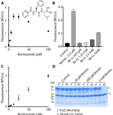
During attempts to identify inhibitors of TtClpP that could be used for further structural biology studies, we observed that bortezomib did not inhibit, but actually stimulated, TtClpP peptidase activity at concentrations from 3 to 100 mM (Fig. 1A and fig. S3A). Bortezomib is an N-protected dipeptide with a boronic acid instead of a carboxylic acid that forms adducts with activated threonines or serines (24). It was developed as a proteasome inhibitor targeting specifically the chymotrypsin-like sites of this complex with nanomolar affinity while exhibiting limited reactivity to the other active sites of the complex. We selected this molecule because it was recently identified in an in vivo cell screen as an inhibitor of GFPssrA degradation by the MtbClpXP1P2 complex and was proposed as a potential antituberculosis candidate (25). The bortezomib dependence of the TtClpP peptidase activity displays a bell shape, with maximum activation at 12.5 mM (about 10- to 20-fold activation), while higher drug concentrations reduced the enzymatic activity (Fig. 1A). A time-lag phase for the activation of TtClpP was consistently observed and likely resulted from the
neces-sary fluorimeter temperature equilibrium because the assays were executed at 45° to 60°C. Bortezomib activation derives from intrinsic properties of TtClpP, and when tested against EcClpP peptidase activity, it behaved as a powerful inhibitor with an apparent IC50(median
inhib-itory concentration) of 1.6 mM (fig. S3B).
Although paradoxical, activation of some ClpP complexes by in-hibitors has already been reported (3, 22, 26–28). Akopian and col-leagues (3) have shown that the MtbClpP complex (MtbClpP1P2), otherwise inactive, is activated by small peptide aldehydes. The most potent of these activators in Mtb, an N-terminally blocked dileucine (Bz-LL), was also capable to activate TtClpP, albeit with lower efficiency (Fig. 1B). Conversely, bortezomib could also activate the peptidase ac-tivity of the MtbClpP1P2 complex. We applied a set of biochemical, bio-physical, and structural studies to understand the mechanistic and functional foundation of this inhibitor-based activation.
Bortezomib activatesTtClpP unfolded protein degradation After establishing the bortezomib-induced activation of TtClpP for degradation of small peptides, we investigated its potential to stim-ulate protein degradation. Contrary to small peptides, which can
50 37 25 20 15 10 F luor esc enc e (RFU/s) A B C D Control Bortez. 12.5 μMBz-LL 625 μM Bz-LL 1.25 mM Bz-LL 2.5 mM Bz-L L 5m M 0.0 0.1 0.2 0.3 0 0 50 100 1 2 3 Bortezomib (μM) F luor esc enc e (RFU/s) 0 0 50 100 1 2 Bortezomib (μM) N N O O N H N H OH OH B 1: FtsZ (44.4 kDa) 2: TtClpP (22.7 kDa) 0′ 5′ 45′ 0′ 5′ 45′ 0′ 5′ 45′ 0′ 5′ 45′Control 20 μM Bortez.200 μM Bortez.2 mM Bortez.
F luor e sc enc e (RFU/s) kDa 1 2
Fig. 1. Bortezomib activatesTtClpP for peptide and intrinsically disordered pro-tein degradation. (A) TtClpP (1 mM complex) peptidase activity was measured with the substrate PKMamc (100 mM) in the presence of bortezomib at the indicated con-centrations. As the peptide is cleaved, 7-amino-4-methylcoumarin is released, result-ing in an increase in the measured fluorescence. Initial rates are plotted as a function of the bortezomib concentration. RFU, relative fluorescence units. (B) The activating effect of bortezomib is compared with the one observed with Bz-LL, a previously described activator of MtbClpP1P2. (C) The degradation of the unfolded protein sub-strate FITC-casein by TtClpP (0.1 mM complex) was measured in the presence of the indicated bortezomib concentrations. Initial degradation rates following temperature equilibration were plotted as a function of the bortezomib concentration. (D) TtClpP (1 mM complex) can degrade the intrinsically disordered E. coli FtsZ. The degradation of FtsZ by TtClpP was monitored by SDS-gel electrophoresis. While no degradation was observed with apoTtClpP, degradation of FtsZ was observed in the presence of 200 mM and 2 mM bortezomib.
independently diffuse into the proteolytic chamber, the basal degrada-tion of proteins by ClpP is low because the flexible N-terminal tails of ClpP act as a barrier to protein entry into the chamber. Our as-say used as proteolytic target fluorescein isothiocyanate–labeled casein (FITC-casein), a proline-rich protein lacking stable secondary struc-tures. Protease-catalyzed hydrolysis of FITC-casein leads to highly flu-orescent dye–labeled peptides. Similarly to the peptidase activity, addition of bortezomib did not result in inhibition of FITC-casein degradation, but instead, a striking activation was observed (Fig. 1C and fig. S4A). Furthermore, contrary to what was observed with the peptidase activity, concentration dependence of this effect was linear and no reduction of the activation was observed up to 100 mM (Fig. 1C). In addition to casein, we tested whether bortezomib could also promote the degradation of other substrates. The bacterial guanosine triphos-phatase (GTPase) FtsZ forms a cytokinetic ring at mid-cell, recruits the division machinery, and coordinates membrane and peptidoglycan cell wall invagination. An FtsZ monomer contains an intrinsically disordered C-terminal linker, and the degradation of FtsZ by ClpP ac-tivated by the natural antibiotic acyldepsipeptides (ADEPs) has been proposed to trigger growth inhibition (29). We therefore tested the ef-fect of bortezomib ClpP-catalyzed proteolysis on FtsZ and found that FtsZ was, even at low temperature for TtClpP (37°C), degraded by ClpP when in the presence of bortezomib (Fig. 1D) (29). These experiments establish that bortezomib acts as a TtClpP activator for the degradation of different disordered protein substrates or small peptides.
ClpX enhances peptidase activity ofTtClpP
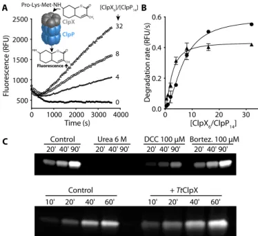
Because ClpP acts in association with specific chaperones in vivo (ClpX, ClpA, or ClpC), we proceeded by assessing the effect of TtClpX association on TtClpP peptidase activity. When we measured the deg-radation of PKMamc by TtClpP in the presence of TtClpX, we observed a marked increase in peptidase activity that was dependent on TtClpX concentration (Fig. 2, A and B). Similarly to TtClpP activation by bortezomib, a lag phase was observed, which could result either from temperature equilibrium or from oligomerization of TtClpX hexamers from monomers in the presence of adenosine 5′-triphosphate (ATP).
Because bortezomib and ClpX are both able to activate TtClpP, we tested whether the effects of bortezomib and ClpX were synergistic. Bortezomib (10 mM) did not induce further activation but instead acted as an inhibitor (fig. S4B) in the presence of ClpX. However, in the presence of bortezomib, a marked feature of the ClpX activation curve was a certain level of cooperativity, i.e., less TtClpX concentration was required to achieve maximal activity, suggesting that bortezomib binding can modulate the affinity of ClpP for ClpX (Fig. 2B). In the presence of bortezomib, half-maximal activation was achieved with a TtClpX6/ClpP14ratio of 2.4, corresponding to 120 nM TtClpX6. This
value for TtClpX-TtClpP association is on the same order of magnitude as the one reported for E. coli using an indirect ATPase assay (27).
The large activation of ClpP-catalyzed degradation of small peptides by ClpX was unexpected, because auxiliary chaperones are required for unfolding, translocation, and opening of the N-terminal pores facilitat-ing entry of protein substrates into the ClpP proteolytic chamber, while small peptides can just freely diffuse into the proteolytic chamber. The hydrolysis rate of small peptides (dipeptides) in EcClpP has been inde-pendently shown to be unaffected by ClpX association (30, 31). Activa-tion by EcClpX is, however, essential for bigger peptides and proteins, and ClpX activation increases as a function of peptide molecular weight. ClpX binding has also been shown to stimulate active-site modification of ClpP by fluorophosphates, but only to the level predicted from faster
diffusion of the inhibitor into the ClpP chamber (30). Nonetheless, it was recently proposed that, in addition to the opening of the ClpP gate, chaperones can also allosterically activate the ClpP catalytic sites by inducing a shift of ClpP from the inactive compressed state, in which the catalytic triad is distorted, to the extended state, in which the cata-lytic residues are positioned for enzymatic action (23).
Because the small molecular weight of the substrate used in our as-says is not an obstacle to its free diffusion into the proteolytic chamber (molecular weight of 574 Da) (30), we tested whether TtClpX binding to TtClpP could affect the protease active sites. To confirm the effect of co-chaperone or bortezomib on the active site, we used an assay based on fluorophosphonate (FP) conjugated tetramethylrhodamine (TAMRA-FP, molecular weight of 680 Da), which specifically and covalently labels activated Ser active sites with a fluorescent dye. TAMRA-FP can be used to quantify the reactivity of a given protease active site, e.g., if the cata-lytic triad is in the correct geometry for proper serine activation. The presence of an activated Ser can thus be detected using fluorescent gel imaging. We incubated TtClpP and TAMRA-FP with either urea, bor-tezomib, dichloroisocoumarin (DCC; an inhibitor of serine proteases), or TtClpX. While urea (by unfolding the complex) and DCC (by ir-reversibly blocking the active site) reduced the amount of TAMRA bound to ClpP, ClpX association resulted in a significant increase of active-site acetylation—a clear indication that ClpX association acti-vates the ClpP active site. Although bortezomib also leads to an activa-tion of ClpP, we failed to observe any increase of TAMRA-FP labeling (Fig. 2C). This could result from competition between the two inhibitors
Degradation rate (RFU/s
) 0 1000 2000 3000 4000 500 1000 1500 2000 2500 Time (s) 0 10 20 30 0.0 0.2 0.4 0.6 [ClpX 6/ClpP14] Fluorescence (RFU) A B C 20' 40' 90' 20' 40' 90' 20' 40' 90' 20' 40' 90' 20' 10' 40' 60' 10' 20' 40' 60'
Control Urea 6 M DCC 100 μM Bortez. 100 μM
Control + TtClpX 4 8 32 0 [ClpX6]/[ClpP14] Pro-Lys-Met-NH O O CH3 NH2 O O CH3 ClpX ClpP
Fig. 2. TtClpX activates TtClpP peptidase activity. (A) TtClpP (0.1 mM complex) peptidase activity was measured in the presence of TtClpX at the indicated ClpX6/
ClpP14ratios. (B) The activation of TtClpP peptidase activity in the absence (circles)
or presence (triangles) of bortezomib (10 mM) is plotted as a function of the molar ClpX6/ClpP14ratio. Half-maximal peptidase activity was obtained at 6.5 ± 0.5 and
2.4 ± 0.3 ClpX6/ClpP14ratio for the apo and bortezomib-loaded TtClpP, respectively.
(C) Activation of the TtClpP active site by bortezomib (100 mM) and TtClpX (1 mM), monitored by labeling with TAMRA-FP. TAMRA-FP (5 mM) was incubated with TtClpP (1 mg/ml), and aliquots of the reaction mixture were removed at the indi-cated time points. Labeling of the active-site serines is reflected by the increase in fluorescence observed.
(bortezomib and TAMRA-FP). Therefore, we cannot exclude that TAMRA-FP binding also results in TtClpP activation. However, con-trary to bortezomib, TAMRA-FP forms irreversible adducts with TtClpP, thereby preventing activity measurements.
Bortezomib binds to the serine of ClpP active site
To understand bortezomib’s mechanism of action, we examined its binding site using NMR spectroscopy. With a molecular weight of 300 kDa, NMR studies of TtClpP are challenging. In solution, the slow tumbling leads to severe line broadening, to the extent that stan-dard solution NMR studies are not feasible. The most sensitive route for observing solution NMR signals of proteins of the size of ClpP is the specific labeling of methyl groups in an otherwise deuterated background (32). Although being a very powerful approach for probing interactions and dynamics, methyl-directed NMR is able to observe only the subset of methyl-bearing amino acid types, and the sequence-specific assignment of NMR resonances is challenging, generally requiring multiple mutants.
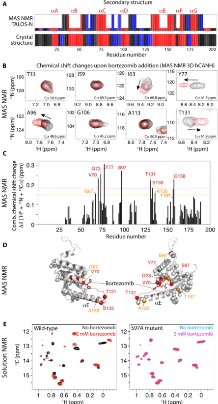
MAS solid-state NMR does not suffer from this inherent size lim-itation and is able to detect all amino acid types. We have used samples of TtClpP that were either obtained from sedimenting ClpP from so-lution into MAS NMR sample tubes (1.3-mm rotors) or obtained by addition of methyl-pentane-diol, an oft-used precipitant in crystallog-raphy. Spectra acquired from these two preparations are very similar. We have used proton-detected three-dimensional (3D) and 4D MAS NMR approaches (33) to obtain the sequence-specific resonance as-signment for (1HN,15N,13Ca,13CO) nuclei of 97 residues spread throughout the molecule, including the active site and the central helix aE, which undergoes most changes in the compressed-to-extended transition. The N-terminal gate region was not assigned, presumably due to its structural heterogeneity and dynamics, which lead to in-efficient NMR coherence transfers and line broadening (16). Figure 3A shows the residue-wise secondary structure obtained from these MAS NMR resonance assignments. As expected, the location of secondary structures matches well the information inferred from crys-tallography (see below).
3D spectra correlating intra-residue1H-13Ca-15N frequencies (re-ferred to as hCANH) of sedimented TtClpP with and without bortezomib are reported in Fig. 3 (B and C). The observed changes are modest, which is likely linked to the relatively low affinity of bortezomib, making it difficult to saturate the protein in the highly concentrated sedimented sample. Nevertheless, chemical shift perturbations (CSPs) are ob-served for a number of residues in the vicinity of the active site, includ-ing Val70, Gly73, Tyr77, and the catalytic Ser97, as well as residues in the neighboring helix aE (Fig. 3D). These data reveal that bortezomib in-teracts with the active site, and suggest that the interaction induces also small changes in the structure and the dynamics of helix aE, which is involved in mediating the compressed-to-extended transition.
To independently confirm the direct involvement of the active site for bortezomib binding in solution, we have performed addition-al NMR experiments, making use of perdeuterated, Ile-d1-CH3–
labeled TtClpP. Spectra of apo TtClpP, and of the latter in the presence of 2 mM bortezomib, are shown in Fig. 3E (left). Although we did not attempt to assign the resonances of the 18 isoleucines per ClpP subunit, the clear differences in the spectra without or with bortezomib un-ambiguously confirms binding. In a control experiment performed with the catalytic S97A mutant, no spectral changes were observed, corroborating that the bortezomib interaction requires the presence of the catalytic serine (Fig. 3E, right). The absence of any substantial
CSP in the S97A mutant demonstrates that all spectral changes are a result of the direct binding of bortezomib to the active site and its associated conformational changes—not from nonspecific binding to other parts of the protein.
Together, MAS NMR and solution NMR provide direct evidence that bortezomib binds to the catalytic site. CSPs outside the active site (particularly in helix aE) suggest that binding may lead to further struc-tural changes, possibly connected to the observed TtClpP activation. Structural basis for bortezomib activation
To understand the structural details of the TtClpP-bortezomib interac-tion initially characterized by NMR, we used x-ray crystallography and determined the crystal structures of TtClpP in the absence and presence of bortezomib at a resolution of 1.95 and 2.7 Å, respectively (Fig. 4).
The overall structure of TtClpP is essentially identical to the “ex-tended” state (34–37) of ClpP described for other species {root mean square deviation (RMSD) to EcClpP tetradecamer [Protein Data Bank (PDB): 6MT6] = 2.115 Å for 1451 aligned Ca atoms and RMSD to EcClpP monomer = 0.73 Å for 176 aligned Ca atoms}, which is in agreement with its high sequence similarity to ClpP orthologs (% iden-tity to EcClpP = 58.25). The structure of one TtClpP monomer is presented in Fig. 4B and depicts an a/b-fold composed of two domains: a head domain comprising six repeats of a/b units (helices A to D, F, and G and strands 1 to 5, 7, and 8) and a handle domain (helix E and strand 6). The N terminus of TtClpP forms a short antiparallel b-hairpin that protrudes from the apical surface of the barrel. This N-terminal b-hairpin is constituted of residues 5 to 8 and residues 15 to 18 of TtClpP, with missing electron density for residues 9 to 14. While the overall monomer structure of ClpP is highly conserved among the structures solved to date, a high degree of variability exists in the conformation of the N-terminal regions. Several ClpP crystal structures feature N-terminal b-hairpin structures of varying lengths and degrees of flexibility, some of which appear to be stabilized by crystal-packing contacts (38). Solution NMR studies have demon-strated that the N termini of ClpP are structurally heterogeneous and adopt multiple conformations (16, 39), which might explain the missing density near the end of the N-terminal b-hairpins in the TtClpP structure.
Unexpectedly, in the TtClpP crystals without added bortezomib, we consistently observe a continuous stretch of electron density near the active-site serine (Ser97) of each TtClpP monomer, which, based on its shape, suggests the presence of a tripeptide (Fig. 4C and fig. S5). Because no peptide was added, the nature of this molecule was un-known. We, therefore, built it in the electron density as a tri-L-alanine
(see also omit and polder maps presented in fig. S5). The presence of short peptides in Helicobacter pylori ClpP structures resulting from proteolysis during crystallization has been previously described (40). While in that particular case the observed tri- and tetrapeptides are caused by proteolysis of added heptapeptides before crystallization, the origin of the tripeptides observed in our TtClpP crystals remains unclear. Matrix-assisted laser desorption/ionization–time-of-flight (MALDI-TOF) measurements exclude the presence of these peptides in fresh purified TtClpP. We speculate that they are the result of au-toproteolysis of unfolded or partially unfolded TtClpP in the crystal-lization drops followed by the formation of TtClpP:peptide crystals. The serendipitous presence of peptides near the active-site Ser97 pro-vides valuable insights into how TtClpP binds its substrates (Fig. 4C, inset 1). Apart from the active-site Ser97, the terminal carboxy group of the peptide (residue P3) is further stabilized by hydrogen bonds
6.8 7.0 7.2 106 108 T33 I63 Y77 G106 A96 T131 I59 A113 9.0 9.2 9.4 9.6 120 122 124 8.0 8.2 8.4 8.6 118 120 122 7.4 7.6 7.8 8.0 122 124 8.2 8.4 8.6 110 112 8.6 8.8 9.0 9.2 128 130 6.8 7.0 7.2 102 104 7.4 7.6 7.8 8.0 116 118 15N (ppm) 15N (ppm) 1H (ppm) 1H (ppm) 1H (ppm) 1H (ppm) 25 50 75 100 125 150 175 200 Residue number A C V70 G73 Y77 S97 T131 S133 A136 T157
Chemical shift changes upon bortezomib addition (MAS NMR 3D hCANH)
D E 0 0.1 0.2 0.3 G158 G67 Secondary structure MAS NMR TALOS-N Crystal structure B 25 50 75 100 125 150 175 200 Residue number M A S NMR M A S NMR M A S NMR S o lution NMR 0 1 0.8 0.6 0.4 0.2 1 0.8 0.6 0.4 0.2 0 12 13 14 15 12 13 14 15 13C (ppm) 1H (ppm) 1H (ppm) T131 S133 A136 T131 A136 Bortezomib V70 G67 Y77 G67 G73 S97 V70 T157 S97A mutant Wild-type No bortezomib 2 mM bortezomib No bortezomib 2 mM bortezomib δ C omb . chemical shif t change (ppm)
Fig. 3. Bortezomib binding to TtClpP investigated by MAS NMR and solution NMR. (A) Secondary structures (loop, black; a-helix, red; b-strand, blue) as a function of sequence number obtained from the resonance assignments obtained by MAS NMR and analysis with TALOS-N (24) (tall bars). For residues for which no assignment has been obtained, the small bars are obtained by TALOS-N from a database approach. The bottom panel shows the comparison with the secondary structure obtained from the crystal structure (see Fig. 5). (B) Zooms onto 2D1H-15N excerpts from 3D hCANH MAS NMR correlation spectra of TtClpP sedimented in the presence of 10 mM
bortezomib (red) or without bortezomib (black). Chemical shift changes are indicated with arrows. Note that the CSPs are relatively small, presumably due to the difficulty of saturating the binding site in a sedimented sample, given the high Kdvalue, resulting in incompletely occupied binding sites. ppm, parts per million. (C) Combined CSPs
upon addition of bortezomib from the added peak shifts in1H,13Ca, and15N dimensions obtained from the spectra shown in (B). (D) Location of residues with substantial CSPs mapped onto the x-ray structure of the TtClpP:bortezomib complex obtained from the data shown in (C). (E) Solution-state NMR spectra of wild-type (left) and S97A (right) TtClpP in the absence and presence of bortezomib, showing large spectra changes for the former due to binding, but no effect on the mutant.
B 90° TtClpP TtClpP' C A TtClpP + tripeptide TtClpP + bortezomib Inset 1 Inset 2 N C αA αB αC αD αE αF αG βN1 βN2 β1 β2 β3 β5 β4 β6 β8 β9 β7 β7 αE αE β8 β8 β6 β6 αD αD αC αC Trp125 His122 Asp171 Ser97 Val70 Gly68 Met98 Trp25 Asp171 Ser97 Val70 Gly68 Met98 His122 β4 β4 Inset 1 Inset 2 αA βN1 βN2 αA N C αB αC αD αE αF αG β1 β2 β8 β4β6 β7 β9 β3 β7 β5 MD simulation MAS NMR MD simulation RMSF (Å) 1.4 0.4 50 0 MAS NMR Spin relaxation 20 40 60 80 100 120 140 160 180 20 40 60 80 100 120 140 160 180 Residue number Residue number 0 1 2 3 4 RMSF (Å) MAS NMR 15 N R1ρ (s −1) 0 10 20 30 40 50 60 70 80 R1ρ (s−1) D E F G
Fig. 4. Structure and dynamics ofTtClpP from x-ray crystallography, MAS NMR, and MD simulations. (A) Side and top views of the TtClpP 14-mer. One TtClpP monomer per heptameric ring (light and dark gray) is highlighted in light and dark blue, respectively. (B) Cartoon representation of the TtClpP monomer in peptide-bound (left) and bortezomib-peptide-bound (right) states. Helices are named by letters, and strands are indicated by numbers. A zoom of the ligands present in the active sites (dashed boxes) is shown in (C). (C) Substrate-binding pocket of TtClpP. The residues involved in the binding to the model peptide (inset 1) and bortezomib (inset 2) are shown as sticks. (D and E) Residue-wise MAS NMR amide15N R1arelaxation rate constants. High R1arate constants point to enhanced nanosecond-to-millisecond
motions and are found primarily in loop regions and in helix aE, as shown in (C). (F and G) MD-derived root mean square fluctuations (RMSFs) over the 1-ms-long MD trajectory of the assembled 14-mer ClpP.
with the Ne2 atom of His122(part of the ClpP catalytic triad) and the backbone amide groups of Met98and Gly68. The Nd1 atom of His122 on its turn forms an additional hydrogen bond with Asp171, the third residue of the ClpP catalytic triad. The amino groups of peptide re-sidues P2 and P3 are stabilized by backbone interactions with Gly68 and Trp125, respectively, while the carbonyl groups are stabilized by in-teractions with amino groups of Trp125and Val70(Fig. 4C, inset 1). The geometry and distances between the catalytic triad residues (Ser97, His122, and Asp171) are consistent with a functional TtClpP catalytic triad because their distances are in the range of the ones observed in classical serine proteases and active ClpPs (40). Figure 4B displays the 2.7-Å resolution structure of the bortezomib-bound state (see also fig. S5). Similarly to the peptides present in the TtClpP:peptide struc-ture, bortezomib tightly fits between b-strand 6 and a small b-turn formed by Gly68and Val70, thereby forming an antiparallel b-sheet. This conformation is characterized by a hydrogen bond network al-most identical to the one observed in the TtClpP:peptide interaction (Fig. 4C, inset 2). Unlike the TtClpP:peptide complex, where Ser97is forming hydrogen bonds with the terminal carboxy group of the pep-tide, bortezomib seems to be covalently bound to the active-site Ser97 via its boron atom. Other significant differences are observed in the conformation of Trp125, which is displaced toward bortezomib and forms an aromatic interaction with the bortezomib phenylalanyl group. In addition, the nearby Thr131residue, which in peptide-bound TtClpP interacts with Arg170of a neighboring ClpP monomer, now forms a hy-drogen bond with Gln123of an opposing ClpP monomer in the other heptameric ring. Despite the rearrangement of the Trp125side chain, the catalytic triad geometry is again consistent with a fully active protease. To probe the flexibility of TtClpP, we used MAS NMR spin relaxa-tion experiments and MD simularelaxa-tions. Site-specific amide15N R1r
experiments probe the local amplitudes and time scales of backbone motion and are particularly sensitive to nanosecond-to-millisecond motions (41). The sites with highest15N R1rrate constants are found
not only in the loop regions but also at the tip of helix aE (Fig. 4, D and E). One-microsecond-long MD simulations of the TtClpP tetradecamer in the apo and peptide-bound states corroborate this observation, albeit with a greater flexibility at the tip of the helix (Fig. 4, F and G). Enhanced flexibility in this helix has been reported also in EcClpP by methyl-directed solution-state NMR and suggested by other methodologies (16–19, 34, 35). Because of the lack of reliable parametrization of the boronic acid moiety of bortezomib, we focused on simulating tripep-tides bound to the active site. It is noteworthy that over the length of three independent simulations of ClpP initially loaded with tri-alanine at all active sites, only a fourth of the peptides remain associated to their designated active sites (fig. S6), consistent with the idea that the products of TtClpP are necessarily weak binders, which ought to be re-leased rapidly to leave the catalytic sites ready for the next degradation reaction.
Bortezomib induces the transition from a tense state to a relaxed state with higher affinity for the substrate
Having identified the bortezomib-binding site of TtClpP by NMR and x-ray crystallography, we turned to ITC to characterize thermo-dynamically the interaction at different temperatures ranging from 25° to 45°C. At 25°C, far from the ideal catalytic temperature of TtClpP (65°C), only a moderate affinity and symmetric binding iso-therm was observed. The binding enthalpy was negative and rather small (see Table 1), thus indicating that binding was mainly entropy-driven. However, when the temperature was increased to 35° and 45°C,
considerable changes were observed in the binding isotherm. The asymmetry of the curve and the appearance of two different phases in the binding isotherm along the titration, within the low ligand satu-ration region, were clear indicators of positive cooperativity (Fig. 5A). Because of the architecture of TtClpP, comprising two heptameric rings forming a tetradecameric assembly, the ITC data were fitted to the Monod-Wyman-Changeux (MWC) model (Fig. 5B), which considers two intrinsically noncooperative conformations, with low and high binding affinity, denoted as tense (T) state and relaxed (R) state, respec-tively. In the MWC model, ligand binding shifts the equilibrium toward the high-affinity conformation (R) in a concerted cooperative way (i.e., all subunits change their conformation simultaneously; Table 1). This model has been previously transposed to other enzymatic systems, where activation at low ligand concentration and inactivation at high ligand concentration were also observed (42).
Fitting of the ITC data revealed that in the absence of the ligand, 76% of TtClpP exists in the T state, which has a lower affinity for bortezomib (Kd,T= KT−1= 40 mM). Bortezomib binding induces a
concerted transition from the T state to the R state, with a higher affinity for substrate (Kd,R= KR−1= 15 mM). A model in which we assumed
that there is no ligand binding to the subunits in the T conformation (KT= 0, DHT= 0) can be ruled out based on significantly worse
fitting. The higher affinity of the ligand for the R conformation drives the conversion of T subunits into R subunits. In addition, the compul-sory concerted conformational change within a given protein oligomer (i.e., all subunits in a given protein oligomer must undergo the confor-mational change at once, even if not all of them are occupied by ligand) will further promote the conversion of T subunits into R subunits, thereby increasing the total fraction of R subunits. The Hill coefficient [maximal slope in the log(nLB/(14− nLB)) versus log[ligand] plot] of
TtClpP is 1.3, which is rather small for a tetradecameric protein but was expected considering the small value for the conformational equilibrium constant g.
Although the MWC model was used 40 years ago for the calorimet-ric study of the interaction of trout hemoglobin with carbon monoxide using a gas-liquid reaction calorimeter (43), the present work represents the first detailed mathematical description and implementation of the MWC model in ITC, for which the experimental methodology and the mathematical formalism are not the same as those outlined in the afore-mentioned seminal work.
The MWC model provides a rationale for TtClpP activation by bortezomib. Activation of TtClpP by the inhibitor bortezomib at
Table 1. Thermodynamic parameters of bortezomib binding from ITC and fitting to the MWC model. KRand KTrefer to the bortezomib
as-sociation constants for the relaxed and tense state. DHR, DHT, and DHg
refer to the binding enthalpy of the R state, T state, and conformational enthalpy between R and T conformations. g is the equilibrium constant between R and T conformations in the absence of the ligand. N is the fraction of active or binding-competent protein. Relative errors in equi-librium constants are 30%. Absolute error in enthalpies is 0.3 kcal/mol.
T (°C) N KR (M−1) DHR (kcal/mol) KT (M−1) DHT (kcal/mol) g DHg (kcal/mol) 25°C 0.97 4.5 × 10⁴ −0.5 No apparent cooperativity 35°C 0.97 8.0 × 10⁴ −0.9 2.8 × 10⁴ −0.1 3.3 −0.3 45°C 1.00 6.5 × 10⁴ −1.5 2.5 × 10⁴ −0.3 3.2 −0.2
T
R
0 5 10 15 20 25 30 10 15 20 25 30 0 10 20 30 40 50 0 5 0 10 20 30 40 50 0 5 10 15 20 25 30 −2.5 −2.0 −1.5 −1.0 −0.5 0.0 −0.6 −0.4 −0.2 0.0 0.2 0 10 20 30 40 50 A B 0.01 0.1 1 10 100 1000 0.0 0.2 0.4 0.6 0.8 1.0 C 0.0 0.5 1.0 1.5 2.0 0.0 0.5 1.0 1.5 DDegradation rate (RFU/s)
PKMamc (mM) 25°C 35°C 45°C
...
...
+L KT +L KR +L +LMolar ratio Molar ratio Molar ratio
Time (min) Time (min) Time (min)
dQ
/d
t (μcal/s)
Q
(kcal/ mol of injec
tant)
F
R,TOTALF
T,TotalF
R,FreeF
T,FreeF
R,BoundF
T,BoundF
T or R,Bound Bortezomib (μM) Molar fractionFig. 5. Cooperative bortezomib binding detected by ITC experiments. (A) Calorimetric titrations for the interaction of TtClpP (10 mM in the calorimetric cell) with bortezomib (1.4 mM in the injecting syringe) in 50 mM Hepes (pH 7.6) and 50 mM NaCl. Experiments were performed at three different temperatures: 25°, 35°, and 45°C. Thermograms (thermal power as a function of time) are displayed in the upper plots, and binding isotherms (ligand-normalized heat effects per injection as a function of the molar ratio, [L]T/[P]T) are displayed in the lower plots. The binding isotherms were analyzed with the MWC model for TtClpP, consisting of 14 identical subunits,
each one containing a single ligand-binding site. Nonlinear least squares regression analysis allows the determination of the following binding parameters (see Table 1): association constants for the R and T states (KR, KT), binding enthalpies to the R and T states (DHR, DHT), conformational equilibrium constant and conformational
enthalpy change between states R and T (g, DHg), and fraction of active protein (N). (B) MWC model for a 14-mer oligomeric protein. The protein can populate only
two conformational states in equilibrium (with equilibrium constant g): all subunits in R (relaxed, ellipsoidal shape) conformation or all subunits in T (tense, spherical shape) conformation. Subunits have a single ligand-binding site, exhibiting the R conformation a higher binding affinity (KR> KT). Ligand binding occurs through an
independent (noncooperative) fashion within an oligomer (ligand-free subunits in light blue and ligand-bound subunits in dark blue). T conformation is favored at low ligand concentration; the higher ligand-binding affinity for R conformation promotes a highly cooperative compulsory concerted conformational change driven by ligand binding and involving all subunits within an oligomer, displacing the equilibrium toward the R conformation. (C) Molar fraction of the different protein species (total, ligand-free, and ligand-bound R and T conformations) as a function of ligand concentration: total fraction of subunits in R conformation (continuous black line), total fraction of subunits in T conformation (continuous red line), fraction of subunits in bound R conformation (dashed black line), fraction of subunits in ligand-bound T conformation (dashed red line), fraction of ligand-free subunits in R conformation (dotted black line, highlighted with an arrow), and fraction of ligand-free subunits in T conformation (dotted red line). In addition, the fraction of ligand-bound subunits in either R or T conformation is shown (dashed green line). It is obvious that the contribution of subunits in T conformation to the ligand binding is very small (dashed red line). At very low ligand concentration, 77% of the protein subunits are in T conformation and 23% are in R conformation, according to the value of the equilibrium constant g equal to 3.3. At low ligand concentration, the total fraction of subunits in R conformation increases, while the total fraction of subunits in T conformation decreases due to the T→R conversion. However, both the fractions of ligand-bound subunits in R and T conformation increase due to ligand binding (although the increment in ligand-bound subunits in T conformation is negligible). At high ligand concentration, the total fraction of subunits in R conformation further increases, while that of total subunits in T conformation decreases, and the fraction of ligand-bound subunits in R conformation further increases due to ligand binding, but the fraction of ligand-bound subunits in T conformation decreases due to the T→R conversion. The fraction of ligand-free subunits in R conformation dominates this region, with a maximal population of 60%. This is due to the concerted con-version of subunits within a given protein oligomer (all subunits within an oligomer undergo the conformational change T→R, although not all of the subunits bind a ligand). At very high ligand concentration, the fraction of ligand-bound subunits in R conformation dominates the conformational ensemble, reaching a maximal population of 100%. The R↔T equilibrium shows a switchover or crossover point (R and T are equally populated) at around 2 mM free bortezomib concentration. (D) TtClpP (1 mM complex) peptidase activity was measured as a function of PKMamc concentration in the presence (15 mM) and absence of bortezomib.
moderate concentrations occurs as a consequence of the concerted conformational change within the protein tetradecamer, which makes the total fraction of subunits in the enzymatically active R conforma-tion (FR,Total) much larger than the fraction of ligand-bound subunits
in R conformation (FR,Bound), i.e., there is no proportionality between
FR,Totaland FR,Bound(Fig. 5C). At intermediate bortezomib
concentra-tion, the fraction of inhibitor-free (active) subunits in an R confor-mation is maximal and predominates in the speciation distribution (Fig. 5C). Thus, the extents of cooperativity for binding and coop-erativity for activity are different [a phenomenon previously reported for allosteric proteins in (44)]. If the conformational changes were not concerted, then the activity of TtClpP would continuously decrease with increasing bortezomib concentration, with no maximal activity at intermediate bortezomib concentration. The increase in the fraction of inhibitor-free R subunits at moderate inhibitor concentration is the molecular event underlying the increase in activity. Similarly, the de-crease in the fraction of inhibitor-free R subunits at high inhibitor concentration is the molecular event underlying the decrease in activ-ity at high inhibitor concentration.
The ITC-derived model predicts accurately the activity data. Ac-cording to the peptide degradation activity measurements (Fig. 1A), the maximal TtClpP activity is found around 12.5 mM of total borte-zomib. The ITC-derived MWC model predicts that the maximum fraction of inhibitor-free TtClpP in the R conformation (FR,Free)
should be around 6 mM of free bortezomib (Fig. 5C). Considering the population of bound TtClpP at 6 mM free bortezomib concentra-tion (FTorR,Bound= 0.3; Fig. 5C), and taking into account the TtClpP
subunit concentration of 10 mM (used in our activity measurements), one can calculate that the concentration of bortezomib bound to TtClpP is 3 mM. Consequently, the total concentration of bortezomib for achieving 6 mM free bortezomib (i.e., the concentration for max-imal bortezomib-free subunits in R conformation) is around 9 mM, in excellent agreement with the activity measurements reporting a maximum activity for 12.5 mM bortezomib concentration (Fig. 1A). Furthermore, to verify that the bortezomib-induced R state has a higher affinity for the substrate, we measured peptide hydrolysis as a function of substrate concentration in the apo state and in the presence of nonsaturating concentrations of bortezomib (15 mM). While there are differences between Kd, a thermodynamic equilibrium constant,
versus a purely kinetic constant as KM, in the presence of bortezomib,
TtClpP displayed an apparent KMaround 400 mM. In its absence, no
indications of substrate saturation were observed (Fig. 5D), reflecting the major differences between the bortezomib-bound and apo states. Put together, the activity measurements and ITC data reveal that acti-vation of TtClpP by an inhibitor is achieved by shifting the conforma-tional population toward a higher-affinity R state.
Off-equilibrium MD simulations provide rationale for stabilization of extended state
Activity measurements, ITC data, x-ray structures, and NMR spec-troscopic data have established that, by binding to a subset of TtClpP active sites, bortezomib and peptide substrates favor a catalytically more active particle by keeping both the bound and free subunits in an active state, with the catalytic triad poised for catalysis. We at-tempted to bridge the functional and thermodynamic information from biochemistry and ITC with the structural view by performing in silico experiments. In a nutshell, in these MD simulations, we in-vestigated the energetics of the transition from the extended (active) to the compressed (inactive) state and interrogated whether the
pres-ence of a molecule bound to the active site alters the relative stability of the two states. Near-equilibrium MD experiments were performed, in which the two heptameric rings were slowly brought from the extended state to the compressed state using the centers of mass of the two rings as single variable for the steered MD protocol (see Materials and Methods). Figure 6 (A and B) displays the force required for this extended-to-compressed transition, obtained from five inde-pendent in silico experiments, as a function of the distance of the two half-rings. Figure 6B shows the work (i.e., the integrated force) for this process. The present simulations have been performed for both the apo state (gray) and a substoichiometrically peptide-loaded state in which only 2 or 3 of the 14 binding sites were occupied with tri-alanine tide. During the compression process, none of the initially bound pep-tides were expulsed from their target catalytic sites, and all the secondary structure elements were preserved (Fig. 6, C to E). Compressing ClpP from the extended conformation requires a stronger force in the pres-ence of a substoichiometric amount of alanine tripeptides compared to the empty protein (Fig. 6, A and B).
Together, these data provide a rationale for the enhanced stability of the extended conformation in the presence of a ligand: Even sub-stoichiometric amounts of the ligand, i.e., only partially occupied sites, lead to an overall stabilization of all subunits in the active state. The simulations were performed with peptides rather than bortezomib due to the lack of reliable force field parameters for boronic acid and reveal the substrate-induced stabilization of the active extended state. It can be expected that this stability difference of extended over compressed state is even more pronounced with the covalently bound bortezomib.
The MD simulations support the idea that bound ligand, even at substoichiometric amounts, favors a stable extended form, which we believe is the R state modeled in the ITC data, while the apo state is a compressed state. We attempted to validate the compressed-to-extended state transition observed from the MD experiments by performing dynamic light scattering (DLS). Figure 6G shows the hydrodynamic radii (Rhyd) of TtClpP in the absence and presence of 5 mM bortezomib,
revealing that addition of bortezomib causes an increase of approxi-mately 8 Å in Rhyd(or 16 Å in diameter) of TtClpP. The
experimen-tally obtained hydrodynamic radii for TtClpP without and with 5 mM bortezomib (5.95 and 6.67 nm, respectively) are in excellent agree-ment with the theoretical hydrodynamic radii obtained for compressed and extended states of ClpP, supporting the view that the T-to-R transition modeled from the ITC data corresponds to a compressed-to-extended transition.
DISCUSSION
Activation of different ClpPs by previously identified inhibitors has been independently reported (3, 22), but to date, no mechanism for these paradoxical results has been proposed. Bortezomib, just like N-blocked peptide aldehyde Mtb activators reported previously, does not follow the canonical definition for an allosteric activator, i.e., they bind to the protease active site (14, 15) and their effect is not univer-sal to all ClpP homologs. For instance, we show here that although bortezomib activates TtClpP (up to 100 mM), it is, in the same con-centration range, a good inhibitor of EcClpP. This result is rather puzzling, considering the high sequence and structural similarity be-tween the two proteins (58% sequence identity).
We show in the present contribution that ClpP activation by in-hibitors derives from the intrinsically cooperative character of the
ClpP tetradecameric structure. Bortezomib, although blocking a num-ber of active sites, can overall increase the catalytic rate by promoting a concerted transition in the complex, stabilizing the active-state con-formation. In line with this observation, the near-equilibrium MD simulations performed in our study suggest that compression of the extended-active conformation of TtClpP is hampered when a frac-tion of the catalytic sites is occupied by unspecific tripeptide substrates. In the presence of a stronger specific binder such as bortezomib, it could be reasonably anticipated that compression toward the inactive con-formation is even more hindered. A comparable mechanism likely underlies MtbClpP1P2 activation, because it has been shown that the peptide activator would sterically clash with a non-active ClpP1 complex, thereby forcing the complex into an active conformation (22). The similarity between the bortezomib TtClpP structure and the structure of TtClpP with peptides in the active site strongly suggests that our findings reflect an inherent substrate-dependent activation mechanism of the enzyme. Through this mechanism, the presence
of the substrate in the ClpP active site promotes the activation of the remaining subunits, allowing efficient and processive protein degra-dation. Although we were unable to obtain a crystal structure of apo TtClpP, the DLS experiments performed here are consistent with a bortezomib-induced transition from a compressed or compacted state to an extended TtClpP conformation. Addition of bortezomib altered chemical shifts in MAS NMR experiments, possibly pointing also toward the changes in the equatorial region of ClpP, expected for a compressed/extended transition (Fig. 3D).
Our results also support the previously described bidirectional cross-talk between chaperone binding and protease active sites (14, 22, 23, 26–28), because bortezomib is no longer an activator in the presence of the co-chaperone TtClpX and TtClpP partially saturated with bortezomib binds strongly to TtClpX. Significant dif-ferences were observed between the activation of TtClpP peptidase activity and proteolytic activity by bortezomib. While peptidase ac-tivation displayed a bell-shaped curve, protease acac-tivation showed a
−8 −6 −4 −2 0 Δζ (Å) −70 −60 −50 −40 −30 −20 −10 0 Fapplied (kcal/mol Å) Apo Peptide-loaded A −8 −6 −4 −2 0 0 50 100 150 200 250 300 w (kcal/mol) Δζ (Å) Apo Peptide-loaded B C E D F 80 60 40 20 7 10 In te nsit y (%) Rhyd (nm) No bortezomib 5 mM bortezomib G 6 5 8 9 4 5 6 7 25°C 45°C 25°C 45°C 5 mM bortezomib No bortezomib Calc. TtClpP extended Calc. TtClpP compressed Rhyd (nm)
Fig. 6. In silico pulling experiment ofTtClpP from the extended state to the compressed state. (A and B) Profile of the force (A) and work (B) required for compressing the two rings toward each other as a function of the distance z between the rings (shown in the inset, with exaggerated distance for better visibility). Three independent 200-ns simulations were performed for apo TtClpP or TtClpP, in which 2 or 3 of the 14 binding sites were occupied with trialanine peptide (see Materials and Methods). (C and D) Conformations of TtClpP extended (C) and SaClpP compressed (PDB: 3QWD) (D) states observed in crystal structures. (E) Starting conformation of TtClpP in the steered MD simulations (shown as sphere representation). (F) Final conformation of the compressed state at the conclusion of the steered MD. Note that the peptides are still bound and that the structure is in good overall agreement with the compressed state from x-ray diffraction shown in (D). (G) DLS analysis of TtClpP (3 mM) supplemented without or with 5 mM bortezomib. The upper graph shows one representative curve of three replicates measured at 25°C for TtClpP without (red curve) or with 5 mM bortezomib (green curve), with the relative intensity (in %) plotted versus particle size [hydrodynamic radius, Rhyd(in nm)]. The
average of the three replicates is indicated using a dotted line. The lower graph depicts average hydrodynamic radii (Rhyd) calculated from three independent
experiments measured at 25° and 45°C (0 mM bortezomib, 25°C: 5.946 ± 0.036 nm; 0 mM bortezomib, 45°C: 5.940 ± 0.102 nm; 5 mM bortezomib, 25°C: 6.673 ± 0.192 nm; 5 mM bortezomib, 45°C: 6.702 ± 0.176 nm). Theoretical calculated hydrodynamic radii of compressed TtClpP (6.05 nm) and the extended TtClpP:bortezomib complex (6.47 nm) are indicated using a dotted black line (see also Materials and Methods).
linear rise in the range of the used bortezomib concentration. Al-though the process underlying bortezomib activation in the two ac-tivities should be common, important differences exist between the two assays. Protein degradation is strictly dependent on substrate access to the proteolytic chamber, because larger substrates cannot diffuse freely toward the proteolytic chamber. Our results showing protein degradation activated by bortezomib demonstrate that mod-ulation of protein entry can also be achieved via binding to the ClpP active site and not exclusively by ClpX or ADEP binding. Protein access to the proteolytic chamber leads to high local concentrations of the substrate present at the ClpP active sites. Although bortezomib activates protein degradation at low concentrations, it must compete with high concentrations of substrate in the proteolytic activity as-say, thereby rationalizing the requirement for higher concentrations of drug to achieve activation. The range of bortezomib concentrations used in our proteolytic activity assay likely corresponds to the rising part of the bell curve monitored in the peptidase activity assay and also ex-plains why several ClpP inhibitors are 10-fold more potent in peptidase assays compared with protease assays (45).
It is unclear why the action of bortezomib and activators is not uniformly conserved between different ClpPs. Differences in enzymat-ic activity are generally more diffenzymat-icult to decode than major structural differences because allosteric pathways often result from subtle changes in conformational dynamics that affect population distributions, which are not readily observed from comparisons of structures. For example, single mutations or truncations in EcClpP N-terminal have been shown to induce reversible inactivation of ClpP (16), and using an in-direct methodology based on the association of an EcClpP E14W mu-tant with EcClpA, the equilibrium consmu-tant between an EcClpP active state (R) and inactive state (T) was previously quantified as Keq= 7.5
(37°C) (46), i.e., a value similar to the one we describe here for TtClpP Keq= 3.3 (45°C). This finding is particularly interesting, as it suggests
that in the absence of mutations, EcClpP preferentially populates active states, while TtClpP is preferentially inactive (16). The energy barrier between the two states is, however, rather small, with an energy gap of 0.7 kcal/mol between the R and the T conformations in TtClpP. Given the high sequence identity between EcClpP and TtClpP, the key to the difference in the equilibria between active and non-active states must necessarily result from small sequence changes. EcClpP and SaClpP contain a conserved Q131XT/S133 motif at the tip of the turn between helix aE and the b-strand 6 in the handle domain (fig. S7). Here, Gln131 engages in hydrogen bonds with the h1 and h2 nitrogens of Arg170on a neighboring subunit of the same heptamer ring (intra-ring) and with the backbone oxygen of Glu169and e1 oxygen of Gln123on a monomer of the other ring (inter-ring). The side chain of the conserved Thr133 forms hydrogen bonds with the nitrogen atoms of Gln123and Lys146 (inter-ring). Gln123is part of the conserved His-Gln-Pro (HQP) motif, which includes the catalytic triad His122, while Arg170is the vicinal residue of the catalytic triad Asp171. It is clear that this extended hydro-gen bond network restricts the degrees of freedom of the aforemen-tioned catalytic triad residues. It is noteworthy that these residues are conserved neither in TtClpP nor in MtbClpP1. In TtClpP, Gln131is replaced by a threonine (Thr131), which interacts either with the e1 ni-trogen of Arg170(intra-ring) or with Gln123(inter-ring) in the TtClpP: peptide or TtClpP:bortezomib structures, respectively. Ser133of TtClpP contacts Gln123and Lys146(inter-ring) in a similar way as EcClpP Thr133(fig. S7). The replacement of the conserved glutamine Q131
by the shorter threonine in TtClpP or serine in MtbClpP1 leads to modifications in the hydrogen network that normally sustains the
extended helix aE. Structural and MD studies in SaClpP have shown that these key residues are essential for keeping the long helix aE in a straight conformation. In TtClpP, the exclusive hydrogen bond accep-tor character and shorter side chain of Thr131does not allow the for-mation of a network that simultaneously coordinates two of the members of the catalytic triad. This reduced H-bonding capacity is expected to result in an increased flexibility in that region and likely to an equilibrium shift toward nonfunctional conformations, supported by the flexibility measurements reported here by MAS NMR and MD simulations (Fig. 5). We propose that bortezomib binding through preferential stabilization of the active site (R form) shifts the entire population toward a fully active enzyme.
A different hydrogen bond network is also observed for MtbClp1. In this case, Gln131is replaced by serine, while Thr133is replaced by alanine. When purified alone, ClpP1 is an inactive enzyme, and the x-ray structure of the ClpP1 tetradecamer shows that the catalytic triad is not in the active state (13). However, in complex with ClpP2, ClpP1 has been shown to be fully active, likely because MtbClpP2, which contains the conserved QFT motif at the tip of the helix aE, can stabilize ClpP1 by forming a similar network, as has been observed in EcClpP (3). Cooperativity allows multimeric systems to adapt rapidly to envi-ronmental changes by more efficiently converting a biochemical input, i.e., the substrate/ligand concentration, into a biochemical response. Cooperative response to incoming substrates has been reported for other machineries of the protein quality control system. Because protein degradation is key to cell homeostasis, several levels of regulation have evolved to prevent uncontrolled proteolysis that would be deleterious to the cell. ClpP, as well as other complexes like the 20S proteasome, hides their active sites inside an inaccessible catalytic chamber to prevent un-controlled proteolysis. This architecture is fine-tuned by ClpP-specific co-chaperones, which add another layer of regulation in protease con-trol, either by“opening” the axial pores of the cylinder and translocating unfolded substrates into the chamber or by directly activating the ClpP active sites (2). Several studies have reported that ClpX is able to revert the conformational inhibition of ClpP induced by chemical compounds (14) or even by mutations known to promote ClpP inactive states (23). The fact that one of these inactive-state promoting mutations, namely, the Asp172Asn SaClpP mutant, corresponds to the native form of LmClpP1 and several other ClpPs suggests that this mechanism could be relevant for in vivo ClpP regulation in these species (47). Our data support a similar mechanism of conformational control for TtClpP. Our observation that TtClpX and EcClpX (data not shown) can activate TtClpP further suggests that this enzyme is conformationally attenu-ated, i.e., in the absence of the chaperone, it is in a latent inactive state. The nonphysiological activation observed with bortezomib is both a consequence of the inherent cooperativity of the protease and the fact that, in the absence of activators, its predominant state is the non-active T state (Fig. 5C). The small molecule–induced conformational shift opens new possibilities for drug development, particularly as not only peptide degradation but also protein degradation is stimulated, as we report here (Fig. 1C).
In summary, using a host of biochemical, biophysical, and struc-tural approaches, we have proposed a mechanism for the activation of ClpP by inhibitors with a key element, namely, a concerted acti-vating conformational change in the supramolecular ClpP tetradeca-meric structure elicited by the binding of inhibitors at subsaturating concentrations. Hints to a similar allosteric behavior have been re-ported for the proteasome in the presence of bortezomib (47). While bortezomib inhibits efficiently the chymotrypsin site of the proteasome,
it has been observed that it induces a paradoxical activation of the trypsin site of the complex (24).
MATERIALS AND METHODS Cloning and mutagenesis
TtClpP DNA was directly amplified from T. thermophilus HB8 DNA and cloned in to a pET41c (Novagen) expression vector using Ned I and Xho I restriction enzymes. The expressed gene contained the ad-ditional residues LEHHHHHHHH at its C terminus to allow affinity chromatography purification. Full-length ClpX DNA was first cloned into a pET28 vector using Ned I and Hind III restriction enzymes, re-sulting in residues MGSSHHHHHHSSGLVPRGSH added to the N ter-minus. While full-length ClpX was insoluble, similar to ClpX orthologs from other species, removal of its N-terminal domain (residues 1 to 54) allowed expression of a soluble ClpX construct (DTtClpX) (48). Re-moval of the TtClpX N terminus was performed by GeneCust using Ned I and Hind III as restriction enzymes. TtClpP and DTtClpX were expressed in E. coli Bl21RIL cells following overnight expression at 20°C after induction with 1 mM isopropyl-b-D-thiogalactopyranoside
(IPTG). A TtClpP mutant containing an S97A mutation was con-structed using the QuikChange II Site-Directed Mutagenesis Kit from Thermo Fisher Scientific.
Protein purification
GFPssrA was purified as previously described (49). Casein FITC from bovine milk was obtained from Sigma (C0528) and loaded onto a G25 column (GE Healthcare) to remove free fluorescein (49). TtClpP and DTtClpX were purified using native NiNTA (nickel-charged nitrilotriacetic acid) affinity chromatography followed by SEC using a 16/600 S200 200 pg Superdex column and a final SEC buffer containing 100 mM tris (pH 8.5), 100 mM NaCl, and 5% glycerol. U-[2H,15N],Ile-d1-[13CH3]TtClpP was expressed as described (33).
When expressed in D2O, TtClpP was insoluble and the protein was
re-covered from E. coli inclusion bodies using denaturing conditions. Briefly, cells were harvested and resuspended in denaturing buffer containing 8 M urea followed by sonication and centrifugation to re-move cell debris. The resulting supernatant was loaded onto a NiNTA column and washed several times with denaturing buffer. The column was then equilibrated in refolding buffer [100 mM sodium phosphate (NaPi) (pH 8), 5% glycerol, and 100 mM NaCl] followed by elution with elution buffer [100 mM NaPi (pH 8), 5% glycerol, 100 mM NaCl, and 400 mM imidazole]. The eluted fractions were loaded into a 16/600 S200 200 pg Superdex column, and the fractions corresponding to native ClpP were pulled. EcClpP was purified as described in (50). FtsZ was a gift of T. Hosek and was purified as previously described (50).
Biochemical assays
Concentrations of ClpP and ClpX were calculated using the molec-ular weight of a tetradecamer and a hexamer, respectively. PKMamc degradation by TtClpP and EcClpP was measured as described (45). Briefly, TtClpP in Hepes buffer [50 mM Hepes (pH 7.6) and 50 mM NaCl] was mixed with indicated concentrations of PKMamc and bortezomib/Bz-LL [or respective dimethyl sulfoxide (DMSO) volume] at 60°C (or indicated temperature), and fluorescence (340-nm excita-tion, 462-nm emission) was measured at defined time intervals. FITC-casein degradation by TtClpP was measured as described (45); in a typical assay, FITC-casein (0.15 mM) was mixed with TtClpP14
(0.1 mM) and fluorescence increase (440-nm excitation, 509-nm
emis-sion, 495-nm cutoff) was monitored. GFPssrA (0.5 mM) degradation by TtClpX6(0.2 mM) and TtClpP14(0.1 mM) in the presence of ATP
(10 mM) was monitored, taking advantage of the intrinsic fluores-cence of GFPssra (440-nm excitation, 509-nm emission, 495-nm cutoff) at the indicated temperatures. FtsZ degradation by TtClpP was measured at 37°C. Briefly, TtClpP14(1 mM) was mixed with
pur-ified FtsZ (20 mM) in the presence of the indicated concentrations of bortezomib. Aliquots were removed at the indicated time points, and the samples were loaded in a 15% SDS agarose gel. It is important to note the substantial differences in TtClpP concentration used for the different techniques, with 10-fold more protease being used for the FtsZ protein degradation study compared with FITC-casein degrada-tion, as a consequence of the lower sensitivity of SDS–polyacrylamide gel electrophoresis staining versus fluorescein fluorescence methods. TtClpP reaction with ActivX TAMRA-FP was executed according to the manufacturer’s instructions (Thermo Fisher Scientific). TtClpP14
(1 mg/ml) was incubated in the indicated conditions with 1 ml of TAMRA-FR reagent (final concentration of 5 mM). At defined time intervals, aliquots were removed and loaded in a 4 to 20% SDS gel. Gels were visualized in a Bio-Rad ChemiDoc XRS system. TtClpP (20 mM) unfolding was measured in a Varian Cary Eclipse spectro-fluorimeter monitoring tryptophan intrinsic fluorescence (280-nm ex-citation, 350-nm emission). TtClpP14analytical SEC was performed
using a Superdex 200 10/300 GL column. NMR experiments
MAS solid-state NMR experiments were performed on a Bruker Avance III spectrometer with a 1.3-mm MAS NMR probe tuned to1H,13C,15N and2H frequencies at a MAS frequency of 55 kHz and an effective sample temperature of 30°C. Backbone resonance assignment was achieved with proton-detected triple-resonance experiments described earlier (33). The assignment was obtained for 97 residues; many more spin systems were observable in the spectra, but spectral overlap challenged further assignment, which likely requires additional high-dimensional experiments. Given that in the present context we were interested in probing interactions and dynamics, the current extent of assignment was sufficient here.
For the measurement of CSPs upon addition of bortezomib, we prepared two samples of U-[2H,13C,15N]–labeled ClpP in H2O-based
buffer [100 mM tris (pH 8.5), 5% glycerol, and 100 mM NaCl] con-taining either 10 mM bortezomib and 5% DMSO or only 5% DMSO. In both cases, the protein (10 mg/ml) was sedimented into a 1.3-mm Bruker MAS NMR rotor, and hCANH spectra were collected in ca. 2.5 days per spectrum. The peak positions in the two spectra were extracted for all assigned backbone sites using CCPN (Collaborative Computing Project for NMR) software (51).
Spin relaxation measurements (15N R1r) were performed with
proton-detected 3D hCANH experiments. The 3D approach was chosen to re-solve sites that would be overlapped in 2D1H-15N correlation spectra. The experiment comprised a15N spin-lock element of 15-kHz
radio-frequency field strength and delays of 2 and 55 ms. Total experimental times were 3 days per 3D experiment. All spectra were acquired with TopSpin v3.5 and analyzed with CCPN. Relaxation rate constants were obtained using in-house written Python scripts using the peak intensities extracted from the 3D spectra, and error bars were determined using Monte Carlo analysis based on the spectral noise level (determined in CCPN).
Solution-state NMR experiments were performed on a Bruker Avance III spectrometer, equipped with cryogenically cooled TCI