HAL Id: hal-01063096
https://hal.archives-ouvertes.fr/hal-01063096
Submitted on 12 Sep 2014
HAL is a multi-disciplinary open access
archive for the deposit and dissemination of sci-entific research documents, whether they are pub-lished or not. The documents may come from teaching and research institutions in France or abroad, or from public or private research centers.
L’archive ouverte pluridisciplinaire HAL, est destinée au dépôt et à la diffusion de documents scientifiques de niveau recherche, publiés ou non, émanant des établissements d’enseignement et de recherche français ou étrangers, des laboratoires publics ou privés.
Proteomic investigation of enzymes involved in
2-ethylhexyl nitrate biodegradation in Mycobacterium
austroafricanum IFP 2173.
Elodie Nicolau, Lauriane Kuhn, Rémy Marchal, Yves Jouanneau
To cite this version:
Elodie Nicolau, Lauriane Kuhn, Rémy Marchal, Yves Jouanneau. Proteomic investigation of en-zymes involved in 2-ethylhexyl nitrate biodegradation in Mycobacterium austroafricanum IFP 2173.. Research in Microbiology, Elsevier, 2009, 160 (10), pp.838-47. �10.1016/j.resmic.2009.09.017�. �hal-01063096�
Proteomic investigation of enzymes involved in 2-Ethylhexyl nitrate biodegradation in 1
Mycobacterium austroafricanum IFP 2173
2 3
Elodie Nicolaua,c,d, Lauriane Kuhnb, Rémy Marchald, Yves Jouanneaua,c * 4
5 6
aLaboratoire de Chimie et Biologie des Métaux and bLaboratoire d’Etude de la Dynamique des
7
Protéomes, CEA, iRTSV, 38054 Grenoble, France 8
cCNRS-UMR5249, 38054 Grenoble, France
9
dDépartement de Biotechnologie, Institut Français du Pétrole, 92852 Rueil-Malmaison Cedex,
10 France 11 12 13 14 15 Corresponding author : 16 Yves Jouanneau 17 LCBM/iRTSV, CEA-Grenoble 18
F-38054 Grenoble Cedex 9, France. 19
Tel. : 33 (0)4 38 78.43.10; Fax : 33 (0)4 38 78.51.85 20
21
E-mail addresses : nicolau.elodie@gmail.com ; lauriane.kuhn@pansanel.net ; 22
Remy.MARCHAL@ifp.fr ; yves.jouanneau@cea.fr (*correspondence and reprints) 23
24 25
Abstract 1
2-Ethyhexyl nitrate (2-EHN) is a synthetic chemical used as a diesel fuel additive, which is 2
recalcitrant to biodegradation. In this study, the enzymes involved in 2-EHN degradation have 3
been investigated in Mycobacterium austroafricanum IFP 2173. Using two-dimensional gel 4
electrophoresis and a shotgun proteomic approach, a total of 398 proteins appeared to be more 5
abundant in cells exposed to 2-EHN than in acetate-grown cells. This set of proteins includes 6
multiple isoenzymes of the β-oxidation pathway, two alcohol and one aldehyde dehydrogenases, 7
as well as four cytochromes P450, including one CYP153 which functions as an alkane 8
hydroxylase. Strain IFP 2173 was also found to contain two alkB-like genes encoding putative 9
membrane-bound alkane hydroxylases. RT-PCR experiments showed that the gene encoding the 10
CYP153 protein, as well as the alkB genes, were expressed on 2-EHN. These findings are 11
discussed in the light of a recently proposed 2-EHN degradation pathway, involving an initial 12
attack by an alkane hydroxylase and one turn of β-oxidation, leading to the accumulation of a γ-13
lactone as a dead-end product. 14
15
Keywords : alkane hydroxylase; cytochrome P450; CYP153; 2-ethylhexyl nitrate; 16
Mycobacterium austroafricanum; β-oxidation. 17
18
Abbreviations : ADH : alcohol dehydrogenase ; ALDH : aldehyde dehydrogenase ; EHN : 2-19
ethylhexyl nitrate ; SDR : short-chain dehydrogenase/reductase 20
21 22
1. Introduction 1
2-Ethylhexyl nitrate (2-EHN) is a xenobiotic compound used as a gasoline additive. Due to 2
its explosive properties, 2-EHN is considered as the best cetane improver for diesel oil, including 3
bio-diesels that might be used in the near future [1; 25]. In case of accidental release, 2-EHN is a 4
serious health hazard, as humans exposed to this chemical were found to suffer from various 5
symptoms, including headache, dizziness, chest discomfort, palpitations or nausea [7]. Although 6
2-EHN was considered not readily biodegradable by US EPA [26]), it was recently reported that 7
it could be degraded by Mycobacterium austroafricanum [24]. 8
Soil Mycobacteria have been described for their ability to degrade a wide range of aliphatic 9
and aromatic hydrocarbons, including polycyclic aromatic hydrocarbons [3; 10; 33], and fuel 10
additives such as methyl tertio-butyl-ether [5; 13]. These bacteria are well equipped to degrade 11
hydrocarbons, which they used as carbon sources. Their bacterial wall, rich in mycolic acids, 12
confers resistance to toxic hydrophobic pollutants and, on the other hand, may facilitate access to 13
hydrocarbons [12; 22]. Moreover, soil Mycobacteria contain oxygenases of different types, 14
which play a crucial role in the degradation of both aliphatic and aromatic hydrocarbons. For 15
instance, monooxygenases catalyze the first step in the degradation of alkanes. C5-C16 alkanes are
16
substrates of two kinds of enzymes, either integral-membrane non-heme diiron monooxygenases 17
(AlkB) [29], or cytochromes P450 [14]. Growth on alkanes requires metabolic adaptation, as 18
shown through a proteomic analysis of the marine bacterium Alcanivorax borkumensis SK2 [18]. 19
This study revealed that alkane utilisation may proceed via different pathways, involving two 20
AlkB hydroxylases, one putative flavin monooxygenase, and three P450 cytochromes. Moreover, 21
bacterial adaptation to alkane utilisation resulted in a strongly modified metabolism, with 22
consequences for carbon flow and membrane lipid composition [18]. In recent years, high-23
throughput proteomics was implemented to identify whole sets of enzymes involved in complex 24
bacterial metabolic pathways, such as the biodegradation of aromatic hydrocarbons [9]. 25
Combined with functional genomics, proteomics helps understand cell response to environmental 1
stimuli and may prove useful to develop efficient bioremediation strategies [34]. 2
In previous studies, M. austroafricanum IFP 2173 was isolated on iso-octane [23], then 3
selected for its ability to degrade 2-EHN [24]. Degradation of 2-EHN was found to be 4
incomplete, yielding a 6-carbon γ-lactone, which accumulated as a dead-end product. A 5
degradation pathway was proposed involving hydroxylation of the methyl group in distal 6
position, then oxidation to the carboxylic acid, and further metabolism through one cycle of β-7
oxidation [17]. In order to identify the enzymes involved in this pathway, we have undertaken a 8
proteomic analysis of cells exposed to 2-EHN. Because the genome sequence of strain IFP 2173 9
is unknown, we tentatively identified relevant proteins by comparing their peptide sequences to 10
those of orthologs found in the data bases. Currently, 21 genome sequences of Mycobacterium 11
strains are available, six of which are from fast-growing strains isolated from soil, and genome 12
annotation of M. smegmatis and related species has been assessed by proteomic analysis [6]. 13
Besides focusing on enzymes involved in 2-EHN degradation, this study gives an insight into 14
proteins possibly involved in the response of bacteria to exposure to a toxic and hydrophobic 15
xenobiotic compound. 16
17
2. Material and Methods 18
2.1 Bacterial strain and growth conditions 19
M. austroafricanum strain IFP 2173 was grown on a mineral salts medium at 30°C as described 20
previously [17]. The carbon source was sodium acetate (4 g/l) or 2-EHN (500 mg/l). Growth was 21
monitored by measurements of the optical density (OD) at 600 nm. To prepare 2-EHN-induced 22
cells, acetate-grown cells were washed and resuspended to an OD600 of 1.5 in culture medium,
23
then incubated for five days with 2-EHN in conical flasks sealed with Teflon-coated screw caps. 24
2.2 In vivo 35S labelling of proteins 1
For labelling experiments, bacteria were incubated with 2-EHN or acetate (control) in the 2
presence of a mixture of 35S-labelled methionine and cysteine (0.1 mCi, Easytag Express protein-3
labelling mix; NEN Life Science Products). Bacteria were incubated for 6 h on acetate or 30 h 4
on 2-EHN at 30°C. In a control experiment, bacteria were incubated without exogenous C-source 5
for 30h. Protein extracts were prepared as described below and analyzed by 2D electrophoresis 6
and SDS-PAGE. 7
2.3 Preparation of protein extracts 8
Cell-free extracts were prepared by ultrasonication as described previously [10]. 9
Ultracentrifugation at 240,000 Í g for 1 h was performed to separate soluble proteins from the
10
membrane fraction, using an Optima TLX Ultracentrifuge (Beckman Instruments). Supernatant 11
fractions were treated with benzonase (2,000 U; Merck), and subsequently dialysed for 4-5 h at 12
4°C against 5 mM phosphate buffer, pH 7.5, containing 1 mM MgCl2, then overnight against
13
ultrapure water. Samples were immediately processed as described below or stored at -20°C. 14
2.4 Two dimensional gel electrophoresis 15
Two-dimensional (2D) gel electrophoresis was carried out as described previously [10], with 16
minor modifications. Briefly, 400 µg protein samples (or labelled extracts equivalent to 4.2 104 17
cpm) were applied to 18-cm IPG strips (ReadyStrip; Biorad) and fractionated in the pH range 4 18
to 7 by isoelectric focusing for a total of approximately 70 kVh. Second dimension 19
electrophoresis was carried out on 12.5 % polyacrylamide gels in a Protean II xi cell (Biorad) at 20
20 mA per gel for 15-16 h, using a Tris-glycine buffer system [11]. The proteins were visualised 21
by colloidal blue G-250 staining as described by Neuhoff [16], except that ethanol replaced 22
methanol. 35S labelled gels were stained, dried, and exposed to X-ray films for 3 weeks (Kodak 23
BioMax MR). All gels were performed in triplicate, except for gels containing labelled proteins. 24
Comparison of 2D gel patterns and spot intensities was carried out by visual inspection of gels. 25
Only spots that were absent in control extracts or that repeatedly showed an estimated intensity at 1
least twice as high on 2-EHN extracts compared to control extracts were taken into consideration. 2
Protein spots of interest were excised from the gel and processed for in-gel protein digestion and 3
LC-MS/MS analysis as described below. Proteins up-regulated in acetate-grown cells are not 4
discussed in this study. 5
2.5 SDS-PAGE of membrane fractions 6
Protein pellets from high speed ultracentrifugation were homogenized in a volume equivalent to 7
1/10 the initial volume of extract with 25 mM HEPES pH 7.5, containing 10 % of ethylene 8
glycol. Protein samples were adjusted to 1 % SDS, 2.5 % β-mercaptoethanol, 10% glycerol, 9
0.001 % bromophenol blue and 150 mM Tris-HCl, pH 7.0, prior to separation by SDS-PAGE on 10
a 12.5 % polyacrylamide gel in a Tris-Tricine buffer system [20]. Proteins were stained with 11
colloidal blue G-250 as described above. 12
2.6 Protein digestion 13
Protein bands were manually excised from the gels and processed in 96-well microtitration plates 14
using an automatic platform (EVO150, Tecan). For shotgun analysis, the protein track resulting 15
from SDS PAGE was cut into 13 slices. Gel slices were washed in 25 mM NH4HCO3 for 15 min
16
and then in 50 % (v/v) acetonitrile containing 25mM NH4HCO3 for 15 min. This washing
17
procedure was repeated three times. Gel pieces were then dehydrated with 100 % acetonitrile and 18
then incubated with 7 % H2O2 for 15 min before being washed again as described above. 0.15 µg
19
of modified trypsin (Promega, sequencing grade) in 30µl of 25 mM NH4HCO3 was added to each
20
gel slice for an overnight incubation at 37°C. Peptides were then extracted from gel pieces in 21
three 15 min sequential extraction steps in 30 µL of 50% acetonitrile, 30 µL of 5% formic acid 22
and finally 30 µL of 100% acetonitrile. The pooled supernatants were then dried under vacuum. 23
2.7 Nano-LC-MS/MS analysis. 1
For nano-LC-MS/MS analysis, the dried extracted peptides were resuspended in water containing 2
2.5 % acetonitrile and 2.5 % trifluoroacetic acid. A nano-LC-MS/MS analysis was then 3
performed (Ultimate 3000, Dionex and LTQ-Orbitrap, Thermo Fischer Scientific). The system 4
included a 300 µm x 5 mm PepMap C18 precolumn and a 75 µm x 150 mm C18 Gemini column. 5
The column was developed at a flow rate of 300 nL/min with a 60-minute gradient from solvent 6
A (5% acetonitrile and 0.1% formic acid in water) to solvent B (80% acetonitrile and 0.08% 7
formic acid in water). MS and MS/MS data were acquired using Xcalibur (Thermo Fischer 8
Scientific) and processed automatically using Mascot Daemon software (Matrix Science). 9
Consecutive searches against the SwissProt/TrEMBL database were performed for each sample 10
using an intranet version of Mascot 2.0. Peptide modifications allowed during the search were N-11
acetylations, mono- and dioxidations (methionine), conversions to cysteic acid and methionine 12
sulphone. Proteins showing at least two peptides with a score higher than the query threshold (p-13
value <0.05) were automatically validated. If one set of peptides yielded two or more proteins, 14
and proteins were from the same organism, only the protein ranked first in the alphabetic order 15
was validated (rejection of redundant proteins). When the proteins were from different 16
Mycobacterium strains, that from M. vanbalenii PYR-1 was arbitrarily chosen. Proteins identified 17
by only one peptide were checked manually using the classical fragmentation rules. The rate of 18
false-positive protein identifications was estimated to be about 1.2% by performing a search with 19
a SwissProt/TrEMBL decoy database according to a published procedure [4]. 20
For each identified protein, the spectral count values were determined, and abundance rates were 21
then calculated as percentages of the whole set of identified proteins according to the formula : 22
Abundance of protein X = (spectral count protein X) / (Σ spectral counts for all proteins) x 100. 23
2.8 Cloning of the CYP153 and alkB genes 1
DNA fragments containing alkB1 alone , alkB1-rubA1-rubA2, alkB1-rubA1-rubA2-tetR, and 2
alkB2 were separately PCR-amplified using appropriate primers and genomic DNA from 3
IFP2173 prepared as previously described [8]. CYP153 was amplified using 5’-4
GCATATGACCGAAATGACGGTG and 5’-CGGATCCTCAGGCGTTGATGCGCAC as 5
forward and reverse primers, respectively. The amplicons were purified, cloned into pDRIVE 6
(Qiagen) and sequenced on both strands. Gene sequences were validated when sequencing of 7
replicate amplicons gave identical results. Details on the amplification and cloning procedures 8
are available upon request. Sequence analysis was performed using BLAST. 9
2.9 RNA extraction and RT-PCR analysis 10
Total RNA was extracted from 50-mL cultures of strain IFP 2173 using standard procedures 11
[19]. Bacteria were grown on acetate to an OD600 of 0.7 (control cells) or washed and
12
resuspended to an OD600 of 0.6, and further incubated with 2-EHN for four days. Bacteria were
13
then centrifuged at 10,000 Í g, and resuspended in 200 µL of 20 mM Tris-HCl, 5 mM EDTA,
14
pH 8, containing lysozyme and lysostaphin, 1.5 and 0.025 mg/mL, respectively (Sigma Life 15
Science). After 10 min at 37°C, RNA was extracted using the RiboPureTM-Bacteria kit (Ambion,
16
Austin, Texas). Crude RNA samples (2 µg) were treated with Turbo DNase (Ambion) and the 17
resulting RNA preparations were quantified using a Nanodrop apparatus (NanoDrop 18
Technologies). PCR was performed with 10 ng of RNA preparation using the One step RT-19
PCR kit (Promega, France). PCR amplification of internal gene sequences was carried out with 20
the following primer pairs: for alkB1, alkB1-F (5’-CGTGATCATGGGTGCCTAC-3’) and 21
alkB1-R (5’-CCAGAACGTCTCACCGAAG-3’); for alkB2, alkB2-F (5’-22
CCTGATGTTCCTCGTGATCC-3’) and alkB2-R (5’-CTTGTCGACGTCGCTCATC-3’); for 23
CYP153, P450fw1 and P450rw3 [30]; for the aldehyde dehydrogenase encoding gene (alkH), 24
ALDH1-F (5’-GCACCGTGCTGATCATCGGTGC-3’) and ALDH1-R
CCAGGCGATGCGCTTGGCG-3’), for the 16S RNA gene, P16S-F (5’-1
GGTCTAATACCGAATACACCCTTCT-3’) and P16S-R (5’-CCAGGAATTCCAGTCTCCC-2
3’). RT-PCR reactions were carried out as follows: 45 min at 45°C, 3 min at 95°C, then 32 cycles 3
of 30 s at 95°C, 30 s at 62°C and 30 s at 72°C, 5 min final elongation at 72°C. Products were 4
analyzed by electrophoresis on 2% agarose gels. 5
2.10 Nucleotide sequences 6
The nucleotide sequences of alkB1rubA1rubA2tetR, alkB2, CYP153, alkH (partial) were 7
deposited under accession number FJ009005, FJ009004, FJ009003, FJ207472, respectively. 8
3. Results 9
3.1 Identification of cytoplasmic proteins up-regulated on 2-EHN 10
In order to identify proteins up-regulated on 2-EHN, protein profiles of cells incubated with this 11
compound were compared to those of cells grown on acetate. Cytoplasmic proteins were 12
prepared and analysed by 2D gel electrophoresis while membrane fractions from the high-speed 13
centrifugation pellet of cell extracts were separated by SDS-PAGE. As discussed below, 14
membrane fractions possibly included proteins loosely associated to membranes as well as 15
cytoplasmic proteins trapped into membrane vesicles. 16
Comparison of 2D gel protein profiles revealed that 30 protein spots were either absent in 17
acetate-grown cells or at least two-fold more abundant in 2-EHN-grown cells (Fig. 1). To 18
confirm these results, we performed 35S-labeling experiments where cells were exposed to 2-19
EHN for 30 h or to acetate for 6 h. This difference in incubation time was intended to reflect the 20
much slower growth of strain IFP 2173 on 2-EHN compared to acetate. Autoradiographies of the 21
2D gel showed markedly different patterns (Fig. S1 in supplementary data). Most labelled 22
proteins uniquely detected in 2-EHN-exposed cells corresponded to spots previously identified 23
based on comparison of stained gels, but two additional 2-EHN-specific polypeptides were found 24
(E10 and E28). The position of these extra polypeptides has been reported on the 2D image in 25
Fig. 1A. The 32 protein spots of interest were subjected to trypsin digestion followed by LC-1
MS/MS analysis and search for peptide matches in the data bases using Mascot (see Materials 2
and Methods for details). Thanks to the high accuracy and wide dynamic range of the mass 3
spectrometer, several spots were found to contain 2- to 4 imperfectly separated proteins. Spots 4
E2, E3, E4, E7, E12, E18, E22 and E24 yielded two protein identifications, spots E1, E9 and E10 5
yielded three and spot E23 gave four. On the other hand, a few pairs of closely-located spots 6
gave single protein identifications. As a result, the analysis of 32 spots ended up with a total of 7
42 proteins, which matched orthologs found in M. vanbalenii PYR-1 and related Mycobacterium 8
strains from soil (Table 1). One set of induced proteins was clearly associated with the β-9
oxidation of fatty acids. Some of the enzymes involved in this pathway were found in multiple 10
isoforms, including acetyl-CoA acyltransferase (2 copies), acyl-CoA dehydrogenase (7 copies), 11
enoyl-CoA hydratase/isomerase (4 copies). Consistent with this finding, the reference 12
Mycobacterium strains mentioned above were found to contain multiple gene copies coding for 13
enzymes of the β-oxidation in their genome (Table S1). Other proteins up-regulated on 2-EHN 14
included dehydrogenases, diverse metabolic enzymes and proteins involved in cell response to 15
stress (Table 1). 16
17
3.2 2-EHN-induced proteins associated to membrane fractions 18
Since membrane proteins are generally difficult to analyze by regular 2D gel electrophoresis, we 19
chose to separate the proteins of the insoluble high-speed fractions of cell extracts by one 20
dimension SDS-PAGE. When stained protein profiles of 2-EHN versus acetate-grown cells were 21
compared no obvious differences were observed. However, 35S radioactive labelling revealed that 22
some protein bands became clearly labelled upon exposure to 2-EHN, including a prominent 45-23
kDa protein (Fig. S2). In order to identify proteins of interest, protein sets from 2-EHN and 24
acetate grown cells were separated by SDS-PAGE, and subjected to trypsin-digestion and peptide 25
analysis by LC-MS/MS. Data processing using Mascot identified over 1300 proteins, most of 1
which had counterparts in the proteome of M. vanbalenii PYR-1. Search for membrane-bound 2
proteins using the HMMTOP software [27] revealed that about 30% of this set of proteins 3
potentially showed at least one transmembrane segment. In addition, an unknown proportion of 4
the detected proteins were probably membrane-associated through hydrophobic interactions or as 5
part as membrane-bound complexes However, many proteins recovered in the membrane fraction 6
were cytoplasmic, indicating that they might have been trapped in membrane vesicles that 7
formed upon cell lysis. 8
An inventory of proteins found to be common or specific to cells incubated with 2-EHN or 9
acetate is presented in tables S2 and S3 in supplementary material. From the set of common 10
proteins, a subset was selected based on abundance rates more than twice as high for the 2-EHN 11
treated cells as compared to control cells. The proteins of this subset (65 proteins) as well as 12
those found to be specific to the 2-EHN treatment (300 proteins) were tentatively classified in 13
terms of enzyme category or metabolic function, with special emphasis on enzymes related to 14
alkane degradation (Table 2). A comparison of this set of proteins with that found by the 2D gel 15
analysis revealed that only 9 proteins were common to both sets (Table 1). A total of 17 proteins 16
were found to be enzymes of the β-oxidation of fatty acids, including many redundant isoforms, 17
five of which were also detected on 2D gels (A1TCG6, A1TDA6, A1T5U2, A1TE56 and 18
A1TDW4). Consistent with the 2D gel data, numerous proteins up-regulated on 2-EHN were 19
dehydrogenases including 12 short-chain dehydrogenase/reductases (SDR). Several proteins 20
were likely involved in the response to stress, other presumably act as transcriptional regulators. 21
The analysis highlighted two alcohol dehydrogenases (ADH) and one aldehyde dehydrogenase 22
(ALDH) possibly implicated in the early steps of the 2-EHN degradation pathway (see below). 23
Besides, enzymes of the central metabolism previously shown to be essential for alkane 24
assimilation [18] have been detected, including two phosphoenolpyruvate synthases involved in 25
gluconeogenesis. Enzymes related to the metabolism of lipids were also identified, suggesting 1
that membrane modifications might occur as part of the bacterial adaptation to growth on a 2
hydrophobic substrate. 3
Four cytochromes P450 were identified, two of which belong to the CYP153 subclass of P450, 4
potentially capable of alkane hydroxylation. The most abundant of the two enzymes, identified 5
by 11 peptides (36% coverage), was found to be closely related to the CYP153 enzyme from 6
Mycobacterium sp. XHN-1500 [31]. 7
Membrane proteins relevant to the metabolism of alkanes were not detected in either protein 8
extract. This is the case for the trans-membrane AlkB hydroxylase that is known to catalyze the 9
initial oxidation reaction of alkanes in many bacteria [29]. 10
11
3.3 Occurrence of several putative alkane monooxygenases in strain IFP 2173 12
Our proteomic analysis revealed that one cytochrome P450 with close similarity with a well 13
characterized alkane hydroxylase (CYP153) was 2-EHN-specific. In order to learn more about 14
this protein, its structural gene was PCR-amplified using genomic DNA from strain IFP 2173 and 15
specific oligonucleotides designed based on the gene sequence of CYP153A from strain XHN-16
1500 [31]. A 1261 bp gene was obtained, which displayed high sequence similarity with its 17
counterpart from strain XHN-1500 (99 % identities), resulting in a predicted protein having only 18
two amino acid changes compared to CYP153A. 19
In a previous study, a gene potentially involved in isoalkane degradation was found in strain IFP 20
2173 and identified as an alkB gene based on partial sequence determination [23]. A BLAST 21
search showed than this gene was closely related to alkB from M. vanbaalenii PYR-1. This strain 22
has two alkane monooxygenase genes, one of which is associated with two genes encoding 23
rubredoxins. Primers were designed after the alkB gene sequences of M. vanbaalenii PYR-1, and 24
used to amplify corresponding genes from strain IFP 2173 genomic DNA. Two alkB-like genes 25
were found in two separate loci, which displayed exactly the same gene arrangement as that 1
found in M. vanbaalenii PYR-1. In particular, the alkB1 gene was followed by two rubredoxin 2
genes, named rubA1 and rubA2, almost identical to counterpart genes of M. vanbaalenii PYR-1 3
(99% identity). The second alkB gene (alkB2) was 98% identical to its counterpart in strain PYR-4
1. 5
3.4 RT-PCR evidence for the expression of three alkane hydroxylases in 2-EHN fed cells 6
Since none of the AlkB-like hydroxylases was detected in extracts of strain IFP 2173 upon 7
proteomic analysis, we carried out RT-PCR experiments to determine whether the corresponding 8
genes were expressed under the growth conditions used in this study. Transcripts of the alkB1 9
and alkB2 genes were equally detectable in acetate and 2-EHN-fed cells (Fig. 2). Further analysis 10
showed that a transcript specific for the gene encoding the CYP153 hydroxylase described above 11
was also detected in both 2-EHN and acetate-grown cells (Fig. 2). This finding is consistent with 12
the fact that the enzyme was clearly identified by proteomic analysis in 2-EHN-fed cells but 13
contrasted with the finding that it was not found in acetate-grown cells. Perhaps cells growing on 14
acetate produce the CYP153 protein at a low level or in a transient manner during a particular 15
phase of growth, so that it passed undetected. 16
A single ALDH appeared to be up-regulated in 2-EHN-fed cells to a level at least 2-fold as high 17
as in acetate-grown cells. Using primers designed after the gene encoding an orthologous ALDH 18
from strain PYR-1 (A1P1A6), a DNA fragment that perfectly matched the target gene sequence, 19
was PCR-amplified from IFP 2173 genomic DNA. RT-PCR indicated that this gene was 20
transcribed in both acetate and 2-EHN fed cells (Fig. 2). The deduced sequence of the closely 21
related PYR-1 enzyme displayed 39% sequence identity with the product of the alkH gene from 22
P. putida GPo1. 23
4. Discussion 1
The present study deals with the metabolic adaptation of a bacterium which was forced to grow 2
on a xenobiotic compound being a poor carbon source and a toxic substance. Our proteomic 3
analysis identified over 1300 proteins based on sequence information available in the data bases 4
even though the genome of strain IFP 2173 was unknown. Most proteins were identified as 5
orthologs from strain M. vanbaalenii PYR-1 or related Mycobacterium species, thus reflecting 6
the high degree of conservation of protein sequences in the proteomes from fast-growing 7
Mycobacterium species isolated from various places around the world. Besides catabolic 8
enzymes enabling the bacterium to utilize 2-EHN as carbon source, many up-regulated proteins 9
were found to be involved in lipid metabolism, regulation and response to stress, and might help 10
bacteria to adapt to the toxic and/or hydrophobic character of 2-EHN. The following discussion 11
focuses on enzymes that might be implicated in 2-EHN degradation. 12
In a previous study, we showed that strain IFP 2173 partially degraded 2-EHN to a compound 13
identified as 4-ethyltetrahydrofuran-2(3H)-one, and we proposed a degradation pathway outlined 14
in figure 3 [17]. Every step in the pathway can be assigned at least one enzyme found among the 15
proteins up-regulated on 2-EHN, except for the last step of the β-oxidation which is catalyzed by 16
a thiolase. Since three thiolase genes are present in the genomes of three related Mycobacterium 17
species (Table S1), at least one thiolase is expected to be produced by strain IFP 2173 grown on 18
2-EHN. Perhaps, the enzyme was synthesized in small amounts and passed undetected in our 19
proteomic analysis. 20
We identified four alkane hydroxylases that might catalyze the first step in 2-EHN degradation 21
i.e. the hydroxylation of the distal methyl group. Two enzymes are similar to the classical AlkB 22
membrane-bound enzymes that were found to play a pivotal role in alkane degradation by P. 23
putida GPo1 [28] and A. borkumensis SK2 [21]. The AlkB proteins were not detected in the 24
membrane fraction of IFP 2173 in either growth conditions, even when searching the proteomic 25
data for the expected peptides derived from their deduced protein sequence. Nevertheless, 1
specific transcripts for the corresponding genes were found in cells grown on acetate or exposed 2
to 2-EHN suggesting that the proteins were synthesized. Perhaps, the AlkB proteins were poorly 3
solubilized in SDS or yielded few tryptic peptides detectable by LC-MS/MS. The two other 4
plausible enzymes that can initiate 2-EHN degradation are soluble cytochromes P450 of the 5
CYP153 subfamily. Although many genes encoding cytochromes P450 are present in the 6
genomes of related Mycobacterium species (Table 2), sequence alignments indicated that none of 7
the gene products was related to the CYP153 (data not shown). In addition, the CYP153 proteins 8
identified in strain IFP 2173 were found to be mainly produced on 2-EHN, suggesting that at 9
least one of these enzymes takes part in the degradation. CYP153 cytochromes hydroxylate linear 10
or cyclic alkanes with medium chain length [31]. For instance, the CYP153A6 from 11
Mycobacterium sp. XHN1500, which is the closest ortholog of the major CYP153 from IFP 12
2173, preferentially utilizes octane [31], but can also hydroxylate substrates with a bulky 13
structure like limonene [31]. CYP153-like genes have been found in other Actinomycetes as well 14
as in α and β-proteobacteria, and three groups were distinguished based on phylogenetic 15
considerations [29]. Like other bacterial cytochromes, the CYP153 enzymes function with two 16
electron carriers, a NAD(P)H-oxidoreductase and a ferredoxin. In this respect, a FAD-binding 17
oxidoreductase that might be functionally associated with one of the CYP153 from IFP 2173 has 18
been identified by the 2D gel approach (Table 1), and four such reductases were detected by the 19
shotgun approach (Tables S2 & S3). 20
An esterase is also required to hydrolyze the nitro-ester bond of 2-EHN. A chloride peroxidase 21
was found among the 2-EHN-induced proteins in the 2D gel analysis (A1T5E7), which showed 22
the classical consensus sequence (GXSXG) typical for the active site of esterases. This type of 23
enzyme is active on carboxylic esters [2], but it is unknown whether it could remove the nitro 24
group of 2-EHN. Four other putative esterases up-regulated on 2-EHN can potentially catalyze 25
this reaction (Tables S2 & S3). Since strain IFP 2173 can use 2-ethylhexanol and transform it to 1
2-ethylhexanoic acid and 4-ethyltetrahydrofuran-2(3H)-one [17], it may be inferred that 2
hydrolysis of the ester bond precedes the hydroxylation of the distal methyl group in the 3
biodegradation pathway. 4
The subsequent step in the degradation pathway is the conversion of the 2-ethylpentane-1,5-diol 5
to an aldehyde by an ADH. Three 38-kDa ADH were apparently associated to 2-EHN 6
metabolism, which are predicted to have a zinc-binding domain and a GroES-like structure. A 7
different and larger ADH (AlkJ; 61 kDa) is involved in alkane oxidation in P. putida GPo1. 8
However, a deletion of alkJ did not affect alkane degradation, indicating that this reaction does 9
not require a specific ADH [28]. 10
A single 2-EHN-specific ALDH was detected by SDS-PAGE and peptide analysis. Examination 11
of the sequence of the orthologous enzyme of strain PYR-1 (A1T1A6) showed that it might be 12
composed of a catalytic domain and a LuxC-like domain [15]. The detected ALDH showed 39 % 13
sequence identity with AlkH encoded by the alk operon, which is involved in alkane 14
biodegradation in P. putida GPo1 [32]. 15
The biosynthesis of multiple isoenzymes of the β-oxidation pathway in response to cell exposure 16
to 2-EHN is intriguing. Many Mycobacterium species are known to thrive on alkanes, but the 17
redundancy of β-oxidation enzymes had not been previously reported, although it could be 18
predicted from the abundance of genes coding for such enzymes in available genome sequences 19
of Mycobacteria (Table S1). In contrast, the hydrocarbonoclastic bacterium A. borkumensis SK2 20
produced relatively few specific enzymes when growing on alkanes [18]. Perhaps, the greater 21
number of isoenzymes found in soil Mycobacteria reflects a more versatile metabolism, adapted 22
to a diet made of diverse hydrocarbons present in their environment. 23
In our study, we identified most of the enzymes possibly involved in 2-EHN degradation by 24
strain IFP 2173. Since these enzymes have counterparts in other soil Mycobacteria, the question 25
arises whether these bacteria can degrade 2-EHN. We recently found that this ability is in fact 1
restricted to a few M. austroafricanum strains [21]. Hence, 2-EHN degradation might depend on 2
the catalytic activity of some specific enzyme such as a CYP153 hydroxylase, which is present in 3
strain IFP 2173 but absent in related Mycobacteria including strain PYR-1. Accordingly, we 4
observed that strain PYR-1 cannot utilize 2-EHN as carbon source (unpublished results). 5
6
Acknowledgements 7
This work was supported by grants from the Centre National de la Recherche Scientifique, the 8
Commissariat à l’Energie Atomique and University J. Fourier to UMR5249. E. Nicolau thanks 9
the Association Nationale de la Recherche Technique for a doctoral fellowship. We thank C. 10
Meyer and M. Louwagie for technical assistance and J. C. Willison for critical reading of the 11
manuscript. 12
References 1
1 Ban-Weiss, G.A., Chen, J.Y., Buchholz, B.A., and Dibble, R.W. (2007). A numerical 2
investigation into the anomalous slight NOx increase when burning biodiesel; A new (old) 3
theory. Fuel Process. Technol. 88, 659-667. 4
2 Bornscheuer, U.T. (2002). Microbial carboxyl esterases: classification, properties and 5
application in biocatalysis. FEMS Microbiol. Rev. 26, 73-81. 6
3 Dean-Ross, D., and Cerniglia, C.E. (1996). Degradation of pyrene by Mycobacterium 7
flavescens. Appl Microbiol. Biotechnol. 46, 307-312. 8
4 Elias, J.E., and Gygi, S.P. (2007). Target-decoy search strategy for increased confidence 9
in large-scale protein identifications by mass spectrometry. Nature Methods 4, 207-214. 10
5 Francois, A., Mathis, H., Godefroy, D., Piveteau, P., Fayolle, F., and Monot, F. (2002). 11
Biodegradation of methyl tert-butyl ether and other fuel oxygenates by a new strain, 12
Mycobacterium austroafricanum IFP 2012. Appl. Environ. Microbiol. 68, 2754-2762. 13
6 Gallien, S., Perrodou, E., Carapito, C., Deshayes, C., Reyrat, J.M., Van Dorsselaer, A., 14
Poch, O., Schaeffer, C., and Lecompte, O. (2009). Ortho-proteogenomics: multiple proteomes 15
investigation through orthology and a new MS-based protocol. Genome Res. 19, 128-135. 16
7 Gołofit-Szymczak, M. (2005). 2-EHN Documentation. In: International Journal Of 17
Occupatioinal Safety dand Ergonomics, CIOP PIP, Warszawa, Czerniakowska. 18
8 Heiss-Blanquet, S., Benoit, Y., Marechaux, C., and Monot, F. (2005). Assessing the role 19
of alkane hydroxylase genotypes in environmental samples by competitive PCR. J Appl 20
.Microbiol. 99, 1392-1403. 21
9 Kim, S.J., Kweon, O., and Cerniglia, C.E. (2009). Proteomic applications to elucidate 22
bacterial aromatic hydrocarbon metabolic pathways. Curr. Opin. Microbiol. 12, 301-309. 23
10 Krivobok, S., Kuony, S., Meyer, C., Louwagie, M., Willison, J.C., and Jouanneau, Y. 1
(2003). Identification of pyrene-induced proteins in Mycobacterium sp. strain 6PY1: evidence for 2
two ring-hydroxylating dioxygenases. J. Bacteriol. 185, 3828-3841. 3
11 Laemmli, U.K. (1970). Cleavage of structural proteins during the assembly of the head of 4
bacteriophage T4. Nature 227, 680-685. 5
12 Lee, M., Kim, M.K., Kwon, M.J., Park, B.D., Kim, M.H., Goodfellow, M., and Lee, S.T. 6
(2005). Effect of the synthesized mycolic acid on the biodegradation of diesel oil by Gordonia 7
nitida strain LE31. J. Biosci. Bioeng. 100, 429-436. 8
13 Lopes Ferreira, N., Maciel, H., Mathis, H., Monot, F., Fayolle-Guichard, F., and Greer, 9
C.W. (2006). Isolation and characterization of a new Mycobacterium austroafricanum strain, IFP 10
2015, growing on MTBE. Appl. Microbiol. Biotechnol. 70, 358-365. 11
14 Maier, T., Forster, H.H., Asperger, O., and Hahn, U. (2001). Molecular characterization 12
of the 56-kDa CYP153 from Acinetobacter sp. EB104. Biochem. Biophys. Res. Commun. 286, 13
652-658. 14
15 Marchler-Bauer, A., Anderson, J.B., Derbyshire, M.K., DeWeese-Scott, C., Gonzales, 15
N.R., Gwadz, M., Hao, L., He, S., Hurwitz, D.I., Jackson, J.D., et al. (2007). CDD: a conserved 16
domain database for interactive domain family analysis. Nucleic Acids Res. 35, D237-240. 17
16 Neuhoff, V., Arold, N., Taube, D., and Ehrhardt, W. (1988). Improved staining of 18
proteins in polyacrylamide gels including isoelectric focusing gels with clear background at 19
nanogram sensitivity using Coomassie Brilliant Blue G-250 and R-250. Electrophoresis 9, 255-20
262. 21
17 Nicolau, E., Kerhoas, L., Lettere, M., Jouanneau, Y., and Marchal, R. (2008). 22
Biodegradation of 2-ethylhexyl nitrate by Mycobacterium austroafricanum IFP 2173. Appl. 23
Environ. Microbiol. 74, 6187-6193. 24
18 Sabirova, J.S., Ferrer, M., Regenhardt, D., Timmis, K.N., and Golyshin, P.N. (2006). 1
Proteomic insights into metabolic adaptations in Alcanivorax borkumensis induced by alkane 2
utilization. J. Bacteriol. 188, 3763-3773. 3
19 Sambrook, J., Fritsch, E.F., and Maniatis, T., eds. (1989). Molecular Cloning: a laboratory 4
manual. Second edition. Cold Spring Harbor Laboratory Press, Cold Spring Harbor, NY. 5
20 Schagger, H. (2006). Tricine-SDS-PAGE. Nat. Protoc. 1, 16-22. 6
21 Schneiker, S., Dos Santos, V.A., Bartels, D., Bekel, T., Brecht, M., Buhrmester, J., 7
Chernikova, T.N., Denaro, R., Ferrer, M., Gertler, C., et al. (2006). Genome sequence of the 8
ubiquitous hydrocarbon-degrading marine bacterium Alcanivorax borkumensis. Nat. Biotechnol. 9
24, 997-1004. 10
22 Sokolovska, I., Rozenberg, R., Riez, C., Rouxhet, P.G., Agathos, S.N., and Wattiau, P. 11
(2003). Carbon source-induced modifications in the mycolic acid content and cell wall 12
permeability of Rhodococcus erythropolis E1. Appl. Environ. Microbiol. 69, 7019-7027. 13
23 Solano-Serena, F., Marchal, R., Heiss, S., and Vandecasteele, J.P. (2004). Degradation of 14
isooctane by Mycobacterium austroafricanum IFP 2173: growth and catabolic pathway. J. Appl. 15
Microbiol. 97, 629-639. 16
24 Solano-Serena, F., Nicolau, E., Favreau, G., Jouanneau, Y., and Marchal, R. (2008). 17
Biodegradability of 2-ethylhexyl nitrate (2-EHN), a cetane improver of diesel oil. Biodegradation 18
20, 85-94. 19
25 Szybist, J.P., Boehman, A.L., Taylor, J.D., and McCormick, R.L. (2005). Evaluation of 20
formulation strategies to eliminate the biodiesel NOx effect. Fuel Proces. Technol. 86, 1109-21
1126. 22
26 The American Chemistry Council Petroleum additives panel (2006). High Production 23
Volume Challenge program Final Submission for Nitric Acid, 2-Ethylhexyl Ester, pp. 11. 24
27 Tusnady, G.E., and Simon, I. (2001). The HMMTOP transmembrane topology prediction 1
server. Bioinformatics 17, 849-850. 2
28 van Beilen, J.B., Eggink, G., Enequist, H., Bos, R., and Witholt, B. (1992). DNA 3
sequence determination and functional characterization of the OCT-plasmid-encoded alkJKL 4
genes of Pseudomonas oleovorans. Mol. Microbiol. 6, 3121-3136. 5
29 van Beilen, J.B., and Funhoff, E.G. (2007). Alkane hydroxylases involved in microbial 6
alkane degradation. Appl. Microbiol. Biotechnol. 74, 13-21. 7
30 van Beilen, J.B., Funhoff, E.G., van Loon, A., Just, A., Kaysser, L., Bouza, M., 8
Holtackers, R., Rothlisberger, M., Li, Z., and Witholt, B. (2006). Cytochrome P450 alkane 9
hydroxylases of the CYP153 family are common in alkane-degrading eubacteria lacking integral 10
membrane alkane hydroxylases. Appl. Environ. Microbiol. 72, 59-65. 11
31 van Beilen, J.B., Holtackers, R., Luscher, D., Bauer, U., Witholt, B., and Duetz, W.A. 12
(2005). Biocatalytic production of perillyl alcohol from limonene by using a novel 13
Mycobacterium sp. cytochrome P450 alkane hydroxylase expressed in Pseudomonas putida. 14
Appl. Environ. Microbiol. 71, 1737-1744. 15
32 van Beilen, J.B., Panke, S., Lucchini, S., Franchini, A.G., Rothlisberger, M., and Witholt, 16
B. (2001). Analysis of Pseudomonas putida alkane-degradation gene clusters and flanking 17
insertion sequences: evolution and regulation of the alk genes. Microbiology 147, 1621-1630. 18
33 Wang, R.F., Cao, W.W., and Cerniglia, C.E. (1995). Phylogenetic analysis of polycyclic 19
aromatic hydrocarbon degrading mycobacteria by 16S rRNA sequencing. FEMS Microbiol. Lett. 20
130, 75-80. 21
34 Zhao, B., and Poh, C.L. (2008). Insights into environmental bioremediation by 22
microorganisms through functional genomics and proteomics. Proteomics 8, 874-881. 23
24 25 26
Legends to figures 1
Figure 1: 2D gel map of soluble proteins from M. austroafricanum IFP 2173 induced by 2-EHN. 2
Isoelectric focusing was performed in the pH range 4 to 7. A: cells grown on 2-EHN, B: cells 3
grown on acetate. 2-EHN-specific protein spots are numbered in panel A. 4
5
Figure 2 : Expression of genes relevant to 2-EHN degradation as analyzed by RT-PCR. 6
Reactions were performed as described under Materials and Methods using primers specific to an 7
internal region of the indicated genes. alkB1 and alkB2 designate genes encoding two alkane 8
hydroxylases, while alkH refers to a gene coding for an aldehyde dehydrogenase (see text). RNA 9
used as template was extracted either from 2-EHN exposed cells (lanes 3 & 4) or from acetate-10
grown cells (lanes 5 & 6). The content of each RNA preparation was checked by carrying out a 11
RT-PCR of a portion of 16S RNA. Lanes 3 and 5 are control reactions in which reverse 12
transcriptase was omitted. Lane 1, DNA ladder ; lane 2, PCR amplicon generated from gDNA. 13
14
Figure 3 : Proposed pathway and enzymes involved in 2-EHN degradation 15
Products: a: 2-ethylhexyl nitrate; b: 2-ethylpentane-1,5-diol; c: 5-(hydroxymethyl)heptanoic 16
acid; d: 5-(hydroxymethyl)heptanoyl CoA; e: 5-(hydroxymethyl)heptan-2-enoyl CoA; f: 3-17
hydroxy-5-(hydroxymethyl)heptanoyl CoA; g: 5-(hydroxymethyl)-ketoheptanoyl CoA; h: 3-18
(hydroxymethyl)-pentanoyl CoA; i: 3-(hydroxymethyl)-pentanoic acid; j: 4-ethyldihydrofuran-19
2-(3H)one 20
Enzymes: 1: alkane hydroxylase (AlkB1, AlkB2 or CYP153) and esterase, 2: alcohol 21
dehydrogenase and aldehyde dehydrogenases, 3 and 8: acyl CoA acyltransferase, 4: Acyl CoA 22
dehydrogenase, 5: Enoyl CoA hydratase, 6: Hydroxyacyl CoA dehydrogenase, 7: Thiolase, 9: 23
spontaneous cyclisation. Indicates that β-oxidation is blocked by the ethyl chain in β 24
position. 25
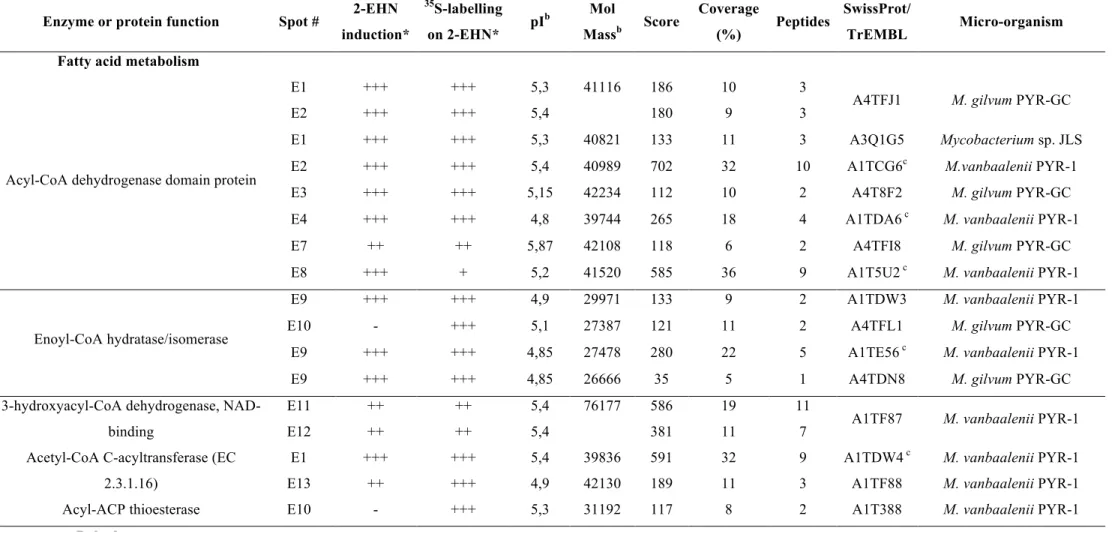
Table 1: Cytoplasmic proteins up-regulated upon incubation of M. austroafricanum IFP 2173 on 2-EHN
Enzyme or protein function Spot # 2-EHN
induction* 35S-labelling on 2-EHN* pI b Mol Massb Score Coverage (%) Peptides SwissProt/ TrEMBL Micro-organism
Fatty acid metabolism
Acyl-CoA dehydrogenase domain protein E1 E2 +++ +++ +++ +++ 5,3 5,4 41116 186 180 10 9 3
3 A4TFJ1 M. gilvum PYR-GC
E1 +++ +++ 5,3 40821 133 11 3 A3Q1G5 Mycobacterium sp. JLS
E2 +++ +++ 5,4 40989 702 32 10 A1TCG6c M.vanbaalenii PYR-1
E3 +++ +++ 5,15 42234 112 10 2 A4T8F2 M. gilvum PYR-GC
E4 +++ +++ 4,8 39744 265 18 4 A1TDA6 c M. vanbaalenii PYR-1
E7 ++ ++ 5,87 42108 118 6 2 A4TFI8 M. gilvum PYR-GC
E8 +++ + 5,2 41520 585 36 9 A1T5U2 c M. vanbaalenii PYR-1
Enoyl-CoA hydratase/isomerase
E9 +++ +++ 4,9 29971 133 9 2 A1TDW3 M. vanbaalenii PYR-1
E10 - +++ 5,1 27387 121 11 2 A4TFL1 M. gilvum PYR-GC
E9 +++ +++ 4,85 27478 280 22 5 A1TE56 c M. vanbaalenii PYR-1
E9 +++ +++ 4,85 26666 35 5 1 A4TDN8 M. gilvum PYR-GC
3-hydroxyacyl-CoA dehydrogenase, NAD-binding E11 E12 ++ ++ ++ ++ 5,4 5,4 76177 586 381 19 11 11
7 A1TF87 M. vanbaalenii PYR-1 Acetyl-CoA C-acyltransferase (EC
2.3.1.16)
E1 +++ +++ 5,4 39836 591 32 9 A1TDW4 c M. vanbaalenii PYR-1
E13 ++ +++ 4,9 42130 189 11 3 A1TF88 M. vanbaalenii PYR-1
Acyl-ACP thioesterase E10 - +++ 5,3 31192 117 8 2 A1T388 M. vanbaalenii PYR-1
Dehydrogenases
Alcohol dehydrogenase GroES domain protein E14 E4 E5 +++ +++ +++ +++ +++ +++ 4,75 4,77 4,8 38681 370 409 307 19 26 18 5 5 4
Enzyme or protein function Spot # 2-EHN induction* 35S-labelling on 2-EHN* pI b Mol Massb Score Coverage (%) Peptides SwissProt/ TrEMBL Micro-organism
FAD-dependent pyridine nucleotide-disulphide oxidoreductase E15 E7 +++ +++ +++ + 5,55 5,8 42755 319 152 18 9 5
3 A4TFL9 M. gilvum PYR-GC
Short-chain dehydrogenase/reductase E17 +++ ++ 4,9 29920 164 15 3 A1T1A7
c M. vanbaalenii PYR-1
E18 +++ +++ 4,9 30143 102 10 2 Q1BFX1 Mycobacterium sp. MCS
(S)-2-hydroxy-acid oxidase E30 +++ - 7 42022 169 10 3 A1T4N1 M. vanbaalenii PYR-1
Dihydrolipoamide dehydrogenase E22 ++ ++ 5,6 49719 370 18 6 A1T382 M. vanbaalenii PYR-1
Lysine biosynthesis
Dihydrodipicolinate synthase E24 ++ - 5,4 31436.7 163 12 3 A1T7Q1 M. vanbaalenii PYR-1
Dihydrodipicolinate reductase (EC
1.3.1.26) E23 ++ - 4,8 25816 59 8 1 A1T7N8 M. vanbaalenii PYR-1
Protein synthesis
Serine-tRNA ligase (EC 6.1.1.11) E25 +++ +++ 4,8 60542.4 128 6 2 A1TGX4 M. vanbaalenii PYR-1
Ketol-acid reductoisomerase (EC 1.1.1.86) E3 +++ +++ 5,2 36513 158 5 2 Q1BAR7 Mycobacterium sp. MCS
Nitrogen assimilation
Alanine dehydrogenase (EC 1.4.1.1) E28 - +++ 5,2 38907 203 11 3 A1T7L9 M. vanbaalenii PYR-1
Oxidative phosphorylation ATP synthase epsilon chain (EC 3.6.3.14)
(ATP synthase F1 sector epsilon subunit) E29 +++ +++ 4,8 13330 85 9 1 P45822 M. leprae TN
CO2 hydratation
Carbonic anhydrase E31 ++ ++ 4,8 18225 272 33 6 A1TDF0 M. vanbaalenii PYR-1
Glycolysis / glyconeogenesis
Phosphoglycerate kinase (EC 2.7.2.3) E32 E6 +++ +++ +++ +++ 4,7 4,75 42102 435 572 24 28 6
Enzyme or protein function Spot # 2-EHN induction* 35S-labelling on 2-EHN* pI b Mol Massb Score Coverage (%) Peptides SwissProt/ TrEMBL Micro-organism Stress response
Heat shock protein Hsp20 E26 +++ +++ 4,8 15648.4 177 27 3 A1T4V8 c M. vanbaalenii PYR-1
UspA E24 ++ - 5,4 31354 85 5 1 A1T4W2 M. vanbaalenii PYR-1
Miscellaneous
Putative esterase precursor E18 +++ +++ 5 35010 79 6 1 A1T6C2 M. vanbaalenii PYR-1
Chloride peroxidase (EC 1.11.1.10) E19 ++ - 5,7 30410 173 15 3 A1T5E7 M. vanbaalenii PYR-1
Antibiotic biosynthesis monooxygenase E16 +++ +++ 4,8 11741 56 16 1 Q1B2M9 Mycobacterium sp. MCS
Allophanate hydrolase subunit 1 E10 +++ +++ 5,3 25092 142 10 2 A1T1V3 M. vanbaalenii PYR-1
Fumarate lyase E20
E21 +++ +++ ++ ++ 5,1 5,1 49944 49944 579 538 24 27 10 8 A1TE24 c M. vanbaalenii PYR-1
HpcH/HpaI aldolase E23 ++ - 4,8 29032 202 13 4 A1TCG4 M. vanbaalenii PYR-1
Ribonuclease PH (EC 2.7.7.56) E23 ++ - 4,8 27449 75 5 2 A1T7Q1 M. vanbaalenii PYR-1
Glycyl-tRNA synthetase, alpha2 dimer E22 ++ ++ 5,6 59543 405 18 7 A1TBP9 M. vanbaalenii PYR-1
3-hydroxyisobutyrate dehydrogenase
precursor E23 ++ - 4,8 29262 195 16 3 A1T4U4 M. vanbaalenii PYR-1
Cyclic nucleotide-binding:regulatory
protein, Crp E27 +++ - 9,6 24776 409 40 7 A1T6A5 M. vanbaalenii PYR-1
Phosphoribosyltransferase: Erythromycin
esterase E12 ++ ++ 5,4 74587 49 2 1 A1T4X7
c M. vanbaalenii PYR-1
*Spot intensity was estimated from visual inspection of stained gels or autoradiographies : +, ++, +++ stand for small, medium size and large spots, respectively. (-) means undetected spot.
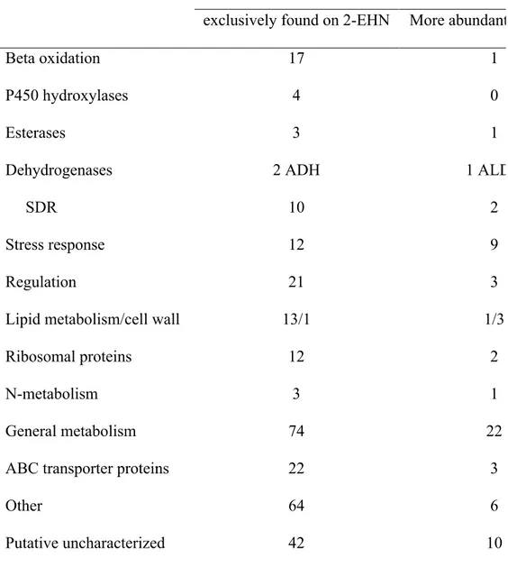
Table 2: Functional classification of proteins more abundant or exclusively detected on 2-EHN
Number of proteins
exclusively found on 2-EHN More abundant on 2-EHN
Beta oxidation 17 1 P450 hydroxylases 4 0 Esterases 3 1 Dehydrogenases SDR 2 ADH 10 1 ALDH 2 Stress response 12 9 Regulation 21 3
Lipid metabolism/cell wall 13/1 1/3
Ribosomal proteins 12 2
N-metabolism 3 1
General metabolism 74 22
ABC transporter proteins 22 3
Other 64 6