HAL Id: hal-02182313
https://hal.archives-ouvertes.fr/hal-02182313
Submitted on 26 May 2020
HAL is a multi-disciplinary open access
archive for the deposit and dissemination of
sci-entific research documents, whether they are
pub-lished or not. The documents may come from
teaching and research institutions in France or
abroad, or from public or private research centers.
L’archive ouverte pluridisciplinaire HAL, est
destinée au dépôt et à la diffusion de documents
scientifiques de niveau recherche, publiés ou non,
émanant des établissements d’enseignement et de
recherche français ou étrangers, des laboratoires
publics ou privés.
Copyright
a nutrient by the bacterial pathogen Agrobacterium
fabrum
Thibault Meyer, Armelle Vigouroux, Magali Aumont-Nicaise, Gilles Comte,
Ludovic Vial, Céline Lavire, Solange Moréra
To cite this version:
Thibault Meyer, Armelle Vigouroux, Magali Aumont-Nicaise, Gilles Comte, Ludovic Vial, et al.. The
plant defense signal galactinol is specifically used as a nutrient by the bacterial pathogen
Agrobac-terium fabrum. Journal of Biological Chemistry, American Society for Biochemistry and Molecular
Biology, 2018, 293 (21), pp.7930–7941. �10.1074/jbc.RA118.001856�. �hal-02182313�
The plant defense signal galactinol is specifically used as a
nutrient by the bacterial pathogen Agrobacterium fabrum
Received for publication, January 13, 2018, and in revised form, March 27, 2018 Published, Papers in Press, March 30, 2018, DOI 10.1074/jbc.RA118.001856 Thibault Meyer‡1,2, Armelle Vigouroux§1, Magali Aumont-Nicaise§, Gilles Comte‡, Ludovic Vial‡, Ce´line Lavire‡3, andX Solange More´ra§4From the‡UMR Ecologie Microbienne, CNRS, INRA, VetAgro Sup, UCBL, Universite´ de Lyon, F-69622, Villeurbanne, Lyon, France and§CNRS CEA Universite´ Paris Sud, Universite´ Paris-Saclay, Institute for Integrative Biology of the Cell (I2BC), Avenue de la Terrasse, 91198 Gif-sur-Yvette, France
Edited by Joseph M. Jez
The bacterial plant pathogen Agrobacterium fabrum uses periplasmic-binding proteins (PBPs) along with ABC transport-ers to import a wide variety of plant molecules as nutrients. Nonetheless, how A. fabrum acquires plant metabolites is incompletely understood. Using genetic approaches and affinity measurements, we identified here the PBP MelB and its trans-porter as being responsible for the uptake of the raffinose family of oligosaccharides (RFO), which are the most widespread
D-galactose– containing oligosaccharides in higher plants. We also found that the RFO precursor galactinol, recently described as a plant defense molecule, is imported into Agrobacterium via MelB with nanomolar range affinity. Structural analyses and binding mode comparisons of the X-ray structures of MelB in complex with raffinose, stachyose, galactinol, galactose, and melibiose (a raffinose degradation product) revealed how MelB recognizes the nonreducing end galactose common to all these ligands and that MelB has a strong preference for a two-unit sugar ligand. Of note, MelB conferred a competitive advantage to A. fabrum in colonizing the rhizosphere of tomato plants. Our integrative work highlights the structural and functional characteristics of melibiose and galactinol assimilation by
A. fabrum, leading to a competitive advantage for these bacteria
in the rhizosphere. We propose that the PBP MelB, which is highly conserved among both symbionts and pathogens from
Rhizobiace family, is a major trait in these bacteria required for
early steps of plant colonization.
The plant–rhizospheric microbial population interaction is dynamic and largely influenced by root exudates, with either beneficial or harmful consequences for plant growth
develop-ment and health (1). The germinating seeds in contact with the
surrounding soil and microorganisms have strong influences on the rhizosphere composition and favor fast-growing micro-organisms able to exploit carbon released, to resist to antimi-crobial compounds, and to outcompete other surrounding
bac-teria (2–4). Raffinose and stachyose from the raffinose family of
oligosaccharides (RFO)5accumulate in plant seeds as
energy-storage metabolites, and are released during plant germination
(2, 5). The precursor of RFO synthesis, namely galactinol, a
D-galactose bound to an inositol, is produced by the plant
enzyme galactinol synthase (GolS) (5, 6) (Fig. 1). Galactinol
plays an important role in plant health, being involved in plant
resistance against abiotic (drought and temperature) (7, 8) and
biotic stresses (9 –11). Indeed, this molecule, which
accumu-lates in plants in response to bacterial inoculation, is involved in
the induced systemic resistance to phytopathogens (9).
Raffi-nose and stachyose are synthesized from sucrose by the subse-quent addition of activated galactose moieties donated by galactinol using plant raffinose and stachyose synthases,
respectively (Fig. 1). Therefore, RFOs are ␣-(1,6)-galactosyl
extensions of sucrose.
Periplasmic-binding proteins (PBPs) associated with their ATP-binding cassette (ABC) transporter are essential for
trans-port (12). A PBP-mediated transport system is responsible for
RFO uptake from seed exudates into bacterial cells as
previ-ously shown in Ensifer meliloti 1021 (13, 14). RFOs, which are
degraded by␣-galactosidases in this latter strain, are used as
nutrients, and their assimilation may be involved in bacterial
survival in plant rhizosphere (13, 14). In more detail, raffinose
and stachyose can be degraded into melibiose and fructose, and
raffinose and galactose, respectively (15, 16). Bacterial
assimi-lation of RFOs and melibiose was associated with trophic
advantage in plant– bacteria interaction (14, 17), whereas
noth-ing was known for galactinol. Agrobacteria are telluric and rhi-zosphere bacteria, commonly isolated from roots of numerous plants as commensal bacteria. They can also be pathogenic with
the presence of the tumor-inducing plasmid (18, 19). They are
then able to create their own ecological niche after plant cell transformation that leads to tumor formation in a wide range of This work was supported by CNRS (Mission pour l’interdisciplinarite´,
Agrom-ics 2014 –2016) (to T. M., A. V., S. M., and C. L.). The authors declare that they have no conflicts of interest with the contents of this article.
This article containsFigs. S1 and S2 and Tables S1 and S2.
The atomic coordinates and structure factors (codes6EPY,6EQ1,6EQ8,6EQ0, and6EPZ) have been deposited in the Protein Data Bank (http://wwpdb.org/).
1This work was submitted to fulfill the requirements for a doctorate of
biol-ogy at ED341-E2M2 from Universite´ de Lyon, granted from the French Ministe`re de l’Education Nationale, de l’Enseignement Supe´rieur et de la Recherche.
2These authors contributed equally to this work.
3To whom correspondence may be addressed. E-mail:
celine.lavire@univ-lyon1.fr.
4To whom correspondence may be addressed. E-mail: solange.morera@
i2bc.paris-saclay.fr.
5The abbreviations used are: RFOs, raffinose family of oligosaccharides;
PBP, periplasmic-binding protein; YPG medium, yeast peptone glucose medium; ITC, isothermal titration microcalorimetry; RMSD, root mean square deviation; dpi, days post inoculation; eGFP, enhanced GFP.
cro
ARTICLE
7930
J. Biol. Chem. (2018) 293(21) 7930 –7941at INRA Institut National de la Recherche Agronomique on April 25, 2019
http://www.jbc.org/
plants (18). An in silico analysis of␣-galactosidases distribution in bacteria indicated that Agrobacterium fabrum C58 strain contains an operon putatively involved in RFO transport and
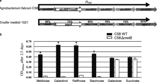
degradation (20 –22). This operon that we named mel is similar
to the agp operon of E. meliloti (13, 14), and encodes the PBP
MelB (Atu4661) which shares 73% sequence identity with the PBP AgpA, its associated ABC transporter (Atu4662–Atu4664), and
two␣-galactosidases (Atu4660 and Atu4665). All these latter
pro-teins display between 74 and 86% sequence identity with their E.
meliloticorresponding homologues (Fig. 2a).
We hypothesized that this mel operon was responsible for
the transport and assimilation of␣-galactosides in A. fabrum.
Here, we focused on its transport function, and investigated the genetic and molecular role of the PBP MelB through an inte-grative approach using a defective mutant in cellulo and
in planta, crystallography, and affinity measurements. We showed that MelB was the PBP responsible for RFO, melibiose, and galactinol import into A. fabrum C58, displaying the high-est affinity for galactinol in nanomolar range and preferring to bind a 2-unit ligand. We structurally characterized the binding mode of MelB for its different ligands. Overall, our work
high-lights how the capacity of agrobacteria to assimilate plant
␣-gal-actosides confers on them an advantage in colonizing efficiently the plant tomato rhizosphere, explaining why the PBP MelB is highly conserved among symbionts and pathogen rhizobiales.
Results
The PBP MelB is responsible for galactinol, melibiose, and RFOs (raffinose and stachyose) uptake
The growth profiles of A. fabrum C58 wildtype (WT) and
C58⌬melB-defective mutant for MelB were compared in rich
(YPG) and minimal medium containing RFOs, their derivatives or succinate (as control) as the sole source of carbon. The
C58⌬melB mutant has the same growth rate as the WT strain in
minimum medium with succinate and in rich medium.
How-ever, the C58⌬melB mutant did not grow on galactinol,
melibi-ose, raffinmelibi-ose, and stachymelibi-ose, in contrast to WT (Fig. 2b).
Therefore, MelB associated to its ABC transporter is the trans-port system responsible for the uptake and is necessary for the assimilation of these four molecules in pure culture.
MelB exhibits a high affinity for galactinol
Binding of galactinol, melibiose, raffinose, and stachyose to the purified recombinant mature protein MelB was explored using tryptophan fluorescence spectroscopy (MelB possesses 16 tryptophans) and isothermal titration microcalorimetry (ITC). Intrinsic protein fluorescence titration experiments
yielded apparent dissociation constant KDvalues of 10⫾ 1 nM
and 72 ⫾ 4 nM with galactinol and melibiose, respectively,
showing that MelB is very efficient for galactinol binding.
Figure 1. RFO synthesis and degradation. Reactions for synthesis (a) are represented with black arrows whereas those for degradation (b) are with gray
arrows. The first step of RFOs biosynthesis starts with the formation of galactinol from UDP-galactose and inositol catalyzed by the plant enzyme galactinol synthase. Raffinose, stachyose, and verbascose are synthesized from sucrose by the subsequent addition of activated galactose moieties donated by galactinol using plant raffinose and stachyose synthases and galactan:galactan galactosyltransferase (GGT), respectively. Cleavage, shown by scissors, of raffinose by an ␣-galactosidase leads to the formation of either melibiose and fructose or sucrose and galactose. For RFOs of higher degree of polymerization (DP), an ␣-galactosidase activity results in RFOs of lower DP and galactose.
at INRA Institut National de la Recherche Agronomique on April 25, 2019
http://www.jbc.org/
Reducing the ligand size to a monosaccharide (galactose) or increasing it resulted in a substantial affinity reduction
com-pared with galactinol: KDvalues of 35-fold higher for the
raffi-nose, and over 1000-fold higher for both galactose and
stachyose, respectively (Table 1andFig. S1). The KD values
were slightly higher using ITC than those determined by
auto-fluorescence, but this increased KDwas consistent (Table 1and
Fig. S1). Because MelB was not stable at high concentration during the time course of ITC experiment, we were not able to measure an interpretable signal for the stachyose binding. The ITC data confirmed the 1:1 binding stoichiometry for all ligands and revealed a high enthalpy of binding for galactinol and melibiose meaning that both ligands use the same binding mechanism mainly involving polar interactions. In contrast, the binding mode of raffinose was characterized by an unfavorable enthalpy contribution and a strong entropy term indicating that hydrophobic interactions may play a predominant role and/or a displacement of water molecules occurs upon ligand binding. The galactose interaction adopts an intermediate behavior with a high entropy term, accompanied by a weakly favorable enthalpy of binding, suggesting that polar bonds are less important for galactose alone compared with a 2-unit ligand, resulting in a lower enthalpy, thus a weaker affinity.
MelB is specific for␣-(1,6)-galactosides (RFOs). Indeed, no
interaction could be measured with glucose, sucrose,
cello-biose, lactose, and␣-(1,6)-glucosides.
Thermal denaturation experiments revealed a contribution
of more than 3 °C for two ligands for protein stability (Fig.
S2). Indeed, adding galactinol or melibiose led to a melting
temperature (Tm) of over 46 °C compared with the 43 °C for
the unliganded protein in agreement with the measured KD
values. Galactose or stachyose binding did not stabilize MelB whereas the raffinose binding produced a slight effect with a
Tmof 45 °C.
MelB is a PBP from cluster C
The mature MelB expression plasmid was a synthetic gene lacking the first 18 signal sequence residues that serve for local-ization to bacterial periplasm. The numbering used for the description of residues corresponds to the mature protein of 677 amino acids. Because MelB is the biggest PBP so far and shares low sequence identity (around 20%) compared with PBPs with known three-dimensional structures, we first solved the structure of seleniated MelB in complex with raffinose at 2 Å resolution by single wavelength anomalous dispersion method. The asymmetric unit is composed of four similar MelB-raffinose complexes (average root mean square deviation (RMSD) value of 0.4 Å). By the molecular replacement method, we then solved the structure of MelB in complex with galacti-nol, melibiose, galactose, and stachyose at 2.2, 1.8, 2.5, and 2.1 Å
resolution, respectively (Table 2). The asymmetric unit of the
galactinol and melibiose complexes also contains four very sim-ilar molecules with RMSD values between monomers ranging from 0.2 to 0.4 Å whereas that of the galactose and stachyose complexes possesses two identical molecules. Moreover, the five ligand-bound structures are very similar with an average RMSD value of 0.4 Å. They all adopt a closed conformation. MelB fold is monomeric, composed of two lobes, each formed
by a central-sheet flanked by ␣-helices (Fig. 3a). The biggest
lobe (lobe 1) consists of residues 8 –354 and 619 – 678 and the smallest (lobe 2) comprises the residues 364 – 610. Two short
segments (Fig. 3a) define the hinge region connecting the two
lobes. MelB possesses a typical fold of cluster C within the PBP
structural classification (12) as SSM-EBI (http://www.ebi.
ac.uk/msd-srv/ssm)6reports: RMSD between MelB and similar
PBP structures binding oligopeptide are over 2.6 Å for 450 C␣
6Please note that the JBC is not responsible for the long-term archiving and
maintenance of this site or any other third party hosted site.
Figure 2. The mel operon structure and MelB involvement in galactinol, melibiose, and RFO consumption. a, atu4660-atu4665 genes belong to the same transcription unit (operon prediction by Westover et al. (22) that we called the mel operon). The mel promoter Pmelindicates the gene transcription direction.
Both atu4660 and atu4665 genes are annotated as two␣-galactosidases, atu4661 as the PBP MelB and atu4662-atu4664 genes as the associated ABC trans-porter. Comparison is between the mel operon genes and their homologues in E. meliloti 1021 (13, 14). the percentages of sequence identity between each homologous protein are indicated; for example, the PBPs AgpA and MelB share 73% sequence identity. b, 2.5 days growth (A at 600 nm) of A. fabrum C58 WT strain (in white) and the C58⌬melB mutant (in black) in AT minimal medium supplemented with different carbon sources. Standard deviations were calculated from five technical and two biological replicates. Asterisks indicated significant differences (Mann-Whitney p value⫽ 0.05).
Galactinol and melibiose bound to MelB
at INRA Institut National de la Recherche Agronomique on April 25, 2019
http://www.jbc.org/
atoms. Nonetheless, a detailed structural comparison is irrelevant because MelB presents a distinct ligand-binding site.
Ligand-binding site of MelB
All ligands are bound between the two closed lobes of MelB. The size and the volume of the ligand-binding site constrain the conformation of bound RFOs. Indeed, raffinose and stachyose
bind in a very compact form (Fig. 3b), and the addition of a
galactosyl moiety at the nonreducing end of stachyose corre-sponding to verbascose will abolish its binding, because the pocket is not large enough to accommodate a pentasaccharide. All ligands are well defined in their electron density maps except the fructose moiety of the stachyose likely responsible
for its low affinity (Fig. 3, c–h). They share a buried nonreducing
end galactosyl unit wedged between two aromatic residues
(Trp317and Trp639) which superimposes very well with the
bound galactose alone (Fig. 3, c–h). These galactoses at position
1 make 10 similar protein contacts involving the main chain
amino group of Gly111and the side chains of Arg320, Asn333,
Glu335, and Glu641from lobe 1 and both side chains of Tyr487
and Arg533from lobe 2. The O6 atom of this pyranose interacts
with a conserved water molecule observed in each complexed structure (except in molecule B of MelB-galactose complex), which in turn makes hydrogen bonds with the amino group of
Gly112and the side chain of Asp114. In both structures of MelB
in complex with galactose and stachyose, the galactose at
posi-tion 1 does not interact with Asn333but because of two
hydro-gen bonds with the side chain of Glu335, it conserves 10
inter-actions with MelB. Modeling a glucosyl unit at the nonreducing end (position 1) creates steric hindrance between the equatorial
C4-OH and the Trp639indol explaining the specificity of MelB
for a galactosyl unit at position 1 as shown by the affinity measurement.
In contrast to position 1, at position 2, the glucosyl moieties of melibiose and raffinose, the inositol moiety of galactinol and the galactose moiety of stachyose do not superimpose and can shift up to 3.5 Å to allow, for example, the fructose or the sucrose accommodation of raffinose and stachyose in the
ligand-binding site, respectively (Fig. 3, b and c). A
conforma-tional change less than 1 Å for Trp110and Tyr487side chains is
observed to accommodate a glucosyl unit at position 2 (Fig. 3c).
More arrangements from amino acids of lobe 2 can occur to
accommodate the glucosyl units at positions 3 and 4 (Fig. 3c).
For example, Trp557is pushed away by more than 1 Å compared
with the other liganded structures to find a room for the fruc-tose at position 4. All units at position 2 have in common the
stacking onto the aromatic indol of Trp110, with the optimum
one for the inositol because of the shorter link (one carbon shorter) between the two subunits of galactinol compared with melibiose and RFOs. The melibiose’s glucose and the inositol Table 1
Affinity measurement for MelB
KDvalues were measured by intrinsic protein fluorescence titration (Fluorescence) and by isothermal titration microcalorimetry (ITC).
Fluorescence ITC
KD R
2 K
D n Enthalpy (⌬H) Entropy (⌬S) Entropic contribution (ⴚT⌬S) Free enthalpy (⌬G)
M M cal/mol cal/mol/deg cal/mol cal/mol
Galactinol 0.010⫾ 0.001 0.99 0.12⫾ 0.03 0.83 ⫺6908 8 ⫺2356 ⫺9264
Melibiose 0.072⫾ 0.004 0.99 0.76⫾ 0.11 0.94 ⫺6412 6.1 ⫺1794 ⫺8206
Raffinose 0.347⫾ 0.42 0.99 2.9⫾ 0.5 0.97 2115 32.5 ⫺9527 ⫺7412
Galactose 13.8⫾ 3 0.99 24⫾ 2 1 ⫺797 18.4 ⫺5393 ⫺6190
Stachyose 24.6⫾ 2.4 0.99 Not determined
Table 2
Crystallographic data and refinement parameters
Values for the highest resolution shell are in parentheses.
MelB
Raffinose (SeMet) Galactinol Melibiose Galactose Stachyose
PDB code 6EPY 6EQ8 6EPZ 6EQ0 6EQ1
Space group C2 C2 C2 C2 C2 Cell parameters (Å, °) a⫽ 354.3 a⫽ 355.3 a⫽ 351.6 a⫽ 107.8 a⫽ 108.2 b⫽ 74.3 b⫽ 73.7 b⫽ 73.7 b⫽ 73.9 b⫽ 74 c⫽ 108.2 c⫽ 108.1 c⫽ 107.6 c⫽ 171.1 c⫽ 171.4  ⫽ 105.5  ⫽ 105.5  ⫽ 105.4  ⫽ 92.5  ⫽ 92.4 Resolution (Å) 50–2 (2.17–2) 50–2.2 (2.3–2.2) 48–1.8 (1.9–1.8) 50–2.5 (2.59–2.5) 50–2.1 (2.2–2.1)
No. of observed reflections 1,143,584 (175,894) 725,141 (106,000) 1,633,125 (250,861) 314,407 (48,297) 541,503 (82,382)
No. of unique reflections 333,472 (51,505) 137,636 (20,717) 246,319 (38,629) 49,700 (7682) 79,391 (12,494)
Rsym(%) 7 (52.8) 17.1 (112.5) 10 (77.2) 18.2 (100) 15.4 (200) Completeness (%) 99.8 (94.4) 98.7 (92.6) 99.4 (97.1) 99.2 (95.7) 99.6 (97.9) I/ 10.8 (2) 7.5 (1.5) 11.4 (1.9) 9 (1.6) 8.12 (0.7) CC1/2 99.8 (80.1) 99.3 (57.9) 99.8 (80) 99.1 (51.2) 99.7 (50.1) Rcryst(%) 17.5 18.5 17.7 19.4 19.1 Rfree(%) 19.9 21.7 19.5 24.2 22.5 RMS bond deviation (Å) 0.01 0.01 0.01 0.01 0.01 RMS angle deviation (°) 1.0 1.09 1 1.16 1.13 Average B (Å2 ) protein 44.3 45.6 33.7 55.9 52.6 ligand 41.2 36.6 30.5 40.5 44.7 solvent 51.7 49.6 39.2 55 52.2
CC1/2⫽ percentage of correlation between intensities from random half-dataset (40).
at INRA Institut National de la Recherche Agronomique on April 25, 2019
http://www.jbc.org/
Galactinol and melibiose bound to MelB
at INRA Institut National de la Recherche Agronomique on April 25, 2019
http://www.jbc.org/
share only one protein contact: an oxygen interacts with the NH
of the Trp639side chain (Fig. 3, e and f). The inositol makes three
additional hydrogen bonds with Asn484, Tyr487, and Ser515from
lobe 2 whereas the rest of the oxygen of the melibiose’s glucose interacts with the protein side chains via water molecules only. In contrast, the glucose moiety of raffinose and the galactose moiety of stachyose at position 2 make an additional H-bond
compared with inositol involving the side chains of Asp519(Fig.
3, g and h). The fructose at position 3 in raffinose interacts with
the main chain of Arg533from lobe 2 and the NH of the Trp639
indol from lobe 1 (Fig. 3g). In contrast, Arg532side chain makes
two H-bonds with the glucose moiety at position 3 in stachyose whereas only one is present between the fructose moiety at
position 4 and the Trp639side chain (Fig. 3h).
MelB is highly conserved among rhizobiales
Searching for MelB conservation in the bacterial kingdom (protein database at NCBI), and subsequent phylogenetic
anal-ysis revealed⬎310 PBPs above 65% sequence identity (Fig. 4).
Galactinol-, melibiose-, and raffinose-binding signatures
share 10 amino acids Trp110–Gly111–Trp317–Arg320–Asn333–
Glu335–Tyr487–Arg533–Trp639–Glu641. Stachyose binding shares
only nine of these latter because Asn333 does not interact
with stachyose. Two additional residues (Asn484and Ser515)
are involved in galactinol, raffinose, and stachyose binding compared with melibiose binding. One additional residue
Asp519 belongs to the raffinose and stachyose signatures.
A last additional residue Arg532defines the
stachyose-bind-ing signature composed of 13 residues in total. Members of the MelB subgroup (67 PBPs) display more than 90% identity sequence with the conserved binding signature. They all belong to Rhizobium and Agrobacterium genera. Outside the MelB cluster, the signature slightly degenerates for galactinol and raffinose binding. Nonetheless, modeling indicates that their binding would not be affected.
Remark-Figure 3. Ribbon representation of MelB structures and ligand-binding site. a, raffinose in magenta is located in the cleft between lobes 1 and 2 shown in
slate and in pink, respectively, and the hinge region is in red. b, superposition of the bound galactose, melibiose, galactinol, raffinose, and stachyose shown in green, yellow, orange, magenta, and blue sticks, respectively, in the binding site of MelB. c, same figure as in b showing the stacking between ligands and tryptophan (Trp639, Trp317, and Trp110). Except Trp639and Trp317, all the other labeled amino acids mainly from lobe 2 can move up to 1 Å upon ligand binding.
d–h, galactose (d), melibiose (e), galactinol (f), raffinose (g), and stachyose (h) bound to the binding site of MelB are shown in the same code color as in b. Hydrogen bonds between MelB and each ligand are shown as dashed lines in black (distances are up to 3.2 Å). A water molecule forming a hydrogen bond with each ligand is shown as a red circle. Each ligand is shown in its annealing Fo-Fc omit map contoured at 4.
Figure 4. MelB phylogeny and binding signature. For each protein clade, the residues, which are identical to (black) and different from (red) those involved in the galactinol/melibiose/raffinose binding of A. fabrum C58 MelB are indicated. Number in bracket represents the number of MelB-relative PBPs per clade.
at INRA Institut National de la Recherche Agronomique on April 25, 2019
http://www.jbc.org/
ably, all these PBPs belong to soil- and plant-interacting genera.
Galactinol and melibiose are inducers of mel operon genes
We constructed the C58 pOT1e-Pmelreporter fusion strain
to study in cellulo gene expression of mel operon in the pres-ence of commercial compounds. Compared with succinate, slight but significant inductions were observed with raffinose, galactose, and stachyose (2-, 1.6-, and 1.25-fold change values,
respectively) (Fig. 5a). In contrast, galactinol and melibiose
are efficient inducers with 4.8- and 6.5-fold change values, respectively.
mel operon genes are expressed in early plant colonization At two early stages of plant colonization, in planta expres-sion of mel operon has been studied in the WT strain harboring
the pOT1eM-Pmelplasmid reporter fusion (m-cherry
constitu-tive expression and egfp inducible expression). 48 h after seed imbibition and inoculation, most bacteria in contact with the radicle cells expressed mel operon, as shown by the yellow cells inFig. 5, b and c. 14 days after seed imbibition and inoculation, among bacterial cells present on plant roots (red and yellow cells), some of them were still expressing RFO uptake and
deg-radation genes (yellow cells inFig. 5d). Thus, mel operon was
more expressed at the beginning of plant colonization.
The PBP MelB confers a competitive advantage in colonizing tomato rhizosphere
The colonization of plant rhizosphere by A. fabrum C58 WT
and C58⌬melB mutant was evaluated at 2 days post inoculation
(dpi) and 14 dpi (Fig. 6). When tomato seeds were inoculated
with each strain individually, the bacterial colonization level did not significantly differ at 2 dpi (Mann-Whitney p value of 0.09) whereas at 14 dpi, this was slightly higher for the WT strain
(Mann-Whitney p value⬍2.2e–16e) (Fig. 6a). When A. fabrum
C58 WT and C58⌬melB mutant were co-inoculated, a slight
and a drastic reduced fitness was observed for the C58⌬melB
mutant at 2 and 14 dpi, respectively (Fig. 6b), revealing a
selec-tive advantage conferred by galactinol/melibiose/RFO exploi-tation under a competitive challenge.
Discussion
This work reveals the molecular and ecological roles, and structural basis of the PBP MelB, encoded by the linear chro-mosome of A. fabrum C58.
At the beginning of the study, we made the straightforward assumption that MelB was behaving like its homologous PBP
AgpA from E. meliloti described as an ␣-galactoside
trans-porter (13). Our gene expression analyses and growth assays
experiments showed that similarly to what has been reported
for expression of agp operon genes in E. meliloti,␣-galactosides
Figure 5. Expression of mel operon genes in cellulo and in tomato radicle and root. a, comparison of mel operon gene expression in AT minimal medium supplemented with different carbon sources. Standard deviations were obtained from four technical and two biological replicates. Letters above histograms indicate significant different -fold change values (Tukey’s test, p value⫽ 0.05). b–d, bacterial mel operon gene expression at two early stages of plant growth. Gene expression was monitored using the pOT1eM-Pmeltranscriptional reporter fusion by confocal microscopy at 2 (b and c) and 14 (d) dpi, corresponding to
radicle emergence and root elongation stages, respectively. Representative pictures from five plants per stages are shown. Red fluorescence from M-cherry indicates the presence of bacteria, whereas yellow fluorescence shows bacteria that were both active and able to express Pmel-egfp. Plant auto fluorescence,
represented in green allows distinguishing different types of plant cells, small compact cells from the radicle and elongated cells from the growing root. The scale is represented in white. Most cells expressed Pmel-egfp at 2 dpi, whereas at 14 dpi, few cells only expressed the transcriptional fusion.
Galactinol and melibiose bound to MelB
at INRA Institut National de la Recherche Agronomique on April 25, 2019
http://www.jbc.org/
induce expression of mel operon genes in A. fabrum, and are used as carbon sources after being imported by MelB. Indeed, in contrast to the WT strain, a MelB defective mutant is unable to
grow on␣-galactosides. Moreover, using two different
biophys-ical methods, we demonstrated that MelB can bind melibiose, raffinose, and stachyose. Because of the small volume cavity of the ligand binding, stachyose displays a weak affinity for MelB (micromolar range) compared with melibiose and raffinose (nanomolar range). The accommodation of stachyose requires drastic conformational constraints on the ligand when bound to the protein. Indeed, only small local rearrangements of few
protein side chains (Trp110and Trp557) forming the binding site
can occur. With an affinity in the micromolar range, MelB also
binds galactose, which is the sugar common to all
␣-galacto-sides present at the nonreducing end. Nonetheless, the low affinity of MelB for galactose prevents galactose from compet-ing with melibiose and raffinose. From our results, MelB can appear as an alternative galactose transporter suspected by
Kemner et al. (23), which allowed a chvE-gguABC defective
mutant to grow on galactose (23, 24). Conversely, the presence
of the ChvE-GguABC sugar transporter can explain why melB defective mutant was still able to grow on galactose.
An unexpected outcome of our study was that galactinol,
which is the precursor of␣-galactosides production in plants,
was uptaken into agrobacteria via the MelB-mediated transport system. Moreover, MelB displays a preference for galactinol with high affinity (nanomolar range) indicating that this mole-cule must be efficiently imported into A. fabrum, in line with the gene expression results. Remarkably, MelB recognizes sim-ilarly the nonreducing end galactose common to all tested ␣-galactosides and galactinol. Overall, using genetic, structural, and affinity data, this work demonstrates that the
MelB-medi-ated transport system contributes to the import of
␣-galacto-sides with a strong preference for a 2-unit ligand (melibiose) and mainly contributes to that of galactinol. To our knowledge, this is the first description of a bacterial PBP allowing galactinol import.
The imported sugars are used to sustain bacterial growth.
Galactinol- and␣-galactoside–rich environments would
facil-itate the settlement of bacteria capable to assimilate these plant compounds efficiently. From in planta competition assays on tomato between the A. fabrum WT and melB defective mutant, we showed that MelB confers a marked selective advantage in colonizing tomato rhizosphere, and since early time. This observation correlates with the presence of melibiose and raf-finose in the plant rhizosphere, as they are highly abundant in
the seeds (25) and known to be released during seed
germina-tion (14). Hence, the competitive advantage of the WT in the
tomato rhizosphere could be because of a trophic advantage of the strain during seed germination. A germinating seed can indeed be considered as a new environment to be colonized. The community composition of the mature plant is influenced by historical contingency (timing and order of arrival) of the seed community members and their ability to efficiently settle
in that environment (26), as the first establishing species are
known to affect the ability of potential immigrants to establish.
This is called the priority effect (27). Thus, the ability of bacteria
to compete and settle in germinating seed environment by
growing on galactinol or␣-galactosides released at this time
could have long effect on their ability to persist and colonize plant rhizosphere. This is consistent with our findings that at 14 dpi, the competitive advantage of the WT strain is even higher than the one measured at 2 dpi.
Besides the trophic advantage linked to the mel operon, this operon could be associated to another aspect of bacterial plant interactions, linked to plant defense signaling and protection against pathogens. Indeed galactinol is a plant compound
involved in plant defense (9 –11). For example, Pseudomonas
chlororaphis O6-mediated induced systemic resistance was shown associated with an elevation of galactinol content within plants, which conferred disease resistance against pathogen
attack (9). The disease resistance was associated with induction
of the expression of a set of pathogen-responsive genes (9, 10).
Moreover, in Arabidopsis, deletion of enzymes that decrease the galactinol and/or raffinose content has been shown to increase plant resistance against the phytopathogenic
nema-Figure 6. MelB confers a competitive advantage in tomato roots. a,
A. fabrum bacterial concentration (cfu/mg dry roots) in tomato roots (at 2 and 14 dpi) infected with either A. fabrum C58 WT or C58⌬melB mutant. Standard deviations were calculated from three technical and five biological replicates. b, proportion of A. fabrum genotypes (%) in inoculum and tomato root at 2 and 14 dpi infected with a mixture (1:1 ratio) of A. fabrum C58 WT and C58⌬melB mutant. Standard deviations were calculated from 4 biological replicates and 10 independent assays. Asterisks indicate significant differ-ences (Mann-Whitney test).
at INRA Institut National de la Recherche Agronomique on April 25, 2019
http://www.jbc.org/
tode Heterodera schachtii (11). Similarly, Agrobacterium mel-mediated activity could modify the level of plant galactinol and/or raffinose, which could either drive bacterial recognition by the plant or reduce plant defense signaling through the fall of galactinol content. Agrobacterium is known to be able to bypass
and overcome plant defenses (28). It would thus be of interest to
study the involvement of the mel operon in that situation. In this study, we defined the galactinol-/melibiose-/raffi-nose-binding signature and found out that this is strictly con-served in MelB homologues in Agrobacterium and Rhizobium, which share a high sequence identity over 90% with MelB. Moreover, phylogenetic and structural data showed that this wide occurrence is extended among Rhizobiaceae, all plant-interacting genera (Mesorhizobium, Allorhizobium, Ensifer,
Martelella, Pleomorphomonas, Kaistia, and Devosia). There-fore, whatever advantage it gives, it is tempting to speculate that galactinol/melibiose (and to a lesser extent raffinose) may be associated with a selective pressure toward the acquisition of binding, transport, and degradation functions in microorgan-isms, making the PBP MelB a major trait in the first step of tomato colonization and likely of other plant species.
Experimental procedures Bacterial culture conditions
Bacteria and plasmids used in this study are shown inTable
S1. Escherichia coli strains were grown at 37 °C in LB medium
supplemented when it was necessary with appropriate
antibi-otics (tetracycline 10g/ml, gentamicin 15 g/ml, ampicillin
100g/ml). A. fabrum C58 strain and its derivatives were
cul-tivated at 28 °C in YPG (yeast extract, 5 g per liter, peptone, 5 g per liter, glucose, 10 g per liter, and pH adjusted to 7.2) rich medium supplemented when required with neomycin (25 g/ml), kanamycin (25 g/ml), and/or gentamycin (20 g/ml). In growth assays, AT minimal medium supplemented with 10
mMammonium sulfate and 10 mMcarbon sources was used.
200l were inoculated in Bioscreen honeycomb 100-well
ster-ile plates and incubated in a Bioscreen C Reader (Labsystems, Helsinki, Finland) at 28 °C during 5 days. Cell growth was mea-sured every 20 min. Analyses were performed in five technical replicates and in three biological replicates.
Construction of melB defective mutant in A. fabrum C58 and transcriptional fusion
The A. fabrum C58⌬melB defective mutant was constructed
as described previously (29) without marker exchange. Briefly,
the recombinant region containing the upstream and down-stream region flanking the melB gene (amplified by PCR using
primers listed inTable S2) was inserted into pJQ200sk vector
(30) leading to a nonpolar mutant. The resulting plasmid was
introduced into A. fabrum C58 by electroporation. Bacteria were spread on YPG medium plates containing gentamicin (20 g/ml) for the first selection and gentamicin-resistant colonies were spread on YPG plates supplemented with 5% sucrose for the second selection. The deletion of melB was verified by sequencing (GenoScreen, Lille, France).
The C58 pOT1e-Pmeltranscriptional reporter fusion strain
was obtained as follows: the promoter region of
atu4660-atu4665 genes named Pmelwas PCR amplified (using primer
listed inTable S2) and the PCR fragment obtained was ligated
into ClaI-SalI digested vector pOT1e. pOT1eM-Pmelplasmid
was obtained by cloning Pmelinto SpeI digested pOT1eM
vec-tor as described previously (31). Transcriptional reporter
con-structions were introduced into A. fabrum by electroporation. Cloning, expression, and purification of mature MelB
The mature MelB expression plasmid was chemically syn-thesized using codon optimization for the expression in E. coli and inserted into pET-9a plasmid using NdeI and BamHI re-striction enzyme (GenScript, Piscataway, NJ). E. coli BL21 com-petent cells transformed with pET9a-MelB were grown in LB
media at 37 °C until A600of 0.6. 0.5 mMof isopropyl-D
-thio-galactopyranoside (IPTG) was added to the culture for over-night expression at 20 °C. The cells were pelleted by
centrifu-gation at 4000⫻ g for 15 min at 4 °C, resuspended in 50 mM
Tris-HCl, pH 8, 300 mMNaCl, and 20 mMimidazole, and
dis-rupted by sonication. After centrifugation at 25,000⫻ g for 30
min, the filtered supernatant was injected on a nickel affinity column (HiTrap 5 ml, GE Healthcare). After a washing step
of 6% 50 mMTris-HCl, pH 8, 300 mM NaCl, and 300 mM
imidazole (Buffer B), the protein was eluted with Buffer B and injected on a gel filtration Superdex 200 26/60 (GE
Healthcare) using 50 mMTris-HCl, pH 8, and 150 mMNaCl.
The protein fractions were pooled, concentrated at 10.7
mg/ml, and stored at⫺80 °C.
Expression and purification of mature seleniated MelB
The E. coli BL21 cells transformed with the plasmid pET9a-MelB were grown overnight at 28 °C in M9 media
supple-mented with 0.4% glucose; 2 mM MgSO4; 1 M CaCl2; 100
mg/liter of lysine, threonine, and phenylalanine; and 50 mg/li-ter of leucine, valine, isoleucine, and methionine. The pelleted cells were resuspended in fresh M9 media (same as above) with 100 mg/liter of selenomethionine instead of methionine for 1 h
at 37 °C before inducing the expression with 0.5 mMisopropyl
-D-thiogalactopyranoside overnight at 20 °C. The cells were
centrifuged at 4000⫻ g for 15 min at 4 °C. The purification
protocol was the same as described above. Crystallization and data collection of MelB
Crystallization conditions for seleniated MelB in the
pres-ence of 2 mMraffinose were screened using Qiagen kits
(Valen-cia, CA) with a Cartesian NanoDrop robot (Genomic Solu-tions). The crystals were manually reproduced in hanging drops experiments by mixing equal volumes of protein solution and
the precipitant solution 25% PEG 4000, 0.2MNaCl, 0.1MMes,
pH 6.5, and 0.2MCaCl2. For the four other complexes, a similar
condition without CaCl2and 0.6 MNaCl was used. Crystals
were transferred to a cryoprotectant solution (mother liquor supplemented with 25% PEG 400) and flash-frozen in liquid nitrogen. X-ray diffraction data sets were collected at 100 K on the Proxima 1 or 2 beamlines (SOLEIL synchrotron, Saint-Au-bin, France). Data processing was performed using the XDS
package (32) (Table 2).
Structure determination and refinement of MelB
The crystal structure of the MelB-raffinose complex was determined by SAD method from selenomethionine-labeled
Galactinol and melibiose bound to MelB
at INRA Institut National de la Recherche Agronomique on April 25, 2019
http://www.jbc.org/
protein and refined at 2 Å resolution. Solvent content analysis using CCP4 (Collaborative Computational Project, Number 4) indicated the presence of four monomers in the asymmetric unit. The positions of 12 over 15 selenium atoms per monomer
were found using SHELX suite program (33) The phases were
calculated using PHASER (34) and density modification was
performed by PARROT (CCP4 suite). An iterative process of manual building in COOT combined with phase calculation where a partial model was used as input, allowed the modeling of the complete polypeptide chain. The structures of all other liganded MelB were solved using the SeMet-MelB monomer as a search model. Refinement of each structure was performed
with BUSTER-2.10 (34), NCS restraints, and TLS group.
Because of the strong anisotropy of the crystals of MelB-stachyose, the DEBYE and STARANISO programs developed by Global Phasing Ltd. were applied to the data scaled with
AIMLESS using the STARANISO server (http://staraniso.
globalphasing.org).6These programs perform an anisotropic cut-off of merge intensity data on the basis of an analysis of local
I/s(I); compute Bayesian estimates of structure amplitudes,
taking into account their anisotropic fall-off; and apply an anisotropic correction to the data. The corrected anisotropic amplitudes were used for further refinement of the MelB-stachyose structure with BUSTER-2.10. Inspection of the density maps and manual rebuilding were performed using
COOT (35). The three-dimensional models of stachyose and
galactinol were generated with the ProDRG webserver (36),
whereas those of melibiose and raffinose were found in the Protein Data Bank. Refinement details of each structure are
shown inTable 2. Molecular graphics images were generated
using PyMOL.
Fluorescence titration measurements of MelB
Each ligand bound to MelB was monitored by autofluores-cence by exciting the protein at a wavelength of 295 nm and monitoring the quenching of fluorescence emission of trypto-phans at 335 nm. All experiments were performed at 22 °C in 96-well plates (1/2 Area Plate-96F, PerkinElmer Life Sciences) using Tecan Infinite M1000 (Tecan, Ma¨nnedorf, Switzerland)
in 25 mMTris-HCl, pH 8.0, and 150 mMNaCl with a fixed
amount of proteins (1 M) and increasing concentrations of
ligand. Each ligand has no emission signal at 335 nm. The data were analyzed using Origin® 7 software and fitted to the follow-ing equation.
f⫽ ⌬Fluorescencemax⫻ abs共x兲/共KD⫹ abs共x兲兲 (Eq. 1)
Isothermal titration microcalorimetry measurements of MelB Isothermal titration microcalorimetry experiments were performed with an ITC200 isothermal titration calorimeter from MicroCal (GE Healthcare). The experiments were carried out at 20 °C. Protein concentration in the microcalorimeter cell
(0.2 ml) varied from 10 to 300M. Nineteen injections of 2l of
ligand solution (raffinose, stachyose, melibiose, galactose, and
galactinol) concentration from 0.1 to 2.8 mMwere performed at
intervals of 180 s while stirring at 500 rpm. The experimental data were fitted to theoretical titration curves with software supplied by MicroCal (ORIGIN®). This software uses the
rela-tionship between the heat generated by each injection and⌬H
(enthalpy change in kcal mol⫺1), Ka(the association binding
constant in mol⫺1), n (the number of binding sites), total
pro-tein concentration, and free and total ligand concentrations
(37).
Differential scanning calorimetry
Thermal stability of the WT and liganded MelB (13Mand
50Mfor protein and ligand, respectively) was studied by
dif-ferential scanning calorimetry (DSC) on a MicroCal model VP-DSC in a standard buffer. Each measurement was preceded by a baseline scan with the standard buffer. All solutions were degassed just before loading into the calorimeter. Scans were
performed at 1 K䡠min⫺1between 20 and 90 °C. The heat
capac-ity of the buffer was subtracted from that of the protein sample before analysis. Thermodynamic parameters were determined by fitting the data to the following equation,
⌬Cp共T兲 ⫽
Kd共T兲 ⌬Hcal⌬HvH
关1 ⫹ Kd共T兲兴2RT2
(Eq. 2)
where Kdis the equilibrium constant for a two-state process,
⌬HvHis the enthalpy calculated on the basis of a two-state
pro-cess, and⌬Hcalis the measured enthalpy.
Phylogenetic analysis
Sequences were analyzed using BlastP from NCBI (https://
blast.ncbi.nlm.nih.gov/) and MicrosScope (https://www. genoscope.cns.fr/).6 Alignments of MelB and related se-quences were conducted using ClustalW software. Relation-ship tree was build using Mega software, version 7. The boot-strap consensus tree inferred from 1000 replicates was taken to represent the evolutionary history of the taxa analyzed. The evolutionary distances were computed using the Poisson cor-rection method and are in units of the number of amino acid substitutions per site.
Measurement of mel operon gene expression
Expression of mel operon genes was measured in the C58
pOT1e-Pmelstrain. Quantification of fluorescence was carried
out in a microplate filled with 200l of AT medium,
supple-mented with different carbon sources at a final concentration of
10 mM. Microplate wells were inoculated with overnight
cul-tures to obtain an A600 of 0.2. A TECAN apparatus (Tecan
SparkTM 15 M, Ma¨nnedorf, Switzerland) was used to read
microplates after 24 h of incubation at 28 °C. The following parameters were used: absorbance at 600 nm, fluorescence excitation at 488 nm, and emission at 510 nm. Results were
normalized by the A600and -fold change values were obtained
by dividing the fluorescence by the corresponding value obtained from the empty pOT1e vector. The fluorescence level
comparison was carried out using the Tukey’s test (p value⫽
0.05) and computed with the “vegan” package in the R v3.1.3 statistical software environment (R Core Team, 2014). Plant inoculation
For bacterial colonization, competition assays, and confocal observation studies, tomato seeds (Solanum lycopersicum
at INRA Institut National de la Recherche Agronomique on April 25, 2019
http://www.jbc.org/
“Marmande”) were sterilized as described (38). Seeds were plated on 0.8% agar plant cell culture supplemented with 1.5 g/liter of the Plant-Prod 15-15-30 High K nutrient solution (Master Plant-Prod Inc., Brampton, Ontario, Canada). They
were inoculated with 10l of overnight culture (106cfu/ml) of
a single strain (A. fabrum C58 pTiatu6148:Kmderivative of the
WT strain (39) or C58⌬melB mutant), or with a mixture of both
at 1:1 ratio (competition). Petri dishes were placed 2 days in the dark and then in a climatic chamber at 24 °C with 18/8 h for light/dark and 65% of humidity. To determine bacterial colonization level, roots were ground at 2 and 14 dpi. Serial dilutions of crushed roots were plated on the YPG medium and colonies were counted after 2 days of incubation at 28 °C. Significant difference in the population level resulting from five plants per strain, with enumeration of three Petri dishes for each plant, was evaluated with Mann-Whitney test
(p value⫽ 0.05) performed with the R v3.1.3 statistical
soft-ware environment.
In competition experiments, the colonized bacteria were nonselectively recovered at 2 and 14 dpi. To that end, crushed roots were first plated with a spiral plater (EasySpiral®, Inter-science, Saint-Nom-la-Brete`che, France) on YPG medium without antibiotics to enable a biologically unbiased recovery of
both C58 pTiatu6148:Km WT strain and C58⌬melB mutant
(kanamycin sensitive). Two hundred individual colonies were then plated in YPG medium with kanamycin/neomycin to
determine the relative proportions of C58 pTiatu6148:Kmand
C58⌬melB mutant strains (output ratio). The determination of
the initial strains ratio of the inoculum was realized using the same protocol. The experimental assays were performed with 10 independent assays and repeated four times. The propor-tions of WT strains between the initial strain ratio and in planta output ratios were compared with the test of equal or given
proportions (p value⫽ 0.05).
Confocal microscopy analyses
Visualization of reporter bacterial cells harboring
pOT1eM-Pmelon tomato radicles and roots was performed using a
con-focal laser scanning microscope (LSM 800 Meta Concon-focal Microscope, Zeiss, Oberkochen, Germany). In the reporter strain, M-Cherry (red color) is constitutively expressed and
enhanced GFP (eGFP) (green color) is expressed under Pmel
control. The red color indicates the bacteria presence (red cells)
whereas eGFP reports the induction of Pmelshown as
yellow-green cells. At 2 and 14 dpi, tomato radicles and roots were mounted between a slide and a coverslip in a commercial mounting fluid (Aqua Poly/Mount, Polysciences, Inc., War-rington, PA). The eGFP and the M-cherry were excited with argon laser at 488 nm and 584 nm, respectively, and fluores-cence was captured at 528 nm and 607 nm. Analyses of images (five plants per condition) were performed thanks to LSM 800 software (Zeiss, Oberkochen, Germany).
Coordinates
The atomic coordinates and structure factors have been deposited at the Protein Data Bank (PDB) under accession codes 6EPY (seleniated MelB with raffinose), 6EQ1 (MelB with
stachyose), 6EQ8 (MelB with galactinol), 6EPZ (MelB with mel-ibiose) and 6EQ0 (MelB with galactose).
Author contributions—T. M., L. V., and C. L. performed all the microbiology work. A. V. and S. M. performed all the crystallography work. A. V. performed the fluorescence assays. A. V. and M. A. N. performed the microcalorimetry experiments. A. V., T. M., C. L., and S. M. performed the phylogenetic analysis. S. M. and C. L. wrote the manuscript. All the authors discussed the results and contributed to the writing of the manuscript.
Acknowledgments—This work has benefited from the I2BC crystalli-zation and microcalorimetry platforms supported by FRISBI ANR-10-INSB-05-01 as well as from the “Centre Technologique des Micro-structures” and the “Serre et chambres climatiques” platforms, supported by the FR BioEnviS Research Federation. We acknowledge SOLEIL for provision of synchrotron radiation facilities (proposals ID 20130869, 20140774, and 20160782) in using Proxima beamlines. We thank Andrew Saurin for critical reading of the manuscript.
References
1. Bais, H. P., Weir, T. L., Perry, L. G., Gilroy, S., and Vivanco, J. M. (2006) The role of root exudates in rhizosphere interactions with plants and other organisms. Annu. Rev. Plant Biol. 57, 233–266CrossRef Medline 2. Nelson, E. B. (2004) Microbial dynamics and interactions in the
spermo-sphere. Annu. Rev. Phytopathol. 42, 271–309CrossRef Medline 3. Nelson, E. B. (2018) The seed microbiome: Origins, interactions, and
im-pacts. Plant Soil 422, 7–34CrossRef
4. Barret, M., Briand, M., Bonneau, S., Pre´veaux, A., Valie`re, S., Bouchez, O., Hunault, G., Simoneau, P., and Jacquesa, M.-A. (2015) Emergence shapes the structure of the seed microbiota. Appl. Environ. Microbiol. 81, 1257–1266CrossRef Medline
5. Sengupta, S., Mukherjee, S., Basak, P., and Majumder, A. L. (2015) Signif-icance of galactinol and raffinose family oligosaccharide synthesis in plants. Front. Plant Sci. 6, 656CrossRef Medline
6. Nishizawa, A., Yabuta, Y., and Shigeoka, S. (2008) Galactinol and raffinose constitute a novel function to protect plants from oxidative damage. Plant Physiol. 147,1251–1263CrossRef Medline
7. Taji, T., Ohsumi, C., Iuchi, S., Seki, M., Kasuga, M., Kobayashi, M., Yama-guchi-Shinozaki, K., and Shinozaki, K. (2002) Important roles of drought-and cold-inducible genes for galactinol synthase in stress tolerance in Arabidopsis thaliana. Plant J. 29,417– 426CrossRef Medline
8. Iba´n˜ez, C., Collada, C., Casado, R., Gonza´lez-Melendi, P., Aragoncillo, C., and Allona, I. (2013) Winter induction of the galactinol synthase gene is associated with endodormancy in chestnut trees. Trees 27, 1309 –1316 CrossRef
9. Kim, M. S., Cho, S. M., Kang, E. Y., Im, Y. J., Hwangbo, H., Kim, Y. C., Ryu, C.-M., Yang, K. Y., Chung, G. C., and Cho, B. H. (2008) Galactinol is a signaling component of the induced systemic resistance caused by Pseu-domonas chlororaphisO6 root colonization. Mol. Plant Microbe Interact. 21,1643–1653CrossRef Medline
10. Cho, S. M., Kang, E. Y., Kim, M. S., Yoo, S. J., Im, Y. J., Kim, Y. C., Yang, K. Y., Kim, K. Y., Kim, K. S., Choi, Y. S., and Cho, B. H. (2010) Jasmonate-dependent expression of a galactinol synthase gene is involved in priming of systemic fungal resistance in Arabidopsis thaliana. Botany 88, 452– 461 CrossRef
11. Siddique, S., Endres, S., Sobczak, M., Radakovic, Z. S., Fragner, L., Grun-dler, F. M. W., Weckwerth, W., Tenhaken, R., and Bohlmann, H. (2014) Myo-inositol oxygenase is important for the removal of excess myo-ino-sitol from syncytia induced by Heterodera schachtii in Arabidopsis roots. New Phytol. 201,476 – 485CrossRef Medline
12. Berntsson, R. P.-A., Smits, S. H. J., Schmitt, L., Slotboom, D.-J., and Pool-man, B. (2010) A structural classification of substrate-binding proteins. FEBS Lett. 584,2606 –2617CrossRef Medline
Galactinol and melibiose bound to MelB
at INRA Institut National de la Recherche Agronomique on April 25, 2019
http://www.jbc.org/
13. Gage, D. J., and Long, S. R. (1998)␣-Galactoside uptake in Rhizobium meliloti: Isolation and characterization of agpA, a gene encoding a periplasmic binding protein required for melibiose and raffinose utiliza-tion. J. Bacteriol. 180, 5739 –5748Medline
14. Bringhurst, R. M., Cardon, Z. G., and Gage, D. J. (2001) Galactosides in the rhizosphere: Utilization by Sinorhizobium meliloti and development of a biosensor. Proc. Natl. Acad. Sci. U.S.A. 98, 4540 – 4545CrossRef Medline 15. Liljestro¨m, P. L., and Liljestro¨m, P. (1987) Nucleotide sequence of the melAgene, Coding for␣-galactosidase in Escherichia coli K-12. Nucleic Acids Res. 15,2213–2220CrossRef Medline
16. Charaoui-Boukerzaza, S., and Hugouvieux-Cotte-Pattat, N. (2013) A fam-ily 3 glycosyl hydrolase of Dickeya dadantii 3937 is involved in the cleavage of aromatic glucosides. Microbiology. 159, 2395–2404CrossRef Medline 17. Liu, Y., Chen, L., Wu, G., Feng, H., Zhang, G., Shen, Q., and Zhang, R. (2017) Identification of root-secreted compounds involved in the commu-nication between cucumber, the beneficial Bacillus amyloliquefaciens, and the soil-borne pathogen Fusarium oxysporum. Mol. Plant Microbe Interact. 30,53– 62CrossRef Medline
18. Nester, E. W. (2014) Agrobacterium: nature’s genetic engineer. Front. Plant Sci. 5,730CrossRef Medline
19. Abarca-Grau, A. M., Penyalver, R., Lo´pez, M. M., and Marco-Noales, E. (2011) Pathogenic and non-pathogenic Agrobacterium tumefaciens, A. rhizogenesand A. vitis strains form biofilms on abiotic as well as on root surfaces: Biofilms formed by Agrobacterium spp. Plant Pathol. 60, 416 – 425CrossRef
20. Hall, B. G., Pikis, A., and Thompson, J. (2009) Evolution and biochemistry of family 4 glycosidases: Implications for assigning enzyme function in sequence annotations. Mol. Biol. Evol. 26, 2487–2497CrossRef Medline 21. Wood, D. W., Setubal, J. C., Kaul, R., Monks, D. E., Kitajima, J. P., Okura,
V. K., Zhou, Y., Chen, L., Wood, G. E., Almeida, N. F., Jr., Woo, L., Chen, Y., Paulsen, I. T., Eisen, J. A., Karp, P. D., et al. (2001) The genome of the natural genetic engineer Agrobacterium tumefaciens C58. Science. 294, 2317–2323CrossRef Medline
22. Westover, B. P., Buhler, J. D., Sonnenburg, J. L., and Gordon, J. I. (2005) Operon prediction without a training set. Bioinformatics 21, 880 – 888 CrossRef
23. Kemner, J. M., Liang, X., and Nester, E. W. (1997) The Agrobacterium tumefaciensvirulence gene chvE is part of a putative ABC-type sugar transport operon. J. Bacteriol. 179, 2452–2458CrossRef Medline 24. Cornish, A., Greenwood, J. A., and Jones, C. W. (1989)
Binding-protein-dependent sugar transport by Agrobacterium radiobacter and A. tumefa-ciensgrown in continuous culture. J. Gen. Microbiol. 135, 3001–3013 CrossRef Medline
25. Andersen, K. E., Bjergegaard, C., Møller, P., Sørensen, J. C., and Sørensen, H. (2005) Compositional variations for␣-galactosides in different species of leguminosae, brassicaceae, and barley: A chemotaxonomic study based on chemometrics and high-performance capillary electrophoresis. J. Ag-ric. Food Chem. 53,5809 –5817CrossRef Medline
26. Kristin, A., and Miranda, H. (2013) The root microbiota—a fingerprint in the soil? Plant Soil 370, 671– 686CrossRef
27. Fukami, T., Martijn Bezemer, T., Mortimer, S. R., and van der Putten, W. H. (2005) Species divergence and trait convergence in experimental plant community assembly. Ecol. Lett. 8, 1283–1290CrossRef
28. Veena Jiang, H., Doerge, R. W., and Gelvin, S. B. (2003) Transfer of T-DNA and Vir proteins to plant cells by Agrobacterium tumefaciens in-duces expression of host genes involved in mediating transformation and suppresses host defense gene expression. Plant J. 35, 219 –236CrossRef 29. Lassalle, F., Campillo, T., Vial, L., Baude, J., Costechareyre, D., Chapulliot,
D., Shams, M., Abrouk, D., Lavire, C., Oger-Desfeux, C., Hommais, F., Gueguen, L., Daubin, V., Muller, D., and Nesme, X. (2011) Genomic spe-cies are ecological spespe-cies as revealed by comparative genomics in Agro-bacterium tumefaciens. Genome Biol. Evol. 3,762–781CrossRef 30. Quandt, J., and Hynes, M. F. (1993) Versatile suicide vectors which allow
direct selection for gene replacement in gram-negative bacteria. Gene 127,15–21CrossRef Medline
31. Meyer, T., Renoud, S., Vigouroux, A., Miomandre, A., Gaillard, V., Ker-zaon, I., Prigent-Combaret, C., Comte, G., More´ra, S., Vial, L., and Lavire, C. (2018) Regulation of hydroxycinnamic acid degradation drives Agro-bacterium fabrum lifestyles. Mol. Plant Microbe Interact. CrossRef Medline
32. Kabsch, W. (2010) XDS. Acta Crystallogr. D Biol. Crystallogr. 66, 125–132 CrossRef Medline
33. Sheldrick, G. M. (2008) A short history of SHELX. Acta Crystallogr. A. 64, 112–122CrossRef Medline
34. McCoy, A. J., Grosse-Kunstleve, R. W., Adams, P. D., Winn, M. D., Sto-roni, L. C., and Read, R. J. (2007) Phaser crystallographic software. J Appl. Crystallogr. 40,658 – 674CrossRef Medline
35. Emsley, P., and Cowtan, K. (2004) Coot: Model-building tools for mo-lecular graphics. Acta Crystallogr. D Biol. Crystallogr. 60, 2126 –2132 CrossRef Medline
36. Schu¨ttelkopf, A. W., and van Aalten, D. M. F. (2004) PRODRG: a tool for high-throughput crystallography of protein-ligand complexes. Acta Crys-tallogr. D Biol. CrysCrys-tallogr. 60,1355–1363CrossRef Medline
37. Wiseman, T., Williston, S., Brandts, J. F., and Lin, L. N. (1989) Rapid measurement of binding constants and heats of binding using a new titra-tion calorimeter. Anal. Biochem. 179, 131–137CrossRef Medline 38. Vacheron, J., Moe¨nne-Loccoz, Y., Dubost, A., Gonc¸alves-Martins, M.,
Muller, D., and Prigent-Combaret, C. (2016) Fluorescent Pseudomonas strains with only few plant-beneficial properties are favored in the maize rhizosphere. Front. Plant Sci. 7, 1212CrossRef Medline
39. Lang, J., Planamente, S., Mondy, S., Dessaux, Y., More´ra, S., and Faure, D. (2013) Concerted transfer of the virulence Ti plasmid and companion At plasmid in the Agrobacterium tumefaciens-induced plant tumour. Mol. Microbiol. 90,1178 –1189CrossRef Medline
40. Karplus, P. A., and Diederichs, K. (2012) Linking crystallographic model and data quality. Science. 336, 1030 –1033CrossRef Medline
at INRA Institut National de la Recherche Agronomique on April 25, 2019
http://www.jbc.org/
Vial, Céline Lavire and Solange Moréra
Thibault Meyer, Armelle Vigouroux, Magali Aumont-Nicaise, Gilles Comte, Ludovic
Agrobacterium fabrum
pathogen
The plant defense signal galactinol is specifically used as a nutrient by the bacterial
doi: 10.1074/jbc.RA118.001856 originally published online March 30, 2018 2018, 293:7930-7941.
J. Biol. Chem.
10.1074/jbc.RA118.001856
Access the most updated version of this article at doi: Alerts:
When a correction for this article is posted
•
When this article is cited
•
to choose from all of JBC's e-mail alerts
Click here
http://www.jbc.org/content/293/21/7930.full.html#ref-list-1
This article cites 40 references, 7 of which can be accessed free at
at INRA Institut National de la Recherche Agronomique on April 25, 2019
http://www.jbc.org/