HAL Id: hal-01637191
https://hal-univ-rennes1.archives-ouvertes.fr/hal-01637191
Submitted on 28 Nov 2017
HAL is a multi-disciplinary open access
archive for the deposit and dissemination of
sci-entific research documents, whether they are
pub-lished or not. The documents may come from
teaching and research institutions in France or
abroad, or from public or private research centers.
L’archive ouverte pluridisciplinaire HAL, est
destinée au dépôt et à la diffusion de documents
scientifiques de niveau recherche, publiés ou non,
émanant des établissements d’enseignement et de
recherche français ou étrangers, des laboratoires
publics ou privés.
Local Anesthetics Inhibit the Growth of Human
Hepatocellular Carcinoma Cells
G. Le Gac, G. Angenard, B. Clément, B. Laviolle, C. Coulouarn, H. Beloeil
To cite this version:
G. Le Gac, G. Angenard, B. Clément, B. Laviolle, C. Coulouarn, et al.. Local Anesthetics Inhibit the
Growth of Human Hepatocellular Carcinoma Cells. Anesthesia and Analgesia, Lippincott, Williams
& Wilkins, 2017, 125 (5), pp.1600-1609. �10.1213/ANE.0000000000002429�. �hal-01637191�
Revised
manuscript
Surgical tumor resection is a main treatment of solid cancers. However, surgery itself is associated with an increased risk of tumor cell dissemination and recur-rence.1 Several reports suggested that the type of anesthesia
chosen for surgery could be crucial and may influence the fate of the disease. Thus, a benefit of local anesthetic (LA) administration during cancer surgery has been suggested in several solid tumors (eg, in prostate cancer).2 LAs have
long been used for their capacity to block nociceptive input. They are routinely used during and after surgery for their analgesics and anti-inflammatory properties. Perineural, perimedullar, or intravenous (only for lidocaine) admin-istration of LAs were reported to improve postoperative rehabilitation by shortening postoperative ileus, length of
stay, and improving analgesia.3 Retrospective studies have
suggested that the administration of LAs during cancer sur-gery could reduce cancer recurrence.2,4,5 A meta-analysis of
14 studies showed a better overall survival when general anesthesia was associated with an epidural analgesia dur-ing cancer surgery.6 Large-scale prospective clinical studies
are currently recruiting to further investigate this potential benefit of LAs. Besides, experimental studies have reported an inhibitory effect of LAs on tumor cell growth in lung and colon cancer.7,8 LAs were notably reported to activate
cas-pases and to decrease estimated glomerular filtration rate activity. These effects vary with the type and the concentra-tion of LAs used, and the type of cancer.7 While studies in
several solid tumors (eg, lung) have been published, very few reports were related to hepatocellular carcinoma (HCC) so far. Interestingly, one of these studies reported that pro-caine exhibits growth inhibitory and DNA demethylat-ing effects on HCC cells.9 Notably, procaine was shown to
restore the expression of tumor suppressor gene CDKN2A/ P16, which is frequently silenced by promoter hypermethyl-ation in cancer.9 However, procaine is rarely used in clinical
medicine. HCC is a frequent and aggressive cancer with lim-ited therapeutic options. According to the Barcelona Centre Liver Cancer classification,10 surgery is recommended for
early-stage HCC (Barcelona Centre Liver Cancer classifi-cation 0). In this context, patients are stringently selected, and surgery is restricted to patients with solitary tumors and very well-preserved liver function, as normal bilirubin with either hepatic venous pressure gradient ≤10 mm Hg or platelet count ≥100,000.10 However, the benefit of
sur-gery is limited by a high risk of tumor recurrence (70% after BACKGROUND: Hepatocellular carcinoma (HCC) is an aggressive cancer with limited
therapeu-tic options. Retrospective studies have shown that the administration of local anesthetherapeu-tics (LAs) during cancer surgery could reduce cancer recurrence. Besides, experimental studies reported that LAs could inhibit the growth of cancer cells. Thus, the purpose of this study was to investi-gate the effects of LAs on human HCC cells.
METHODS: The effects of 2 LAs (lidocaine and ropivacaine) (10−2 to 10–6 M) were studied after
an incubation of 48 hours on 2 HCC cell lines, namely HuH7 and HepaRG. Cell viability, cell cycle analysis, and apoptosis and senescence tests were performed together with unsuper-vised genome-wide expression profiling and quantitative real-time polymerase chain reaction for relevant genes.
RESULTS: We showed that LAs decreased viability and proliferation of HuH7 cells (from 92% [P < .001] at 5 × 10⁻³ M to 40% [P = .02] at 10−4 M with ropivacaine and from 87%
[P < .001] to 37% [P = .02] with lidocaine) and HepaRG progenitor cells (from 58% at 5 × 10⁻³
M [P < .001] to 29% at 10−4 M [P = .04] with lidocaine and 59% [P < .001] with ropivacaine
5 × 10⁻³ M) in concentration-dependent manner. LAs have no effect on well-differentiated
HepaRG. Ropivacaine decreased the mRNA level of key cell cycle regulators, namely cyclin A2, cyclin B1, cyclin B2, and cyclin-dependent kinase 1, and the expression of the nuclear marker of cell proliferation MKI67. Lidocaine had no specific effect on cell cycle but increased by 10× the mRNA level of adenomatous polyposis coli (P < .01), which acts as an antagonist of the Wnt/β-catenin pathway. Both LAs increased apoptosis in Huh7 and HepaRG progenitor cells (P < .01). CONCLUSIONS: The data demonstrate that LAs induced profound modifications in gene expres-sion profiles of tumor cells, including modulations in the expresexpres-sion of cell cycle–related genes that result in a cytostatic effect and induction of apoptosis. (Anesth Analg 2017;XXX:00–00)
Local Anesthetics Inhibit the Growth of Human
Hepatocellular Carcinoma Cells
Grégoire Le Gac, MD,
*† Gaëlle Angenard, BS,* Bruno Clément, PhD,* Bruno Laviolle, MD, PhD,‡
Cédric Coulouarn, PhD,
* and Hélène Beloeil, MD, PhD*†
From the *INSERM, UMR 991, and Université de Rennes 1, Rennes, France; †CHU Rennes, Pôle Anesthésie et Réanimation, Inserm CIC 1414, Rennes, France; and ‡CHU Rennes, Clinical Pharmacology Department and Inserm CIC 1414, Université de Rennes 1, Rennes, France.
Accepted for publication July 25, 2017.
Funding: This research was supported by INSERM, University of Rennes 1, Ligue Contre le Cancer InCa/Canceropole and SFAR (French society of Anesthesia and Intensive Care) France.
The authors declare no conflicts of interest.
Supplemental digital content is available for this article. Direct URL citations appear in the printed text and are provided in the HTML and PDF versions of this article on the journal’s website (www.anesthesia-analgesia.org). Address correspondence to Hélène Beloeil, MD, PhD, Pôle d’Anesthésie Réanimation Chirurgicale, CHU Rennes, 2 Rue Henri Le Guilloux, 35033 Rennes Cedex 9, France. Address e-mail to helene.beloeil@chu-rennes.fr.
Revised
manuscript
5 years).11 In the present study, we investigated the effects of
2 LAs (ropivacaine and lidocaine) on the growth of HuH712
and HepaRG human HCC cells. HepaRG cell line exhibits a unique property to differentiate into well-differentiated hepatocytes from highly proliferative progenitor cells.13
We have previously shown that HepaRG cells represent a suitable model in HCC carcinogenesis.14,15 We hypothesized
that LAs would specifically inhibit the viability and prolif-eration of HCC cells.
METHODS
Cell Lines and Experimental Procedure
HuH7 and HepaRG cell lines were established and main-tained as previously described.12,16 HuH7 cells were grown
in Dulbecco’s Modified Eagle’s medium (with l-gluta-mine-d glucose without pyruvate sodium) supplemented with 10% fetal bovine serum, 1% penicillin/streptomycin. HepaRG cells were grown in William’s E medium supple-mented with 10% fetal bovine serum, 1% penicillin/strep-tomycin, 5 μg/mL insulin, and 50 μmol/L hydrocortisone hemisuccinate. Differentiation of HepaRG cells from pro-liferative progenitors to mature well-differentiated hepato-cytes was achieved in 4 weeks by culturing the cells in the supplemented medium in the presence of 2% dimethyl sulf-oxide for the last 2 weeks as previously described.14 All cell
cultures were conducted at 37°C in a 5% CO2 atmosphere.
Independent culture experiments were performed at least in triplicate. It was not feasible for the experimenters (G.L.G., H.B., and G.A.) to be blind to the experimental conditions.
Cells were incubated with or without LAs for 24, 48, or 72 hours. Ropivacaine (7.5 mg/mL) (Kabi, Heudebouville, France) and lidocaine (10 mg/mL) (Aguettant, Lyon, France) were diluted with the corresponding cell culture medium depending on the cell line (see above) to achieve the tested concentrations (from 10−2 to 10−5 M). Concentrations of LAs
were fixed over time (for 24 or 48 hours) except for 1 set of experiments in which the effects of decreasing LA concen-trations (10−3 M [day 1], 10−4 M [day 2], 10−5 M [day 3]) were
assessed over 72 hours. Concentrations, number of cells, and number of experiments were chosen in accordance with previous published data.7,14
Cell Viability and Proliferation
Cells were seeded into 96-well plates (20,000 cells per well for HuH7 and HepaRG progenitors and 50,000 cells per well for differentiated HepaRG cells). Viability was assessed at 24, 48, or 72 hours with a MTT colorimetric assay.17
Cytotoxicity of ropivacaine and lidocaine on Huh7 and HepaRG progenitors was evaluated by measuring lactae deshydrogenase (LDH) activity (Pierce LDH cytotoxicity assay; Thermofisher). LDH assay determines the release of cytoplasmic enzyme lactate dehydrogenase due to plasma membrane damage. LDH assay results were expressed in percentage of cytotoxicity by subtracting the LDH activ-ity of the spontaneous LDH release control (water treated) from the chemical-treated sample LDH activity, divided by the total LDH activity [(maximum LDH release control activity) − (spontaneous LDH release control activity)], and multiplied by 100, following manufacturer instructions. DNA synthesis was assessed using the Click-iT Plus Edu
Assay (Molecular Probes). Edu assay results were expressed in percentage of marked cell, by dividing the number of stained cell by the number of nucleus assessed by Hoechst.
Cell Cycle Analysis
Cell cycle phases were analyzed using a Cellomics Arrayscan Vti (Thermoscientific Laboratory, Villebon-sur-Yvette, France). Cells were seeded into 96-well plates with or without LAs. After 48 hours, cells were fixed for 20 minutes at 4°C with an ace-tic alcohol solution. Fixative was removed and each well was washed with PBS. Cells were then incubated with a Hoechst solution for 20 minutes, washed, and dried. Images were cap-tured using an Olympus microscope and Cellomics Arrayscan Vti, allowing cell quantification in different phases of the cell cycle. Experiments were performed on HuH7 and HepaRG progenitors cells treated with 10−4 M lidocaine and ropivacaine.
Apoptosis Test
The activity of caspase-3/7 (CPP32/apopain)–like proteases was determined using the EnzChek Caspase-3 Assay Kit fol-lowing manufacturer instructions. Briefly, 5 × 10⁵ HepaRG cells
on 6-well plates were incubated with or without LAs follow-ing the same procedures as the MTT assay. After 48 hours, the cells were washed with PBS, lysed, and caspase activity in the extracts was measured. Fluorescent product of the Z-DEVD-rhodamine 110 substrate generated by caspase-3–like proteases was detected by a Polarstar Omega fluorometer with excita-tion/emission at 496/520 nm. Background fluorescence was determined by following the same procedures without cells and subtracted from the total. Negative control was performed by including a specific caspase-3 inhibitor (Ac-DEVD-CHO) in HepaRG cells. Positive control was performed by treating HepaRG cells with doxorubicin (50 ng/mL).18 Apoptosis was
also detected in situ by using a Click-iT Plus TUNEL assay (Molecular Probes, Villebon-sur-Yvette, France). Results were expressed in percentage of marked cell by dividing the number of stained cells by the number of nuclei assessed by Hoechst.
Senescence Test
Senescence-associated β-galactosidase activity was detected with a Cellular Senescence Assay Kits KAA002 (Merck Millipore, Saint Quentin-en-Yvelines, France). Cells were grown with LAs, washed in PBS, fixed for 3–5 minutes at room temperature in 2% formaldehyde/0.2% glutaralde-hyde, washed, and incubated at 37°C (No. C02) with fresh senescence–associated (3-Gal [SA-, 3-Gal]) stain solution (1 mg of 5 bromo-4-chloro-3-indolyl P3-D-galactoside [X Gal] per mL/40 mM citric acid/sodium phosphate, pH 6.0/5 mM potassium ferrocyanide/5 mM potassium ferricya-nide/150 mM NaCl/2 mM MgCl2). Then stained cells were
count under optical microscopy.
Microarray Analysis
Total RNA was purified from cells with miRNeasy Mini Kit (Qiagen, Courtaboeuf, France). Quantity and quality of RNA were evaluated with a Nanodrop ND-1000 spectrophotom-eter (Nyxor, Palaiseau, France). Genome-wide expression profiling was conducted using a 1-color, low-input Quick Amp Labelling Kit and human SurePrint G3 8x60K pange-nomic microarrays (Agilent Technologies, Les Ulis, France),
Revised
manuscript
as previously described.19 Briefly, differentially expressed
genes were identified by a 2-sample univariate t test with a random variance model. Individual genes were selected on the basis of both statistical significance (P < .01) and fold change (FC) difference between the compared groups (FC > 1.5). Microarray experiments were performed on HuH7 cells treated with 10−3 M lidocaine and ropivacaine for 48
hours. Microarray data mining was performed as previ-ously described using Gene Set Enrichment Analysis and Gene Ontology data mining tools.19
Real-Time Reverse Transcriptase Polymerase
Chain Reaction
Relevant genes from microarray data were chosen to con-firm the effects of LAs (10−3 M and 10−4 M) on HuH7 and
HepaRG cells. Gene expression was measured by quanti-tative real-time polymerase chain reaction (QRT-PCR), as done previously.14 Quantitative analysis of PCR data was
conducted with the 2−ΔΔCt method using β-actin Ct
val-ues for normalization. Melting analysis was conducted to validate the specificity of PCR products. The list of oligo-nucleotides used for QRT-PCR experiments is provided in Supplemental Digital Content 1, Table 1, http://links.lww. com/AA/B968.
Western Blot
Protein extraction was performed after 48 hours of LA treatment using a RIPA Lysis and Extraction Buffer (Life Technologies, Marly-le-Roi, France). Protein concentration was determined with a Pierce bovine serum albumine (BSA) Protein Assay kit (Thermo Scientific) by absorbance mea-surement at 562 nm. NuPAGE LDS Sample Buffer (4X) and NuPAGE Sample Reducing Agent (10X) (Life Technologies) were mixed with 30 μg of protein; H2O was added for a
final volume of 15 μL. The mixture was incubated for 10 minutes at 70°C. After gel migration for 45 minutes at 200 V (NuPAGE Novex 4%–12% and 20X NuPAGE MOPS SDS Running Buffer, Life Technologies), proteins were trans-ferred into a membrane using a Blot Dry Blotting System (Invitrogen, Paris, France). Nonspecific sites were saturated with an ECL Advance Blocking Agent (GE Healthcare, Velizy, France). Primary and secondary antibodies were diluted (1/10,000) in BSA 3% and tris buffered saline (TBS) 1%. Antibodies were incubated for at least 1 hour (APC Antibody [C-20], sc-896 Santa Cruz Biotechnology; antirab-bit Antibody, DAKKO; Cyclin antibody sampler Kit #9869; Cell Signal Technology, Saint Quentin-en-Yvelines, France). The detection was performed with an ECL Advance Western Blotting Detection Kit (GE Healthcare). Quantification was achieved by densitometry analysis.
Statistical Analysis
Normal distribution of data was assessed with a Shapiro-Wilk test. The effects of the different concentrations of LAs on HCC viability were compared using a 2-way (cell type, LAs concentration) analysis of variance. In case of significant concentration or cell effect or of cell × concentration interac-tion (when comparing the effects of the different concentra-tions of LAs on HCC viability), pair-wise comparisons were performed using the Tukey test to control the overall rate of type I error due to multiple comparisons. Other comparisons
between quantitative variables were performed using the Student t test or Wilcoxon rank sum test when needed. In these analyses, P < .05 after adjustment for multiple com-parisons was considered statistically significant. Results are presented as percentages of variation between the mean of the group of interest versus control. In the microarray anal-ysis, we identified genes that were differentially expressed among the 2 classes using a random-variance t test. The random-variance t test is an improvement over the standard separate t test as it permits sharing information among genes about within-class variation without assuming that all genes have the same variance.20 The genes were considered to be
differentially expressed between the 2 conditions (treated versus control) when P < .01 and a FC > 1.5. A more strin-gent statistical threshold was also applied for ropivacaine versus control: P < .001 and FC > 2. For experiment on cell viability (Figure 1), a sample size of at least 7 experiments per group allowed to have 97% power to detect at the 0.050 level a difference in means characterized by a variance of means of 0.042 (corresponding to expected values of 1.3 in HuH7 group, 1 in HeparRG, and 0.8 in HepaRG progenitors cells), assuming that the common standard deviation is 0.200. These differences corresponded to an expected benefit of at least 25% with ropivacaine and 60% with lidocaine as compared with placebo. With our results, 6 experiments per group for lidocaine and 8 per group for ropivacaine were sufficient to detect the observed difference with 90% power. For experi-ments on mRNAs levels of genes of interest (Figure 3), QRT-PCR (Figures 4 and 5), and caspase activity, a sample size of at least 3 experiments per group allowed to have 90% power to detect an effect size of at least 3.6 for ropivacaine and lido-caine at the 0.050 level using a 2-group t test. The statistical analysis was performed using SAS statistical software V9.3 (SAS Institute, Cary, NC).
RESULTS
Lidocaine and Ropivacaine Reduce Cell Viability
of Proliferative Tumor Cells
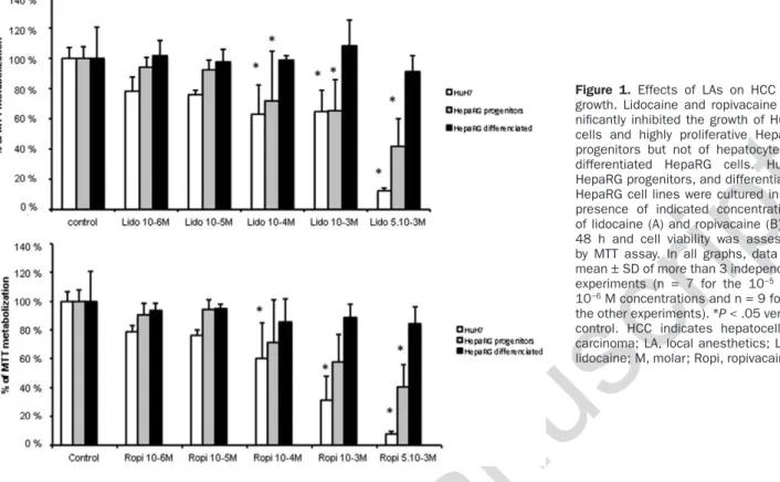
The effects of LAs on cell viability were first evaluated on HuH7 and HepaRG cell lines at 48 hours (Figure 1). There was a significant cell × concentration interaction (P < .001). Pair-wise comparisons showed that as compared with con-trol, lidocaine and ropivacaine (concentrations ranging from 5 × 10⁻³ M to 10−4 M) significantly decreased the growth of
HuH7 cells as follows: by 87% (P < .001) and 92% (P < .001) at 5 × 10⁻³ M, 35% (P = .02) and 67% (P < .001) at 10−3 M, 37%
(P = .02) and 40% (P = .02) at 10−4 M, respectively. Lidocaine
and ropivacaine also decreased the proliferation of highly proliferative HepaRG progenitors (at concentrations of 5 × 10⁻³ M [58%; P < .001], 10−3 M [35%; P < .01], and 10−4 M
[29%; P = .04] for lidocaine, and only at concentration of 5 × 10⁻³ M [59%; P < .001] for ropivacaine) but did not affect
viability of hepatocyte-like differentiated HepaRG cells. Lidocaine and ropivacaine at 10−2 M induced an important
cytotoxicity on HuH7 cells (Supplemental Digital Content 2, Figure 1, http://links.lww.com/AA/B969) and therefore this concentration was not further used in the experiments. No effect on cell viability and proliferation of the cell lines were observed with the lowest concentrations (10−5 and
10−6 M). Cell damage assessed with LDH release test
Revised
manuscript
on HuH7 and HepaRG progenitors cell lines (Supplemental Digital Content 3, Table 2A, http://links.lww.com/AA/ B970). Inhibition of cell proliferation was supported by a LAs-dependent inhibition of DNA synthesis (Supplemental Digital Content 3, Table 2B, http://links.lww.com/AA/ B970). After 24 hours, the effect was less pronounced: The only significant effect was observed at 10−3 M of lidocaine
and ropivacaine, which significantly decreased the growth of HuH7 cells by 13% (P < .001) and 14% (P < .001), respec-tively. When testing the effects of decreasing concentrations of LAs over time, lidocaine and ropivacaine significantly decreased the growth of HuH7 cells by 50% (P = .01) and 77% (P < .001) at 72 hours, respectively.
Lidocaine Inhibits the Growth of HCC Cells by
Increasing the Caspase 3 Activity
The observed reduced cell viability induced by LAs on highly proliferative cells prompted us to determine whether LAs impact the cell cycle. Lidocaine had no significant effect on the cell cycle but was associated with an increase in the number of apoptotic bodies (Figure 2). Unsupervised genome-wide expression profiling showed in Huh7 cells that lidocaine treat-ments (10−3 M) for 48 hours resulted in the deregulation of 194
genes (P < .01; FC > 1.5) (Supplemental Digital Content 4, Figure 2A, http://links.lww.com/AA/B971 and Supplemental Digital Content 3, Table 2, http://links.lww.com/AA/B970). No drastic effect of lidocaine on mRNA levels of cell cycle regu-lators or cyclins was observed (Supplemental Digital Content 4, Figure 2, http://links.lww.com/AA/B971 and Supplemental Digital Content 5, Table 3, http://links.lww.com/AA/B972). However, lidocaine increased by 10 times the mRNA levels of adenomatous polyposis coli (APC) (FC > 1.5; P < .01), which acts as an antagonist of the Wnt/β-catenin pathway (Figure
3). Lidocaine had no effect on the expression of apoptosis-related genes and protein such as caspases and poly (adeno-sin diphosphate (ADP) ribose) polymerases but increased mRNA levels of Harakiri (HRK), encoding a proapoptotic protein (Figure 3). QRT-PCR confirmed microarray results for genes coding for CCNA2 (no significant effect), CCNB1 (no significant effect), APC (on HuH7: 1800% increase at 10−4
M [P = .01], 4400% increase at 10−3 M [P < .01] and on HepaRG
progenitors: 400% increase at 10−4 M [P = .03], 300% increase
at 10−3 M [P = .04]), and HRK (on HuH7: 48% increase at 10−4
M [P = .02], 129% increase at 10−3 M [P < .01] and on HepaRG
progenitors: 135% increase at 10−4 M [P = .05], 117% increase at
10−3 M [P < .01]) (Figure 4). Lidocaine-induced gene
deregula-tions were similar for both cell lines (Figure 4). Western-blot analysis showed that lidocaine increases APC protein level (on HuH7: 25% increase at 10−4 M [P < .001], 32% increase at
10−3 M [P < .001] and on HepaRG progenitors: 59% increase
at 10−4 M [P = .05], 54% increase at 10−3 M [P = .02]) but has no
significant impact on the expression of cyclins (Supplemental Digital Content 6, Figure 3, http://links.lww.com/AA/B973). Apoptosis was upregulated by lidocaine as demonstrated by caspase 3 activity (on HuH7: 393% increase at 10−4 M [P < .01],
357% increase at 10−3 M [P < .001] and on HepaRG
progeni-tors: 104% increase at 10−4 M [P < .01], 90% increase at 10−3 M
[P < .01]) (Figure 6) and TUNEL assays (Supplemental Digital Content 3, Table 2, http://links.lww.com/AA/B970). No effect on senescence was observed (Supplemental Digital Content 7, Figure 4, http://links.lww.com/AA/B974).
Ropivacaine Inhibits the Growth of HCC Cells by
Stopping the Cell Cycle in G2 Phase
Cell cycle analysis demonstrated that ropivacaine treat-ment resulted in a drastic enrichtreat-ment of cells in the G2
Figure 1. Effects of LAs on HCC cell growth. Lidocaine and ropivacaine sig-nificantly inhibited the growth of HuH7 cells and highly proliferative HepaRG progenitors but not of hepatocyte-like differentiated HepaRG cells. HuH7, HepaRG progenitors, and differentiated HepaRG cell lines were cultured in the presence of indicated concentrations of lidocaine (A) and ropivacaine (B) for 48 h and cell viability was assessed by MTT assay. In all graphs, data are mean ± SD of more than 3 independent experiments (n = 7 for the 10−5 and
10−6 M concentrations and n = 9 for all
the other experiments). *P < .05 versus control. HCC indicates hepatocellular carcinoma; LA, local anesthetics; Lido, lidocaine; M, molar; Ropi, ropivacaine.
Revised
manuscript
phase for both cell lines, suggesting a cell cycle blockade before mitosis. This observation coincided with the absence of mitotic nuclei (Figure 2). Unsupervised genome-wide expression profiling in HuH7 cells showed that ropivacaine treatment (10−3 M for 48 hours) resulted in the
deregula-tion of 221 genes (P < .01; FC > 1.5) (Supplemental Digital Content 4, Figure 2B, http://links.lww.com/AA/B971 and Supplemental Digital Content 8, Table 4, http://links. lww.com/AA/B975). Interestingly, ropivacaine was asso-ciated with a decrease in the expression of key cell cycle
regulator genes, especially involved in the G2-M transition phase, namely cyclin A2 (CCNA2) (63% decrease; P < .01), cyclin B1 (CCNB1) (64% decrease; P = .02). Ropivacaine also decreased the expression of MKI67 a nuclear marker of cell proliferation (61% decrease; P < .01) (Figure 3). Gene Set Enrichment Analysis confirmed the negative enrichment of cell cycle–associated gene signatures in HuH7 cells treated with ropivacaine (Supplemental Digital Content 9, Figure 5, http://links.lww.com/AA/B976). QRT-PCR validated the microarray data for CCNA 2 (on HuH7: 81% decrease at
Figure 2. LAs induced cell cycle alterations. Cell cycle phases were analyzed using Cellomics Arrayscan Vti with or without (control) 10−4 M
lidocaine or ropivacaine after 48-h culture of HuH7 and HepaRG progenitors cells. Control: presence of mitotic nuclei and mitosis. Lido 10−4
M: reduction of mitotic nuclei and no significant effect on the cell cycle. Ropi 10−4 M: absence of mitotic nuclei and accumulation of cells in
Revised
manuscript
Figure 3. mRNAs levels of genes of interest detected by microarray after 48-h culture of HuH7 cells with or without 10−3 M lidocaine and
ropivacaine. Lidocaine significantly increased the mRNA levels of CCNDBP1 and APC. Besides MKI67, a nuclear marker of cell proliferation, lidocaine has no impact on the expression of cell cycle–associated genes. Ropivacaine decreased the mRNA level of key cell cycle regulators: CCNA2, CCNB1, CCNB2, and CDK1 and of MKI67. Both LAs increased the mRNA levels of HRK, apro-apoptotic protein. Data from 3 indepen-dent experiments shown as scatter plot (dashed line = 1, representing control). Horizontal line indicates mean for each group. *P < .05 versus control. APC indicates adenomatous polyposis coli; CCNA2, cyclin A2; CCNB1, cyclin B1; CCNDBP1, cyclin D binding protein 1; HRK, Harakiri.
Figure 4. Quantitative real-time polymerase chain reaction confirmed the microarray data on HuH7 (A) and HepaRG progenitor (B) cells treated with lidocaine. Lidocaine induced a significant upregulation of APC and HRK. Lidocaine-induced gene deregulations were similar for both cell lines. Data from 3 independent experiments shown as scatter plot (dashed line = 1, representing control). Horizontal line indicates mean for each group.*P < .05 versus control. APC indicates adenomatous polyposis coli; CCNA2, cyclin A2; CCNB1, cyclin B1; HRK, Harakiri.
Revised
manuscript
10−4 M [P < .01], 44% decrease at 10−3 M [P = .01] and on
HepaRG progenitors: not significant), CCNB1 (on HuH7: 98% decrease at 10−4 M [P = .01], 68% decrease at 10−3 M
[P = .02]; on HepaRG progenitors: 25% decrease at 10−4 M
[P = .02], 40% decrease at 10−3 M [P = .04]), APC (on HuH7:
200% increase at 10−4 M [P = .05], 55% increase at 10−3 M
[P = .03]; and on HepaRG progenitors: not significant), and
HRK (on HuH7: 180% increase at 10−4 M [P = .04], 220%
increase at 10−3 M [P < .01] and on HepaRG progenitors:
58% increase at 10−4 M [P = .02], 137% increase at 10−3 M
[P < .01]) genes, not only in HuH7 cells but also in HepaRG cells treated with different concentrations of ropivacaine (Figure 5). Decreased expression CCNB1 and increased expression of HRK were more pronounced in HuH7 cells as compared to HepaRG cells (Figure 5). Western-blot analysis confirmed a decrease in the expression of cyclin A (on HuH7: 30% decrease at 10−4 M [P = .05], 50% decrease at
10−3 M [P = .04] and on HepaRG progenitors: 34% decrease
at 10−3 M [P = .02]) and cyclin B (on HuH7: 70% decrease
at 10−4 M [P = .01], 80% decrease at 10−3 M [P < .01] and
on HepaRG progenitors: 35% decrease at 10−4 M [P = .02],
65% decrease at 10−3 M [P < .01]) at a protein level in both
cell lines (Supplemental Digital Content 6, Figure 3, http:// links.lww.com/AA/B973). Increased expression of pro-apoptotic HRK genes (Figure 5) after ropivacaine treatment correlated with an increased caspase activity and apoptosis (on HuH7: 173% increase at 10−4 M [P < .01], 217% increase at
10−3 M [P < .001] and on HepaRG progenitors: 53% increase
at 10−4 M [P = .03], 69% increase at 10−3 M [P < .01]) (Figure 6;
Supplemental Digital Content 3, Table 2, http://links.lww. com/AA/B970). No effect on senescence was observed (Supplemental Digital Content 7, Figure 4, http://links. lww.com/AA/B974). Altogether, these results indicated
that ropivacaine treatment resulted in inhibition of tumor cell growth by inducing cell cycle arrest and apoptosis.
DISCUSSION
To our knowledge, this study is the first report on the antitu-mor effect of LAs on HCC cells. LAs were previously shown to inhibit cell growth and induce cell death in lung,7 colon,21
and pancreatic cancer cells.22
In healthy tissue, cell growth and cell cycle are tightly regulated. The loss of this regulation due to gene mutation (eg, inactivation of the tumor suppressor gene TP53), epi-genetic or genomic deregulation, is a hallmark of cancer cells and results in their uncontrolled proliferation, associated with apoptosis resistance. In the present study, we show that ropivacaine may stop the G2 phase of the cell cycle in HCC cells. Only a few reports have investigated the effects of LAs on the cell cycle. In noncancer cells, Lucchinetti et al23 showed that lidocaine and bupivacaine inhibit the cell
cycle of mesenchymal stem cell at the G1/S phase transi-tion. In colon and pancreatic cancer cell lines, Bundscherer et al22 observed a significant antiproliferative effect with
of high concentrations of ropivacaine and bupivacaine. Ropivacaine was shown to inhibit colon cancer cells’ volt-age-gated sodium channels (NaV1.5) and metastatic colon cancer cell invasion.8 In our study, ropivacaine inhibited
the proliferation of HCC cells by stopping the cell cycle in G2 phase. It decreased the mRNA abundance of key cell cycle regulators, especially involved in the G2-M transition phase, namely cyclin A2, cyclin B1, cyclin B2, and cyclin-dependent kinase 1. Ropivacaine also decreased the expres-sion of MKI67, a nuclear marker of cell proliferation. Indeed, the CDK1–cyclin A complex allows the cell cycle to progress from the S (DNA replication) to the G2 (preparation for cell
Figure 5. Quantitative real-time polymerase chain reaction confirmed the microarray data on HuH7 (A) and HepaRG progenitor (B) cells treated with or without ropivacaine. Ropivacaine significantly down-regulated CCNA2 and CCNB1 and upregulated HRK. Ropivacaine-induced gene deregulations were more pronounced in HuH7 cells. Data from 3 independent experiments shown as scatter plot (dashed line = 1, represent-ing control). Horizontal line indicates mean for each group. *P < .05 versus control. APC indicates adenomatous polyposis coli; CCNA2, cyclin A2; CCNB1, cyclin B1; CCNB2, cyclin B2; HRK, Harakiri; M, molar.
Revised
manuscript
division) phase and the CDK1–cyclin B complex allows the progression from G2 to M (cell division) phase. Moreover, ropivacaine increased apoptosis in HepaRG progenitor cells without any effect on cellular senescence. In our study, the effects of ropivacaine were more pronounced on HuH7 and HepaRG progenitor cells than on differentiated HepaRG cells. These observations suggest an enhanced effect of LA on highly versus poorly proliferated cells.
In noncancer cells, lidocaine at a high concentration has been shown to stop cell cycle at the S phase and to inhibit fibroblast multiplication.24 In breast cancer cells, a
poten-tial antitumor effect of lidocaine was reported, associated with a demethylation effect25 and a sensitization effect to
cisplatin cytotoxicity.26 In vitro, lidocaine enhanced natural
killer cell cytotoxicity against lymphoblast cells at 0.01 and 0.1 μM.27 Recently, Chang et al28 reported that lidocaine and
bupivacaine are cytotoxic for thyroid cancer cells. These 2 LAs damaged the mitochondrial membrane potential, lead to cytochrome C release, activation of caspases 3 and 7, poly(ADP-ribose) polymerase cleavage, and induction of BCI-2 associated X. In our study, lidocaine increased by 10 times the mRNA levels of APC and of DKK1, which both act as antagonists of the Wnt/β-catenin pathway. The effect of lidocaine on DKK1 was more modest than on APC. The Wnt/β-catenin pathway is long known to be involved in carcinogenesis, especially in HCC.29,30 Therefore, lidocaine
could be of interest in HCC, particularly those subtypes with an increase activity of the Wnt/β-catenin pathway.
In our study, the effects and the underlying mecha-nisms of action of lidocaine and ropivacaine were different. Different effects of each LA have been previously reported on different cell lines including T-cells,31 neuronal cells,32
and mesenchymal stromal cells.33 In lung cancer cells,
ropivacaine and lidocaine inhibiting effects on Src were mediated through different pathways.7 Indeed, LAs
pos-sess distinct chemical structures and properties. Jose et al34
previously reported a cell-type– and molecule-type–specific effect. In their study, levobupivacaine triggered a more potent cancer-specific reduction of viability than ropiva-caine on certain cancer cell type. They hypothesized a dif-ferent effect on mitochondrial respiratory chain and ATP synthesis. Moreover, the systemic anti-inflammatory effects of bupivacaine are not mediated through sodium channel inhibition35 indicating that LAs exert their properties by
acting on a variety of targets. The differences observed in previous studies as well as in the present study regarding ropivacaine and lidocaine remain mostly unexplained on a mechanistic level and need further experiments.
The effects of LAs on cell viability, cell cycle, genes, and pathways deregulation are dose dependent.7 We observed
the cytostatic effects of LAs on HCC cells for concentration ranging from 10–2 to 10–5 M. In vitro concentrations vary
Figure 6. Effects of LAs on apoptosis of HepaRG progenitor (A) and Huh7 (B) cells. The apoptotic effect was assessed by determining caspase 3 activity. HuH7 and HepaRG progenitor cells were cultured with or without LAs for 48 h; Caspase 3 activity was upregulated by lidocaine with a stronger effect than ropivacaine; results were confirmed when adjusted on cell viability (MTT assay). Data from 3 independent experiments shown as scatter plot (dashed line = 1, represent-ing control). Data are expressed as percentage of the control *P < .05 compared with control. L indicates lidocaine; LA, local anesthetics; R, ropivacaine.
Revised
manuscript
widely in the previous studies. Effects on cancer cells have been described for concentrations ranging from 10–4 M7 to
13.5 × 10⁻³ M36 for lidocaine and from 10–5 M22 to 4.32 × 10⁻³
M37 for ropivacaine. High concentrations of LAs are
cyto-toxic for noncancer human cells. Indeed, only few stud-ies have tested the viability of control cells when LAs are added. Chang et al36 used mammary epithelial cells as
con-trols and reported toxicity with higher concentrations when compared with breast cancer cells. However concentrations were high in this study.
Many mechanisms by which LAs and regional anesthe-sia could exert an antitumor effect have been suggested in the literature: (1) a decrease in opioid requirement is always associated with the use of LAs and opioid might promote cancer cells proliferation38; (2) regional anesthesia inhibits
axonal transport39 and therefore could stop the
dissemina-tion of cancer cells during surgery; (3) a direct inhibidissemina-tion of cancer cells growth by LAs.7 Clinical studies on the
poten-tial benefit of LAs during cancer surgery have been pub-lished for more than 10 years. Although retrospective and with some methodological bias, these studies lead to the hypothesis that LAs could mitigate perioperative tumor growth and metastasis formation,2,4,5 specifically in the
setting of breast and prostate cancer. These clinical stud-ies are echoing the experimental studstud-ies that have already reported a reduction of tumor cell growth by LAs in the spe-cific settings of thyroid,28 breast,25 lung,7 and colon8 cancer
for example. Moreover, Lucchinetti et al23 showed how LAs
impaired the proliferation of mesenchymal stem cell, which are known to play an important role in tumor progression.40
This effect was associated with a potential detrimental effect on wound healing when LAs are administered directly on the wound. Our work is the first to report a mechanism of the inhibiting effect of 2 LAs on HCC cells. In addition to potential effects in the tumor microenvironment, our results showed that LAs may induce profound modifications in gene expression profiles of tumor cells, notably by modulat-ing cell cycle–related genes resultmodulat-ing in a cytostatic effect and induction of apoptosis. Multiple pathways are involved in the modulation of cell growth. These pathways cross talk to modulate the balance between proliferation and apopto-sis. Based on the literature, it is difficult to determine if there is a waterfall effect induced by LAs on these pathways.23
However, the antiproliferative effect of LAs on HCC cells has to be balanced with the possible risks of LAs toxicity and wound healing impairment. Moreover, our results would transpose with difficulty in the clinical setting. Indeed, many elements such as the absence of stress and/or inflammation and/or opioids and/or pain could eventually interact with the effect of LAs. Both preclinical and clinical studies are required to further confirm the benefit of LAs on the outcome of HCC surgery. Due to its analgesic proper-ties, intravenous lidocaine is already part of most anesthe-sia protocol for abdominal surgery. Studying its effect on preventing cancer recurrence would therefore be feasible in clinical practice.
E
ACKNOWLEDGMENTS
The authors thank Rémy le Guével from the “ImPACcell” platform (Biogenouest, SFR biosit, University of Rennes1) and Isabelle Cannie for technical assistance.
DISCLOSURES
Name: Grégoire Le Gac, MD.
Contribution: This author helped conduct the study.
Name: Gaëlle Angenard, BS.
Contribution: This author helped conduct the study.
Name: Bruno Clément, PhD.
Contribution: This author helped analyze the data and write the manuscript.
Name: Bruno Laviolle, MD, PhD.
Contribution: This author helped analyze the data and correct the statistics.
Name: Cédric Coulouarn, PhD.
Contribution: This author helped conduct the study, analyze the data, and write the manuscript.
Name: Hélène Beloeil, MD, PhD.
Contribution: This author helped conduct the study, analyze the data, and write the manuscript.
This manuscript was handled by: Scott M. Fishman, MD.
REFERENCES
1. Gottschalk A, Sharma S, Ford J, Durieux ME, Tiouririne M. Review article: the role of the perioperative period in recur-rence after cancer surgery. Anesth Analg. 2010;110:1636–1643. 2. Biki B, Mascha E, Moriarty DC, et al. Anesthetic technique for
radical prostatectomy surgery affects cancer recurrence: a retro-spective analysis. Anesthesiology. 2008;109:180–187.
3. Vigneault L, Turgeon AF, Côté D, et al. Perioperative intra-venous lidocaine infusion for postoperative pain control: a meta-analysis of randomized controlled trials. Can J Anaesth. 2011;58:22–37.
4. Exadaktylos AK, Buggy DJ, Moriarty DC, Mascha E, Sessler DI. Can anesthetic technique for primary breast cancer surgery affect recurrence or metastasis? Anesthesiology. 2006;105:660–664.
5. Cummings KC III, Xu F, Cummings LC, Cooper GS. A com-parison of epidural analgesia and traditional pain management effects on survival and cancer recurrence after colectomy: a population-based study. Anesthesiology. 2012;116:797–806. 6. Chen WK, Miao CH. The effect of anesthetic technique on
sur-vival in human cancers: a meta-analysis of retrospective and prospective studies. PLoS One. 2013;8:e56540.
7. Piegeler T, Votta-Velis EG, Liu G, et al. Antimetastatic poten-tial of amide-linked local anesthetics: inhibition of lung adenocarcinoma cell migration and inflammatory Src signal-ing independent of sodium channel blockade. Anesthesiology. 2012;117:548–559.
8. Baptista-Hon DT, Robertson FM, Robertson GB, et al. Potent inhibition by ropivacaine of metastatic colon cancer SW620 cell invasion and NaV1.5 channel function. Br J Anaesth. 2014;113(suppl 1):i39–i48.
9. Tada M, Imazeki F, Fukai K, et al. Procaine inhibits the prolifer-ation and DNA methylprolifer-ation in human hepatoma cells. Hepatol
Int. 2007;1:355–364.
10. European Association for Study of Liver; European Organisation for Research and Treatment of Cancer. EASL-EORTC clinical practice guidelines: management of hepatocellular carcinoma.
Eur J Cancer. 2012;48:599–641.
11. Llovet JM, Fuster J, Bruix J. Intention-to-treat analysis of surgi-cal treatment for early hepatocellular carcinoma: resection ver-sus transplantation. Hepatology. 1999;30:1434–1440.
12. Nakabayashi H, Taketa K, Miyano K, Yamane T, Sato J. Growth of human hepatoma cells lines with differentiated functions in chemically defined medium. Cancer Res. 1982;42:3858–3863. 13. Cerec V, Glaise D, Garnier D, et al. Transdifferentiation of
hepa-tocyte-like cells from the human hepatoma HepaRG cell line through bipotent progenitor. Hepatology. 2007;45:957–967. 14. Coulouarn C, Corlu A, Glaise D, et al. Hepatocyte-stellate cell
cross-talk in the liver engenders a permissive inflammatory microenvironment that drives progression in hepatocellular carcinoma. Cancer Res. 2012;72:2533–2542.
15. Dubois-Pot-Schneider H, Fekir K, Coulouarn C, et al. Inflammatory cytokines promote the retrodifferentiation of tumor-derived hepatocyte-like cells to progenitor cells.
Revised
manuscript
16. Gripon P, Rumin S, Urban S, et al. Infection of a human hepa-toma cell line by hepatitis B virus. Proc Natl Acad Sci U S A. 2002;99:15655–15660.
17. Denizot F, Lang R. Rapid colorimetric assay for cell growth and survival. Modifications to the tetrazolium dye procedure giving improved sensitivity and reliability. J Immunol Methods. 1986;89:271–277.
18. Schneider-Jakob S, Corazza N, Badmann A, et al. Synergistic induction of cell death in liver tumor cells by TRAIL and che-motherapeutic drugs via the BH3-only proteins Bim and Bid.
Cell Death Dis. 2010;1:e86.
19. Sulpice L, Rayar M, Desille M, et al. Molecular profiling of stroma identifies osteopontin as an independent predictor of poor prognosis in intrahepatic cholangiocarcinoma. Hepatology. 2013;58:1992–2000.
20. Wright GW, Simon RM. A random variance model for detec-tion of differential gene expression in small microarray experi-ments. Bioinformatics. 2003;19:2448–2455.
21. Martinsson T. Ropivacaine inhibits serum-induced prolifera-tion of colon adenocarcinoma cells in vitro. J Pharmacol Exp
Ther. 1999;288:660–664.
22. Bundscherer A, Malsy M, Gebhardt K, et al. Effects of ropiva-caine, bupivacaine and sufentanil in colon and pancreatic can-cer cells in vitro. Pharmacol Res. 2015;95–96:126–131.
23. Lucchinetti E, Awad AE, Rahman M, et al. Antiproliferative effects of local anesthetics on mesenchymal stem cells: poten-tial implications for tumor spreading and wound healing.
Anesthesiology. 2012;116:841–856.
24. Desai SP, Kojima K, Vacanti CA, Kodama S. Lidocaine inhib-its NIH-3T3 cell multiplication by increasing the expression of cyclin-dependent kinase inhibitor 1A (p21). Anesth Analg. 2008;107:1592–1597.
25. Lirk P, Hollmann MW, Fleischer M, Weber NC, Fiegl H. Lidocaine and ropivacaine, but not bupivacaine, demethyl-ate deoxyribonucleic acid in breast cancer cells in vitro. Br J
Anaesth. 2014;113(suppl 1):i32–i38.
26. Li K, Yang J, Han X. Lidocaine sensitizes the cytotoxicity of cisplatin in breast cancer cells via up-regulation of RARβ2 and RASSF1A demethylation. Int J Mol Sci. 2014;15:23519– 23536.
27. Ramirez MF, Tran P, Cata JP. The effect of clinically therapeutic plasma concentrations of lidocaine on natural killer cell cyto-toxicity. Reg Anesth Pain Med. 2015;40:43–48.
28. Chang YC, Hsu YC, Liu CL, et al. Local anesthetics induce apoptosis in human thyroid cancer cells through the mitogen-activated protein kinase pathway. PLoS One. 2014;9:e89563. 29. Clevers H, Nusse R. Wnt/β-catenin signaling and disease. Cell.
2012;149:1192–1205.
30. Monga SP. β-catenin signaling and roles in liver homeostasis, injury, and tumorigenesis. Gastroenterology. 2015;148:1294–1310. 31. Boselli E, Duflo F, Debon R, et al. The induction of apoptosis by
local anesthetics: a comparison between lidocaine and ropiva-caine. Anesth Analg. 2003;96:755–756.
32. Perez-Castro R, Patel S, Garavito-Aguilar ZV, et al. Cytotoxicity of local anesthetics in human neuronal cells. Anesth Analg. 2009;108:997–1007.
33. Gray A, Marrero-Berrios I, Ghodbane M, et al. Effect of local anesthetics on human mesenchymal stromal cell secretion.
Nano Life. 2015;5:1550001–1550014.
34. Jose C, Bellance N, Chatelain EH, et al. Antiproliferative activ-ity of levobupivacaine and aminoimidazole carboxamide ribonucleotide on human cancer cells of variable bioenergetic profile. Mitochondrion. 2012;12:100–109.
35. Beloeil H, Ababneh Z, Chung R, et al. Effects of bupivacaine and tetrodotoxin on carrageenan-induced hind paw inflamma-tion in rats (part 1): hyperalgesia, edema, and systemic cyto-kines. Anesthesiology. 2006;105:128–138.
36. Chang YC, Liu CL, Chen MJ, et al. Local anesthetics induce apop-tosis in human breast tumor cells. Anesth Analg. 2014;118:116–124. 37. Werdehausen R, Fazeli S, Braun S, et al. Apoptosis induction by different local anaesthetics in a neuroblastoma cell line. Br J
Anaesth. 2009;103:711–718.
38. Afsharimani B, Cabot P, Parat MO. Morphine and tumor growth and metastasis. Cancer Metastasis Rev. 2011;30:225–238. 39. Deruddre S, Combettes E, Estebe JP, et al. Effects of a
bupiva-caine nerve block on the axonal transport of tumor necrosis fac-tor-alpha (TNF-alpha) in a rat model of carrageenan-induced inflammation. Brain Behav Immun. 2010;24:652–659.
40. Wong RS. Mesenchymal stem cells: angels or demons? J Biomed