HAL Id: hal-03087735
https://hal.archives-ouvertes.fr/hal-03087735
Submitted on 28 Dec 2020HAL is a multi-disciplinary open access
archive for the deposit and dissemination of sci-entific research documents, whether they are pub-lished or not. The documents may come from teaching and research institutions in France or abroad, or from public or private research centers.
L’archive ouverte pluridisciplinaire HAL, est destinée au dépôt et à la diffusion de documents scientifiques de niveau recherche, publiés ou non, émanant des établissements d’enseignement et de recherche français ou étrangers, des laboratoires publics ou privés.
The many implications of actin filament helicity
Antoine Jegou, Guillaume Romet-Lemonne
To cite this version:
Antoine Jegou, Guillaume Romet-Lemonne. The many implications of actin filament helicity. Sem-inars in Cell & Developmental Biology, 2020, 102, pp.65-72. �10.1016/j.semcdb.2019.10.018�. �hal-03087735�
The many implications of actin filament helicity
Antoine Jegoua and Guillaume Romet-Lemonnea
a Université de Paris, CNRS, Institut Jacques Monod, 15 rue Hélène Brion, 75013 Paris, France
corresponding authors:
Antoine Jegou, email: antoine.jegou@ijm.fr Guillaume Romet-Lemonne, email: romet@ijm.fr
Permanent address: Institut Jacques Monod, CNRS, Université de Paris, 15 rue Hélène brion, F-75013 Paris, France
Abstract
One of the best known features of actin filaments is their helical structure. A number of essential properties emerge from this molecular arrangement of actin subunits. Here, we give an overview of the mechanical and biochemical implications of filament helicity, at different scales. In particular, a number of recent studies have highlighted the role of filament helicity in the adaptation to and the generation of mechanical torsion, and in the modulation of the filament’s interaction with very different actin-binding proteins (such as myosins, cross-linkers, formins, and cofilin). Helicity can thus be seen as a key factor for the regulation of actin assembly, and as a link between biochemical regulators and their mechanical context. In addition, actin filament helicity appears to play an essential role in the establishment of chirality at larger scales, up to the organismal scale. Altogether, helicity appears to be an essential feature contributing to the regulation of actin assembly dynamics, and to actin’s ability to organize cells at a larger scale.
Keywords (max 6)
mechanotransduction; cytoskeleton; actin assembly dynamics; torque; twist
Abbreviations
1. Introduction: Helical structure of the actin filament
Helicity is a common feature in biopolymers. In the case of actin filaments, helicity is an essential characteristic which can be linked to several key properties of the filaments. We begin here by introducing the main parameters and numbers used to describe actin filament helicity. The following sections will address the mechanical implications of actin filament helicity (section 2), the role of helicity in the interactions of filaments with actin-binding proteins (section 3) and the role it plays in transmitting chirality across scales (section 4).
In the years that followed the discovery of actin filaments by the laboratory of Albert Szent-Györgyi in the 1940's (see Bugyi and Kellermayer [1] for a review of these fascinating early days of actin research), X-ray diffraction and electron microscopy studies detected a periodicity (larger than subunit size) in the structure of actin fibers [2,3]. The helical structure suggested by these early observations [2] was nicely confirmed by negative staining electron microscopy a few years later [4– 6] and its right-handedness (viewing the filament as a double helix) was clearly established [7]. Since then, with the advent of more sensitive detectors and refined algorithms, more studies using EM or X-ray diffraction have consolidated our description of actin filaments as helices [8–13]. Today, helicity is a textbook feature of actin filaments.
The actin filament can be described as a single, left-handed helix, with a short pitch (indicated by orange lines in figure 1A) where each subunit is rotated by -167° with respect to its neighbor. It is along this helix that subunits are added at the end of the filament, as it polymerizes. The actin filament can also be viewed as a double helix, which is perhaps easier to visualize, where two strands of actin subunits are intertwined (red lines in Figure 1A). They are stabilized by interactions between neighboring subunits of the same strand (intra-strand, or longitudinal interactions), and between strands (inter-strand, or lateral interactions). Based on assembly kinetics at filament ends [14,15], thermodynamic considerations and modeling have been used to estimate the energy of longitudinal contacts to be 12-20 kT and that of lateral contacts to be 4-8 kT [16–18]. The standard value for the half-pitch (or crossover distance) of the double helix is 36 nm, a distance which embraces 13 actin subunits. The double helix thus makes one full turn every 72 nm, i.e. it makes 13.9 turns per micrometer. Within each strand, each subunit is rotated by approximately +27° with respect to its longitudinal neighbor. Unless specified otherwise, in this review we are always referring to the right-handed, two-start, long pitch helix.
The numbers we have just cited characterize the canonical actin filament structure. However, one should bear in mind that actin filaments may adopt slightly different conformations. Early EM studies [4] reported a significant variability of the helical pitch of actin filaments, and later studies have indicated that this variability in pitch was not due to sample preparation, but was an intrinsic property of the actin filament [19,20], and could be linked to variations in the conformation of the subunits, in particular that of their DNAse1-binding loop which is highly variable [21]. The resulting different structural states differ by a few degrees in their subunits orientations, but they all remain right-handed double-stranded helices, thereby underlining the robustness of this basic feature. The idea of structural polymorphism has been disputed, notably by cryo-EM studies reporting a very low variability in helicity [11]. An explanation for this apparent discrepancy may come from a reduction of the fluctuations in helical pitch upon application of a tensile force [22–24].
Remarkably, the nucleotide state of the actin subunits, which has a strong impact on the filament’s depolymerization rate [14,25,26] and on its mechanical properties [27] does not seem to affect its helical parameters [13,28]. Similarly, pH and ionic strength, which are known to affect filament stability and rigidity [29–32] do not appear to significantly affect the filament’s helical parameters
[29]. It has been proposed, however, that variability in the occupation of specific cation-binding sites located at the longitudinal interface of subunits could account for the aforementioned structural polymorphism [33]. Mechanical tension has also been proposed as a means to alter the
conformation of actin filaments [34].
In summary, actin filaments are always observed to have a right-handed double-helical structure, and it is not entirely clear to what extent the helical parameters can be altered in the absence of ABPs. In contrast, as we will see in section 3, ABPs can induce significant changes of the helical pitch, which provides a handle to modulate filament properties.
2. Mechanical implications of actin filament helicity
Before discussing how ABPs can modulate filament helicity (section 3) and what consequences it can have at larger scales (section 4), let us briefly summarize some of the physical consequences of helicity.
An obvious consequence of helicity is that it gives filaments an inherent periodicity, other than that of the subunits themselves. This simple feature imposes geometrical constraints when packing filaments into tight bundles (section 3.1).
Another natural consequence of helicity is that it couples different mechanical stresses applied to actin filaments. Experimentally, it has been reported more than 20 years ago that a moderate torque, over-twisting or under-twisting an actin filament by as little as 0.1 turn/µm, was enough to reduce twofold the tensile force required for filament rupture [35]. To our knowledge, this fascinating result has never been reproduced. Spectacular experiments where stronger local stresses were applied, by tying knots in actin filaments with optical tweezers, reduced the tensile force for rupture by two orders of magnitude [36]. Theoretical studies have shown that, due to helicity, mechanical twist (torque) is coupled to tension and bending [37–39]. Twist-bend coupling, in particular, can be quite strong and is likely to affect the mechanical properties of the filaments [40]. Stretch-twist coupling appears to be more moderate, requiring high tensile forces in order to alter the helical pitch of the filament [24]. Our recent data [30], showing that the action of cofilin is barely affected by filament tension up to 30 pN (see section 3.4), seem consistent with this view.
The application of a mechanical torque is naturally linked to helicity. Of course, torque can be applied to non-helical filaments (such as linear synthetic polymers), however, as we will see in section 3, the actin filament’s helical structure makes it easier for ABPs to apply a mechanical torque to the filament. Also, a direct consequence of an applied mechanical torque will be a modification of the helical pitch of the filament. If the two ends of a filament are twisted, a torque is applied uniformly along the filament (just as pulling on the ends would result in a uniform tension along the filament). Filament regions of identical composition (e.g., bare or decorated by an ABP) will thus be identically deformed by the applied torque, and be uniformly under- or over-twisted.
Several papers, including [41,42], have reported a torsional rigidity of approximately 2x10^-27 Nm2 for actin filaments (corresponding to a torsional persistence length of approximately 500 nm), while others [35,43] find filaments to be 10- to 40-fold stiffer. Our recent work [30,44], using the polarized emission of fluorescence to monitor the orientation of actin subunits around the axis of filaments aligned by microfluidics, finds that this orientation remains steady several micrometers away from the filament’s anchoring point. These observations would agree with the higher values of torsional
stiffness. A recent mesoscopic length scale model [45] seems to reconcile the different torsional stiffness measurements, by distinguishing a high “filament torsional rigidity” and a weaker “intersubunit torsional rigidity”. This model also predicts that actin filaments are easier to under-twist than to over-under-twist, as the latter deformation strains longitudinal contacts more than lateral contacts.
3. Crosstalk between helicity and ABPs
The fact that actin filaments are helical has consequences for several, very different ABPs. They are both affected by helicity and able to modify it. Helicity and the mechanical constraints modifying helicity are thus important parameters for the regulation of actin assembly and the organization of filament networks.
3.1 Cross-linkers
Proteins that cross-link actin filaments play an important role in shaping actin networks and cells. As they connect filaments together, they can impose a specific filament polarity within the network and they affect the network’s visco-elastic properties in non-trivial ways [46], which eventually determine the mechanical properties of cells and tissues.
As we have already mentioned in section 2, filament helicity gives rise to periodicity. This is clearly apparent when filaments are tightly bundled by small proteins such as fascin or espin [47–49], as bundling puts the binding sites on two neighboring filaments in register (Figure 2B). Note that, depending on the bundling protein, neighboring filaments in the bundle can be parallel or
antiparallel. The geometrical packing of the filaments by small cross-linkers have been reported to induce changes in the conformation of the filaments and to depend on crosslinker elasticity [50,51]. In the case of fascin and espin, these changes in filament helicity are either continuous or discrete, respectively. The angle between longitudinal actin subunit neighbors was reported to decrease by as much as ~2° (thereby increasing the half-pitch of the double helix to ~39 nm) [51].
The binding energy of an ABP is typically a few kT, significantly larger than the energy required to moderately alter the filament’s helicity (twisting a 36 nm segment by 5 degrees, for example, will cost between 0.06 kT and 0.7 kT, depending on the value chosen for the torsional rigidity - see section 2). Nevertheless, when bundling actin filaments, their helical structure represents a significant geometrical constraint and imposes an additional energy cost. As a result, bundle size seems to reach a maximum of ~ 20 filaments for fascin-induced bundles in vitro, though slightly larger bundles have been observed in the filopodia of glioma cells [52]. Theoretical studies propose that bundle twist spontaneously arises from filament bundling, depending on the relative elasticity of filaments and bundlers. Bundle assembly can thus generate a torque at the scale of both individual filaments and the whole bundle [53]. The energy cost associated with filament twisting may play a role in the function of the acrosomal bundle of Limulus sperm (cross-linked by scruin), where the energy stored in the deformed filaments is rapidly released and drives the penetration of the egg [54]. A recent theoretical study shows that, when cross-linking filaments, the geometrical constraints caused by filament helicity can lead to the storage of elastic energy by the cross-linkers, and
proposes that this mechanism could be relevant in the context of endocytosis [55].
Myosins are a large family of cytoskeletal motor proteins, involved in most cellular processes, as they power muscle contraction, transport cargo, and participate in actin-based membrane deformation. (see [56] for a review). While the motor domain, which comprises the actin and ATP binding sites, is quite conserved, the tail domain (lever arm) of myosins is highly divergent and specifies the
localization and motor function of the different myosin family members. Myosins use actin filaments as tracks, along which they move by converting the chemical energy of ATP into mechanical work. With the exception of myosin VI, all studied myosins thus far move towards actin filament barbed ends. Two-headed myosins can move processively.
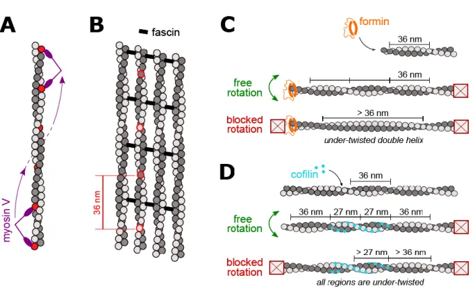
Actin filaments thus present a set of discrete binding sites for myosins, arranged in a helicoidal fashion. When stepping along the filament, a limited set of sites will be accessible to the myosin for each step, depending on the myosin’s lever arm and step size. This has important consequences for myosin movement. For example, myosin V, which has a step size slightly shorter than the filament’s half-pitch, alternates steps on each of the two actin filament strands and moves along a left-handed spiral [57] (see figure 2A).
When filaments are organized into a parallel bundle, myosins cannot rotate around the filaments. This additional geometrical constraint makes it difficult for most myosin motors to walk along filament bundles, except for myosin X which seems optimized for this task. First, it is able to take 36 nm steps and walk straight along a single filament [58,59](Figure 2B). Second, myosin X’s ability to take steps of different sizes (up to 52 nm) allows it to step onto neighboring filaments in the bundle, and thereby increase its velocity and run-length, compared to single filaments [59]. Side-stepping has been confirmed in cells, for myosin X walking in filopodia [60].
When myosins are not transporting cargo, they can be anchored and exert pulling forces on actin filaments. Due to the helical nature of the filaments, myosins can also easily exert a torque on the filaments [61]. In a gliding assay, this torque can cause filaments to rotate around their main axis [62] or to supercoil [63,64] (Note that one should be cautious, however, when conducting such
experiments since a change of helicity can be artificially photo-induced on actin filaments stabilized with labeled phalloidin [65]). Applying a torque to actin filaments may also modify their helical pitch, and alter their interactions with other ABPs. The myosin II motor domain has been reported to have an increased affinity for stretched filaments in cells [66]. This observation might be related to the stretch-twist coupling of actin filaments mentioned in section 2.
3.3 Formins
Formins are involved in the generation of several actin filament networks in cells [67]. Formins are protein homodimers that can track the actin filament barbed end and promote its elongation from profilin-actin [68]. The fact that a formin remains processively bound to the growing barbed end [69– 71] raises the question of its ability to rotate in order to follow the filament helix [72]. In cells, formin-elongated filaments are typically connected to other filaments within networks, and are thus likely to be unable to rotate around their axis. Formin anchoring to membranes, either directly or via regulatory proteins, can potentially allow formin rotation, which appears to be required in order to generate actin filaments with their natural helicity.
Monitoring the polarized emission of fluorescence from a single labeled actin subunit [62], Mizuno et al [73] were able to directly observe the rotation of actin filaments growing from artificial formin mDia1 aggregates. More recently, using EM, the same authors reported that filaments growing from such formin aggregates while their pointed ends were anchored, thereby preventing their rotation,
had a slightly larger helical pitch [74]. In principle, a non-rotating formin elongating an anchored filament could generate a large amount of under-twisting, as the newly added subunits are added without helicity (see Figure 2C). The very mild under-twisting observed by Mizuno et al. could indicate that filaments were very briefly elongated by formins before stalling, or perhaps that formins elongated these filaments by alternating between torque-generating and torque-relaxing steps, as proposed theoretically [72]. The details of formin anchoring, and their ability to rotate, certainly play an important role.
When applying tension to filaments elongated by formin mDia1, Yu et al. observed very different elongation rates depending on how they anchored formins [75,76]. The elongating filaments were not free to rotate, and these results could be consistently interpreted in terms of formin rotational freedom, with the seemingly more permissive anchoring leading to the faster elongation rate. Recently, we have monitored the impact of filament bundling by fascin, which effectively blocks filament rotation, on the activity of formin mDia1 at their barbed ends [44]. We found that, when filaments were bundled, the amount of rotational and translational freedom of the anchored formins mattered greatly: while the activity of freely rotating formins anchored on lipid bilayers was not affected by filament bundling, rotation-constrained formins anchored on glass elongated the
bundled filaments more slowly, and for a shorter amount of time before detaching from their barbed ends. The efficient cross-talk between geometrical constraints on filaments (bundling) and on
formins (anchoring), several micrometers apart, indicates that actin filaments can transmit torsional stress over long distances.
Notably, Zimmermann et al observed that filament elongation by yeast fomin cdc12 was not affected when filament rotation was blocked, in the absence of a pulling force [77]. This result suggests that cdc12 was able to rotate around its anchoring point and applied no torque to the filament, or that cdc12 is better than mDia1 at coping with torsional stress.
The filament elongation rate as well as the formin detachment rate we measured for mDia1 did not appear to vary over time, nor to depend on the formin-to-bundle distance, suggesting that the torque did not increase continuously over the course of elongation. This may indicate that, beyond a certain amount of torsional stress, the formin was able to rotate around its anchoring point, thereby imposing a limit on the torque. It may also indicate, like the EM observations of Mizuno et al. [74], that the processive elongation by formins includes steps where torsional stress is relaxed, as proposed theoretically [72].
Together, these recent papers indicate that, due to filament helicity, anchored formins can apply a torque when elongating rotation-hindered actin filaments. Such situations are likely to be
encountered in cells, where formins are anchored to surfaces and filaments are interconnected. The applied torque has a strong impact on the formin’s ability to efficiently elongate the filaments (at least for formin mDia1). A consequence of this torque is that the elongating filament is under-twisted. At this stage the amplitude of this under-twisting is unclear, and should be addressed by future studies.
3.4 Cofilin
Proteins of the actin depolymerizing factor (ADF)/cofilin family are the central players of actin
filament disassembly. Cofilin disassembles filaments by binding to the sides of ADP-actin filaments, in a cooperative manner, leading to the formation of cofilin domains (Figure 2D). These segments of cofilin-decorated actin (referred to as “cofilactin”) adopt a specific conformation [12,78,79], where
each subunit is tilted by an additional ~9.5° with respect to its longitudinal neighbors. The length of the filament is not significantly affected but the helical pitch of cofilactin is shorter, with a half-pitch of approximately 27 nm (thus the cofilactin double helix makes 18.5 full turns per micrometer, compared to 13.9 turns per micrometer for the bare actin filament. See figure 2D). In comparison, the reported change in helicity induced by fascin or espin bundling is much smaller (see section 3.1). Compared to bare actin, cofilactin filaments have lower torsional and flexural rigidities [41,80]. Whether the cofilin-induced change in helicity propagates beyond the boundaries of the cofilin domain is debated. Atomic Force Microscopy measurements were reported to show that the helical pitch is shortened by cofilin up to 13 subunits away from the cofilin domain towards the pointed end of the filament [81] while recent EM measurements indicate that the conformation changes sharply at the domain boundary [82].
Severing is enhanced at, or near, the boundaries of cofilin domains [83,84]. Cofilin-saturated
filaments do not sever. Their pointed ends depolymerize faster than that of bare actin filaments, and their barbed ends hardly stop depolymerizing even in the presence of fresh actin monomers and capping protein [30,85].
The action of cofilin thus appears to be intimately related to helicity. A consequence of the cofilin-induced change of helicity, is that cofilin can apply a torque as it decorates actin filaments that are not able to rotate around their axis. This is the case, for example, for filaments that are bound to a surface or to other filaments, as many filaments in cells certainly are. Manipulating single actin filaments in vitro, we have recently shown that this cofilin-induced torque did not affect cofilin binding, but that it greatly increased the rate of severing at domain boundaries [30]. As a
consequence, cross-linked filaments are severed by cofilin far more efficiently than free filaments [30,86,87].
Consistent with the notion that twisting and bending are coupled [39] we found that imposing a strong local curvature also enhanced severing by cofilin [30]. However, we found that tension (up to 30 pN) had almost no effect on cofilin binding nor on cofilin-induced severing, in contradiction with an earlier study [88]. It thus appears that this range of tension does not significantly affect filament helicity.
When the torque is applied by cofilin itself as it decorates an anchored filament, like in our experiment [30], the filament will be globally under-twisted by up to approximately 4.6 turns per micrometer (Figure 2D) (Both bare and cofilin-decorated regions are under-twisted, compared to their natural helicity. The maximum undertwist of 4.6 turns per micrometer corresponds to a fully decorated filament). Remarkably, a recent meso-scale model by Schramm et al [45] proposes that the application of a stronger torque could reduce the affinity of cofilin for the filament. For example, the cofilin off-rate is predicted to increase significantly when filaments are under-twisted by more than 6 turns per micrometer, and the effect is stronger when filaments are over-twisted.
As we have discussed in section 3.3, Mizuno et al [74] have used anchored formins and filaments to mildly under-twist filaments. They report that this mild under-twisting is enough to protect the filaments from the action of cofilin. This results appears to be in contradiction with our study on torque-induced cofilin severing [30] and with the Schramm model [45]. More experiments are required to solve this apparent discrepancy.
Taken together, these results show that under-twisting or over-twisting actin filaments can modulate the action of ABPs in non-trivial ways. Importantly, torsional stress can be applied by the same ABPs or by other ABPs, and depends on the mechanical context of the filaments.
4. From actin filament helicity to cellular and organismal chiralities
Over the past years, filament helicity and helicity-sensitive ABPs have been linked to the generation of chirality and asymmetry at larger scales, which are fundamental aspects of life. The establishment of right asymmetry, in particular, is an important and challenging question. In development, left-right asymmetry appears after anterior/posterior and dorsal/ventral asymmetries, which are
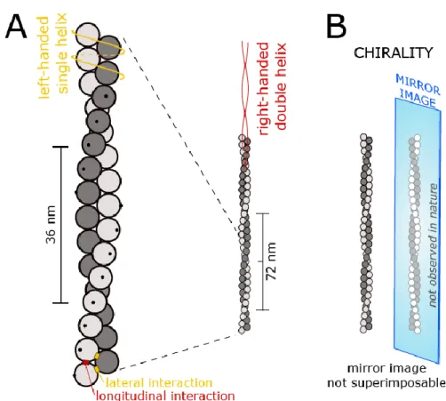
determined at the time of fertilization. Proteins and most biological molecules are chiral (and actin filaments themselves are chiral, see Figure 1B), but understanding how cell-scale chirality can emerge from chiral molecular components is not straightforward [89]. Today it appears that cytoskeletal dynamics and mechanics are key elements of this process [90]. In the following paragraphs, we summarize some recent observations that attribute a role to actin helicity and ABPs in the establishment of cell- and tissue-scale chirality.
Myosins are a clear link between actin helicity and larger-scale chirality. Recent spectacular work by Lebreton et al [91] on Drosophila development shows that the ectopic expression of myosin-1d induces the directional polarization of cells, a pronounced dextral twisting of the trachea, and a dextral twisting of the whole larval body. In contrast, myosin-1c has an antagonistic effect (sinistral twisting). Strikingly, the difference between the two motors is already apparent at the single filament level in in vitro gliding assay, as myosin-1d propels filaments in a clear counter-clockwise circular movement, and not myosin-1c. The mechanistic details linking these different scales are not clearly known.
Another example can be found in the cortex of C. elegans embryos, where actomyosin contraction generates torques and drives chiral cortical flows in one-cell embryos, which later allow the establishment of left-right asymmetry [92,93].
In the context of left-right brain asymmetry, the spiral stepping of Myosin V on actin filaments (figure 2A) has been proposed to drive the left helical movement of growth cone filopodia, and to explain the clockwise growth of neurites in two dimensions [94]. This model is corroborated by the
observation of rotational retrograde flow of the actin shaft inside filopodia [95]. Although there is no direct evidence of their implication in the rotational movement of filopodia, formins and bundling proteins are good candidates to generate the observed twisted super-helical actin bundles, resulting in the chiral movements of filopodia.
Formins have been linked to the establishment of large-scale chirality, in various contexts. For example, Davison et al. [96] have identified diaphanous-related formins as essential to induce morphological asymmetry during the early stages of embryo development in snails and frogs. Another fascinating example is the appearance of rotating radial fibers in fibroblasts plated
isotropically on 2D adhesive disk patterns [97]. Rotation is asymmetrical and its direction is reversed by overexpression of alpha-actinin (interestingly, alpha-actinin has also been linked to the actin-dependent, asymmetrical rotation of epithelial cells confined between two matrigel layers [98]). With keratinocytes, the same chiral swirling could not develop when cells lacked the formin DIAPH1, but was facilitated in cells lacking VASP, possibly through an increased activity of DIAPH1 since both proteins are sometimes described as antagonists [99]. The mechanistic interpretation is that the elongation of the filaments in the radial fibers, driven by anchored formins, causes their rotation around their axes, and drives the swirling of the fiber network. These assays have identified myosin II in transverse fibers and formins at focal adhesion sites as the active molecular players required for the emergence of chiral swirling in single cells [97,99]. However, the details linking formin-induced
filament rotation to the generation of a rotational movement of the fibers, which are complex filament bundles, are still missing.
Globally, the mechanistic, molecular details of the symmetry-breaking mechanisms we have summarized here are not fully understood. Nonetheless, these few examples illustrate how actin filaments, being helical and thus chiral, may be the necessary intermediate to bridge chirality across scales.
5. Conclusion and future directions
As we have discussed in section 3, actin filament helicity is sensed and modified by various types of ABPs. Helicity thus provides an essential handle for ABPs to modulate each other’s regulatory
actions. Interestingly, ABPs’ ability to affect the filament’s helicity depends strongly on the filaments’ geometrical organization. For example, details of filament and formin anchoring have an impact on the generation of torque, and on the action of regulatory proteins such as cofilin. This provides a clear means to regulate protein activity as a function of the local mechanical context. As such, torsional stress and the modulation of helicity efficiently contribute to filament-level
mechanotransduction, for which only a few mechanisms have been identified [100,101]. While tension has been proposed as a means to alter the filament conformation [34] it appears that applying a torque is a direct way to modify the filament’s helical pitch, and to alter the action of ABPs. For these reasons, the modulation of helicity appears to be a powerful and efficient way to regulate actin assembly and organization in cells.
The modulation of helicity as a regulatory parameter is still an emerging concept, and a lot of questions remain open. As we have discussed in section 3.3, the amplitude of the torque a formin can apply to a filament, and the amplitude of the resulting under-twisting, are still unclear. Also, the different studies we have cited were mostly focused on formin mDia1, and other formins are likely to differ in their ability to apply torque and in their response to an applied torque.
Several important ABPs have not been mentioned here and their response to changes in helicity should be studied in the future. We are confident that more regulatory mechanisms involving actin helicity and torsional stress will be uncovered in the coming years. For example, side-branching by the Arp2/3 complex, which has been shown to be sensitive to the local curvature of the mother filament [102] is likely to be sensitive to changes in the helical pitch of the mother filament. Tropomyosins constitute a large family of coiled-coil proteins with as many as 40 isoforms in
mammals. Today, tropomyosins appear to play a central role in regulating the binding of many other ABPs to actin filaments [103] and to drive actin network differentiation in cells [104]. Tropomyosins bind to the sides of actin filaments by spanning over several subunits (up to half a pitch per protein for the longest tropomyosin isoforms) and closely follow the filament strands. It thus appears that tropomyosins should be sensitive to small changes in helicity, even though structural data [105] indicate that tropomyosin binding does not modify the filament’s helical pitch. We therefore see tropomyosins as promising candidates for helicity-sensing, and anticipate that this property may be key to understanding their regulatory function.
As we have discussed in section 4, the exploitation of the helical nature of actin filaments by ABPs now appears essential to establish asymmetries at larger scales. We can view helicity as filament-scale chirality, and as a necessary link between molecular-filament-scale chirality and cell-to-tissue-filament-scale chirality. However, the mechanistic details of how filament helicity translates into larger-scale
asymmetries are still mysterious. The fact that the action of several ABPs is coupled to filament helicity and torque suggests that complex, integrated molecular mechanisms are at play.
For instance, in their study of organismal chirality in Drosophila, Lebreton et al [91] report that the body-twisting phenotypes induced by the ectopic expression of the myosins were reduced or suppressed when profilin expression was reduced. This observation suggests that formins, being torque-sensitive and a major interactor of profilin, may be involved in the process.
Finally, microtubules may also participate in the transmission of chirality across scales, even though their contribution to symmetry breaking is usually described without invoking their intrinsic chirality [106]. For example, dynein motors can have helical trajectories along microtubule and generate a torque [107]. Several recent studies show that actin filaments and microtubules are often
co-regulated [108,109]. A thorough understanding of how actin filament helicity organizes asymmetry in cells will certainly require inclusion of microtubules in the picture.
Conflicts of interest
The authors declare no conflict of interest.
Acknowledgements
We acknowledge funding from the French ANR (Grants ‘MuscActin’ and ‘Conformin’ to G.R.-L.) and the European Research Council (Grant StG-679116 to A.J.).
FIGURES
Figure 1. Schematic representation of an actin filament.
A. Actin monomers are sketched as spheres, the two strands are represented in different shades of gray, for clarity. The black dots are here to indicate the orientation of the monomers, which are rotated by +27° with respect to their longitudinal neighbors. The pitch of the double helix is 72 nm, corresponding to 13.9 turns per micrometer. It is straightforward to see the actin filament as this right-handed, two-start, long-pitch helix (indicated by the red lines), but it can also be described as a left-handed, one-start, short-pitch helix (indicated by the orange line).
Figure 2. Filament helicity and ABPs.
A. Myosin V takes steps that are shorter than the half-pitch of the actin double helix (the subunits marked in red indicate myosin V’s stepping positions). As a consequence, myosin V follows a left-handed helix as it moves along the filament.
B. Actin filaments bundled by fascin adopt a regular geometrical packing. Motors (like myosin X) must take 36 nm steps in order to walk along the bundle in a straight line (stepping on the subunits that are circled in red). The change in helical pitch induced by fascin bundling is not visible on this sketch. Note that bundles are typically in 3 dimensions, adding constraints for motors to walk on filaments having more than 2 neighbors.
C. A formin tracking the elongating barbed end of an anchored actin filament must be free to rotate in order to follow the helical pitch. If the formin cannot rotate, the filament cannot incorporate new turns and becomes under-twisted (potentially missing up to a maximum of 13.9 turns/µm, if the filament is fully assembled straight). If formins cannot rotate but are able to relax torsional stress, filaments may be mildly undertwisted (a situation intermediate to the two extremes that are sketched here).
D. Cofilin forms domains as it binds cooperatively to the sides of an actin filament. The pitch of cofilactin (cofilin-decorated actin) filaments is shorter than that of actin filaments. If the filament is free to rotate as it gets decorated by cofilin, bare filament regions and cofilin-decorated regions are both able to adopt their natural helicity. If the filament cannot rotate, no new turns can be added as cofilin binds, and both regions will be under-twisted.
REFERENCES
[1] B. Bugyi, M. Kellermayer, The discovery of actin: “to see what everyone else has seen, and to think what nobody has thought,” J. Muscle Res. Cell Motil. (2019). doi:10.1007/s10974-019-09515-z.
[2] C.C. Selby, R.S. Bear, The structure of actin-rich filaments of muscles according to x-ray diffraction, J. Biophys. Biochem. Cytol. 2 (1956) 71–85. doi:10.1083/jcb.2.1.71.
[3] W.T. Astbury, S.V. Perry, R. Reed, L.C. Spark, An electron microscope and X-ray study of actin: I. Electron microscope, Biochim. Biophys. Acta. 1 (1947) 379–392.
doi:10.1016/0006-3002(47)90149-2.
[4] J. Hanson, Axial Period of Actin Filaments, Nature. 213 (1967) 353–356. doi:10.1038/213353a0. [5] J. Hanson, J. Lowy, The structure of F-actin and of actin filaments isolated from muscle, J. Mol.
Biol. 6 (1963) 46–IN5. doi:10.1016/S0022-2836(63)80081-9.
[6] H.E. Huxley, ELECTRON MICROSCOPE STUDIES ON THE STRUCTURE OF NATURAL AND SYNTHETIC PROTEIN FILAMENTS FROM STRIATED MUSCLE, J. Mol. Biol. 7 (1963) 281–308. doi:10.1016/S0022-2836(63)80008-X.
[7] R.H. Depue Jr, R.V. Rice, F-ACTIN IS A RIGHT-HANDED HELIX, J. Mol. Biol. 12 (1965) 302–303. doi:10.1016/S0022-2836(65)80306-0.
[8] E.H. Egelman, The structure of F-actin, J. Muscle Res. Cell Motil. 6 (1985) 129–151. doi:10.1007/BF00713056.
[9] K.C. Holmes, D. Popp, W. Gebhard, W. Kabsch, Atomic model of the actin filament, Nature. 347 (1990) 44–49. doi:10.1038/347044a0.
[10] T. Oda, M. Iwasa, T. Aihara, Y. Maéda, A. Narita, The nature of the globular- to fibrous-actin transition, Nature. 457 (2009) 441–445. doi:10.1038/nature07685.
[11] T. Fujii, A.H. Iwane, T. Yanagida, K. Namba, Direct visualization of secondary structures of F-actin by electron cryomicroscopy, Nature. 467 (2010) 724–728. doi:10.1038/nature09372.
[12] K. Tanaka, S. Takeda, K. Mitsuoka, T. Oda, C. Kimura-Sakiyama, Y. Maéda, A. Narita, Structural basis for cofilin binding and actin filament disassembly, Nat. Commun. 9 (2018) 1860.
doi:10.1038/s41467-018-04290-w.
[13] S.Z. Chou, T.D. Pollard, Mechanism of actin polymerization revealed by cryo-EM structures of actin filaments with three different bound nucleotides, Proc. Natl. Acad. Sci. U. S. A. (2019). doi:10.1073/pnas.1807028115.
[14] M.F. Carlier, D. Pantaloni, E.D. Korn, Evidence for an ATP cap at the ends of actin filaments and its regulation of the F-actin steady state, J. Biol. Chem. 259 (1984) 9983–9986.
https://www.ncbi.nlm.nih.gov/pubmed/6236218.
[15] T.D. Pollard, Rate constants for the reactions of ATP- and ADP-actin with the ends of actin filaments, J. Cell Biol. 103 (1986) 2747–2754. http://www.ncbi.nlm.nih.gov/pubmed/3793756. [16] H.P. Erickson, Co-operativity in protein-protein association. The structure and stability of the
actin filament, J. Mol. Biol. 206 (1989) 465–474. doi:10.1016/0022-2836(89)90494-4.
[17] D. Sept, J.A. McCammon, Thermodynamics and kinetics of actin filament nucleation, Biophys. J. 81 (2001) 667–674. doi:10.1016/S0006-3495(01)75731-1.
[18] E.M. De La Cruz, J.-L. Martiel, L. Blanchoin, Mechanical heterogeneity favors fragmentation of strained actin filaments, Biophys. J. 108 (2015) 2270–2281. doi:10.1016/j.bpj.2015.03.058. [19] E.H. Egelman, N. Francis, D.J. DeRosier, F-actin is a helix with a random variable twist, Nature.
298 (1982) 131–135. doi:10.1038/298131a0.
[20] E.H. Egelman, D.J. DeRosier, Image analysis shows that variations in actin crossover spacings are random, not compensatory, Biophys. J. 63 (1992) 1299–1305.
doi:10.1016/S0006-3495(92)81716-2.
[21] V.E. Galkin, A. Orlova, G.F. Schröder, E.H. Egelman, Structural polymorphism in F-actin, Nat. Struct. Mol. Biol. 17 (2010) 1318–1323. doi:10.1038/nsmb.1930.
[22] Y. Inoue, T. Adachi, Mechanosensitive kinetic preference of actin-binding protein to actin filament, Phys. Rev. E. 93 (2016) 042403. doi:10.1103/PhysRevE.93.042403.
[23] V.E. Galkin, A. Orlova, M.R. Vos, G.F. Schröder, E.H. Egelman, Near-atomic resolution for one state of F-actin, Structure. 23 (2015) 173–182. doi:10.1016/j.str.2014.11.006.
[24] S. Matsushita, Y. Inoue, M. Hojo, M. Sokabe, T. Adachi, Effect of tensile force on the mechanical behavior of actin filaments, J. Biomech. 44 (2011) 1776–1781.
doi:10.1016/j.jbiomech.2011.04.012.
[25] I. Fujiwara, D. Vavylonis, T.D. Pollard, Polymerization kinetics of ADP- and ADP-Pi-actin determined by fluorescence microscopy, Proc. Natl. Acad. Sci. U. S. A. 104 (2007) 8827–8832. doi:10.1073/pnas.0702510104.
[26] A. Jégou, T. Niedermayer, J. Orbán, D. Didry, R. Lipowsky, M.-F. Carlier, G. Romet-Lemonne, Individual actin filaments in a microfluidic flow reveal the mechanism of ATP hydrolysis and give insight into the properties of profilin, PLoS Biol. 9 (2011) e1001161.
doi:10.1371/journal.pbio.1001161.
[27] H. Isambert, P. Venier, A.C. Maggs, A. Fattoum, R. Kassab, D. Pantaloni, M.F. Carlier, Flexibility of actin filaments derived from thermal fluctuations. Effect of bound nucleotide, phalloidin, and muscle regulatory proteins, J. Biol. Chem. 270 (1995) 11437–11444.
http://www.ncbi.nlm.nih.gov/pubmed/7744781.
[28] F. Merino, S. Pospich, J. Funk, T. Wagner, F. Küllmer, H.-D. Arndt, P. Bieling, S. Raunser,
Structural transitions of F-actin upon ATP hydrolysis at near-atomic resolution revealed by cryo-EM, Nat. Struct. Mol. Biol. 25 (2018) 528–537. doi:10.1038/s41594-018-0074-0.
[29] A.H. Crevenna, N. Naredi-Rainer, A. Schönichen, J. Dzubiella, D.L. Barber, D.C. Lamb, R. Wedlich-Soldner, Electrostatics Control Actin Filament Nucleation and Elongation Kinetics, J. Biol. Chem. 288 (2013) 12102–12113. doi:10.1074/jbc.M113.456327.
[30] H. Wioland, A. Jegou, G. Romet-Lemonne, Torsional stress generated by ADF/cofilin on cross-linked actin filaments boosts their severing, Proc. Natl. Acad. Sci. U. S. A. 116 (2019) 2595–2602. doi:10.1073/pnas.1812053116.
[31] H. Kang, M.J. Bradley, B.R. McCullough, A. Pierre, E.E. Grintsevich, E. Reisler, E.M. De La Cruz, Identification of cation-binding sites on actin that drive polymerization and modulate bending stiffness, Proc. Natl. Acad. Sci. U. S. A. 109 (2012) 16923–16927. doi:10.1073/pnas.1211078109. [32] C.T. Zimmerle, C. Frieden, Effect of pH on the mechanism of actin polymerization, Biochemistry.
27 (1988) 7766–7772. doi:10.1021/bi00420a027.
[33] H. Kang, M.J. Bradley, W.A. Elam, E.M. De La Cruz, Regulation of Actin by Ion-Linked Equilibria, Biophys. J. 105 (2013) 2621–2628. doi:10.1016/j.bpj.2013.10.032.
[34] V.E. Galkin, A. Orlova, E.H. Egelman, Actin Filaments as Tension Sensors, Curr. Biol. 22 (2012) R96–R101. doi:10.1016/j.cub.2011.12.010.
[35] Y. Tsuda, H. Yasutake, A. Ishijima, T. Yanagida, Torsional rigidity of single actin filaments and actin–actin bond breaking force under torsion measured directly by in vitro micromanipulation, Proceedings of the National Academy of Sciences. 93 (1996) 12937–12942.
http://www.pnas.org/content/93/23/12937.abstract.
[36] Y. Arai, R. Yasuda, K. Akashi, Y. Harada, H. Miyata, K. Kinosita Jr, H. Itoh, Tying a molecular knot with optical tweezers, Nature. 399 (1999) 446–448. doi:10.1038/20894.
[37] H. Yamaoka, T. Adachi, Coupling between axial stretch and bending/twisting deformation of actin filaments caused by a mismatched centroid from the center axis, Int. J. Mech. Sci. 52 (2010) 329–333.
https://sci-hub.tw/https://www.sciencedirect.com/science/article/pii/S0020740309001982.
[38] O.N. Yogurtcu, J.S. Kim, S.X. Sun, A mechanochemical model of actin filaments, Biophys. J. 103 (2012) 719–727. doi:10.1016/j.bpj.2012.07.020.
[39] E.M. De La Cruz, J. Roland, B.R. McCullough, L. Blanchoin, J.-L. Martiel, Origin of twist-bend coupling in actin filaments, Biophys. J. 99 (2010) 1852–1860. doi:10.1016/j.bpj.2010.07.009. [40] E.M. De La Cruz, M.L. Gardel, Actin Mechanics and Fragmentation, J. Biol. Chem. 290 (2015)
[41] E. Prochniewicz, N. Janson, D.D. Thomas, E.M. De la Cruz, Cofilin increases the torsional flexibility and dynamics of actin filaments, J. Mol. Biol. 353 (2005) 990–1000.
doi:10.1016/j.jmb.2005.09.021.
[42] J.N. Forkey, M.E. Quinlan, Y.E. Goldman, Measurement of single macromolecule orientation by total internal reflection fluorescence polarization microscopy, Biophys. J. 89 (2005) 1261–1271. doi:10.1529/biophysj.104.053470.
[43] R. Yasuda, H. Miyata, K. Kinosita Jr, Direct measurement of the torsional rigidity of single actin filaments, J. Mol. Biol. 263 (1996) 227–236. https://www.ncbi.nlm.nih.gov/pubmed/8913303. [44] E.L. Suzuki, B. Guichard, G. Romet-Lemonne, A. Jégou, Geometrical constraints greatly hinder
formin mDia1 activity, bioRxiv. (2019) 671842. doi:10.1101/671842.
[45] A.C. Schramm, G.M. Hocky, G.A. Voth, L. Blanchoin, J.-L. Martiel, E.M. De La Cruz, Actin Filament Strain Promotes Severing and Cofilin Dissociation, Biophys. J. 112 (2017) 2624–2633.
doi:10.1016/j.bpj.2017.05.016.
[46] J.H. Shin, M.L. Gardel, L. Mahadevan, P. Matsudaira, D.A. Weitz, Relating microstructure to rheology of a bundled and cross-linked F-actin network in vitro, Proc. Natl. Acad. Sci. U. S. A. 101 (2004) 9636–9641. doi:10.1073/pnas.0308733101.
[47] R. Ishikawa, T. Sakamoto, T. Ando, S. Higashi-Fujime, K. Kohama, Polarized actin bundles formed by human fascin-1: their sliding and disassembly on myosin II and myosin V in vitro, J.
Neurochem. 87 (2003) 676–685. https://www.ncbi.nlm.nih.gov/pubmed/14535950. [48] R.A. Edwards, H. Herrera-Sosa, J. Otto, J. Bryan, Cloning and expression of a murine fascin
homolog from mouse brain, J. Biol. Chem. 270 (1995) 10764–10770. doi:10.1074/jbc.270.18.10764.
[49] S. Jansen, A. Collins, C. Yang, G. Rebowski, T. Svitkina, R. Dominguez, Mechanism of actin filament bundling by fascin, J. Biol. Chem. 286 (2011) 30087–30096.
doi:10.1074/jbc.M111.251439.
[50] M.M.A.E. Claessens, C. Semmrich, L. Ramos, A.R. Bausch, Helical twist controls the thickness of F-actin bundles, Proceedings of the National Academy of Sciences. 105 (2008) 8819–8822. doi:10.1073/pnas.0711149105.
[51] H. Shin, K.R. Purdy Drew, J.R. Bartles, G.C.L. Wong, G.M. Grason, Cooperativity and frustration in protein-mediated parallel actin bundles, Phys. Rev. Lett. 103 (2009) 238102.
doi:10.1103/PhysRevLett.103.238102.
[52] S. Aramaki, K. Mayanagi, M. Jin, K. Aoyama, T. Yasunaga, Filopodia Formation by Cross-linking of F-actin with Fascin in Two Different Binding Manners, Cytoskeleton . (2016).
doi:10.1002/cm.21309.
[53] C. Heussinger, G.M. Grason, Theory of crosslinked bundles of helical filaments: intrinsic torques in self-limiting biopolymer assemblies, J. Chem. Phys. 135 (2011) 035104.
doi:10.1063/1.3610431.
[54] M.F. Schmid, M.B. Sherman, P. Matsudaira, W. Chiu, Structure of the acrosomal bundle, Nature. 431 (2004) 104–107. doi:10.1038/nature02881.
[55] R. Ma, J. Berro, Structural organization and energy storage in crosslinked actin assemblies, PLoS Comput. Biol. 14 (2018) e1006150. doi:10.1371/journal.pcbi.1006150.
[56] M.A. Hartman, J.A. Spudich, The myosin superfamily at a glance, J. Cell Sci. 125 (2012) 1627– 1632. doi:10.1242/jcs.094300.
[57] M.Y. Ali, S. Uemura, K. Adachi, H. Itoh, K. Kinosita Jr, S. ’ichi Ishiwata, Myosin V is a left-handed spiral motor on the right-handed actin helix, Nat. Struct. Biol. 9 (2002) 464–467.
doi:10.1038/nsb803.
[58] Y. Sun, O. Sato, F. Ruhnow, M.E. Arsenault, M. Ikebe, Y.E. Goldman, Single-molecule stepping and structural dynamics of myosin X, Nat. Struct. Mol. Biol. 17 (2010) 485–491.
doi:10.1038/nsmb.1785.
[59] V. Ropars, Z. Yang, T. Isabet, F. Blanc, K. Zhou, T. Lin, X. Liu, P. Hissier, F. Samazan, B. Amigues, E.D. Yang, H. Park, O. Pylypenko, M. Cecchini, C.V. Sindelar, H.L. Sweeney, A. Houdusse, The myosin X motor is optimized for movement on actin bundles, Nat. Commun. 7 (2016) 12456.
doi:10.1038/ncomms12456.
[60] O. Sato, H.S. Jung, S. Komatsu, Y. Tsukasaki, T.M. Watanabe, K. Homma, M. Ikebe, Activated full-length myosin-X moves processively on filopodia with large steps toward diverse
two-dimensional directions, Sci. Rep. 7 (2017) 44237. doi:10.1038/srep44237.
[61] A. Vilfan, Twirling motion of actin filaments in gliding assays with nonprocessive Myosin motors, Biophys. J. 97 (2009) 1130–1137. doi:10.1016/j.bpj.2009.06.008.
[62] I. Sase, H. Miyata, S. Ishiwata, K. Kinosita Jr, Axial rotation of sliding actin filaments revealed by single-fluorophore imaging, Proc. Natl. Acad. Sci. U. S. A. 94 (1997) 5646–5650.
https://www.ncbi.nlm.nih.gov/pubmed/9159126.
[63] Y. Tanaka, A. Ishijima, S. Ishiwata, Super helix formation of actin filaments in an in vitro motile system, Biochim. Biophys. Acta. 1159 (1992) 94–98. doi:10.1016/0167-4838(92)90079-s. [64] T. Nishizaka, T. Yagi, Y. Tanaka, S. Ishiwata, Right-handed rotation of an actin filament in an in
vitro motile system, Nature. 361 (1993) 269–271. doi:10.1038/361269a0.
[65] T. Sanchez, I.M. Kulic, Z. Dogic, Circularization, photomechanical switching, and a supercoiling transition of actin filaments, Phys. Rev. Lett. 104 (2010) 098103.
doi:10.1103/PhysRevLett.104.098103.
[66] T.Q.P. Uyeda, Y. Iwadate, N. Umeki, A. Nagasaki, S. Yumura, Stretching Actin Filaments within Cells Enhances their Affinity for the Myosin II Motor Domain, PLoS One. 6 (2011) e26200. doi:10.1371/journal.pone.0026200.g005.
[67] K. Rottner, J. Faix, S. Bogdan, S. Linder, E. Kerkhoff, Actin assembly mechanisms at a glance, J. Cell Sci. 130 (2017) 3427–3435. doi:10.1242/jcs.206433.
[68] J.B. Moseley, I. Sagot, A.L. Manning, Y. Xu, M.J. Eck, D. Pellman, B.L. Goode, A conserved mechanism for Bni1- and mDia1-induced actin assembly and dual regulation of Bni1 by Bud6 and profilin, Mol. Biol. Cell. 15 (2004) 896–907. doi:10.1091/mbc.E03-08-0621.
[69] C. Higashida, T. Miyoshi, A. Fujita, F. Oceguera-Yanez, J. Monypenny, Y. Andou, S. Narumiya, N. Watanabe, Actin polymerization-driven molecular movement of mDia1 in living cells, Science. 303 (2004) 2007–2010. doi:10.1126/science.1093923.
[70] D.R. Kovar, T.D. Pollard, Insertional assembly of actin filament barbed ends in association with formins produces piconewton forces, Proc. Natl. Acad. Sci. U. S. A. 101 (2004) 14725–14730. doi:10.1073/pnas.0405902101.
[71] S. Romero, C. Le Clainche, D. Didry, C. Egile, D. Pantaloni, M.-F. Carlier, Formin is a processive motor that requires profilin to accelerate actin assembly and associated ATP hydrolysis, Cell. 119 (2004) 419–429. doi:10.1016/j.cell.2004.09.039.
[72] T. Shemesh, T. Otomo, M.K. Rosen, A.D. Bershadsky, M.M. Kozlov, A novel mechanism of actin filament processive capping by formin: solution of the rotation paradox, J. Cell Biol. 170 (2005) 889–893. doi:10.1083/jcb.200504156.
[73] H. Mizuno, C. Higashida, Y. Yuan, T. Ishizaki, S. Narumiya, N. Watanabe, Rotational movement of the formin mDia1 along the double helical strand of an actin filament, Science. 331 (2011) 80– 83. doi:10.1126/science.1197692.
[74] H. Mizuno, K. Tanaka, S. Yamashiro, A. Narita, N. Watanabe, Helical rotation of the diaphanous-related formin mDia1 generates actin filaments resistant to cofilin, Proc. Natl. Acad. Sci. U. S. A. (2018). doi:10.1073/pnas.1803415115.
[75] M. Yu, X. Yuan, C. Lu, S. Le, R. Kawamura, A.K. Efremov, Z. Zhao, M.M. Kozlov, M. Sheetz, A. Bershadsky, J. Yan, mDia1 senses both force and torque during F-actin filament polymerization, Nat. Commun. 8 (2017) 1650. doi:10.1038/s41467-017-01745-4.
[76] M. Yu, S. Le, A.K. Efremov, X. Zeng, A. Bershadsky, J. Yan, Effects of Mechanical Stimuli on Profilin- and Formin-Mediated Actin Polymerization, Nano Lett. (2018).
doi:10.1021/acs.nanolett.8b02211.
[77] D. Zimmermann, K.E. Homa, G.M. Hocky, L.W. Pollard, E.M. De La Cruz, G.A. Voth, K.M. Trybus, D.R. Kovar, Mechanoregulated inhibition of formin facilitates contractile actomyosin ring assembly, Nat. Commun. 8 (2017) 703. doi:10.1038/s41467-017-00445-3.
actin filament dynamics and cellular function, J. Cell Biol. 138 (1997) 771–781. http://www.ncbi.nlm.nih.gov/pubmed/9265645.
[79] V.E. Galkin, A. Orlova, D.S. Kudryashov, A. Solodukhin, E. Reisler, G.F. Schröder, E.H. Egelman, Remodeling of actin filaments by ADF/cofilin proteins, Proceedings of the National Academy of Sciences. (2011). doi:10.1073/pnas.1110109108.
[80] B.R. McCullough, L. Blanchoin, J.-L. Martiel, E.M. De la Cruz, Cofilin increases the bending flexibility of actin filaments: implications for severing and cell mechanics, J. Mol. Biol. 381 (2008) 550–558. doi:10.1016/j.jmb.2008.05.055.
[81] K.X. Ngo, N. Kodera, E. Katayama, T. Ando, T.Q.P. Uyeda, Cofilin-induced unidirectional cooperative conformational changes in actin filaments revealed by high-speed atomic force microscopy, Elife. 4 (2015). doi:10.7554/eLife.04806.
[82] A. Huehn, W. Cao, W.A. Elam, X. Liu, E.M. De La Cruz, C.V. Sindelar, The actin filament twist changes abruptly at boundaries between bare and cofilin-decorated segments, J. Biol. Chem. (2018). doi:10.1074/jbc.AC118.001843.
[83] B.R. Mccullough, E.E. Grintsevich, C.K. Chen, H. Kang, A.L. Hutchison, A. Henn, W. Cao, C. Suarez, J.-L. Martiel, L. Blanchoin, E. Reisler, E.M. De La Cruz, Cofilin-Linked Changes in Actin Filament Flexibility Promote Severing, Biophys. J. 101 (2011) 151–159. doi:10.1016/j.bpj.2011.05.049. [84] C. Suarez, J. Roland, R. Boujemaa-Paterski, H. Kang, B.R. McCullough, A.-C. Reymann, C. Guérin,
J.-L. Martiel, E.M. De la Cruz, L. Blanchoin, Cofilin tunes the nucleotide state of actin filaments and severs at bare and decorated segment boundaries, Curr. Biol. 21 (2011) 862–868.
doi:10.1016/j.cub.2011.03.064.
[85] H. Wioland, B. Guichard, Y. Senju, S. Myram, P. Lappalainen, A. Jégou, G. Romet-Lemonne, ADF/Cofilin Accelerates Actin Dynamics by Severing Filaments and Promoting Their Depolymerization at Both Ends, Curr. Biol. 27 (2017) 1956–1967.e7.
doi:10.1016/j.cub.2017.05.048.
[86] D. Pavlov, A. Muhlrad, J. Cooper, M. Wear, E. Reisler, Actin filament severing by cofilin, J. Mol. Biol. 365 (2007) 1350–1358. doi:10.1016/j.jmb.2006.10.102.
[87] D. Breitsprecher, S.A. Koestler, I. Chizhov, M. Nemethova, J. Mueller, B.L. Goode, J.V. Small, K. Rottner, J. Faix, Cofilin cooperates with fascin to disassemble filopodial actin filaments, J. Cell Sci. 124 (2011) 3305–3318. doi:10.1242/jcs.086934.
[88] K. Hayakawa, H. Tatsumi, M. Sokabe, Actin filaments function as a tension sensor by tension-dependent binding of cofilin to the filament, J. Cell Biol. 195 (2011) 721–727.
doi:10.1083/jcb.201102039.
[89] N.A. Brown, L. Wolpert, The development of handedness in left/right asymmetry, Development. 109 (1990) 1–9. https://www.ncbi.nlm.nih.gov/pubmed/2209459.
[90] G. McDowell, S. Rajadurai, M. Levin, From cytoskeletal dynamics to organ asymmetry: a
nonlinear, regulative pathway underlies left--right patterning, Philos. Trans. R. Soc. Lond. B Biol. Sci. 371 (2016) 20150409. https://royalsocietypublishing.org/doi/abs/10.1098/rstb.2015.0409. [91] G. Lebreton, C. Géminard, F. Lapraz, S. Pyrpassopoulos, D. Cerezo, P. Spéder, E.M. Ostap, S.
Noselli, Molecular to organismal chirality is induced by the conserved myosin 1D, Science. 362 (2018) 949–952. doi:10.1126/science.aat8642.
[92] S.R. Naganathan, S. Fürthauer, M. Nishikawa, F. Jülicher, S.W. Grill, Active torque generation by the actomyosin cell cortex drives left--right symmetry breaking, Elife. 3 (2014) e04165.
doi:10.7554/eLife.04165.
[93] S.R. Naganathan, T.C. Middelkoop, S. Fürthauer, S.W. Grill, Actomyosin-driven left-right asymmetry: from molecular torques to chiral self organization, Curr. Opin. Cell Biol. 38 (2016) 24–30. doi:10.1016/j.ceb.2016.01.004.
[94] A. Tamada, S. Kawase, F. Murakami, H. Kamiguchi, Autonomous right-screw rotation of growth cone filopodia drives neurite turning, J. Cell Biol. 188 (2010) 429–441.
doi:10.1083/jcb.200906043.
[95] N. Leijnse, L.B. Oddershede, P.M. Bendix, Helical buckling of actin inside filopodia generates traction, Proc. Natl. Acad. Sci. U. S. A. 112 (2015) 136–141. doi:10.1073/pnas.1411761112.
[96] A. Davison, G.S. McDowell, J.M. Holden, H.F. Johnson, G.D. Koutsovoulos, M.M. Liu, P. Hulpiau, F. Van Roy, C.M. Wade, R. Banerjee, F. Yang, S. Chiba, J.W. Davey, D.J. Jackson, M. Levin, M.L. Blaxter, Formin Is Associated with Left-Right Asymmetry in the Pond Snail and the Frog, Curr. Biol. (2016). doi:10.1016/j.cub.2015.12.071.
[97] Y.H. Tee, T. Shemesh, V. Thiagarajan, R.F. Hariadi, K.L. Anderson, C. Page, N. Volkmann, D. Hanein, S. Sivaramakrishnan, M.M. Kozlov, A.D. Bershadsky, Cellular chirality arising from the self-organization of the actin cytoskeleton, Nat. Cell Biol. (2015). doi:10.1038/ncb3137. [98] A.S. Chin, K.E. Worley, P. Ray, G. Kaur, J. Fan, L.Q. Wan, Epithelial Cell Chirality Revealed by
Three-Dimensional Spontaneous Rotation, Proc. Natl. Acad. Sci. U. S. A. 115 (2018) 12188– 12193. doi:10.1073/pnas.1805932115.
[99] S. Jalal, S. Shi, V. Acharya, R.Y.-J. Huang, V. Viasnoff, A.D. Bershadsky, Y.H. Tee, Actin cytoskeleton self-organization in single epithelial cells and fibroblasts under isotropic confinement, J. Cell Sci. 132 (2019). doi:10.1242/jcs.220780.
[100] G. Romet-Lemonne, A. Jégou, Mechanotransduction down to individual actin filaments, Eur. J. Cell Biol. 92 (2013) 333–338. doi:10.1016/j.ejcb.2013.10.011.
[101] D. Zimmermann, D.R. Kovar, Feeling the force: formin’s role in mechanotransduction, Curr. Opin. Cell Biol. 56 (2019) 130–140. doi:10.1016/j.ceb.2018.12.008.
[102] V.I. Risca, E.B. Wang, O. Chaudhuri, J.J. Chia, P.L. Geissler, D.A. Fletcher, Actin filament curvature biases branching direction, Proceedings of the National Academy of Sciences. (2012). doi:10.1073/pnas.1114292109.
[103] J.R. Christensen, K.E. Homa, A.N. Morganthaler, R.R. Brown, C. Suarez, A.J. Harker, M.E. O’Connell, D.R. Kovar, Cooperation between tropomyosin and α-actinin inhibits fimbrin association with actin filament networks in fission yeast, Elife. 8 (2019).
doi:10.7554/eLife.47279.
[104] P.W. Gunning, E.C. Hardeman, P. Lappalainen, D.P. Mulvihill, Tropomyosin - master regulator of actin filament function in the cytoskeleton, J. Cell Sci. 128 (2015) 2965–2974.
doi:10.1242/jcs.172502.
[105] J. von der Ecken, M. Müller, W. Lehman, D.J. Manstein, P.A. Penczek, S. Raunser, Structure of the F-actin-tropomyosin complex, Nature. 519 (2015) 114–117. doi:10.1038/nature14033. [106] C. Pohl, Cytoskeletal Symmetry Breaking and Chirality: From Reconstituted Systems to
Animal Development, Symmetry . 7 (2015) 2062–2107. doi:10.3390/sym7042062.
[107] S. Can, M.A. Dewitt, A. Yildiz, Bidirectional helical motility of cytoplasmic dynein around microtubules, Elife. 3 (2014) e03205. doi:10.7554/eLife.03205.
[108] F. Farina, J. Gaillard, C. Guérin, Y. Couté, J. Sillibourne, L. Blanchoin, M. Théry, The centrosome is an actin-organizing centre, Nat. Cell Biol. (2015). doi:10.1038/ncb3285. [109] J.L. Henty-Ridilla, A. Rankova, J.A. Eskin, K. Kenny, B.L. Goode, Accelerated actin filament
polymerization from microtubule plus ends, Science. 352 (2016) 1004–1009. doi:10.1126/science.aaf1709.