HAL Id: inserm-00723845
https://www.hal.inserm.fr/inserm-00723845
Submitted on 14 Aug 2012
HAL is a multi-disciplinary open access
archive for the deposit and dissemination of
sci-entific research documents, whether they are
pub-lished or not. The documents may come from
teaching and research institutions in France or
abroad, or from public or private research centers.
L’archive ouverte pluridisciplinaire HAL, est
destinée au dépôt et à la diffusion de documents
scientifiques de niveau recherche, publiés ou non,
émanant des établissements d’enseignement et de
recherche français ou étrangers, des laboratoires
publics ou privés.
Anne Murati, Mandy Brecqueville, Raynier Devillier, Marie-Joelle
Mozziconacci, Véronique Gelsi-Boyer, Daniel Birnbaum
To cite this version:
Anne Murati, Mandy Brecqueville, Raynier Devillier, Marie-Joelle Mozziconacci, Véronique
Gelsi-Boyer, et al.. Myeloid malignancies: mutations, models and management.. BMC Cancer, BioMed
Central, 2012, 12 (1), pp.304. �10.1186/1471-2407-12-304�. �inserm-00723845�
R E V I E W
Open Access
Myeloid malignancies: mutations, models
and management
Anne Murati, Mandy Brecqueville, Raynier Devillier, Marie-Joelle Mozziconacci, Véronique Gelsi-Boyer
and Daniel Birnbaum
*Abstract
Myeloid malignant diseases comprise chronic (including myelodysplastic syndromes, myeloproliferative neoplasms
and chronic myelomonocytic leukemia) and acute (acute myeloid leukemia) stages. They are clonal diseases arising
in hematopoietic stem or progenitor cells. Mutations responsible for these diseases occur in several genes whose
encoded proteins belong principally to five classes: signaling pathways proteins (e.g. CBL, FLT3, JAK2, RAS),
transcription factors (e.g. CEBPA, ETV6, RUNX1), epigenetic regulators (e.g. ASXL1, DNMT3A, EZH2, IDH1, IDH2,
SUZ12, TET2, UTX), tumor suppressors (e.g. TP53), and components of the spliceosome (e.g. SF3B1, SRSF2).
Large-scale sequencing efforts will soon lead to the establishment of a comprehensive repertoire of these mutations,
allowing for a better definition and classification of myeloid malignancies, the identification of new prognostic
markers and therapeutic targets, and the development of novel therapies. Given the importance of epigenetic
deregulation in myeloid diseases, the use of drugs targeting epigenetic regulators appears as a most promising
therapeutic approach.
Introduction
Myeloid malignancies are clonal diseases of hematopoietic
stem or progenitor cells. They result from genetic and
epi-genetic alterations that perturb key processes such as
self-renewal, proliferation and differentiation. They comprise
chronic stages such as myeloproliferative neoplasms
(MPN), myelodysplastic syndromes (MDS) and chronic
myelomonocytic leukemia (CMML) and acute stages, i.e
acute myeloid leukemia (AML). AML can occur de novo
(~80% of the cases) or follow a chronic stage (secondary
AML). According to the karyotype, AMLs can be
subdi-vided into AML with favorable, intermediate or
unfavor-able cytogenetic risk [1]. MPNs comprise a variety of
disorders such as chronic myeloid leukemia (CML) and
non-CML MPNs such as polycythemia vera (PV),
essen-tial thrombocythemia (ET) and primary myelofibrosis
(PMF).
Molecular biology has always been important in
hematology, especially myeloid malignant diseases.
Cur-rently however, except in some specific examples such as
the BCR-ABL1 fusion in CML, and NPM1 or FLT3
mutations in de novo AML, molecular data are not
asso-ciated with optimal clinical and therapeutic exploitation
in the clinic. This may change with the flurry of new
data that are being generated. It all started with the
dis-covery of the JAK2V617F mutation in MPNs [2–5]. Like
the characterization of the BCR-ABL1 fusion kinase,
which has led to the development of an efficient targeted
therapy [6], this breakthrough showed how much
pro-gress can be made by the identification of a single
mo-lecular event regarding disease definition, understanding
and classification, prognosis assessment, clinical
moni-toring and treatment. Since then, many new mutated
genes have been identified. They affect various cell
pro-cesses such as signaling, regulation of gene transcription
and epigenetics, mRNA splicing and others. The aim of
this review is not to describe these results in detail; this
has been done in several excellent recently-published
reviews [7–16]. Without putting emphasis on a
particu-lar gene, disease or cell process, it is more to discuss
how the new data may improve our global vision of
leukemogenesis and may be used for progress in at least
three directions.
* Correspondence:[email protected]
Centre de Recherche en Cancérologie de Marseille, laboratoire d’Oncologie Moléculaire; UMR1068 Inserm, Institut Paoli-Calmettes, 27 Bd. Leï Roure, BP 30059, Marseille 13273, France
© 2012 Murati et al.; licensee BioMed Central Ltd. This is an Open Access article distributed under the terms of the Creative Commons Attribution License (http://creativecommons.org/licenses/by/2.0), which permits unrestricted use, distribution, and reproduction in any medium, provided the original work is properly cited.
Review
Understanding molecular leukemogenesis
Identification of new mutations
The genetic events involved in leukemogenesis have been
deciphered by using two approaches. First, genomic
alterations have been identified by using karyotype
ana-lysis and DNA hybridization onto oligonucleotide arrays
(SNP-arrays, array-CGH); several types of genomic
pro-files have been found: lack of detectable changes,
unipar-ental disomies (UPD), losses of chromosomes or large
chromosomal regions, trisomies, losses or gains of small
regions or genes. Second, small gene mutations have
been detected by classical Sanger sequencing [17–22] or,
more recently, by the use of new technologies such as
next generation sequencing (NGS) [23–31].
These studies, together with previous ones that had
identified JAK2, NPM1, MPL, RAS and RUNX1
muta-tions, among others, led to the discovery of several
major players in leukemogenesis: ASXL1 [21], BCORL1
[25], CBL [19], DNMT3A [24,32], EZH2 [20,22], IDH1/
IDH2
[26], TET2 [18] and UTX [33]. The mutational
fre-quencies of these genes range from a few percent to
more than 50%, or even virtually 100%, depending on
the gene, the disease and the series studied. Thus, almost
all cases of PV have a mutation of JAK2 [34,35]. Not
counting the latter, mutations in ASXL1 and TET2 are
frequently observed throughout the whole myeloid
spectrum (Figure 1), reaching 40-50% in CMML [33,36].
Mutations in DNMT3A and IDH1/2 are rare in the
chronic stages but reach 15-20% in AML and exhibit a
strong association with monocytic features [30]. Genes
encoding components of the splicing machinery that is
involved in the splicing of introns during pre-mRNA
maturation (mainly SF3B1, SRSF2, U2AF35/U2AF1, and
ZRSR2) have been found frequently mutated in MDSs
and CMML, and more rarely in MPNs and AML
(Fig-ure 1) [31,37–42]. Mutations in splicing factors are
found in more than 60% of MDS with ring sideroblasts
and in more than 50% of CMML [31].
Mutations in leukemogenic genes have been described
in detail in recent reviews [7,9,10,12–16,43]; and will not
be reviewed here. We will rather delve on the questions
aroused by these recent data.
Have we already identified the entire repertoire of mutated
genes?
We may have identified (most of ) the major culprits
[14]. First, there are hundreds of background mutations
(i.e. that do not provide selective advantage) but only a
limited number of driver mutations (i.e. that cause the
disease) in each malignant disease. Second, many of the
newly discovered mutated genes may affect the same
pathways or networks as the major mutated genes. For
example, deletions and mutations of NF1, which have
been recently identified [17,44,45], or PTPN11 [46] are
thought to have the same effect as a RAS mutation; a
mutation of the SHKBP1 gene [47] or a duplication of
the SH3KBP1 gene [48], which both encode cytoplasmic
regulators of the CBL pathway, may have the same effect
as a CBL mutation [49,50]. Because EZH2, EED and
a
b
TET2
SRSF2 EZH2
Figure 1Circos diagrams depict the relative frequency and associations of the major mutations in MPNs (a) and MDSs (b), respectively based on data from our work [37] on 127 classic MPNs and from Damm’s study [38] on 221 MDSs. Wild-type means that no disease allele has been detected in the genes listed.
SUZ12 proteins all belong to the same polycomb
com-plex 2 (PRC2) the rare deletions or mutations of the
EED
[23,51] and SUZ12 genes [17,51] could have the
same effect as EZH2 mutations. Third, several genes
(e.g. ETV6 [52] or RUNX1) can be structurally altered by
mechanisms other than mutation, such as deletions and
breakages. Fourth, some important regulatory genes
could be affected not by structural alteration but through
other mechanisms such as abnormal DNA methylation
(e.g. CDKN2A/B [53], TRIM33 [54], CTNNA1 [55],
SOCS1
[56,57]), histone modifications, mRNA splicing,
microRNA or long non-coding RNA (lncRNA)
modula-tion, or product degradation. Fifth, when all known
mutated genes are analyzed in a series of cases, the
per-centage of samples with at least one mutated candidate
driver gene varies from 50% [58] to over 90% (in CMML;
[33]; Gelsi-Boyer et al., submitted). Moreover, most
sam-ples studied by NGS were shown to harbor gene
muta-tions [23,26]. Thus, we are soon approaching the days
where all cases can be defined by combination of several
alterations. The practical definition of leukemogenesis
will then be based on a specific and limited repertoire of
alterations, including translocations, mutations and copy
number changes, affecting a defined set of driver genes.
However, some issues still need be addressed. First,
many genes may be mutated or deleted with a very low
frequency (i.e. under 1%); their involvement and
recur-rence may be hard to demonstrate. Second, because
NGS studies of several malignancies have shown that
hundreds of genes can be mutated in a single tumor,
background mutations should be discarded and driver
genes validated. Third, we still miss information in some
diseases such as essential thrombocythemia (ET), in
which JAK2 mutations are found in only half the cases,
and TET2 mutations in less than 10%. We also lack
knowledge about the targeted genes of some frequent
genomic alterations such as the 20q11-q13 deletion
(ASXL1 and DNMT3B, more centromeric, are not
involved). Fortunately, this lack of information is bound
to disappear. The example of refractory anemia with ring
sideroblasts (RARS) is instructive; in three-quarters of
RARS, mutations have been recently found in SF3B1,
a gene encoding a subunit of a splicing factor (U2
snRNP) and histone acetyltransferase (STAGA)
com-plexes [27,29,31].
Is there some specificity in gene alterations?
Gene fusions (e.g. BCR-ABL1, PML-RARA,
FGFR1-asso-ciated fusions
. . .),
5q deletion and JAK2 mutations are
specific of some forms of myeloid diseases, although
JAK2
mutations occur in three distinct subtypes of
MPN. RUNX1 mutations are frequent in MDSs, CMML
and AML but rare in MPNs. Among splicing factor
genes, mutations in SF3B1 are highly specific of MDS
with ring sideroblasts and SRSF2 mutations are most
fre-quent in CMML [31]. In contrast, some mutated genes
(e.g. ASXL1, DNMT3A, EZH2, TET2) occur in a wide
range of myeloid diseases and with various frequencies.
Future studies may identify mutations or combinations
of mutations that drive a specific phenotype.
What are the functions of the mutated proteins ?
Leukemogenic alterations mainly affect five classes of
pro-teins (Figure 2): signaling pathway components, such as
ABL, CBL, CBLB, FGFR1, FLT3, JAK2, KIT, LNK, MPL,
PDGFRs, PTPN11, PTPRT [23,59] and RAS, transcription
factors (TFs) such as CEBPA, ETV6 [58], GATA2 [30],
IKZF1 [60], RARA and RUNX1, epigenetic regulators (ERs),
such as ASXL1, BCORL1 [25], DAXX [23], DNMT3A,
EZH2 [20,22], MLL, MYST3, NSD1 [30], PHF6 [61], SUZ12
[17,51], TET2 and UTX [28], tumor suppressors (TSG),
such as CDKN2A, TP53, and WT1 and components of the
spliceosome [27,29,31,38,39,41,42]. However, additional
alterations occur in genes encoding proteins that it is too
early to classified into these defined categories, such as
DIS3, DDX41 [23], mitochondrial NAPDH dehydrogenase
ND4 [62], or cohesin complex proteins [23,63].
In chronic stages, alterations in signaling molecules
can be grouped in two major categories, a first one that
is found in MPNs and affects oncogenic tyrosine kinases
(ABL1, JAK2, FGFR1, PDGFRs) and the downstream
JAK-STAT and/or PI3-kinase pathways, and a second
one that is mutated in CMML and affect the RAS-MAP
kinase pathway (RAS, PTPN11, NF1). CBL alterations
occur in a wide variety of myeloid diseases [50].
TFs and ERs constitute the largest classes, which
in-volve several categories of proteins (Figure 3); because
there are many ways to affect gene expression it is
prob-able that not all of these categories are known yet. The
existence of epigenetic alterations in myeloid
malignan-cies has been known for long time [64,65]. For example,
alterations of MLL, a histone methyltransferase (HMT),
and MYST3, a histone acetyltransferase (HAT), have
shown the importance of epigenetic deregulation in
AMLs with translocation [45,64,65]. However, in chronic
diseases and in AMLs with normal karyotype, the extent,
causes, identities, exact roles and consequences of
epi-genetic alterations have long remained elusive.
Molecu-lar studies have recently shown that both DNA
methylation and histone regulation are affected, and that
epigenetic alterations may be due to genetic alterations,
(i.e. mutations in genes encoding epigenetic regulators).
The latter phenomenon has been observed in
genome-wide analyses of many neoplasias [28,66,67]. However,
not all epigenetic alterations may be due to an abnormal
genetic background [1,53,54].
The recent reports of the interrelated functions of IDH1/
2 and TET2 in DNA methylation represent a major
breakthrough in our understanding of leukemogenesis
[68]. It was initially hard to associate mutations of IDH1
and IDH2, two metabolic enzymes, with mutations in
TET2, an unknown gene product, and as hard to suspect
their role on DNA methylation. A very rapid series of
ele-gant studies have shown i) that IDH1/2 and TET2
muta-tions are mutually exclusive in myeloid malignancies [68],
ii)
that
mutated
IDH1
and
IDH2
produce
2-hydroxyglutarate instead of alpha-ketoglutarate (αKG)
[69,70], iii) that TET2 encodes an αKG–dependent methyl
cytosine dioxygenase whose mutation alters the conversion
of 5-methylcytosine (5-mC) to 5-hydroxymethylcytosine
(5-hmC) [68,71] and iv) that both IDH1/2 and TET2
muta-tions impact on DNA methylation and are involved in the
same biochemical pathway [72]. In addition, TET proteins
can generate from hmC formylcytosine and
5-carboxylcytosine, but their roles are currently unknown
[73]. The recent studies on TET proteins suggest a role in
removing aberrant DNA methylation to ensure DNA
methylation fidelity [74]. This has opened a new area of
re-search since first, other factors involved in DNA
demethy-lation may exist and second, several αKG–dependent
enzymes, such as jumonji histone demethylases [75] are
epigenetic regulators; therefore, some of these proteins
could also be involved in malignancies. However, IDH1/2
and TET2 mutations, while mutually exclusive, are not
equivalent because IDH1/2 mutations are more frequent in
acute than in chronic myeloid diseases, whereas it is not
the case for TET2 alterations, which are more evenly
dis-tributed between chronic and acute stages. Inactivation of
TET2 increases self-renewal in hematopoietic stem cells
and induces a disease resembling CMML in mouse models
[76,77]. Mutated IDH1/2 enzymes may impact on
self-renewal but with a different strength. The likely explanation
is that IDH1/2 and TET2 have other, non-overlapping
functions on the regulation of DNA methylation and
his-tone marks. Also, an IDH-mutated product may depend on
another, rate-limiting factor to exert a leukemogenic effect.
DNMT3A is a de novo DNA methyltransferase involved in
the formation of 5-mC and has complex interactions with
polycomb and HMT proteins [78]. How DNMT3A
muta-tions affect DNA methylation remains to be defined
[24,30,79]; they probably do so in a different way from
TET2
or IDH1/2 mutations since they may co-occur with
either of them. A recent study showed that DNMT3A
loss leads to upregulation of hematopoietic stem cell
genes and downregulation of differentiation genes
but is alone insufficient to induce a malignant disease
in a mouse model [79].
Mutations in regulators of histone marks have become
a major subject of research and the relationships
be-tween them are quickly unveiled. Central regulators of
ASXL1 BCORL1
EZH2, EED, SUZ12 PHF6 UTX … DNMT3A IDH1/2 TET2 FLT3 JAK2 KIT MPL NF1 PTPN11 N,KRAS CEBPA ETV6 NPM1 RARA RUNX1 … Class III: Epigenetic regulators CBL CBLB LNK … Class I: Signaling Clas s V : RNA maturation SF3B1 SRSF2 U2AF1 ZRSR2 PRPF40B SF1 SF3A1 U2AF65 … CDKN2A/B TP53 WT1…
Class IV: Tumor suppressor genes
Class II: Transcription factors
Figure 2Schematic representation of five classes of leukemogenic genes. ERs (class III) can be subdivided into two subclasses (DNA methylation-associated and histone-associated).
myelopoiesis and key players in leukemogenesis seem to
be the polycomb regulatory complexes, especially PRC2,
which, in addition to direct defects of its components
(EED, EZH2, SUZ12), could be affected in its concerted
action with several ERs, such as ASXL1, cohesins,
DNMT3A, IDH1/2, MLL, TET2 and UTX. TET proteins
could regulate pluripotency and self-renewal through
interaction with PRC2 [74,80,81]. The cohesin complex is
encoded by four genes (SMC1, SMC3, RAD21 and
STAG2), which have been found mutated [23] and deleted
[63]. A major interactor of cohesin complex is CTCF.
PRC2 is recruited to specific loci through interaction of
SUZ12 with CTCF [82]. Another main leukemogenic
interactor of PRC2 components is ASXL1. A recent study
showed that ASXL1 loss affects PRC2 complexes and
H3K27me3 histone marks, and induces a
strong
hematopoietic phenotype consistent with an MDS in a
conditional knock-out mouse model [83]. ASXL1 would
direct PRC2 to leukemogenic loci such as HOXA genes.
Thus, through direct alterations of its components or of
proteins or lncRNAs [84] that recruit the complex, PRC2
has emerged as a key node in a network regulating
hematopoietic stem cell self-renewal and proliferation and
as a major factor in myeloid leukemogenesis. This is also
true for T-cell leukemogenesis [85]. Correct functioning
of polycomb repressive complex 1 (PRC1) seems also to
be important for myeloid cells since the loss of BMI1 (a
component of PRC1) in the mouse leads to a disease
simi-lar to PMF [86]. Structural alterations of the BMI1 gene
occur but are rare in human myeloid diseases [87].
Whether other chromatin-associated complexes play a
role in leukemogenesis should soon be revealed. ASXL1
could play a role in a cross-talk between major
chroma-tin silencing systems, PRC1/PRC2, HP1α/CBX5
hetero-chromatin repressive complex and polycomb repressive
deubiquitinase (PR-DUB) complex. Mutations in BCOR
and BCORL1 suggest that the RAF/BCOR complex
[84,88] might be involved in AML. The recent
identifica-tion of a mutaidentifica-tion in the DAXX gene in an AML case
[23] further supports a wide participation of
chromatin-regulatory complexes in leukemogenesis and cancer in
general. DAXX and ATRX (which is mutated in
X-linked α-thalassemia) are subunits of a chromatin
re-modeling complex and are both mutated in solid tumors
[89,90].
The importance of the fifth class of mutated genes was
more unexpected. Mutations in components of the
spli-ceosome, which are mutually exclusive, lead to splicing
defects including exon skipping, intron retention and
use of incorrect splice site [31]. A recent study showed
that a consequence of splicing gene mutations is
accu-mulation of unspliced transcripts affecting a specific
subset of mRNAs [41].
What are the effects of the gene mutations?
The dominant-positive effects of oncogenes such as
BCR-ABL1
, mutated FLT3, JAK2 or RAS, have been easy
to apprehend. CBL and LNK mutations inactivate brakes
on signaling pathways and may have a
dominant-negative effect. TET2 is inactivated in the manner of a
tumor suppressor. EZH2 is frequently associated with
UPD and acts as a TSG. A frequent form of defect seems
to be haplo-insufficiency [91], which could be associated
with the (generally) heterozygous loss or mutation of
ASXL1, NF1, NPM1, TP53, RUNX1
or TET2.
Neo-functionalization results from IDH1/2 mutations, which
are always mono-allelic. For genes altered through
differ-ent mechanisms (mutations, deletions or translocations)
such as RUNX1 or with different types of mutations
(hotspot or dispersed) such as DNMT3A, the function
might be variably affected and some mutants may have a
dominant-negative effect. Mutations in spliceosome
genes are mostly missense and could result in proteins
with a modified but not inactivated function.
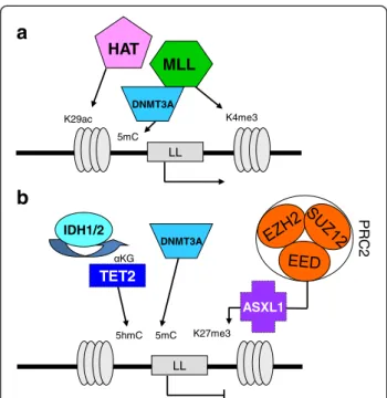
LL LL K29ac
HAT
K4me3MLL
NR K27me3 PRC2 ASXL1TET2
5hmC KG IDH1/2a
b
DNMT3A 5mC DNMT3A 5mCFigure 3Schematic representation of epigenetic regulation of a leukemogenic locus (LL) framed by histone H3. (a) Histone acetyltransferases (HAT; e.g. MYST3) and histone methyltransferases (HMT; e.g. MLL) can activate the locus. (b) Reciprocally, the locus is repressed by polycomb complex PRC2 (which comprised EED, EZH2 and SUZ12 proteins). ASXL1 would direct PRC2 to the locus. Loss-of-function mutations in PRC2 components or in ASXL1 remove PRC2 repression. DNMT3A is involved in the formation of 5-methylcytosines (5mC) from cytosines and interacts with HMTs as well as with PRC2 components. TET2 mediates hydroxylation of 5mC to 5hmC. To function, TET2 requires α-ketoglutarate (αKG), which is provided by IDH1/2 proteins. Aberrant methylation patterns are caused by mutation in TET2 or in IDH1/2, which produces 2-hydroxyglutarate instead of αKG.
Mutations in signaling pathways, transcription
net-works and splicing machinery have many downstream
consequences. Modifications in epigenetic regulation of
DNA and histones may have a strong amplifying effect
since they impact on the transcription of thousands of
genes. This in turn impacts on the properties of
hematopoietic stem cells, favoring self-renewal and
pro-liferation over differentiation, thus promoting
leuke-mogenesis [92]. However, chimeric proteins involving
TFs and ERs (e.g. MLL, MYST3, NSD1
. . .) may induce
a stronger effect than mutations in other TFs and ERs
(such as ASXL1, EZH2 or TET2), which may need to
co-occur with several other alterations to trigger AML,
often after a chronic phase. Perhaps like the difference
between a water jet and a sprinkling rain, this difference
may have to do with the specific functions of TFs and
ERs [64]. TF and ER fusion proteins assemble in
com-plexes that are directly recruited to their target genes
where they modify the local histone marks, drastically
altering transcription. In contrast, mutated ERs may
moderately perturb the epigenetic network, resulting in
global gene deregulation.
Mutations in spliceosome components may lead to
several types of deregulation, including alterations of the
epigenetic control of differentiation and self-renewal;
they may thus result in the same defects as TF and ER
mutations. This may derive from splicing aberrations of
leukemogenic genes (e.g. RUNX1) [41] or from other
specific but indirect defects. SF3B1 for example interacts
with components of the polycomb repressor complex 1
(PRC1) and SF3B1 mutations may compromise PRC1
regulation of leukemogenic loci [93]. Reciprocally, the
function of the pre-mRNA splicing machinery involves
the reading of histone marks, and defective chromatin
regulators may affect splicing [94]. Directly or indirectly,
SF3B1
mutations, which are associated with the presence
of ring sideroblasts, are likely to affect genes involved in
red cell biology and mitochondria function. Because
mutations in splicing genes, in TFs and in ERs are not
mutually exclusive it is probable that the three types of
alterations have additive rather than interchangeable
effects.
Modeling molecular leukemogenesis
Are there preferential combinations and mutual exclusions?
Two driver mutations may never occur together (mutual
exclusion) in the same cell because of epistasis (two hits
in the same pathway are not selected because they do
not provide a growth advantage) or synthetic lethality
(two hits are counter-selected because they compromise
the life of the leukemic cell). Associations and
cooper-ation can occur in all other cases.
Some chronic myeloid malignancies, such as CMML
(myeloproliferative form, MP-CMML) and MPNs, have a
proliferative component. This component is driven by
alterations in signaling molecules, such as CBL, CBLB,
FLT3, JAK2, LNK, MPL, NF1, PTPN11
or RAS. These
mutations are generally mutually exclusive. However,
JAK2
mutations can be found in patients with mutations
of CBL, LNK or MPL [95–97]. In most cases when two
signaling mutations are found in the same patient they are
not in the same cellular clone. Signaling mutations
associ-ate with mutations in genes from the other classes (TSGs,
TFs, ERs). CBL and KIT mutations are more frequent in
AML with t(8;21) and inv(16), i.e. with alterations of the
core binding factor (CBF), a dimeric transcriptional factor
containing the RUNX1 protein [98].
With rare exceptions, mutations in genes encoding
splicing factors do not synergize and are mutually
exclu-sive [31,38,41,42].
As already mentioned, IDH1 or IDH2 mutations are
mutually exclusive with TET2 mutations. Except for this,
TET2
mutations seem to be able to cooperate with either
of the other recurrent alterations. ASXL1 mutations,
which occur preferentially in secondary AML, are
mutu-ally exclusive with NPM1 mutations, which occur in de
novo
AML [99]. Although ASXL1 interacts with PRC2
proteins [83] ASXL1 and EZH2 mutations are not
mutu-ally exclusive [58]. Mutations in EED and SUZ12 may
even be found in the same AML case [23]; however, they
may affect different clones. RUNX1 mutations are
fre-quently associated with ASXL1 defects in MDSs [100].
Mutations in ASXL1 and TET2 can be concomitant
(Fig-ure 1), and each can co-occur with mutations in signaling
molecules [58,100]. In MDSs, U2AF1 mutations are more
frequent in ASXL1-mutated than in ASXL1-wildtype cases
[38,42]. TP53 mutations and losses, likely associated with
genetic instability, are found in MDSs with karyotypic
alterations but not in cases with normal karyotype [58].
DNMT3A
mutations are more frequent in AML with
NPM1
and FLT3 mutations, infrequently found in
ASXL1-mutated cases, and very rare in cases with translocations
[24,101]. Overall, while IDH1/2 and TET2 mutations are
equally distributed, there seem to be two major
associa-tions in AMLs with intermediate cytogenetic risk, ASXL1/
RUNX1
on the one hand (secondary, dysplastic AMLs),
NPM1/FLT3/DNMT3A
on the other hand (primary,
non-dysplastic AMLs) [99]. These and other associations and
exclusions not described here or yet to be discovered will
help understand the major leukemogenic pathways. An
important issue is to demonstrate that mutations found in
the same case are actually cooperating mutations that
co-occur in the same cell progeny and not in different clones.
How many hits are necessary to trigger a malignant
myeloid disease?
Early studies of chronic and acute hematopoietic
malig-nant diseases have shown that some cases may display a
single mutational event whereas others harbor several
hits [100]. This difference may just be due to the low
mu-tational frequency of many driver genes (e.g. NF1A, EED)
[102] and to our current ignorance of other targets.
Actu-ally, NGS studies have shown that the general rule is to
find several altered genes in each case [23,26,59,103] and
murine models have shown that single alterations are,
ex-cept in rare cases, not sufficient to cause AML [104,105].
In the years to come mouse models will have to challenge
many combinations of mutations.
The study of matched chronic and acute stages has
shown that progression is associated with additional
alterations. However, the chronic stages are already
char-acterized by the presence of several mutations. We
found that many cases of CMML have already four
mutations [36], and this was without counting mutations
in splicing factors. JAK2 and TET2 concomitant
muta-tions are frequent in MPNs [16,37]. Whether they are
both necessary for the various phases of the disease and
their order of appearance are a matter of debate [106].
An NGS study indeed showed that the ten mutations
identified in an MDS patient can be detected together in
most studied single cells, suggesting a linear evolution of
the disease and the existence of a dominant clone [103].
Regarding evolution of AML after therapy, a recent NGS
study has revealed two major patterns at relapse [23];
the first pattern is the persistence of a dominant clone
and the second pattern is the selection and expansion of
a minor clone; in both cases the relapse clone had
gained additional mutations. Another recent NGS study
showed that genetic evolution of secondary AML is a
dynamic process shaped by multiple cycles of mutation
acquisition and clonal selection. MDS are oligoclonal
with founding clones; these clones persist in secondary
AML, which shows at least an additional subclone with
new “progression” mutations [107]. Founding mutations
may occur in various genes, such as U2AF1 [39]
or TET2. Many different genes may be involved in
pro-gression. Thus, several steps are necessary to trigger a
myeloid disease, even a so-called chronic one, and
pro-gression involves additional hits.
How many of these steps are there?
A first step in the leukemogenic process is likely to be a
mere clonal expansion. Several gene mutations may play
a role at this stage. Their identity may depend on
whether they target a hematopoietic stem cell or a
pro-genitor. In the first case the initial hit should provide a
proliferation boost, in the second the hit should bestow
self-renewal on the proliferating progenitor [108].
Muta-tions in a TSG, splicing gene, or in some ERs such as
TET2,
could occur at this initial step. It is also possible
that, in a susceptible background, several clones emerge
independently early on [12,109].
Then, because of increasing proliferation and genetic
instability, a cell from the affected clone (or clones)
undergoes various additional mutations (including many
background mutations), leading to an oligoclonal
malig-nant tumor. Some of the early mutations may not be
present in the clone that eventually becomes leukemic.
Thus, for each case, only the determination of all
poten-tial mutations and the reconstitution of the mutation
profile and clonal evolution will help understand the
pathophysiology of the disease. This is now achievable
by using NGS. How many steps can eventually be
indivi-dualized may depend on how many clones are initially
expanded, on the level of genetic instability that results
from the initial hits, and on the impact of the mutations
on self-renewal, differentiation and proliferation. Some
mutations in epigenetic regulators may have a milder
ef-fect on genetic reprogramming than a gene fusion
in-volving a master transcription factor, which will induce a
strong block of differentiation in a hematopoietic
pre-cursor [92]. The latter event is prominent in de novo
AMLs, which accordingly display only few or none of
the other recently-discovered mutated genes.
A previous scheme of leukemogenesis [110] was based
on the minimal cooperation of two oncogene classes,
proliferation-drivers (kinases, RAS) and
differentiation-blockers (mostly transcription factors), to trigger AML.
The ever-increasing molecular complexity of myeloid
ma-lignancies is now obvious and calls for an update of this
model. First, it is now routinely possible to observe the
co-operation, already at the chronic stage, of three, four or
more mutated genes (to speak only of known or suspected
drivers), whose products belong to at least five classes,
class I signaling molecules class II TFs, class III ERs, class
IV TSGs and class V splicing factors [100]. Second, not all
mutations of a class are equivalent; mutations in ASXL1,
RUNX1
or TET2 occur almost as frequently at the chronic
stages as in AML whereas mutations of IDH1/2 or
DNMT3A
are preferentially found at the acute stage. The
reason for this remains obscure but may have to do with
the different intensities in the differentiation block
induced by the mutations. Third, the classes are not well
individualized. For example, EZH2, RUNX1 and TET2 are
both TSGs and TF/ERs. NF1 is both a TSG and a
regula-tor of signaling pathways. Because it induces
phosphoryl-ation
of
histone
H3
and
PRMT5
arginine
methyltransferase, JAK2V617F may also be an ER [111].
Fourth, all classes may not be systematically affected in
each case. Fifth, if classes I and V are relatively well
indivi-dualized, with genes whose mutations are generally
mutu-ally exclusive, the definition of the other classes may
evolve. However, despite all this, the initial schematic
model might not be so far off. The two key processes of
differentiation and self-renewal seem to be always altered
and proliferation is frequently affected. It may just be that
the oncogenic hits required to achieve each step might be
more numerous than initially expected. This model will
apply to cases with intermediate or normal cytogenetic
risk; a different leukemogenesis pathway linked to genetic
instability may be involved in cases with TP53 mutation
and complex karyotype [112].
Considering all this, several pathways to leukemia can
be envisaged (Figure 4). The first pathway could be direct
and trigger de novo AML with a gene fusion as the major
event and few other alterations. The second pathway is
characterized by NPM1 mutations, which are rarely
asso-ciated with mutations in other known TFs or ERs except
in DNMT3A and IDH1/2 [23,101]. AML with complex
karyotype can derive from genetic instability, with or
with-out TP53 mutations. A fourth pathway would be the
accu-mulation of several hits in signaling molecules, TFs, ERs
and splicing factors, which induce either secondary AML
after a chronic phase (Figure 5) or de novo AML; however,
some so-called de novo AMLs with several ER mutations
could actually be secondary to a non-detected chronic
phase. Mutations in TFs and ERs are not major events in
chronic myeloid leukemia (CML), which is triggered by
the BCR-ABL1 fusion; however, mutations in ERs such as
ASXL1, IDH1/2
and TET2 may participate to CML
pro-gression to AML [113,114].
Overall, the development of an AML may follow a “slot
machine” model (Figure 6), in which the late steps would
be, to some point, constrained by the initial ones (clonal
dominance, cooperations/exclusions). Oligoclonality would
be due to several possible draws at each step. It is
import-ant that we determine the exact number of “reels” (hits)
and “symbols” (genes) and the possible combinations.
Utilizing molecular leukemogenesis
Understanding and modeling leukemogenesis will have a
major impact on the management and treatment of
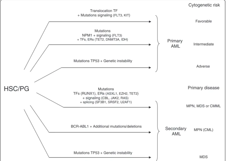
Translocation TF + Mutations signaling (FLT3, KIT)
Mutations TP53 + Genetic instability
Primary
AML
Secondary
AML
Mutations
TFs (RUNX1), ERs (ASXL1, EZH2, TET2) + signaling (CBL, JAK2, RAS) + splicing (SF3B1, SRSF2, U2AF1)
BCR-ABL1 + Additional mutations/deletions Mutations TP53 + Genetic instability
Mutations NPM1 + signaling (FLT3) + TFs, ERs (TET2, DNMT3A, IDH)
Cytogenetic risk
Adverse Favorable IntermediatePrimary disease
MPN, MDS or CMML MDS MPN (CML)HSC/PG
Figure 4Schematic representation of pathways leading to acute myeloid leukemia (AML) from hematopoietic stem cell (HSC) or progenitors (PG). Gene fusions and NPM1 mutation are major events in the induction of primary AMLs with favorable and intermediate cytogenetic risk (they correspond respectively to mutation groups A and B of Ley et al [24], and to mutation groups 2 + 3 and 1 of Shen et al. [101]. Secondary AML following MPN or MDS (see Figure 1) could occur after a series of gene mutations in transcription factors and epigenetic regulators combined with a mutation in a signaling pathway (see Figure 3), after TP53 mutation and a series of mutations and karyotype alterations due to genetic instability, or after additional mutations in BCR-ABL chronic myeloid leukemia.
hematopoietic malignancies. Molecular biology already
helps establish the diagnosis (JAK2), classification
(BCR-ABL1, FGFR1, PDGFRs), prognosis (FLT3, NPM1, CEBPA)
and treatment (BCR-ABL, 5q-, JAK2) of myeloid diseases.
Due to the increasing simplification and accessibility to
clinical laboratories of NGS equipment, the repertoire of
all genetic alterations will soon be determined for any new
case as a routine practice.
The establishment of a precise taxonomy comprising
homogeneous pathophysiological entities is a major goal
in hematology. It relies heavily on molecular data. It
started with the karyotype and has continued with gene
expression profiles [115,116]. Gene mutations will nicely
complete the picture. Other factors such as microRNAs
and long non-coding RNAs status [1], methylation profiles
[117] and histone marks may have to be integrated too.
Several studies have shown that gene mutations have
indeed a major impact on prognosis of myeloid diseases.
This is the case in MDSs for mutations in five genes,
ASXL1, ETV6, EZH2, TP53
and RUNX1 [58]. Mutations
in ASXL1 seem to be associated with an aggressive
phenotype in all myeloid malignancies [8]: they are
fre-quent in high-risk MDSs and correlate with poor
progno-sis in MDSs [118–120] and with acute progression in
CMML [36], they are more frequent in myelofibrosis than
in other MPNs [37,121], and characterize secondary
AML. DNMT3A mutations are frequent in younger
patients with AML and are associated with an
unfavor-able prognosis in MDS and AML [15,24,30,101]. Among
splicing gene mutations, those in U2AF1 and SRSF2 seem
to be associated with aggressive forms of myeloid diseases
and those in SF3B1 with good prognosis [38,39,41,42].
Molecular data will allow the establishment of an
upgraded index of prognosis. For example, in MDSs, it is
highly conceivable that the current prognostic index used
for the evaluation of the disease (IPSS), which already
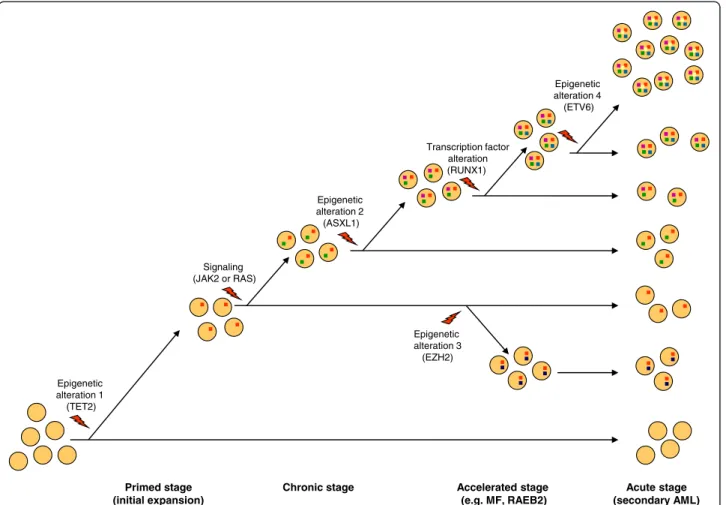
Primed stage (initial expansion) Epigenetic alteration 1 (TET2)
.
. .
.
. .
.
.
.
.
.
.
.
.
.
Epigenetic alteration 2 (ASXL1).
.
.
.
.
.
Signaling (JAK2 or RAS) Chronic stage.
.
.
.
.
.
.
.
.
.
.
.
.
.
.
.
.
.
Acute stage (secondary AML) Epigenetic alteration 4 (ETV6).
.
.
.
.
.
.
.
.
.
.
.
.
.
.
.
.
.
.
.
.
.
.
.
.
.
.
.
.
.
.
.
.
.
.
.
.
.
.
.
.
.
.
.
.
.
.
.
.
Transcription factor alteration (RUNX1). .
. .
. .
. .
. .
. .
.
.
Epigenetic alteration 3 (EZH2).
.
.
.
Accelerated stage (e.g. MF, RAEB2).
.
.
.
.
.
.
.
.
.
Figure 5Schematic representation of a case of malignant myeloid disease evolving in four stages along one pathway. Clones with different gene mutations (color squares in cells) represent various ratios of the oligoclonal leukemia. The order and nature of the mutations (or genome alterations) is given as an example and may differ from one case to another. However, in contrast to JAK2V617F, which has a mild effect on hematopoietic stem cell (HSC) [16], TET2 mutation has the property to initiate the amplification of HSC and to pave the way to secondary mutations [77]. Mutations in signaling molecules, which have a major impact on the disease phenotype, will vary with the type of chronic stage, for example it could affect JAK2 in case of MPN, RAS in case of MP-CMML and be absent in case of MDS. MF: myelofibrosis, RAEB: refractory anemia with excess of blasts, AML: acute myeloid leukemia.
includes karyotypic data, can be improved by a molecular
index regrouping the mutations that impact on the
patient’s outcome [112]. Whether TET2 mutations are to
be included is a matter of debate [122–124]. In AML, a
thorough study of 18 genes, including ASXL1 and
DNMT3A, proposed an updated and precise risk
stratifi-cation based on gene mutations [125].
New therapeutic targets can be found in two of the
five major classes of leukemogenic genes. Following
the successful use of imatinib in CML, abnormal
sig-naling pathways associated with myeloproliferation, be
it the JAK-STAT pathway [126–128] or another
path-way, represent appealing targets. Drugs targeting
epi-genetic modifications, i.e. epidrugs, such as histone
deacetylase
inhibitors
and
hypomethylating
agents
(DNMT inhibitors), are currently developed or used in
clinics, and many new ones are studied in preclinical
assays and clinical trials [1]. Targeting histone
methyl-transferases (e.g. MLL) or lysine acetylmethyl-transferases (e.g.
P300) [129] is also a promising area of development.
The determination of gene mutations and their
conse-quence on gene regulation and cell programming will
help treat myeloid malignancies in providing a
ration-ale for the use and development of new epidrugs, in
directing the choice of the drug cocktails, and in
allowing the design of drug delivery and the
monitor-ing of drug response and disease progression. For
ex-ample, agents directed against TET2-, IDH- and
DNMT3A-associated methylation defects may
repre-sent a new area of development. To date, the use of
TET2
mutations status to evaluate the response to
DNMT inhibitors is still debated [130,131]. Because
many mutations compromise PRC2 function drugs
an-tagonizing this defect hold great promise.
Proteins of two other leukemogenic classes may also
serve as therapeutic targets. For example, the antitumor
macrolide pladienolide targets SF3B1 [132] opening new
opportunities to develop treatments against RARS.
Compounds aiming at restoring a normal P53 pathway
are in development [133,134].
The existence of concomitant mutations is an
incen-tive for combinatorial therapies; for example, therapeutic
synergy may be obtained by the combined use of
signal-ing inhibitors and epidrugs.
Finally, the complete determination of the mutation
repertoire will provide novel therapeutic targets. For
some diseases, such as CMML, it is already possible to
identify at least one target for nearly nine cases out of
ten [33,36]. However, the development of resistance, as
observed with imatinib [135], is a critical issue.
Initial step
Second step
Third step
Fourth step
ER (TET2) TF (RUNX1) Signaling (RAS) Signaling (FLT3) Signaling (CBL) Signaling (JAK2) ER (IDH1) ER (BCORL1) Miscellaneous (Tri 8) ER (ASXL1) ER (EZH2) ER (IDH2) Miscellaneous (20q-) Signaling (NF1) ER (SUZ12) ER (MLL) Splicing (U2AF35) TSG (TP53)
AML
Splicing (SF3B1) TF (inv(16)) TF (NPM1) Signaling (KIT) TF (CEBPA) ER (DNMT3A)AML
Splicing (U2AF1) Signaling (MPL) TF (ETV6) Splicing (SRSF2)Figure 6 “Slot machine” model of leukemogenesis. Alterations in signaling molecules, transcription factors (TFs), epigenetic regulators (ERs), tumor suppressors (TSG), spliceosome components and various genome abnormalities (examples are given) fall into (at least) four “reels” (steps) that combine to induce a malignant myeloid disease. Acute myeloid leukemia (AML) results from one of the allowed combinations of four (at least) cooperating alterations. At chronic stages, the steps are variably combined, some may be absent (e.g. signaling), some may be specific (e.g. SF3B1splicing mutations in RARS). Each step can be achieved by alterations in one of several genes. The initial step leads to expansion of a founding clone. Two examples of draw (plain and dotted lines) leading to AML are shown.
Hopefully, target identification will allow for the
devel-opment of new combinatorial strategies, such as the one
based on synthetic lethality [136,137]. If two mutations
never occur together it may mean that their combined
effect is deleterious. Thus, opportunities for deriving
synthetic lethality drugs could stem from the
observa-tion of exclusions in mutaobserva-tions patterns.
Conclusions
Thus, mutations and models (“M and Ms”) will help
manage myeloid malignancies. The eventual
comprehen-sive determination in any given case and at diagnosis, of
the set of altered genes, underlying affected pathways
and disease stage, will guide towards an optimal
treat-ment based on an appropriate combination of drugs
tar-geting the various affected processes of the disease.
Clinically-oriented laboratories should already be
prepar-ing for that challenge. Meanwhile, there is much to mull
over the “M and Ms” of myeloid malignancies.
Competing interests
The authors have no competing interests. Author’s contributions
All authors have contributed ideas, discussions, and have participated in the writing of the manuscript. All authors read and approved the final manuscript.
Acknowledgements
We are grateful to O. Bernard for his critical reading of the manuscript. Work in our laboratory on this subject is supported by Inserm, Institut Paoli-Calmettes and grants from the Association pour la Recherche contre le Cancer (DB) and Association Laurette Fugain (MJM).
Received: 5 October 2011 Accepted: 30 June 2012 Published: 23 July 2012
References
1. Chen J, Odenike O, Rowley JD: Leukaemogenesis: more than mutant genes. Nat Rev Cancer 2010, 10:23–36.
2. Baxter EJ, Scott LM, Campbell PJ, East C, Fourouclas N, Swanton S, Vassiliou GS, Bench AJ, Boyd EM, Curtin N, Scott MA, Erber WN, Green AR: Acquired mutation of the tyrosine kinase JAK2 in human myeloproliferative disorders. Lancet 2005, 365:1054–1061.
3. James C, Ugo V, Le Couedic JP, Staerk J, Delhommeau F, Lacout C, Garcon L, Raslova H, Berger R, Bennaceur-Griscelli A, Villeval JL, Constantinescu SN, Casadevall N, Vainchenker W: A unique clonal JAK2 mutation leading to constitutive signalling causes polycythaemia vera. Nature 2005, 434:1144–1148.
4. Kralovics R, Passamonti F, Buser AS, Teo SS, Tiedt R, Passweg JR, Tichelli A, Cazzola M, Skoda RC: A gain-of-function mutation of JAK2 in myeloproliferative disorders. N Engl J Med 2005, 352:1779–1790. 5. Levine RL, Wadleigh M, Cools J, Ebert BL, Wernig G, Huntly BJ, Boggon TJ,
Wlodarska I, Clark JJ, Moore S, Adelsperger J, Koo S, Lee JC, Gabriel S, Mercher T, D'Andrea A, Frohling S, Dohner K, Marynen P, Vandenberghe P, Mesa RA, Tefferi A, Griffin JD, Eck MJ, Sellers WR, Meyerson M, Golub TR, Lee SJ, Gilliland DG: Activating mutation in the tyrosine kinase JAK2 in polycythemia vera, essential thrombocythemia, and myeloid metaplasia with myelofibrosis. Cancer Cell 2005, 7:387–397.
6. Druker BJ, Tamura S, Buchdunger E, Ohno S, Segal GM, Fanning S, Zimmermann J, Lydon NB: Effects of a selective inhibitor of the Abl tyrosine kinase on the growth of Bcr-Abl positive cells. Nat Med 1996, 2:561–566.
7. Fathi AT, Abdel-Wahab O: Mutations in epigenetic modifiers in myeloid malignancies and the prospect of novel epigenetic-targeted therapy. Adv Hematol2012, 2012:469592.
8. Gelsi-Boyer V, Brecqueville M, Devillier R, Murati A, Mozziconacci MJ, Birnbaum D: Mutations in ASXL1 are associated with poor prognosis across the spectrum of malignant myeloid diseases. J Hematol Oncol 2012, 5:12.
9. Graubert TA, Walter MJ: Genetics of Myelodysplastic Syndromes: New Insights. Hematology Am Soc Hematol Educ Program 2011, 1:543–549. 10. Marcucci G, Haferlach T, Dohner H: Molecular genetics of adult acute
myeloid leukemia: prognostic and therapeutic implications. J Clin Oncol 2011, 29:475–486.
11. Mercher T, Quivoron C, Couronne L, Bastard C, Vainchenker W, Bernard OA: TET2, a tumor suppressor in hematological disorders. Biochim Biophys Acta2012, 1825:173–177.
12. Schaub FX, Jager R, Looser R, Hao-Shen H, Hermouet S, Girodon F, Tichelli A, Gisslinger H, Kralovics R, Skoda RC: Clonal analysis of deletions on chromosome 20q and JAK2-V617F in MPD suggests that del20q acts independently and is not one of the predisposing mutations for JAK2-V617F. Blood 2009, 113:2022–2027.
13. Takahashi S: Current findings for recurring mutations in acute myeloid leukemia. J Hematol Oncol 2011, 4:36.
14. Tefferi A: Mutations galore in myeloproliferative neoplasms: Would the real Spartacus please stand up? Leukemia 2011, 25:1059–1063. 15. Thol F, Damm F, Ludeking A, Winschel C, Wagner K, Morgan M, Yun H,
Gohring G, Schlegelberger B, Hoelzer D, Lubbert M, Kanz L, Fiedler W, Kirchner H, Heil G, Krauter J, Ganser A, Heuser M: Incidence and Prognostic Influence of DNMT3A Mutations in Acute Myeloid Leukemia. J Clin Oncol 2011, 29:2889–2896.
16. Vainchenker W, Delhommeau F, Constantinescu SN, Bernard OA: New mutations and pathogenesis of myeloproliferative neoplasms. Blood 2011, 118:1723–1735.
17. Brecqueville M, Cervera N, Adélaide J, Rey J, Carbuccia N, Chaffanet M, Vey N, Birnbaum D, Murati A: Mutations and deletions of the SUZ12 polycomb gene in myeloproliferative neoplasms. Blood Cancer Journal 2011, 1:e18.
18. Delhommeau F, Dupont S, Della VV, James C, Trannoy S, Masse A, Kosmider O, Le Couedic JP, Robert F, Alberdi A, Lecluse Y, Plo I, Dreyfus FJ, Marzac C, Casadevall N, Lacombe C, Romana SP, Dessen P, Soulier J, Viguie F, Fontenay M, Vainchenker W, Bernard OA: Mutation in TET2 in myeloid cancers. N Engl J Med 2009, 360:2289–2301.
19. Dunbar AJ, Gondek LP, O'Keefe CL, Makishima H, Rataul MS, Szpurka H, Sekeres MA, Wang XF, McDevitt MA, Maciejewski JP: 250 K single nucleotide polymorphism array karyotyping identifies acquired uniparental disomy and homozygous mutations, including novel missense substitutions of c-Cbl, in myeloid malignancies. Cancer Res 2008, 68:10349–10357.
20. Ernst T, Chase AJ, Score J, Hidalgo-Curtis CE, Bryant C, Jones AV, Waghorn K, Zoi K, Ross FM, Reiter A, Hochhaus A, Drexler HG, Duncombe A, Cervantes F, Oscier D, Boultwood J, Grand FH, Cross NC: Inactivating mutations of the histone methyltransferase gene EZH2 in myeloid disorders. Nat Genet2010, 42:722–726.
21. Gelsi-Boyer V, Trouplin V, Adélaide J, Bonansea J, Cervera N, Carbuccia N, Lagarde A, Prebet T, Nezri M, Sainty D, Olschwang S, Xerri L, Chaffanet M, Mozziconacci MJ, Vey N, Birnbaum D: Mutations of polycomb-associated gene ASXL1 in myelodysplastic syndromes and chronic myelomonocytic leukaemia. Br J Haematol 2009, 145:788–800.
22. Nikoloski G, Langemeijer SM, Kuiper RP, Knops R, Massop M, Tonnissen ER, van der Heijden A, Scheele TN, Vandenberghe P: de WT, van der Reijden BA, Jansen JH: Somatic mutations of the histone methyltransferase gene EZH2 in myelodysplastic syndromes. Nat Genet 2010, 42:665–667. 23. Ding L, Ley TJ, Larson DE, Miller CA, Koboldt DC, Welch JS, Ritchey JK,
Young MA, Lamprecht T, McLellan MD, McMichael JF, Wallis JW, Lu C, Shen D, Harris CC, Dooling DJ, Fulton RS, Fulton LL, Chen K, Schmidt H, Kalicki-Veizer J, Magrini VJ, Cook L, McGrath SD, Vickery TL, Wendl MC, Heath S, Watson MA, Link DC, Tomasson MH, Shannon WD, Payton JE, Kulkarni S, Westervelt P, Walter MJ, Graubert TA, Mardis ER, Wilson RK, DiPersio JF: Clonal evolution in relapsed acute myeloid leukaemia revealed by whole-genome sequencing. Nature 2012, 481:506–510.
24. Ley TJ, Ding L, Walter MJ, McLellan MD, Lamprecht T, Larson DE, Kandoth C, Payton JE, Baty J, Welch J, Harris CC, Lichti CF, Townsend RR, Fulton RS,
Dooling DJ, Koboldt DC, Schmidt H, Zhang Q, Osborne JR, Lin L, O'Laughlin M, McMichael JF, Delehaunty KD, McGrath SD, Fulton LA, Magrini VJ, Vickery TL, Hundal J, Cook LL, Conyers JJ, Swift GW, Reed JP, Alldredge PA, Wylie T, Walker J, Kalicki J, Watson MA, Heath S, Shannon WD, Varghese N, Nagarajan R, Westervelt P, Tomasson MH, Link DC, Graubert TA, DiPersio JF, Mardis ER, Wilson RK: DNMT3A mutations in acute myeloid leukemia. N Engl J Med2010, 363:2424–2433.
25. Li M, Collins R, Jiao Y, Ouillette P, Bixby D, Erba H, Vogelstein B, Kinzler KW, Papadopoulos N, Malek SN: Somatic mutations in the transcriptional corepressor gene BCORL1 in adult acute myelogenous leukemia. Blood 2011, 118:5914–5917.
26. Mardis ER, Ding L, Dooling DJ, Larson DE, McLellan MD, Chen K, Koboldt DC, Fulton RS, Delehaunty KD, McGrath SD, Fulton LA, Locke DP, Magrini VJ, Abbott RM, Vickery TL, Reed JS, Robinson JS, Wylie T, Smith SM, Carmichael L, Eldred JM, Harris CC, Walker J, Peck JB, Du F, Dukes AF, Sanderson GE, Brummett AM, Clark E, McMichael JF, Meyer RJ, Schindler JK, Pohl CS, Wallis JW, Shi X, Lin L, Schmidt H, Tang Y, Haipek C, Wiechert ME, Ivy JV, Kalicki J, Elliott G, Ries RE, Payton JE, Westervelt P, Tomasson MH, Watson MA, Baty J, Heath S, Shannon WD, Nagarajan R, Link DC, Walter MJ, Graubert TA, DiPersio JF, Wilson RK, Ley TJ: Recurring mutations found by sequencing an acute myeloid leukemia genome. N Engl J Med 2009, 361:1058–1066. 27. Papaemmanuil E, Cazzola M, Boultwood J, Malcovati L, Vyas P, Bowen D,
Pellagati A, Wainscoat JS, Hellstrom-Lindberg E, Gambacorti-Passerini C, Godfrey AL, Cross JR, Green AR, Futreal PA, Stratton MR, Campbell PJ: Somatic mutation of SF3B1 in myelodysplasia with ring sideroblasts and other cancers. N Engl J Med 2011, 365:1384–1395.
28. Van Haaften G, Dalgliesh GL, Davies H, Chen L, Bignell G, Greenman C, Edkins S, Hardy C, O'Meara S, Teague J, Butler A, Hinton J, Latimer C, Andrews J, Barthorpe S, Beare D, Buck G, Campbell PJ, Cole J, Forbes S, Jia M, Jones D, Kok CY, Leroy C, Lin ML, McBride DJ, Maddison M, Maquire S, McLay K, Menzies A, Mironenko T, Mulderrig L, Mudie L, Pleasance E, Shepherd R, Smith R, Stebbings L, Stephens P, Tang G, Tarpey PS, Turner R, Turrell K, Varian J, West S, Widaa S, Wray P, Collins VP, Ichimura K, Law S, Wong J, Yuen ST, Leung SY, Tonon G, DePinho RA, Tai YT, Anderson KC, Kahnoski RJ, Massie A, Khoo SK, Teh BT, Stratton MR, Futreal PA: Somatic mutations of the histone H3K27 demethylase gene UTX in human cancer. Nat Genet 2009, 41:521–523.
29. Visconte V, Makishima H, Jankowska A, Szpurka H, Traina F, Jerez A, O'Keefe C, Rogers HJ, Sekeres MA, Maciejewski JP, Tiu RV: SF3B1, a splicing factor is frequently mutated in refractory anemia with ring sideroblasts. Leukemia 2011, 26:542–545.
30. Yan XJ, Xu J, Gu ZH, Pan CM, Lu G, Shen Y, Shi JY, Zhu YM, Tang L, Zhang XW, Liang WX, Mi JQ, Song HD, Li KQ, Chen Z, Chen SJ: Exome sequencing identifies somatic mutations of DNA methyltransferase gene DNMT3A in acute monocytic leukemia. Nat Genet 2011, 43:309–315.
31. Yoshida K, Sanada M, Shiraishi Y, Nowak D, Nagata Y, Yamamoto R, Sato Y, Sato-Otsubo A, Kon A, Nagasaki M, Chalkidis G, Suzuki Y, Shiosaka M, Kawahata R, Yamaguchi T, Otsu M, Obara N, Sakata-Yanagimoto M, Ishiyama K, Mori H, Nolte F, Hofmann WK, Miyawaki S, Sugano S, Haferlach C, Koeffler HP, Shih LY, Haferlach T, Chiba S, Nakauchi H, Miyano S, Ogawa S: Frequent pathway mutations of splicing machinery in myelodysplasia. Nature 2011, 478:64–69.
32. Yamashita Y, Yuan J, Suetake I, Suzuki H, Ishikawa Y, Choi YL, Ueno T, Soda M, Hamada T, Haruta H, Takada S, Miyazaki Y, Kiyoi H, Ito E, Naoe T, Tomonaga M, Toyota M, Tajima S, Iwama A, Mano H: Array-based genomic resequencing of human leukemia. Oncogene 2010, 29:3723–3731. 33. Jankowska AM, Makishima H, Tiu RV, Szpurka H, Huang Y, Traina F, Visconte
V, Sugimoto Y, Prince C, O'Keefe C, Hsi ED, List A, Sekeres MA, Rao A, McDevitt MA, Maciejewski JP: Mutational spectrum analysis of chronic myelomonocytic leukemia includes genes associated with epigenetic regulation: UTX, EZH2 and DNMT3A. Blood 2011, 118:3932–3941. 34. Levine RL, Belisle C, Wadleigh M, Zahrieh D, Lee S, Chagnon P, Gilliland DG,
Busque L: X-inactivation-based clonality analysis and quantitative JAK2V617F assessment reveal a strong association between clonality and JAK2V617F in PV but not ET/MMM, and identifies a subset of JAK2V617F-negative ET and MMM patients with clonal hematopoiesis. Blood2006, 107:4139–4141.
35. Lippert E, Boissinot M, Kralovics R, Girodon F, Dobo I, Praloran V, Boiret-Dupre N, Skoda RC, Hermouet S: The JAK2-V617F mutation is frequently present at diagnosis in patients with essential thrombocythemia and polycythemia vera. Blood 2006, 108:1865–1867.
36. Gelsi-Boyer V, Trouplin V, Roquain J, Adélaide J, Carbuccia N, Esterni B, Finetti P, Murati A, Arnoulet C, Zerazhi H, Fezoui H, Tadrist Z, Nezri M, Chaffanet M, Mozziconacci MJ, Vey N, Birnbaum D: ASXL1 mutation is associated with poor prognosis and acute transformation in chronic myelomonocytic leukaemia. Br J Haematol 2010, 151:365–375.
37. Brecqueville M, Rey J, Bertucci F, Coppin E, Finetti P, Carbuccia N, Cervera N, Gelsi-Boyer V, Arnoulet C, Gisserot O, Verrot D, Slama B, Vey N, Mozziconacci MJ, Birnbaum D, Murati A: Mutation analysis of ASXL1, CBL, DNMT3A, IDH1, IDH2, JAK2, MPL, NF1, SF3B1, SUZ12, and TET2 in
myeloproliferative neoplasms. Genes Chromosomes Cancer 2012, 51:743–755.
38. Damm F, Kosmider O, Gelsi-Boyer V, Renneville A, Carbuccia N, Curtis CH, Della-Valle V, Couronne L, Scourzic L, Chesnais V, Guerci-Bresler A, Slama B, Beyne-Rauzy O, Schmidt-Tanguy A, Stamatoullas-Bastard A, Dreyfus F, Prebet T, De Botton S, Vey N, Morgan M, Cross JR, Preudhomme C, Birnbaum D, Bernard O, Fontenay M: Mutations affecting mRNA splicing define distinct clinical phenotypes and correlate with patient outcome in myelodysplastic syndromes. Blood 2012, 119:3211–3218.
39. Graubert TA, Shen D, Ding L, Okeyo-Owuor T, Lunn CL, Shao J, Krysiak K, Harris CC, Koboldt DC, Larson DE, McLellan MD, Dooling DJ, Abbott RM, Fulton RS, Schmidt H, Kalicki-Veizer J, O'Laughlin M, Grillot M, Baty J, Heath S, Frater JL, Nasim T, Link DC, Tomasson MH, Westervelt P, DiPersio JF, Mardis ER, Ley TJ, Wilson RK, Walter MJ: Recurrent mutations in the U2AF1 splicing factor in myelodysplastic syndromes. Nat Genet 2011, 44:53–57. 40. Lasho TL, Finke CM, Hanson CA, Jimma T, Knudson RA, Ketterling RP,
Pardanani A, Tefferi A: SF3B1 mutations in primary myelofibrosis: clinical, histopathology and genetic correlates among 155 patients. Leukemia 2011, 26:1135–1137.
41. Makishima H, Visconte V, Sakaguchi H, Jankowska AM, Abu KS, Jerez A, Przychodzen B, Bupathi M, Guinta K, Afable MG, Sekeres MA, Padgett RA, Tiu RV, Maciejewski JP: Mutations in the spliceosome machinery, a novel and ubiquitous pathway in leukemogenesis. Blood 2012, 119:3203–3210. 42. Thol F, Kade S, Schlarmann C, Loffeld P, Morgan M, Krauter J, Wlodarski MW,
Kolking B, Wichmann M, Gorlich K, Gohring G, Bug G, Ottmann O, Niemeyer CM, Hofmann WK, Schlegelberger B, Ganser A, Heuser M: Frequency and prognostic impact of mutations in SRSF2, U2AF1, and ZRSR2 in patients with myelodysplastic syndromes. Blood 2012, 119:3578–3584.
43. Jankowska AM, Szpurka H: Mutational determinants of epigenetic instability in myeloid malignancies. Semin Oncol 2012, 39:80–96. 44. Haferlach C, Grossmann V, Kohlmann A, Schindela S, Kern W, Schnittger S,
Haferlach T: Deletion of the tumor-suppressor gene NF1 occurs in 5 % of myeloid malignancies and is accompanied by a mutation in the remaining allele in half of the cases. Leukemia 2011, 26:834–839. 45. Parkin B, Ouillette P, Wang Y, Liu Y, Wright W, Roulston D, Purkayastha A,
Dressel A, Karp J, Bockenstedt P, Al-Zoubi A, Talpaz M, Kujawski L, Liu Y, Shedden K, Shakhan S, Li C, Erba H, Malek SN: NF1 inactivation in adult acute myelogenous leukemia. Clin Cancer Res 2010, 16:4135–4147. 46. Gelsi-Boyer V, Trouplin V, Adélaide J, Aceto N, Remy V, Pinson S, Houdayer
C, Arnoulet C, Sainty D, Bentires-Alj M, Olschwang S, Vey N, Mozziconacci MJ, Birnbaum D, Chaffanet M: Genome profiling of chronic
myelomonocytic leukemia: frequent alterations of RAS and RUNX1 genes. BMC Cancer 2008, 8:299–314.
47. Greif PA, Eck SH, Konstandin NP, Benet-Pages A, Ksienzyk B, Dufour A, Vetter AT, Popp HD, Lorenz-Depiereux B, Meitinger T, Bohlander SK, Strom TM: Identification of recurring tumor-specific somatic mutations in acute myeloid leukemia by transcriptome sequencing. Leukemia 2011, 25:821–827.
48. Adélaide J, Gelsi-Boyer V, Rocquain J, Carbuccia N, Birnbaum DJ, Finetti P, Bertucci F, Mozziconacci MJ, Vey N, Birnbaum D, Chaffanet M: Gain of CBL-interacting protein, a possible alternative to CBL mutations in myeloid malignancies. Leukemia 2010, 24:1539–1541.
49. Grand FH, Hidalgo-Curtis CE, Ernst T, Zoi K, Zoi C, McGuire C, Kreil S, Jones A, Score J, Metzgeroth G, Oscier D, Hall A, Brandts C, Serve H, Reiter A, Chase AJ, Cross NC: Frequent CBL mutations associated with 11q acquired uniparental disomy in myeloproliferative neoplasms. Blood 2009, 113:6182–6192.
50. Makishima H, Cazzolli H, Szpurka H, Dunbar A, Tiu R, Huh J, Muramatsu H, O'Keefe C, Hsi E, Paquette RL, Kojima S, List AF, Sekeres MA, McDevitt MA, Maciejewski JP: Mutations of e3 ubiquitin ligase cbl family members constitute a novel common pathogenic lesion in myeloid malignancies. J Clin Oncol2009, 27:6109–6116.
![Figure 1 Circos diagrams depict the relative frequency and associations of the major mutations in MPNs (a) and MDSs (b), respectively based on data from our work [37] on 127 classic MPNs and from Damm ’ s study [38] on 221 MDSs](https://thumb-eu.123doks.com/thumbv2/123doknet/14614730.546089/3.892.85.807.705.1040/figure-circos-diagrams-relative-frequency-associations-mutations-respectively.webp)