HAL Id: inserm-01322519
https://www.hal.inserm.fr/inserm-01322519
Submitted on 27 May 2016
HAL is a multi-disciplinary open access
archive for the deposit and dissemination of
sci-entific research documents, whether they are
pub-lished or not. The documents may come from
teaching and research institutions in France or
abroad, or from public or private research centers.
L’archive ouverte pluridisciplinaire HAL, est
destinée au dépôt et à la diffusion de documents
scientifiques de niveau recherche, publiés ou non,
émanant des établissements d’enseignement et de
recherche français ou étrangers, des laboratoires
publics ou privés.
Véréna Landel, Pascal Millet, Kévin Baranger, Béatrice Loriod, François Féron
To cite this version:
Véréna Landel, Pascal Millet, Kévin Baranger, Béatrice Loriod, François Féron. Vitamin D interacts
with Esr1 and Igf1 to regulate molecular pathways relevant to Alzheimer’s disease. Molecular
Neu-rodegeneration, BioMed Central, 2015, 11 (1), pp.22. �10.1186/s13024-016-0087-2�. �inserm-01322519�
R E S E A R C H A R T I C L E
Open Access
Vitamin D interacts with Esr1 and Igf1 to
regulate molecular pathways relevant to
Alzheimer
’s disease
Véréna Landel
1, Pascal Millet
1,2, Kévin Baranger
1, Béatrice Loriod
3and François Féron
1*Abstract
Background: Increasing evidence suggests a potential therapeutic benefit of vitamin D supplementation against Alzheimer’s disease (AD). Although studies have shown improvements in cognitive performance and decreases in markers of the pathology after chronic treatment, the mechanisms by which vitamin D acts on brain cells are multiple and remain to be thoroughly studied. We analyzed the molecular changes observed after 5 months of vitamin D3 supplementation in the brains of transgenic 5xFAD (Tg) mice, a recognized mouse model of AD, and their wild type (Wt) littermates. We first performed a kinematic behavioural examination at 4, 6 and 8 months of age (M4, M6 and M8) followed by a histologic assessment of AD markers. We then performed a comparative transcriptomic analysis of mRNA regulation in the neocortex and hippocampus of 9 months old (M9) female mice. Results: Transcriptomic analysis of the hippocampus and neocortex of both Wt and Tg mice at M9, following 5 months of vitamin D3 treatment, reveals a large panel of dysregulated pathways related to i) immune and inflammatory response, ii) neurotransmitter activity, iii) endothelial and vascular processes and iv) hormonal alterations. The differentially expressed genes are not all direct targets of the vitamin D-VDR pathway and it appears that vitamin D action engages in the crosstalk with estrogen and insulin signaling. The misexpression of the large number of genes observed in this study translates into improved learning and memory performance and a decrease in amyloid plaques and astrogliosis in Tg animals.
Conclusions: This study underlies the multiplicity of action of this potent neurosteroid in an aging and AD-like brain. The classical and non-classical actions of vitamin D3 can act in an additive and possibly synergistic manner to induce neuroprotective activities in a context-specific way.
Keywords: Vitamin D, Alzheimer’s disease, Transcriptome, Nervous system, Inflammatory response, Hormonal activity Background
The world is currently facing a global Alzheimer’s disease (AD) pandemic and there is an urgent necessity to find drugs that can halt, or even better, cure this pathology. Clinicians and researchers are actively looking for mole-cules with neuro-protective, regenerative and immuno-modulatory properties [1]. Preferably, to speed up their delivery to AD patients, the selected compounds should be FDA-approved with a well-known toxicity range.
With the intention of unveiling new molecular candi-dates, we elected a recognized animal model for this
disease, namely the 5XFAD transgenic mice [2], that ex-hibit mnesic deficits, increased Amyloid Precursor Protein (APP) processing and an early onset for inflammation and plaque formation [3]. We timely assessed gene misexpres-sion in the hippocampus and neocortex of 5XFAD female mice at presymptomatic, prodromal-like and symptomatic stages of the pathology. As expected, we observed a dysregulated expression of genes involved in inflamma-tion, NADPH oxidase complex, phagocytic processes and interferon-γ-related pathways [4]. Remarkably, we also found a modulated expression of 784 vitamin D-associated transcripts (Additional file 1).
Vitamin D3 is a fat-soluble and seco-steroid hormone. Unlike other vitamins, vitamin D3 is mainly produced after adequate exposure to sunlight. Where this exposure
* Correspondence:francois.feron@univ-amu.fr
1Aix Marseille Université, CNRS, NICN UMR 7259, Marseille 13916, France
Full list of author information is available at the end of the article
© 2016 Landel et al. Open Access This article is distributed under the terms of the Creative Commons Attribution 4.0 International License (http://creativecommons.org/licenses/by/4.0/), which permits unrestricted use, distribution, and reproduction in any medium, provided you give appropriate credit to the original author(s) and the source, provide a link to the Creative Commons license, and indicate if changes were made. The Creative Commons Public Domain Dedication waiver (http://creativecommons.org/publicdomain/zero/1.0/) applies to the data made available in this article, unless otherwise stated.
is lacking, dietary intake of vitamin D (vitamin D2 or vitamin D3) is required. The epidermis contains a chol-esterol metabolite that under ultraviolet light produces a precursor, previtamin D3. After isomerization and two separate hydroxylations, the active 1,25-dihydroxy-vitamin D3 (1,25(OH)2D3 or calcitriol) is produced. 1,25(OH)2D3 operates via both nuclear receptors named Vitamin D Receptor (VDR) (part of the superfamily of ster-oid hormone receptors) and non-genomic systems. The 1,25(OH)2D3/VDR complex interacts with specific gen-omic sequences named Vitamin D Responsive Elements (VDRE) found in promoter regions and has been shown to regulate the transcription of a large number (up to 1000) of target genes [5, 6].
Epidemiological and clinical data have shown that i) high serum levels of 25-hydroxyvitamin D (25(OH)D) as-sociate with better cognitive test performance [7, 8] and ii) vitamin D deficiency is found in patients with Alzheimer’s disease [9–13]. Other studies reported i) associations be-tween VDR polymorphisms and cognitive function in Alz-heimer’s disease patients [14–19] and ii) decreased VDR mRNA levels in the hippocampus of AD patients [20].
1,25(OH)2D3 has also been shown to i) enhance cere-bral clearance of human Amyloid Beta (Aβ) peptide from mouse brain across the blood–brain barrier [21], ii) prevent Aβ-induced alterations in cortical neurons through upregulation of the VDR and downregulation of L-type voltage sensitive calcium channels [22, 23]. Experimental observations confirmed that i) vitamin D deficiency increases spatial learning deficits in a rat model of AD [24] likely by enhancing Aβ deposition through modulation of amyloid processing [25] and ii) vitamin D3 supplementation decreases pathological markers of the disease, such as Aβ deposition, in a transgenic mouse model [26]. However, relatively little is known about the mechanisms of action of this ster-oid hormone in demented brains.
Purposely, we assessed the therapeutic benefit of vitamin D3 in 5XFAD mice, supplemented after the onset of the symptoms, from month 4 (M4) to month 9 (M9). Along the course of the disease, we measured memory abilities and, at the end of the experiment, we quantified amyloid plaque load. In addition, using pangenomic cDNA micro-arrays and bioinformatic tools, we identified the genes that were regulated, directly or indirectly, by a vitamin D3 inter-vention. This study is the first to provide insight into the molecular mechanisms at play after chronic treatment with vitamin D3 in both healthy aging and AD-like brains.
Results
Vitamin D3 supplementation induces an extensive gene dysregulation
To reveal the potential molecular targets of vitamin D3 in specific brain regions, we conducted a series of
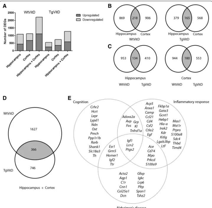
transcriptomic experiments. The study allowed a com-parison of dysregulated genes and associated molecular pathways affected by vitamin D3 supplementation in both a non-AD and AD context, i.e. Wild-type (Wt) versus Transgenic (Tg) animals. Figure 1 reveals the total number of transcripts affected in Wt or Tg ani-mals after 5 months of vitamin D3 supplementation. A total of 2211 genes are dysregulated in Wt animals in both the hippocampus and cortex combined, compared to a total of 1277 Differentially Expressed Genes (DEGs) in Tg mice after vitamin D3 treatment (Fig. 1a). Vitamin D3 supplementation induces a dysregulation of nearly twice as many genes in Wt animals as compared to Tg. Details about these DEGs are given in Additional file 2. There is no significant difference in terms of the number of upregulated or downregulated genes in both genotypes and brain regions (Fig. 1a). When consider-ing each brain area analyzed, there is little difference in the number of dysregulated genes for a given genotype (Fig. 1b). In Wt mice, about 10 % of DEGs are common to both tissues while 13 % shared DEGs can be ob-served in Tg animals (Fig. 1b). Interestingly, a number of DEGs are common to both genotypes in both brain areas (Fig. 1c). Up to 366 genes are commonly dysregu-lated after vitamin D3 treatment in both Wt and Tg mice (Fig. 1d). Using Ingenuity Pathway Analysis (IPA) software, the clustering of the 366 genes indicates that among the top canonical pathways, the ones displaying the largest number of DEGs are: Axon guidance signaling (13 genes), Protein kinase A signaling (13), ILK signaling (9), Huntington’s disease signaling (9), Glioblastoma Multiform signaling (8), Cdc42 signaling (8), IL8 signaling (8), Agranulocyte adhesion and diapedesis (8), Tight junc-tion signaling (7), Embryonic stem cell pluripotency (6), Corticotropin Releasing Hormone signaling (6), Commu-nication between innate and adaptive immune cells (5), Glutamate receptor signaling (5) and Dopamine receptor signaling (5). Furthermore, 39 out of 366 genes are associ-ated to inflammatory response, 26 to cognition and 25 to Alzheimer’s disease (Fig. 1e).
An intriguing expression of hormone transcripts in the cortex and hippocampus
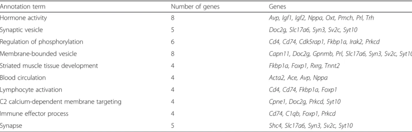
One of the aims of this transcriptomic analysis was to extract information about genes whose expression is modified in the nervous system after 5 months of vita-min D3 supplementation, regardless of the pathological context. One way to obtain such information is to refine the working dataset to include only genes that are com-monly dysregulated in all tested conditions. Table 1 shows the list of genes found to be dysregulated in Wt and Tg mice, in both the hippocampus and cortex. In these four conditions, we found 28 DEGs, with varying fold changes according to the genotype and brain region
studied, that are consistently regulated after 5 months of vitamin D3 supplementation. To gain better sense of the biological functions represented by the differentially expressed genes, we performed a Functional Annotation Clustering using the NIH David tool. This analysis
revealed that the genes in this dataset are related to five different biological clusters: regulation of phosphoryl-ation, hormone activity, membrane trafficking, nucleo-tide binding and metal ion binding. A less restrictive additional analysis was performed on DEGs found in at
Fig. 1 Schematic overview of gene expression in cortex and hippocampus of 9 month-old wild type and transgenic mice reveals that a 5 month vitamin D3 supplementation induces an extensive transcript dysregulation. a Graphical representation of the number of upregulated (dark grey) and downregulated (light grey) DEGs in either the hippocampus, cortex or combined regions in both Wt and Tg animals having been fed a vitamin D3 diet for 5 months. The number of genes specifically dysregulated in wild type mice is twice as large as in transgenic mice. b-c Venn diagrams indicating the number of overlapping (grey) and non overlapping DEGs when the two brain areas are compared in wild type and transgenic mice (b) and when the two strains are compared at the hippocampal or cortex level (c). d Venn diagram showing that, when hippocampal and cortical data are combined, vitamin D3 supplementation triggers the dysregulation of 366 genes in both strains. e List and top associated biological functions of the 366 DEGs common to both strains. Overall, 39 are associated to inflammatory response, 26 to cognition and 25 to Alzheimer’s disease. Fold change (FC) cutoff used for above analyses was -1.5> FC <1.5
least three out of the four conditions. Table 2 represents the top ten biological clusters associated with this data-set. One hundred thirty-six genes were differentially expressed (Additional file 3) and when performing func-tional annotation clustering of these DEGs, the number one biological function is hormone activity (Table 2). Eight genes fall into this ontological category: Avp, Igf1, Igf2, Nppa, Oxt, Pmch, Prl, Trh. Bearing in mind that several of these neuropeptides are largely found in the pituitary axis, it is extremely intriguing to find them dif-ferentially expressed in the cortex or hippocampus of 5XFAD mice.
Among the top ten biological pathways affected by vitamin D3 treatment, two of them concern synaptic
processes and neurotransmission with dysregulation of genes such as Syn3, Syt10 and Sv2c, while two others are related to immune responses including genes such as C1qb, Cd4 and Cd74 (Table 2). A literature search was then performed to determine whether some of these genes contain a putative VDRE in their sequence. Among the 136 genes commonly dysregulated, only four, Fos, Gcnt1, Irak2, St6galnac1, were found to present a known functional VDRE in their promoter region [6].
The effects of vitamin D3 supplementation vary according to the health status of the animal
To move further and comprehensively assess the effect of vitamin D on brain functioning, we analyzed the
Table 1 List of commonly dysregulated genes in the cortex and hippocampus of transgenic and wild type mice
Fold change
Hipocampus Cortex
Gene symbol Entrez gene name WtVitD/Wt TgVitD/Tg WtVitD/Wt TgVitD/Tg Ahcy adenosylhomocysteinase −1,73 −2,07 1,79 2,07 Atg4c autophagy related 4C, cysteine peptidase −2,72 −2,75 4,49 4,82 Avp arginine vasopressin −1,59 2,21 1,73 3,96 C130078n14 uncharacterized protein C130078N14 −2,13 −2,17 3,92 2,77 Cdk5rap1 CDK5 regulatory subunit associated protein 1 −1,78 −2,14 2,66 2,92 Cpne1 copine I −1,56 −1,82 1,6 1,71 Dnajb8 DnaJ (Hsp40) homolog, subfamily B, member 8 −1,85 1,98 −1,92 −1,73 Fam205a family with sequence similarity 205, member A 2,34 2,78 1,61 −1,55 Fkbp1a FK506 binding protein 1A, 12kDa 1,97 1,62 −1,72 −1,52 Gbp6 guanylate binding protein family, member 6 2,19 2,21 −2,65 −3,23 Gm4924 predicted gene 4924 1,68 1,94 2,14 1,53 Hla-drb5 major histocompatibility complex, class II, DR beta 5 1,86 1,53 −2,53 −2,34 Iqcf4 IQ motif containing F4 2,74 2,22 −1,54 −1,73 Iqsec3 IQ motif and Sec7 domain 3 −5,23 4,49 −3,3 1,58 Kcnj6 potassium channel, inwardly rectifying subfamily J, member 6 1,61 1,93 −1,79 −1,67 Kdr kinase insert domain receptor −1,6 1,68 1,53 −1,64 Mirg miRNA containing gene −1,91 1,88 −3,41 −1,68 Nanog Nanog homeobox −2,91 1,53 −1,52 −1,66 Ocel1 occludin/ELL domain containing 1 −3,07 −3,98 3,52 3,21 Omp olfactory marker protein −1,54 −10,41 3,18 1,66 Oxt oxytocin/neurophysin I prepropeptide −2,75 4,04 2,03 4,74 Ppp1r16b protein phosphatase 1, regulatory subunit 16B 2,29 2,39 −1,78 −1,78 Prkcd protein kinase C, delta −2,18 −2,52 1,84 1,65 Prl prolactin 1,93 −1,88 10,07 10,54 Rnase4 ribonuclease, RNase A family, 4 1,61 2,12 −2,14 −2,07 Sdc4 syndecan 4 2,38 1,95 −1,91 −1,81 Serpina3g serine (or cysteine) peptidase inhibitor, clade A, member 3G 2,78 1,94 −1,8 −2,42 Znf669 zinc finger protein 669 −2,08 −1,9 1,84 2,2
Dysregulated transcripts in the hippocampus and cortex of vitamin D3 supplemented wild type and transgenic mice in comparison with unsupplemented littermates. The gene symbol, the full name and the fold change of each gene are indicated
record of dysregulated genes, in accordance with their role in well-described metabolic pathways. For each strain, we combined transcriptomic data from the hippo-campus and the neocortex at M9 and, using Ingenuity software, we listed the main canonical pathways that were modified by the vitamin D3-enriched diet. We then compared the two strains of animals (Wt vs Tg). All per-turbed pathways and the names of their associated DEGs are recorded in Additional file 4. Out of the 90 major modified canonical pathways, 42 were common to both strains. As shown in Fig. 2, 16 of them are related to ei-ther the nervous or the immune system.
In regard to the brain (Fig. 2a), the largest number of dysregulated genes – 53 and 38 for the Wt and Tg ani-mals, respectively – is associated to Axon guidance sig-naling. Within both strains, we observe a modified expression of growth factors (e.g. Bdnf, Bmp1, Bmp6, Egf, Igf1, Figf, Ntf3, Vegfc), chemo-attractive/repulsive agents (e.g. Sema3b, Sema3c, Sema4g, Sema6b, Sema6c), proteases (e.g. Ace, Adam11, Adamts2, Adamts8, Adamts9, Mmp9) and TGF/WNT/β-catenin-associated molecules (e.g. Tgfb1, Tgfbr1, Tgfbr2, Wnt1, Wnt3, Wnt6, Wnt9a, Wnt9b, Wnt16). Two other metabolic pathways - the sig-naling of the corticotropin releasing hormone and the multiform glioblastoma – include large subsets of dys-regulated genes. However, the highest ratio (number of dysregulated genes/number of genes involved in the pathway) is found for the neuroprotective role of THOP1 in Alzheimer’s disease.
With respect to the immune system (Fig. 2b), three canonical pathways – Role of macrophages, fibroblasts and endothelial cells in rheumatoid arthritis, Agranulo-cyte adhesion and diapedesis and Dendritic cell matur-ation - emerge as primarily affected by vitamin D3 supplementation, in both strains. Other modified meta-bolic pathways include Communication between innate and adaptive immune cells, Complement system and
MIF regulation of innate immunity. Nonetheless, the peak ratios are observed for the Role of Il17F in inflam-matory diseases and the role of Il17A in psoriasis.
In addition to this strain independent response to vita-min D3 supplementation, the brains display specific modifications in line with the health status of the ani-mal. For example, vitamin D3-enriched diet modify the expression of glucocorticoid and glutamate receptors in wild type animals and GABA, dopamine and serotonin receptors in transgenic animals (Fig. 2a). Similarly, vita-min D3 alters interleukin signaling in a strain-dependent manner. IL-12 and IL-8 pathways are adjusted in Wt and Tg brains, respectively (Fig. 2b). Other examples of discrepancies can be listed. The non pathological brain responds to vitamin D3 treatment by modifying the expression of i) nitric oxide in macrophages, ii) neuro-trophins and iii) molecules related to Huntington disease whereas the pathological brain reacts by altering the expression of iv) nitric oxide in endothelial cells, v) cathecholamines and vi) molecules associated to Mul-tiple Sclerosis.
Vitamin D3, a potent upstream regulator altering the expression of Alzheimer’s disease-associated genes
To further decipher the detailed role of vitamin D in brain functioning, we narrowed our analysis to the genes that are directly or indirectly regulated by vitamin D or its related-metabolites. Using Ingenuity software, we listed all dysregulated molecules whose expression has been associated, in the literature, to this steroid.
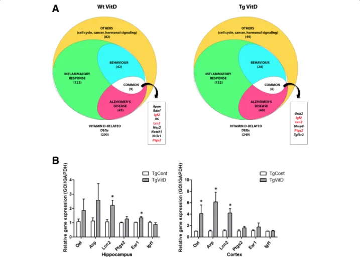
As summarized in Fig. 3, we observed 290 and 249 vitamin D-related DEGs respectively, when comparing vitamin D3-supplemented Wt and Tg mice with their unsupplemented littermates. Within these two pools, 188 and 151 genes are part of the initial list of 784 vita-min D-associated transcripts whose expression is altered in the brain of transgenic animals (Additional file 5).
Table 2 Top ten biological clusters associated with transcripts dysregulated in at least 3 out of 4 conditions: misexpression in the hippocampus and/or cortex of transgenic and/or wild type mice
Annotation term Number of genes Genes
Hormone activity 8 Avp, Igf1, Igf2, Nppa, Oxt, Pmch, Prl, Trh Synaptic vesicle 5 Doc2g, Slc17a6, Syn3, Sv2c, Syt10 Regulation of phosphorylation 6 Cd4, Cd74, Cdk5rap1, Fkbp1a, Irak2, Prkcd
Membrane-bounded vesicle 8 Capn11, Doc2g, Gpnmb, Prl, Slc17a6, Syn3, Sv2c, Syt10 Striated muscle tissue development 4 Fkbp1a, Foxp1, Rxrg, Tnnt2
Blood circulation 4 Acta2, Ace, Avp, Nppa Lymphocyte activation 4 Cd4, Cd74, Fkbp1a, Foxp1 C2 calcium-dependent membrane targeting 4 Cpne1, Doc2g, Prkcd, Syt10 Immune effector process 4 Cd74, C1qb, Foxp1, Prkcd Synapse 5 Shc4, Slc17a6, Syn3, Sv2c, Syt10
Furthermore, when assessing the presence of a VDRE in the promoter region of these DEGs [6], we found 25 (13 %) and 18 (12 %) transcripts in the two above-mentioned pop-ulations (highlighted in grey in Additional file 5). As a comparison, 68 (9 %) out of the 784 genes listed in Additional file 1 harbor a VDRE. We then clustered the two populations of vitamin D-related DEGs and, as shown on Fig. 3, we highlighted three sub-populations, composed of transcripts involved in inflammatory response, Alzhei-mer’s disease and cognition.
At this point, we decided to validate the observed ex-pression of certain DEGs of our dataset, using quantita-tive PCR (qPCR). We chose two hormones, Oxt and Avp, which are, according to the microarray results, dysregulated in the hippocampus and cortex of both Wt and Tg animals. We also focused on four vitamin D-related genes involved in AD commonly dysregulated in the neocortex and hippocampus of Tg animals, namely Lcn2, Ptgs2, Esr1 and Igf1. An overexpression of the transcripts Oxt and Avp is confirmed in the cortex as well as an upregulation of Esr1 in the hippocampus. The gene coding for Lcn2 is overexpressed in both the hippo-campus and the cortex. However, the downregulation of Igf1is not confirmed by qPCR analysis (Fig. 3b).
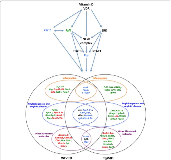
Vitamin D3 supplementation alters the expression of many Alzheimer’s disease-associated genes, even in Wt brains. However, major differences can be observed. As re-ported on Fig. 4, 29 and 27 transcripts are uniquely misex-pressed in Wt and Tg animals, respectively. In addition, 13 out of the 16 vitamin D-related DEGs emerging at the intersection of both strains are inversely expressed. The three transcripts that are similarly misexpressed are Ace, Igf1and Gfap, strongly related to Alzheimer’s disease. All three are underexpressed when compared to unsupple-mented animals (Fig. 4).
The consequences of this discordant response to vita-min D3 treatment are unknown since the Wt mice dis-play no AD symptoms and features. Nevertheless, it allowed us to envision the putative mechanism of action of vitamin D3 or its related metabolites (Fig. 4). Vitamin D3 or 1,25(OH)2D3 and its receptor likely act via four regulators, namely IGF1, ESR1, ERK and NF-κB. Igf1 is underexpressed in both strains while Esr1 is overex-pressed in Tg brains and underexoverex-pressed in Wt brains. The expression of Erk and Nf-κb is unchanged in both strains but many transcripts under their control are mis-expressed in Tg and Wt animals. Downstream gene reg-ulators include Fos, Stat1 and Stat3. The former is overexpressed in Tg and underexpressed in Wt animals.
Vitamin D3 treatment impacts amyloid plaque load and gliosis in transgenic animals
In order to examine whether a 5-month therapeutic intervention with vitamin D3 translated into a positive
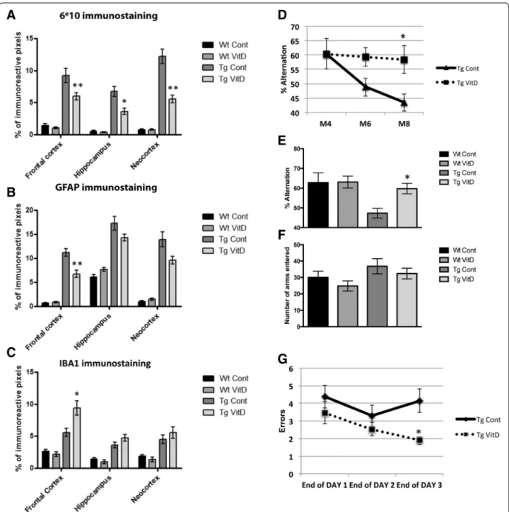
functional outcome, animals were analyzed at the histo-logical and behavioral levels for known markers of AD pathogenesis. At the histological level, vitamin D3 sup-plementation decreased the quantity of plaques in three different brain regions of Tg mice: frontal cortex, hippo-campus and neocortex (Fig. 5a). Astrogliosis, assessed through GFAP-immunostaining, is greatly increased in Tg animals compared to Wt. Vitamin D3 treatment of Tg animals during 5 months significantly decreases GFAP reactivity. However, this significant decrease is only observed in the frontal cortex (Fig. 5b). Microglial activation was assessed through immunostaining with IBA1 antibody and although there is an increase in this microglial marker between Wt and Tg control mice, vitamin D3 supplementation increases IBA1 immunore-activity only in the frontal cortex of Tg animals when compared to their unsupplemented littermates (Fig. 5c).
Four months of vitamin D3-enriched diet is sufficient to rescue cognitive deficits in the 5XFAD model
After 4 months of vitamin D3-supplemented diet, cogni-tive performance was evaluated using two different para-digms: the Y-maze and the 6-radial arm water maze. A longitudinal study was first performed from M4 to M8 using the Y-maze. This study shows that Tg animals display a decreased percent of alternation with time whereas aging Tg animals on a high vitamin D3 diet do not show any cognitive decline between M4 and M8 (Fig. 5d). At M8, we observed a significant decrease in percent alternation performance in Tg mice. This effect is rescued by treatment with vitamin D3 during 4 months with no significant difference in the number of arms entered (Fig. 5e and f ). However, a high vitamin D3 diet does not improve working memory in wild-type animals, even at M8 (Fig. 5e). The RAWM was used in addition to the Y-maze to assess hippocampal-dependent spatial working memory and reference memory. At the end of the learning process (end of day 3), results show a sig-nificant decrease in the total number of errors for Tg animals having been fed a vitamin D3 supplemented diet compared to Tg animals on a control diet (Fig. 5g).
Discussion
Despite growing evidence that vitamin D deficiency is a risk factor for cognitive decline and possibly Alzheimer’s disease, very few studies have investigated the effect of vitamin D supplementation in AD mouse models. To our knowledge, this is the first transcriptomic study to assess the effect of long-term vitamin D3 supplementa-tion in the nervous system of an AD mouse model. This work allowed us to examine the effects of chronic vita-min D3 treatment in both normal aging and AD-like brains, at the transcriptomic, histological and behavioral levels. We observed a very large number of dysregulated
genes in both the pathological and non AD-like cortex and hippocampus of these animals at M9. On the one hand, our results point out that, although there are nearly double the number of DEGs in Wt animals com-pared to Tg, a high number of genes misexpressed after vitamin D3 supplementation are common to both geno-types. On the other hand, when considering the bio-logical pathways affected by these DEGs, the common pathways between Wt and Tg often include different subsets of genes for a given pathway or an inversed ex-pression for the same gene. Our study underlines some well-known actions of vitamin D and related metabolites,
namely its impact on immune and inflammatory processes and cell cycle, along with previously shown effects on ner-vous system molecules such as neurotrophins and nitric oxide. However, our results also comprise unexpected effects on the expression of hormonal transcripts and reveal regulation of genes involved in synaptic func-tion, axon guidance, endothelial and vascular pro-cesses and stem cell pluripotency. A reassuring result is that among the 784 genes predicted to be related to vitamin D action in Tg animals, almost a third of them are indeed regulated after 5 months of supple-mentation. On top of that, the majority of the
Fig. 3 Analysis of vitamin D3-related dysregulated genes indicates an association with inflammatory response, Alzheimer’s disease and behavior. a Overall, 290 and 249 vitamin D-related DEGs are found misexpressed when comparing vitamin D3-supplemented wild type and transgenic mice with their unsupplemented littermates, respectively. Functional classification of these DEGs show importance of inflammatory, behavioural and Alzheimer’s disease related processes. b qPCR validation of several dysregulated transcripts (n = 6 per group). An overexpression of the transcripts oxytocin (Oxt) and arginine vasopressin (Avp) is confirmed in the cortex as well as an upregulation of estrogen receptor 1 (Esr1) in the hippocam-pus. The gene coding for lipocalin 2 (Lcn2) is overexpressed in both the hippocampus and the cortex. * = p < 0.05
(See figure on previous page.)
Fig. 2 Top canonical metabolic pathways associated to gene dysregulation in the cortex and hippocampus of mice after 5 months of vitamin D3 supplementation: common versus strain-specific processes. Metabolic pathways associated to gene expression dysregulation were identified using Ingenuity Pathway Analysis (IPA). Data from both the cortex and hippocampus were analyzed as one dataset and the main metabolic pathways affected were clustered according to their association to the nervous (a) or the immune (b) system
vitamin D-related genes are associated to Alzheimer’s disease, in both the Wt and Tg brains. We show that vitamin D action in the brain of this AD mouse model likely involves signaling through ESR1 and IGF1 and that a 5-month therapeutic intervention duces the histological markers of the disease and re-stores learning and memory capacities.
A common set of brain-associated genes regulated by vitamin D
As mentioned earlier, no pangenomic study previously assessed the effect of vitamin D3 supplementation in a mouse model of Alzheimer’s disease. As a result, no direct comparison can be performed. However, three teams performed a thorough transcriptomic analysis on
Fig. 4 Schematic view of the putative mechanisms of vitamin D3 action in the brain. Vitamin D3 or its related metabolites and their receptors act via four main regulators: IGF1, ESR1, ERK and NF-κB. Igf1 is underexpressed (green) in both strains while Esr1 is overexpressed in transgenic brains and underexpressed in wild type brains (blue). The expression of Erk and NF-κB is unchanged in both strains but many transcripts under their control are misexpressed in transgenic and wild type animals. Downstream gene regulators include Fos, Stat1 and Stat3. Fos is overexpressed in transgenic animals and underexpressed in wild type animals (blue). Overall, 72 vitamin D-related transcripts are associated to either inflammation (19 genes) or amyloidogenesis and amyloid plaques (29) or AD-related features (24). Sixteen genes are dysregulated in both strains (center areas). However, only three of them - Ace, Igf1 and Gfap - are similarly misexpressed (black colour). Specific up- (red) and down-regulated (green) are indicated for each strain
i) brains from adult rats that were vitamin D-deprived during gestation [27] ii) 1,25(OH)2D3-supplemented cultures of neurons and glia [28] or pericytes [29] and iii) hippocampus from middle aged rats after chronic treatment with increasing doses of vitamin D3 [30]. A cross examination of our data with those reported in these studies reveals that the dysregulated expression of 46 genes– Aaas, Aldoa, Atp2b4, Bcam, Bmp1, C3, Cat, Ccl2, Cntn4, Col16a1, Dpp6, Epas1, Fam120c, Fam70a, Fbln1, Fkbp1a, Foxc1, Gfap, Grp, Hspa12a, Htr2c, Igf1, Kdr, Kif1a, Lcat, Lpl, Ndst4, Nmbr, Nr4a2, Pde3b, Pmch, Polr1b, Ppp2r2b, Rab33b, Sdc4, Senp7, Serinc2, Slc17a6, Syt2, Thbd, Tia1, Trhr, Ttc39c, Vcam1, Vdr and Vps13c– was formerly observed. The newly generated list may appear relatively restricted when compared to our initial dataset of 3488 dysregulated genes. However, given the variety of scientific paradigms (depletion vs supplemen-tation), the nature of the samples (cells vs different brain regions), the molecular diversity of the administered molecule (vitamin D3 vs 1,25(OH)2D3) and the hetero-geneous size of the datasets, it is not of minor import-ance to be able to establish an inventory of commonly misexpressed genes in nervous cells or brain areas after vitamin D3 treatment. Moreover, some studies have per-formed targeted analysis of specific gene expression in nervous system cells or tissues. For instance, Kaneko and collaborators demonstrated that calcitriol regulates the expression of two human brain-related genes con-taining VDREs, tryptophan hydroxylase (Tph) and leptin (Lep) [31]. These genes are also modulated in the brains of either Wt or Tg mice in our study. In a set of experi-ments examining the effect of maternal vitamin D defi-ciency on fetal brain development, Hawes and colleagues found that the pups from deficient mothers display a modulated expression of Bdnf, Foxp2, Tgfb1 and Th, which are also affected in certain conditions of our study [32]. Altogether, these results underline the necessity for additional work to be performed on the genomic effects of vitamin D3 or 1,25(OH)2D3 in nervous system cells, as there are clearly multiple targets relevant to brain function and these could potentially explain a causal relationship between vitamin D status and brain aging or AD.
We chose to perform a literature-based analysis of pos-sible VDRE-containing DEGs in our datasets. To avoid discrepancies between study designs, we initially based our search on a large in silico and microarray-based study by Wang and colleagues [6]. This revealed the presence of a very limited number of putative VDREs in our dataset. However, although ChiP-seq and genome-wide studies have found that the most frequent sequence located in VDR binding regions is the classic consensus VDRE motif, these data also suggest the presence of other motifs whose configuration diverge from the classic one and that might or might not interact directly with the VDR [33].
Moreover, there appears to be 1000–10,000 possible chro-matin VDR binding sites, according to the cell-type con-sidered. In contrast the number of primary 1,25(OH)2D3 target genes is thought to be around 100–1000 per tissue, suggesting that some genes are controlled by more than one VDR binding site [33]. It is therefore difficult to con-clude on the complete list of VDR direct targets in our dataset. For instance, the study by Latimer and coworkers, reported putative VDRE sequences in three genes from our dataset– Cntn4, Ppp2r2b and Syt2 - which were not previously identified by Wang and colleagues [6, 30]. These potential VDRE-containing DEGs are involved in synaptic vesicle trafficking and neurotransmission, indi-cating a possible direct modulation of these processes by 1,25(OH)2D3 in the 5XFAD animal model. Since the determination of true VDRE sequences would require further investigation and that vitamin D metabolites are also known to interact with various membrane-based signaling pathways, the search for indirect actions, for instance through modulation of immune processes, is not to be excluded.
A confirmed immuno-modulatory role, several mechanisms of action
The impact of vitamin D3 on inflammatory and immune processes is all but unexpected since it is now well estab-lished that vitamin D is a potent immune modulator [34]. 1,25(0H)2D3 adjusts the expression of inflammatory cyto-kines and prevents the proliferation of proinflammatory cells [35]. As a general rule, 1,25(OH)2D3-treated T-cells produce less proinflammatory Th1 (IL-2, interferon-γ, tumor necrosis factor-α), Th9 (IL-9) and Th22 (IL-22) cytokines and more anti-inflammatory Th2 (3, 4, IL-5, IL-10) cytokines [36–39]. In addition, Th17 activity is affected by vitamin D. Activated T-cells treated with 1,25(OH)2D3 secrete reduced levels of IL-17, interferon-γ and IL-21 [40]. The overall picture remains slightly blurry. However, several key regulatory mechanisms begin to surface.
Characteristically, when calcitriol binds the VDR/RXR, the complex recognizes VDREs located in the promoter region of target genes and activates or inhibits their transcription. Concerning IL8 signaling, one of the top commonly affected processes in both Wt and Tg animals in our study, it is likely that IL8 is under the primary control of 1,25(OH)D and VDR as a study recently dem-onstrated the presence of VDR binding sites, in human monocytes, within the interleukin 8 (IL8) cluster [41]. However, direct activation/repression of gene expression is not the only mechanism by which the vitamin D sys-tem can act on immune processes [42]. As presented in Fig. 2, IL17A and IL17F signaling are affected by vitamin D3 supplementation in both strains. No known VDREs are present in the promoter regions of the
TH17-signature cytokines that are Il17a and Il17f. Recently, it was reported that vitamin D inhibition of Th17 differen-tiation is obtained through modulation of Smads. More specifically, the presence of a negative VDRE in Smad7
promoter leading to Smad7 repression along with a VDR-dependent increase in Smad3 explain the changes observed in Th17 cells during differentiation. In parallel, 1,25(OH)2D3 indirectly regulates the expression of
Fig. 5 Vitamin D3 supplementation reduces amyloid plaque load and cognitive deficits in transgenic animals. a In the three assessed brain regions - frontal cortex, hippocampus and neocortex– the number of amyloid plaques is reduced in transgenic supplemented brains. Vitamin D3 treatment limits astrogliosis (GFAP staining) in the frontal cortex and neocortex (b) and increases microglial activation (IBA1 staining) in the frontal cortex (c) of transgenic mice. Cognitive performance was assessed using the Y-maze and the radial arm water maze. d Vitamin D3 supplemented transgenic mice do not display the decreased percent of alternation (Y maze) observed in unsupplemented transgenic mice over time as measured at M4, M6 and M8. e, f At the end of the supplementation period (M8), a significant decrease in percent alternation performance is observed in transgenic mice. This effect is rescued by vitamin D3 supplementation with no significant difference in the number of arms entered. g Using the radial arm water maze, hippocampal-dependent spatial working memory was assessed. At the end of the learning process (Day 3), a significantly reduced number of errors is observed in supplemented transgenic mice. * = p < 0.05; ** = p < 0.01
Th17-specific genes Il17a and Il17f through activation of ERK [43]. On top of direct transcriptomic action and in-direct signaling through vitamin D receptors, it is also possible that epigenetic mechanisms are also at play. For example, 1,25(OH)2D3 regulates TLR-mediated inflam-mation signaling by downregulating miR-155 in macro-phages [44]. In our study, the modulated expression of several members of the TLR family (Tlr1, Tlr2 and Tlr7), with no known VDREs, could potentially involve such mechanisms [45].
In addition to these various mechanisms, 1,25(OH)2D3 interacts with other transcription factors, such as the glucocorticoid receptor, the nuclear factor of activated T-cells (NFAT) or NF-κB resulting in overall decreased inflammatory states [46]. It has been shown that, when ac-tivated by its ligand, the VDR dimerizes with the gluco-corticoid receptor and together they bind to glucogluco-corticoid responsive elements of genes, leading to an up-regulation of anti-inflammatory molecules and a down-regulation of pro-inflammatory genes [47]. Glucocorticoid receptor sig-naling is one of the top biological functions affected in our study, in both Wt and Tg brains. VDR is also able to bind to an NFAT binding site, inducing a diminished NFAT activity and downstream a repressed expression of IL-2, cyclooxygenase 2 [48] and IL-17 [49]. Similarly, 1,25(OH)2D3 has been shown to inhibit NF-κB activa-tion and signaling through up-regulaactiva-tion of IκB ex-pression [50], interference with DNA binding [51] and by enhancing FBW-7 dependent turnover of NF-κB subunits [52], leading to a reduced production of pro-inflammatory cytokines and pro-pro-inflammatory enzymes such as cyclooxygenase 2 [53]. Deciphering the exact mode of action for 1,25(OH)2D3 modulation of immune and inflammatory pathways, whether it involves direct gene regulation, interactions and cross-talks between vari-ous signaling pathways, and epigenetic mechanisms is be-yond the scope of this study. However, it is clear that in both the healthy and AD-like brain, vitamin D3 supple-mentation induces a strong effect on inflammatory and immune processes.
A background-specific action of vitamin D3
As established during the last 25 years, vitamin D and its metabolites play crucial roles in cell proliferation and differentiation, neuroprotection, neurotransmission and neuroplasticity [54]. However, as observed in the immune system, vitamin D3 action is space- and time-specific. For example, a hypervitaminosis D alters the synthesis of calcium binding proteins in the caudate/ putamen but not in the hippocampus and the cerebral cortex [55]. Likewise, 3 weeks after the induction of an experimental allergic encephalomyelitis, vitamin D3 supplementation provokes a massive downregulation of inducible nitric oxide synthase (iNOS) only in the rat
cerebellum and brainstem [56]. The time window for vitamin D depletion or supplementation can be essen-tial as well. A transient developmental vitamin D defi-ciency induces persistent morphological and molecular changes in the adult rat brain [57–59] while an adult vitamin D deficiency is associated with less profound modifications [60].
An important finding from our transcriptomic analysis is the effect of the pathological context on gene response to vitamin D3 treatment. Indeed, our study provides evi-dence that vitamin D3 has distinct genomic effects in a non-pathologic and an AD-like brain. It is clear that, when given to healthy animals, vitamin D3 induces dys-regulation of genes implicated in an important number of nervous system related pathways, whereas it appears to act preferentially on immune system processes in Tg mice, as illustrated in Fig. 2 and Additional file 4. When considering nervous system related pathways, distinct actions also appear. For instance, the non-pathologic brain expresses genes associated to neurological func-tions such as neurotrophins or tight junction modulation while the pathologic brain activates/represses genes re-lated to catecholamine synthesis or nitric oxide signaling (Fig. 2). Another mean to demonstrate the effect of the pathological background on gene dysregulation is to ob-serve the subtle strain-associated changes in a common canonical pathway. One of the most remarkable is the Wnt/β-catenin signaling. Out of seven genes whose ex-pression is dysregulated in both strains – Dsh, Frizzled, Lef/Tgf, Lrp1, Rar, Tgfbr, Wnt – 5 are inversely misex-pressed, indicating a putative opposite action of vitamin D3. In addition, 12 and 7 genes are exclusively dysregu-lated in the wild type and transgenic brains, respectively (Additional file 6).
Several studies have highlighted the importance of vari-ous factors, such as genetic settings, pathological back-grounds, organ areas, cell subtypes, delivery time windows, molecule concentrations, in determining the re-sponse to vitamin D treatment. We can list a few exam-ples. Conversely to cells from healthy individuals, 1,25(OH)2D3 increases the production of IL-2 in periph-eral blood mononuclear cells from hemodialysis patients [61]. 1,25(OH)2D3 addition in the culture medium of ei-ther monocytes or macrophages induces an up-regulation and a down-regulation of IL-1 and IL-6 mRNA, respect-ively. These results were abolished or modified when cells, before being stimulated with LPS or TNF-α, were pre-incubated with 1,25(OH)2D3 [62]. 1,25(OH)2D3 triggers a lessened expression of IL-10, followed by an increased production, at a later stage [46].
Such regulation variability may explain some observed discrepancies between the mouse strains used in the current study. Genetically similar mice from the same ages were fed with the same diet, during the same time
window, and the same brain areas were analyzed. Yet, one strain differs from the other by carrying two mutated hu-man genes – APP and PSEN1 - that induce molecular, morphological and behavioral changes. The 5XFAD mouse model is an inflammatory model [4, 63] and, as mentioned above, 1,25(OH)2D3 induces a different response when acting on an inflamed tissue. Interestingly, this may even apply to the canonical pathways that are dysregulated in both strains (Additional file 4). In most if not all cases, the two lists of misexpressed genes are not fully identical. This confirms that the pleiotropic action of vitamin D3 and its related metabolites strongly varies according to the health status of the animal.
In addition, the strain-associated differences may be related to the mechanism of action of vitamin D metab-olites at play, whether it is a genomic response or the in-duction of membrane based signaling pathways. In a recent review, Michael Berridge develops the hypothesis that vitamin D acts as a guardian of phenotypic stability in a cell- and context-specific manner [64]. This stability is maintained through modulation of various intracellu-lar signaling pathways that are linked to vitamin D action. For instance, vitamin D is capable of regulating the expression of many components of pathways acti-vated by insulin, TGFB1, WNT and NOTCH [65], whose mRNA expression is affected in our study.
A strong link with Alzheimer’s disease in both Wt and Tg
The 5XFAD model is based on the insertion of two human transgenes leading to a major increase of Aβ 1-42 that accumulates rapidly in the brains of transgenic animals. Overall, this model is both an inflammatory and amyloidogenic model of Alzheimer’s disease [2, 4, 66]. As seen so far, the inflammatory component present in Tg animals is highly addressed by vitamin D3 treat-ment and, to a lesser extent, in Wt animals. Other ef-fects of gene dysregulation in the brain of both Wt and Tg mice relate to Alzheimer’s disease. For instance, one of the commonly dysregulated pathways, described in Additional file 4, concerns the neuroprotective role of THOP1. THOP1, an oligopeptidase whose levels increase upon Aβ exposure, is found overexpressed in human AD brains, and appears to engage in neuroprotective activities against toxic effects of Aβ at early stages of the disease [67]. This pathway is significantly affected in our study, however it is difficult to conclude about the outcome in-duced by such dysregulation at the functional level, espe-cially since the expression of different genes in the pathway is modified according to the genotype.
Within the THOP1 pathway, the expression of angiotensin-converting enzyme (ACE), which converts angiotensin I to angiotensin II, a potent vasoconstrictor, is affected in both wild type and transgenic animals. No study has focused on the effect of vitamin D3
supplementation on ACE expression. However, it has been observed that VDR knockout mice display an increased production of ACE mRNA [68]. Interestingly, this metal-loprotease is involved in Alzheimer’s disease since human ACE increases degradation and cleavage of Aβ [69–72].
Another interesting result is the finding that a number of DEGs relate to vascular processes. Angiogenesis is a complex process that involves the activation and modifi-cation of several signaling pathways within endothelial cells. A key mediator of this process is VEGF. Patho-logical angiogenesis is now considered as a key contribu-tor to the development of AD. Substantial evidence suggests that the neurodegenerative process can be caused by cerebral hypoperfusion which induces reduced oxygen, glucose and nutrient supply to the brain, and damages the parenchymal cells and the blood–brain bar-rier (BBB). BBB dysfunction promotes oxidative stress, inflammation, paracellular permeability and dysregula-tion of nitric oxide [73]. Several signaling pathways such as VEGF signaling, tight junction signaling, nitric oxide signaling in macrophages and eNos signaling are dis-turbed by vitamin D3 treatment, especially in Tg mice.
Conversely, we observed a misexpression of Apoe mRNA only in wild type supplemented animals. This protein is associated to cardiovascular diseases as well as Alzheimer’s disease [74]. No direct interaction be-tween vitamin D3 and ApoE has been unveiled so far. However, several studies indicate putative interplays. For example, the VDR mRNA regulates the phosphoryl-ation and production of STAT1 [75] which in turn is involved in the expression of apoE mRNA [76]. Fur-thermore, the vitamin D system interacts with IGF1 and Esr1 (see below), the latter upregulating Apoe mRNA in rat primary culture of hippocampal neurons [77]. On the other hand, ApoE ɛ4 alleles have been associated with reduced memory function and higher serum 25(OH)D concentrations [78].
Overall, the fact that a large number of vitamin D-related genes are categorized as AD-D-related in both Wt and Tg animals is intriguing. This is suggestive of a close link between vitamin D signaling and AD-related signaling, as has been proposed by researchers [25, 79]. Although we observed a positive effect of vitamin D3 treatment on the amount of amyloid plaques, we never detected a misexpression of the endogenous App tran-script in our transgenic or wild type animals. This comes as a relative surprise since it has been reported that either the cotransfection of VDR or a calcitriol treatment reduces, in a dose dependent manner, APP transcription in neuroblastoma cells [18]. It is hypoth-esized that VDR interacts with SMAD3 [80], a tran-scription factor known to regulate APP trantran-scription through TGF beta signaling [81]. However, our data failed to confirm these findings. In wild type mice,
vitamin D3 supplementation induces an under-expression of Smad6, Tgfb1 and Tgfbr1 transcripts without affecting App expression. In addition, no misexpression of these four mRNAs is observed in transgenic animals. It is there-fore likely that other intermediate molecules, such as those described below, are involved in the reduction of amyloid burden.
An unexpected regulation of oxytocin and arginine vasopressin transcripts in the cortex
One very intriguing result is the mRNA regulation of a number of hormones classically known to be present in the pituitary axis. It is true that OXT and AVP, for in-stance, act as both hormones and central neurotrans-mitters. However, to our knowledge, the presence of local regulation of gene expression in the hippocampus or cortex for both these hormones has never been re-ported before. Binding sites for both hormones have been described in different central regions such as the amygdala, septum and olfactory region [82]. However, while AVP receptors have been reported within the hippocampus no OXT receptor has been found in this area [83]. Both neuropeptides regulate rodent, primate and human social behaviors and stress responses [84]. AVP influences memory processes in the brain by facili-tating memory consolidation and retrieval [85, 86]. Re-garding its role in AD pathology, a significant decrease in AVP concentration was found in the brain of AD pa-tients while application of AVP improves learning and memory in aged people and in rats having received Aβ [87–90]. OXT regulates hippocampal synaptic plasticity and improves hippocampal-dependent cognitive func-tions [91–93]. The direct or indirect regulation of these neuropeptides by vitamin D3 could have important im-plications for a number of behavioral and cognitive processes in both the normal and pathological brain. Differential expression of these two genes (Oxt and Avp) was then measured by qPCR. A significant in-crease for both these genes was only confirmed in the cortex, although the same tendency is observed in the hippocampus. It appears that important inter-individual variations are present among the tested mice, further supporting the idea that vitamin D3 acts not only in a space- and time-specific manner, but also in an individual-specific way.
A putative role of insulin growth factor 1 and estrogen receptor 1 as downstream regulators
Four other gene expressions were assessed with qPCR and, once again, not all were validated in each brain area analyzed. Lcn2 and Ptgs2 are both involved in in-flammatory signaling processes and have been found to contribute to amyloid response in AD [94, 95]. Esr1 and Igf1 induce signaling pathways known to be
affected in AD. Several interactions between the estro-gen and vitamin D endocrine system have been de-scribed so far. 1,25(OH)2D3 may play an important role in estrogen biosynthesis suggested by the fact that serum estrogen levels are decreased in VDR knock-out mice as compared with wild-type mice [96]. The direct action of vitamin D on estrogen signaling is also cell-specific as demonstrated by the fact that 1,25-(OH)2D3 increases estrogen receptor (ER) expression in osteoblast-like cells, while it exerts a negative effect on ER in MCF-7 human breast cancer cells [97, 98]. An alteration in ER distribution in AD hippocampal neurons has been de-scribed, and, more precisely, a shift in cellular localization of these receptors inhibits the development of AD path-ology [99, 100]. Collectively, investigations suggest that levels of estrogen receptors play an important role in neu-roprotection and against neuroinflammation-induced de-generation in AD [101, 102]. ESR1 has also been shown to interact directly with amyloid processing through non-genomic signaling via activation of MAPK/ERK pathway [103]. Recently, two potential negative VDREs have been identified in the promoter region of Esr1 [104] suggesting a direct transcriptional action of vitamin D/VDR on ER leading to an altered estrogen signaling mechanism important to both Wt and AD-like brains. A possible mechanism by which ESR1 confers neuroprotective effects in AD is through interaction with IGF1R. Both in vivo and in vitro studies have revealed interaction between ERs and IGF1R in the promotion of neuronal survival, synaptic plasticity and against Aβ toxicity [105, 106]. Insulin sig-naling is emerging as an important event in neurode-generative processes and aging in general. However, inconsistencies regarding the role of insulin-like signal-ing and its association to AD have appeared. In rodents, reduced insulin/IGF1 signaling slows aging and there-fore prevents development of neurodegenerative dis-eases [107] but normal aging is associated with declining levels of IGF1 and administration of insulin can improve cognitive function in AD patients [108, 109]. Regardless, in vivo evidence shows that AD-like pathology is significantly delayed in IGF1R knock out mouse models of AD [110, 111]. Potential crosstalks between vitamin D and IGF1 signaling have been put forward. VDR knock-out mice display decreased ex-pression of Igf1, Igf1r and Nf- kb [112]. 1,25(OH)2D3 also regulates the expression of a well-known anti-aging gene, Klotho (Kl), whose expression is modified in the hippocampus of both Wt and Tg mice in our study. Kl dysregulation is linked to modulation of Insu-lin/IGF1 signaling pathways [113]. Taken together, these results largely support both direct and indirect action of vitamin D/VDR on ESR1 and IGF1 signaling pathways in both Wt and 5XFAD mice, as a mechanism to reduce brain aging processes.
Convergent data on amyloid plaque load and divergent results on astrogliosis
The bulk of our study focuses on describing the similar-ities or differences induced by chronic vitamin D3 sup-plementation at the transcriptomic level in Wt and Tg animals at M9. Importantly, the altered gene expression translates into a modification of functional outcomes. We note a link between the action of vitamin D3 on gene regulation in our model and its effect at the histo-logical and behavioral levels. We show here that 5 months of vitamin D3 treatment restores learning and memory performance in Tg animals in both the Y-maze and RAWM but does not affect cognitive outcome in Wt mice. This is accompanied by a decrease in the num-ber of amyloid plaques, astrocytic reactivity and subtle changes in microglial activation for Tg animals having been fed a vitamin D3 diet. Several studies have shown an improvement in memory and cognitive function in different models of AD or aging following vitamin D2, vitamin D3 or 1,25(OH)2D3 treatment. In their work, Yu and colleagues used young AβPP transgenic mice and found an amelioration of performance in the Morris Water Maze (MWM) for animals under a high vitamin D3 diet. These behavioral changes were accompanied by a diminished Aβ load, along with an increase in astro-cytic reactivity, NGF levels and decreased TNF-α in the brain of treated mice. This study focused on a preventive action of vitamin D3, which might explain some of the differences observed when comparing their data to ours. Specifically, the increase in astrocytic marker GFAP is inconsistent with the diminished GFAP staining ob-served in our study [26]. This discrepancy might reflect the balance by which vitamin D3 exerts its effects ac-cording to the pathological context and the time of de-livery. An increase in GFAP expression is a hallmark of reactive gliosis and is found in many neuropathological conditions, including AD. The functional changes asso-ciated to this increased expression however are not fully understood. To study the functional outcomes of GFAP dysregulation on the surrounding brain tissue and cells, GFAP knock-outs (KO) have been generated and when combined to brain injury models, studies suggest that GFAP increase is associated to protective mechanisms [114, 115]. When GFAP KO mice were crossed with a mouse model for AD, the deposition of amyloid plaques was accelerated. Furthermore, neuritic dystrophy was ex-acerbated and the interaction of astrocytes with plaques was decreased [116]. These observations lead to the con-clusion that astrogliosis can be considered as a brain defense mechanism attempting to prevent plaque forma-tion and neuronal damage, and this would take place during the onset of the disease. However, we cannot rule out that, at the same time, gliosis affects the synaptic and metabolic support functions of astrocytes [116, 117].
It is then probable that with age and repeated insults, chronic astrocyte reactivity may become deleterious, engaging the opposite effect of vitamin D3 on GFAP expression when compared to a younger tissue. We and others have shown that genes involved in glial reactivity and inflammation are among the earliest affected during disease pathogenesis in the 5xFAD model and that these are correlated with cognitive impairment [4]. We show here that vitamin D3 is able to act specifically on both astrocytic and microglial activation at both the transcriptomic and histological levels. In the AD brain, microglial activation is the driving force for the elaboration of an inflammatory cascade [118] and reports show an upregulation of VDR mRNA in microglia stimulated by Aβ in vitro [119], indi-cating that microglia found around amyloid plaques could increase their responsiveness to vitamin D metabolites. Microglia, unlike the other cells of the nervous system, stems from the myeloid lineage and is considered the macrophages of the brain. As discussed thoroughly here and elsewhere, the vitamin D system displays potent anti-inflammatory roles in immune cells and the increase in microglial activity in the frontal cortex of 5xFAD mice re-flects the capacity of vitamin D3 to balance the pro- and anti-inflammatory cytokine production regulated by micro-glia [118, 120]. In addition, this enhanced activation of microglia at the proteomic level could also reflect increased phagocytic activity of Aβ, a process demonstrated to be promoted by vitamin D3 in the macrophages of AD pa-tients [121, 122]. Once again, it appears that vitamin D3 acts on both nervous system and the immune system path-ways to reduce alteration in neuronal metabolism and fur-ther preserve cognitive outcomes.
An age- and concentration-dependent action on cognition
The study by Yu and colleagues does not present the ef-fect of vitamin D3 on cognitive functions in the Wt coun-terpart, leaving no comparison possible [26]. Accordingly, an 8 week treatment with intraperitoneal injections of 1,25(OH)2D3 in a different mouse model of AD restored learning and memory deficits, assessed by the fear condi-tioning paradigm, compared to vehicle-treated Tg mice but did not affect cognitive performance in non-Tg ani-mals [123]. Yet, another study, which tested a 7 months vitamin D2 supplementation in another AD mouse model, concluded that, in this case, vitamin D2 improved cogni-tive performance more reliably in Wt mice rather than Tg at M9 [124]. And when focusing solely on the implication of vitamin D in normal aging and memory disabilities, Latimer and colleagues showed that a high vitamin D3 diet (10,000 IU/Kg diet) lasting 5 to 6 months prevented cognitive decline in aging rats. The study demonstrates a causal relationship between enhanced learning and mem-ory during aging with vitamin D3 status which is
underlined by enhanced synaptic transmission due to gene expression changes in the brain [30]. At approximately the same time, Briones and Darwish performed subcutaneous injections of 1,25(OH)2D3, for 21 days, to both young and aged rats (6 and 20 months, respectively) and observed an attenuation of cognitive impairment accompanied by en-hanced brain energy metabolism, modulation of inflam-matory and redox state in aged rats after treatment compared to controls [125, 126]. Both these studies underlie a role for vitamin D metabolite supplementation in the preservation of cognitive function in “normal” aging animals. The middle-age period is characterized by the onset of subtle changes in cognitive performance that might not be detected by the straightforward tests that are the Y-maze and RAWM. It is probable that M9 is not old enough to detect memory decline in Wt ani-mals or that tests aiming at detecting more subtle per-formance changes in executive function or processing speed would be required to allow analysis of vitamin D3 treatment on cognitive performance in Wt aging animals.
Conclusions
Classically, investigations into physiological processes and neurodegenerative disease pathogenesis have moved from the whole organism to the molecular level as the advent of genome-wide microarrays and large-scale gen-ome sequencing have becgen-ome powerful tools for probing physiological mechanisms. Vitamin D signaling is ideally suited for genomic analysis as its main receptor VDR is a direct regulator of gene transcription. However, al-though transcriptomic technologies have evolved greatly, there is still no consensus about an optimal way to analyze transcriptomic data. This renders comparison with other studies rather difficult, especially when plat-form and data analysis programs differ. The growing knowledge about epigenetic mechanisms at play in gene regulation and its relevance to vitamin D action in the brain could not be addressed in this study but needs further investigation in future experimental designs. The absence of a clear consensus motif for VDREs also impedes the pinpointing of direct vitamin D targets. Moreover, a 5-month supplementation entails that tran-scriptional activation is not the only mechanism at play, and that membrane-based actions of vitamin D and its related metabolites are to be considered. The interac-tions between the vitamin D system and other signaling pathways are clearly as important as direct transcrip-tional activation or repression of 1,25(OH)2D3 targets, as shown in this study.
The aim of this study was to i) assess the potential therapeutic benefit and the mechanisms of action of a chronic vitamin D3 supplementation in an AD model and ii) extract information about specific signaling
pathways affected by vitamin D3 in the nervous system. Therefore, validating each DEG obtained by microarray assay using qPCR or proteomic approaches was largely beyond the scope of the study but this needs to be fur-ther addressed, especially for those genes put forward as key partners or downstream actors. It is important to note that the discordant results observed between microarray data and qPCR analysis might reflect the im-portance of inter-individual variability in response to vitamin D3 treatment but also the limitations for each method used. Indeed, transcriptomic data are normal-ized to global measures while qPCR results are obtained in reference to one housekeeping gene. Concerning Igf1 mRNA quantification, it is also likely that putatively low levels followed by further reduction in gene expression is more error prone and reaches the limit of qPCR ana-lysis. Moreover, as our study emphasizes the time-, back-ground- and individual-specific action of vitamin D3 in the brain, it is obvious that differential activity at the transcriptomic level is operated between the cortex and hippocampus. Additional analysis of our data would allow for more precise deciphering of vitamin D3 impact in each brain area known to be affected in AD. The study was also designed using a female cohort, and it would seem extremely relevant to repeat such experi-ments in male animals. This could help contribute to the ongoing debate about the relevance of gender differ-ences in the pathogenesis of the disease. Especially since estrogen signaling appears to play an important part in the events observed here, along with the mRNA dysreg-ulation of a certain number of hormones, we cannot exclude a sex-impact to be considered in the way vita-min D3 acts on brain cells.
Lastly, animal studies using vitamin D treatment in AD models, including ours, show improvement in vari-ous markers of the disease but interventional clinical tri-als, to date, failed to show such effects. It is important to bear in mind that animal models of AD do not recapitu-late fully the complex physiopathology of the disease and major differences in regulation by the vitamin D sig-naling system are observed, according to the species concerned [127], possibly further increasing the gap between the results obtained from animal and human studies. Moreover, we chose to place ourselves in a therapeutic context, however, reproducing such a study in a preventive manner would allow refining the analysis of vitamin D3 action in a time-dependent manner. Indeed, one major current problem concerning clinical trials, using vitamin D as a protective agent against cognitive decline and AD, is the time and dose for treat-ment. When comparing our study to others, we high-light the differences in functional outcomes depending on the time of treatment, the dose or metabolite used. As with clinical trials, there is a great need for additional
work in rodents before deciphering the exact mecha-nisms of action of vitamin D in the nervous system. However, we feel confident that the present dataset is a useful public resource which reflects the variety and pleiotropy of vitamin D3 actions in the brain with par-ticular emphasis on the crosstalk between vitamin D3 activated pathways and those affected in AD and should allow future studies to further validate the use of vitamin D3 as a potent therapeutic agent in AD.
Methods
Animals and experimental design
Female 5XFAD transgenic mice were used for this study. These mice overexpress two transgenes bearing five tions linked to familial AD: human APP (Swedish muta-tion K670N, M671L; Florida mutamuta-tion I716V; London mutation V717I) and human presenilin 1 (PSEN1 M146L, L286V), under transcriptional control of the mouse Thy1 promoter. 5XFAD lines from the B6/SJL genetic back-ground were maintained by crossing hemizygous trans-genic mice with B6/SJL F1 breeders. These mice exhibit AD-related symptoms earlier than other animal models and amyloid deposition starts in the cortex and subiculum at 2 months of age [2, 128]. Heterozygous 5XFAD trans-genic animals and wild type (Wt) controls were obtained after breeding of progenitors purchased from the Jackson Laboratory. Newborn pups were genotyped by polymerase chain reaction (PCR) of tail DNA biopsies in order to de-tect the human PSEN1 gene as previously described [128]. Animals were weaned at 4 weeks of age and fed an identical diet until M4. Mice were then allocated to dif-ferent groups: Wt animals on a control diet (1000 IU/kg) (n = 10), Wt animals on a vitamin D3 enriched diet (7500 IU/kg) (n = 11), transgenic animals on a control diet (n = 14) and transgenic animals fed with the vitamin D3 enriched diet (n = 16) (INRA, France). Mice were tested at M4, M6 and M8 in the Y-maze, and at M8 in the 6-radial-arm water maze before euthanasia at M9. Animal experi-ments were approved by the Ethics Committee of the Medical Faculty of Marseille and were carried out in ac-cordance with the guidelines published in the European Communities Council Directive of November 24, 1986 (86/609/EEC). All efforts were made to reduce animal suf-fering and the number of mice needed for the study.
Behavioural testing Y-maze
Spontaneous alternation in the Y-maze was tested accord-ing to the followaccord-ing protocol: each mouse was placed in a random arm of the symmetrical Y-maze and was allowed to explore freely through the maze during an 8 min ses-sion. The sequence and total number of arms entered was recorded. Arm entry was considered complete when the hind paws were completely in the arm. Experiments were
done blind with respect to the genotype and diet of the mice. Washing with water and ethanol was performed between each passage. Percentage of alternation was de-termined as follows: number of triads containing entries into all three arms/maximum possible alternations (total number of arms entered– 2) x 100.
6-Radial arm water maze
The 6-RAWM is a hybrid of the Morris water maze and a radial arm maze. The test allows assessment of spatial and reference memory. The mouse version of this maze uses a circular pool with six swim alleys (arms) radiating out from an open central area with a hidden escape plat-form submerged at the end of one goal arm. The overall design of the study was adapted from Alamed and col-laborators [129], using a 3-day protocol containing 12 sessions per day. Visual cues were displayed around the room and the experimenter remained visible throughout the training sessions. For this procedure the goal arm differed between mice but was held constant for each mouse during all trials with a different start arm on suc-cessive trials. The first day of training comprised of al-ternating between hidden and visible platform. During the 12 trials of the second and third day, the platform remained hidden and the mice were required to use the visual cues to locate the goal arm. When a mouse en-tered a wrong arm, which did not contain the platform, an error was scored. At the end of the procedure, mice were tested in the circular pool using a visible platform, to ensure their visual abilities were satisfactory for inclu-sion in the study.
Tissue isolation
Brain tissues were collected at M9, from all groups. Mice were anesthetized with isoflurane before sacrifice and brains isolated from each animal. The hippocampus and neocortex from a half brain were dissected, snap-frozen in liquid nitrogen and stored at -80 °C until use. The other hemi-brain was placed in 4 % paraformaldehyde during 6 days at 4 °C, for further immunohistological processing.
Tissue processing and immunostaining
Coronal brain sections from hemi-brains previously col-lected and fixed in 4 % paraformaldehyde were serially generated using a cryostat and stored at -20 °C in six-well plates containing a cryoprotectant (30 % glycerol; 30 % ethylene glycol (Sigma Aldrich, Saint-Quentin Fallavier, France) diluted in 0,05 M PBS phosphate buffered saline (pH 7.4) until being processed for immunostaining.
After washing in PBS, floating sections were incubated, during 1 h at room temperature (RT), with a blocking buffer (3 % BSA, 0.1 % Triton X-100 in PBS pH 7.4) and, overnight at 4 °C, with the following primary