HAL Id: hal-02679603
https://hal.inrae.fr/hal-02679603
Submitted on 31 May 2020
HAL is a multi-disciplinary open access
archive for the deposit and dissemination of
sci-entific research documents, whether they are
pub-lished or not. The documents may come from
teaching and research institutions in France or
abroad, or from public or private research centers.
L’archive ouverte pluridisciplinaire HAL, est
destinée au dépôt et à la diffusion de documents
scientifiques de niveau recherche, publiés ou non,
émanant des établissements d’enseignement et de
recherche français ou étrangers, des laboratoires
publics ou privés.
Distributed under a Creative Commons Attribution| 4.0 International License
Heterogeneity of shiga toxin-producing Escherichia coli
strains isolated from hemolytic-uremic syndrome
patients, cattle, and food samples in central france
Nathalie Pradel, Karima Boukhors, Yolande Bertin, Christiane Forestier,
Christine Martin, Valérie Livrelli
To cite this version:
Nathalie Pradel, Karima Boukhors, Yolande Bertin, Christiane Forestier, Christine Martin, et al..
Heterogeneity of shiga toxin-producing Escherichia coli strains isolated from hemolytic-uremic
syn-drome patients, cattle, and food samples in central france. Applied and Environmental Microbiology,
American Society for Microbiology, 2001, 67 (6), pp.2460-2468. �10.1128/AEM.67.6.2460-2468.2001�.
�hal-02679603�
Copyright © 2001, American Society for Microbiology. All Rights Reserved.
Heterogeneity of Shiga Toxin-Producing Escherichia coli Strains
Isolated from Hemolytic-Uremic Syndrome Patients,
Cattle, and Food Samples in Central France
NATHALIE PRADEL,1KARIMA BOUKHORS,2YOLANDE BERTIN,2CHRISTIANE FORESTIER,1
CHRISTINE MARTIN,2ANDVALE´RIE LIVRELLI1*
Groupe de Recherche Pathoge´nie Bacte´rienne Intestinale, Faculte´ de Pharmacie, Universite´ d’Auvergne Clermont-1, Clermont-Ferrand,1and Laboratoire de Microbiologie, Institut National
de la Recherche Agronomique, St.-Gene`s-Champanelle,2France
Received 29 November 2000/Accepted 22 March 2001
A detailed analysis of the molecular epidemiology of non-O157:H7 Shiga toxin-producing Escherichia coli (STEC) was performed by using isolates from sporadic cases of hemolytic-uremic syndrome (HUS), animal reservoirs, and food products. The isolates belonged to the O91 and OX3 serogroups and were collected in the same geographical area over a short period of time. Five typing methods were used; some of these were used to explore potentially mobile elements like the stx genes or the plasmids (stx2-restriction fragment length
polymorphism [RFLP], stx2gene variant, and plasmid analyses), and others were used to study the whole
genome (ribotyping and pulsed-field gel electrophoresis [PFGE]). The techniques revealed that there was great diversity among the O91 and OX3 STEC strains isolated in central France. A close relationship between strains of the same serotype having the same virulence factor pattern was first suggested by ribotyping. However,
stx2-RFLP and stx2variant analyses differentiated all but 5 of 21 isolates, and plasmid analysis revealed further
heterogeneity; a unique combination of characteristics was obtained for all strains except two O91:H21 isolates from beef. The latter strains were shown by PFGE to be the most closely related isolates, with >96% homology, and hence may be subtypes of the same strain. Overall, our results indicate that the combination of stx2-RFLP,
stx2variant, and plasmid profile analyses is as powerful as PFGE for molecular investigation of STEC diversity.
Finally, the non-O157:H7 STEC strains isolated from HUS patients were related to but not identical to those isolated from cattle and food samples in the same geographical area. The possibility that there are distinct lineages of non-O157:H7 STEC, some of which are more virulent for humans, should be investigated further.
Shiga toxin-producing Escherichia coli (STEC) strains have been associated with human diseases ranging from uncompli-cated diarrhea to hemorrhagic colitis and hemolytic-uremic syndrome (HUS). They have been implicated both in out-breaks and in sporadic cases of infection. STEC infections are mainly food borne, and bovine feces are the main source of food contamination. The ability of STEC strains to cause se-rious disease in humans is related to their ability to produce one or more Shiga toxins (Stx1, Stx2, and variants of Stx2) (21; M.S. Jacewicz, H. Trachtman, D. S. Newburg, and D. W. K. Acheson, Abstr. 100th Gen. Meet. Am. Soc. Microbiol., abstr. B-102, p.64, 2000). The variants of Stx2, some of which are thought to be less pathogenic for humans, include the Stx2c variants Stx2vh-a and Stx2vh-b, the Stx2d variants Stx2d-Ount and Stx2d-OX3a, Stx2e, and Stx2f (19, 29, 37, 39). The Shiga toxin-encoding genes (stx) are present in the genomes of tem-perate, lambdoid bacteriophages, which appear to regulate Shiga toxin expression as part of their lytic switch (36).
There seem to be additional factors involved in STEC viru-lence. Among these is the locus of enterocyte effacement, which contains genes encoding proteins responsible for the attaching and effacing lesions in epithelial cells (41). This locus
has been identified as a pathogenicity island, a region of the bacterial chromosome transmitted by horizontal transfer (27). Large plasmids of STEC encode determinants that are thought to be additional virulence factors. These include enterohemo-lysin, which is encoded by the ehxA gene and acts as a pore-forming cytolysin on eukaryotic cells (34), and secreted serine protease (EspP), which can cleave human coaggulation factor V (13). All the virulence factors currently described in STEC are thus encoded by accessory genetic elements, which have probably been acquired by horizontal DNA transfer.
Among the STEC strains, E. coli serotype O157:H7 is the main causative agent of large-scale outbreaks. Recent studies using molecular fingerprinting methods have revealed signifi-cant genomic diversity among O157:H7 isolates possessing the same known virulence determinants (1, 2, 24, 31, 32). A com-parison of human and bovine isolates demonstrated that there are two distinct lineages of E. coli O157:H7 and suggested that one of the lineages was either less virulent for humans or inefficiently transmitted to humans from bovine sources (24). Because of the increasing prevalence of non-O157:H7 sero-types in human diseases, comprehensive data on the molecular epidemiology and virulence properties of STEC strains are needed.
During a 1-year survey performed in the Auvergne region of central France, 220 STEC strains were isolated from bovine feces, food samples, and asymptomatic children (30). Twenty-two isolates were selected and compared with three HUS-* Corresponding author. Mailing address: Groupe de Recherche
Pathoge´nie Bacte´rienne Intestinale, Universite´ d’Auvergne, Faculte´ de Pharmacie, 28 Place Henri-Dunant, 63 001 Clermont-Ferrand, France. Phone: (33) 473 60 80 19. Fax: (33) 473 27 74 94. E-mail: Valerie .Livrelli@u-clermont1.fr.
2460
on October 18, 2017 by INRA-MME CORNIC
http://aem.asm.org/
associated strains isolated from the stools of adult patients in the same geographical area (10). All 25 STEC isolates were
stx2 positive and belonged to serotypes O91:H10, O91:H21, OX3:H⫺, and OX3:H21, which have been shown to be asso-ciated with severe human disease (23; http://www.who.int/emc -documents/zoonoses/whocsraph988c.html). Identical viru-lence gene patterns and identical serotypes suggested that the strains had a common clonal origin. In this study, we per-formed a detailed analysis of the molecular epidemiology of non-O157:H7 STEC strains by using ribotyping, pulsed-field gel electrophoresis (PFGE) typing, stx2-restriction fragment length polymorphism (RFLP) and plasmid analyses. Our aims were (i) to analyze the clonal relatedness among STEC isolates obtained from sporadic cases of HUS, cattle, and food prod-ucts in the same geographical area over a short period of time and (ii) to compare the stx2-RFLP and plasmid profiles with the results of classical subtyping techniques, ribotyping, and PFGE.
MATERIALS AND METHODS
STEC isolates.A total of 25 STEC isolates collected in the same geographical area (central France) were examined in this study. Three human STEC isolates belonging to serotypes O91:H21, O91:H10, and OX3:H⫺were isolated from
sporadic cases of HUS between March 1997 and June 1997 in the teaching hospital in Clermont-Ferrand, France (10). Nineteen isolates (six O91:H10, three O91:H21, one OX3:H⫺, and nine OX3:H21 isolates) were obtained from
the feces of healthy cattle at the city slaughterhouse from November 1997 to September 1998 (30). Three O91:H21 isolates, obtained from food products (two beef samples and one cheese) between December 1997 and June 1998, were included (30). E. coli EDL933 (⫽ ATCC 43895) (serotype O157:H7) was used as a reference strain, and for PFGE experiments NV95, a bovine O157:H7 strain isolated in France, was also used (30).
Ribotyping.Total genomic DNA of STEC isolates were prepared from 10-ml overnight cultures in Muller-Hinton broth (Biokar Diagnostics, Beauvais, France) by the method described by Picard-Pasquier et al. (28). Approximately 3g of genomic DNA was digested independently with two restriction endo-nucleases, EcoRI and HindIII (Boehringer Mannheim, Meylan, France) accord-ing to the manufacturer’s instructions. The digested DNA was separated by electrophoresis in a 0.8% agarose gel at 100 V for 6 h in TAE buffer (40 mM Tris-acetate [pH 8], 5 mM sodium acetate, 2 mM EDTA). DNA restriction fragments were then transferred to Hybond N⫹ nylon membranes (Amersham Pharmacia Biotech, Orsay, France) by standard methods. The rrnB7 probe was prepared from a 7.0-kb restriction fragment carrying the entire rrnB operon of E.
coli K-12 and flanking sequences (3, 11). The 7.0-kb restriction fragment was
purified with a 0.22-m-pore-size filter (SPIN-X; Costar, Cambridge, Mass.) and radiolabeled with [␣-32P]dCTP (Amersham Pharmacia Biotech) by using a
ran-dom primed DNA labeling kit (Boehringer Mannheim) according to the manu-facturer’s specifications. The probe was separated from unincorporated nucleo-tides with a Sephadex G-50 Fine (Amersham Pharmacia Biotech) column.
Southern hybridization was performed by using a rapid hybridization buffer (Amersham Pharmacia Biotech) as indicated by the manufacturer. The hybrid-ized membranes were washed once at 65°C for 20 min with 0.1% sodium dodecyl sulfate–300 mM NaCl–30 mM sodium citrate and twice at 65°C (20 min each) with 0.1% sodium dodecyl sulfate–75 mM NaCl–7.5 mM sodium citrate, and then they were exposed to Hyperfilm MP film (Amersham Pharmacia Biotech) and processed with an automated film developer (Hyperprocessor; Amersham Pharmacia Biotech). On the basis of its restriction fragment profile, each strain was assigned a ribotype pattern. The groups were each identified by a letter (each group differed from the other groups by more than two profile bands), and a number was used to identify minor differences (in one or two profile bands) within a group. The RFLP Extension Ladder System (Life Technologies, Cergy Pontoise, France) was used as the molecular weight marker.
PFGE analysis.Genomic DNA was prepared by using the protocol described by Bohm and Karch (9). Restriction endonuclease digestion was performed with 50 U of XbaI (Life Technologies) at 37°C for 18 h. PFGE was performed in 1.2% agarose gels by using a contour-clamped homogeneous electric field PFGE Gene Navigator apparatus (Pharmacia, Uppsala, Sweden) in 0.5⫻ Tris-borate-EDTA buffer at 14°C and 200 V. The pulse time was increased from 10 to 40 s over a 24-h period. Lambda ladder (Bio-Rad, Ivry, France) was used as the size marker. The Dice similarity coefficient and GelCompar software (Applied Maths, Kor-trijk, Belgium) were used to compare PFGE profiles. Cluster analysis was per-formed by using the hierarchic unweighted pair group arithmetic average algo-rithm.
stx2-RFLP analysis.Genomic DNA restriction fragments digested by EcoRI or
HindIII were Southern blotted as described above and hybridized with an stx2
-specific DNA probe. The probe was prepared from a 587-bp PCR product of the strain EDL933 stx2gene obtained by using the LP43-LP44 primer pair (Table 1)
(15). Probe labeling, hybridization, washing, and the procedure used for auto-radiography of the membranes were performed as described above. We assigned different profiles to the strains tested on the basis of the size and number of bands obtained. The RFLP Extension Ladder System (Life Technologies) was used as the molecular weight marker.
Detection of stx2variants by PCR.The primers used for stx2variant analysis
are shown in Table 1. The VT2-c–VT2-d primer pair was used in a PCR protocol to detect the stx2, and stx2vh-a, and stx2vh-bgenes. RFLP analysis of the amplicons
(PCR-RFLP analysis) identifies the Stx2 and Stx2c variants (Stx2vh-a and Stx2vh-b) (39). The VT2-cm–VT2-f primer pair was used to specifically detect the genes coding for the Stx2d variants (Stx2d-Ount and Stx2d-OX3a) (29). The Stx2e variant was detected with the VT2e-a–VT2e-b primer pair (19). The DNA to be amplified was released from whole organisms by boiling. The amplification reactions were performed with a Perkin-Elmer Cetus DNA thermal cycler 2400. DNA from E. coli EDL933 (O157:H7; Stx2), B2F1 (O91:H21; Stx2vh-a and Stx2vh-b), Fac9 (Stx2e), OX3:H11 (Stx2d), and HB101 (negative control) were included in each PCR analysis.
Preparation of plasmid DNA and detection of ehxA.Plasmid DNA of STEC isolates were prepared from 1.5-ml overnight cultures of bacteria grown in Luria broth (Difco Laboratories, Detroit, Mich.) by the alkaline lysis method described by Kado and Liu (20). Plasmid DNA patterns were obtained by electrophoresing the DNA on 0.7% agarose gels in TAE buffer, and the gels were stained with ethidium bromide and observed under UV light. The molecular sizes of the plasmids were estimated by comparing their mobilities with those of plasmids of known molecular weights extracted from the E. coli EDL933 reference strain. An alphabetical code was used to describe the plasmid profile established for each
TABLE 1. Primer sequences used in PCR to amplify specific fragments from the stx2, ehxA, and espP genes
Target(s) Primer Oligonucleotide sequence (5⬘-3⬘) Location in the gene Size of amplifiedproduct (bp) Refer-ence
stx2 LP43 ATC CTA TTC CCG GGA GTT TAC G bp 57 to 643 of B subunit 587 15
LP44 GCG TCA TCG TAT ACA CAG GAG C
stx2, stx2vh-a, stx2vh-b VT2-c AAG AAG ATG TTT ATG GCG GT bp 4 of B subunit to bp 21
upstream 285 39
VT2-d CAC GAA TCA GGT TAT GCC TC
stx2d-Ount, stx2d-OX3a VT2-cm AAG AAG ATA TTT GTA GCG G bp 4 to 259 of B subunit 256 29
VT2-f TAA ACT GCA CTT CAG CAA AT
stx2e VTe-a CCT TAA CTA AAA GGA ATA TA bp -25 downstream to bp 205
of A subunit 230 19
VTe-b CTG GTG GTG TAT GAT TAA TA
ehxA RH35 CAC ACG GAG CTT ATA ATA TTC TGT CA bp 1645 to 1965 321 18
RH37 AAT GTT ATC CCA TTG ACA TCA TTT GAC T
espP esp-A AAA CAG CAG GCA CTT GAA CG bp 544 to 2373 1,830 13
esp-B GGA GTC GTC AGT CAG TAG AT
VOL. 67, 2001 STEC CHARACTERIZATION BY MOLECULAR SUBTYPING 2461
on October 18, 2017 by INRA-MME CORNIC
http://aem.asm.org/
isolate. The probe used for detection of ehxA was a 321-bp fragment obtained by PCR from strain EDL933 by using primers RH35 and RH37 (Table 1) (18).
Detection of espP by colony blot hybridization.The espP gene was detected by colony blot hybridization by using the classic procedure of Maas (25). The probe used for detection of espP was a 1,830-bp fragment that was obtained by PCR from strain EDL933 by using primers shown in Table 1 and was labeled as described above.
RESULTS
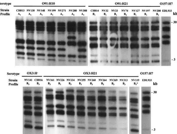
Ribotyping profiles of the STEC isolates.Twenty-two STEC isolates, obtained from cattle and food samples between No-vember 1997 and September 1998 in central France, were analyzed by ribotyping with the rrnB7 probe and were com-pared with three isolates obtained from HUS patients in the same area. All of the isolates belonged to serotype O91:H10, O91:H21, OX3:H⫺, or OX3:H21. Between 8 and 10 fragments were obtained with HindIII-digested genomic DNA, and 10 to 18 fragments were obtained with EcoRI; the fragments ranged from 1 to 30 kb long. Figure 1 shows fragment profiles gener-ated from the isolates with restriction enzyme HindIII. Each strain was assigned a ribotype pattern; a letter indicated the group, and a subscript number indicated minor differences (one or two bands) within a group. When HindIII was used, the isolates were classified into two major groups (groups A and B) comprising six different ribotypes designated ribotypes A1 to A2 and B1 to B4. When the EcoRI enzyme was used, four major groups (groups C to F) comprising five ribotypes were identified for the 25 STEC isolates. Figure 2 shows represen-tative patterns generated with the HindIII and EcoRI enzymes.
Six and four bands were common to all of the strains for the
HindIII and EcoRI profiles, respectively. The profiles obtained
for O157:H7 strain EDL933 differed from the non-O157 STEC profiles by at least three bands.
FIG. 1. Ribotype patterns generated with HindIII-digested genomic DNA. The positions of molecular size standards are indicated on the right. The strains and patterns are shown in Table 2. One additional 1.5-kb fragment could not be shown in pattern B4.
FIG. 2. Representative ribotype patterns obtained for the STEC strains. For each pattern the letter indicates the group and the sub-script number indicates minor differences within a group. The posi-tions of molecular size standards are indicated on the left. (A) Six ribotype patterns obtained with HindIII (patterns A1to A2and B1to B4) for the 25 STEC analyzed in this study compared to the pattern of O157:H7. (B) Five ribotype patterns obtained with EcoRI (patterns C, D1 to D2, E, and F) compared to the pattern of O157:H7. The O157:H7 profile was obtained with the EDL933 reference strain.
on October 18, 2017 by INRA-MME CORNIC
http://aem.asm.org/
When the HindIII and EcoRI ribotypes were combined (Ta-ble 2), eight distinct patterns were identified. Two similar pat-terns were distinguished for the O91:H10 STEC isolates. The NV280 isolate, which was recovered from a bovine sample, produced the A2-C pattern, and the six other O91:H10 isolates (one, CH013, recovered from a patient suffering from HUS and five from bovine samples) produced identical A1-C pat-terns. A major B1-D1pattern was identified for six of the seven O91:H21 isolates (one from a patient with HUS, two from beef samples, and three from bovine feces). The 091:H21 isolate obtained from cheese, which had a specific stx1-stx2-ehxA viru-lence gene profile, had a distinct B2-E pattern. The predomi-nant B3-D2pattern of five of the nine OX3:H21 isolates was the same as that of the OX3:H⫺isolates, which suggests that the two serotypes are closely related. In contrast, 13 OX3:H2 strains had ribotype profiles that were completely distinct from those of the OX3:H⫺strains (data not shown), which indicates a more distant relationship. Because they were clearly different from the HUS-associated OX3:H⫺isolate, the four remaining OX3:H21 and the OX3:H2 strains were not used for further analysis. Taken together, these data indicate that HindIII and
EcoRI ribotyping could reveal only minor differences in
non-O157:H7 strains belonging to the same serotype.
Toxin gene RFLP patterns of STEC isolates. To establish more discriminative clonal relatedness among STEC isolates, the stx2-RFLP method was used. This method has been used
for molecular epidemiological investigations of STEC and has been shown to be sufficiently sensitive to allow interstrain dif-ferentiation of STEC belonging to the same serotype (33). For the RFLP procedure we used the EcoRI and HindIII restric-tion enzymes, which do not have a restricrestric-tion site in the stx2 toxin gene and allow detection of restriction site polymorphism near the toxin genes. Hybridization of digested genomic DNA with the stx2 probe was performed for 21 isolates, including 16 isolates belonging to serotypes O91:H10, O91:H21, and OX3:H⫺ and the 5 OX3:H21 isolates which produced the B3-D2ribotype pattern (Table 3). Ten and 11 DNA band size profiles were obtained with HindIII- and EcoRI-digested genomic DNA, respectively. Between one and three fragments of different sizes (ranging from approximately 3.5 to 30 kb for
HindIII and from 4.5 to 15 kb for EcoRI) were obtained for
STEC isolates with each of the restriction enzymes. For the 21
E. coli isolates, the combined data resulted in 15 distinct RFLP
patterns.
Most of the O91:H10 strains produced stx2 fragments of different sizes; the only exceptions were two cattle isolates (NV148 and NV271), which produced the same pattern (two bands at approximately 26 and 12 kb with HindIII and EcoRI, respectively). The seven O91:H21 strains produced three dis-tinct RFLP patterns; three isolates from food (NV32, NV33, and NV74) and one isolate from cattle (NV200) produced the same pattern (one band at 5.5 kb with HindIII and one band at 4.5-kb with EcoRI), one bovine isolate (NV197) produced a distinct pattern (three bands at 20, 9, and 5.5 kb and three bands at 12, 5, and 4.5 kb with HindIII and EcoRI, respective-ly), and the pattern produced by isolate NV127 was indistin-guishable from that produced by O91:H21 HUS isolate CH014 (26- and 20-kb HindIII bands and 10- and 7.5-kb EcoRI bands). In contrast, the RFLP patterns of the two OX3:H⫺ isolates, one from a HUS patient and one from cattle feces, differed from each other and from those of the OX3:H21 isolates.
Analysis of the stx2 gene variants. To further assess the
diversity of non-O157:H7 STEC, the stx2gene types present in each isolate were identified by specific PCR methods. The results of this analysis are summarized in Table 3. The 21 isolates tested were found to contain stx2, stx2vh-a, or stx2vh-b, but none was found to contain stx2dor stx2e. The strains con-tained either one stx2 gene type or a combination of two or three. The serotypes were not linked to a specific stx genotype. Three of the seven O91:H10 isolates contained only the
stx2vh-atarget sequence, one contained only the stx2vh-bgene, and two contained both stx2vh-a and stx2vh-b genes. Interest-ingly, the isolate from a HUS patient, CH013, contained a combination of the three sequences. Among the O91:H21 STEC, the three isolates obtained from food samples con-tained only the stx2vh-b gene. Two isolates from cattle con-tained the stx2and stx2vh-bsequences, while the other bovine isolate contained stx2vh-a and stx2vh-b. The O91:H21 isolate obtained from a HUS patient contained the three variants. When the OX3:H⫺and OX3:H21 serotypes were examined, only one stx2gene type was found for each isolate. One OX3:H isolate from a HUS patient and two OX3:H21 isolates from cattle contained only stx2vh-a, while the other OX3:H⫺isolate and three OX3:H21 isolates, all obtained from cattle, con-tained stx2vh-b. No correlation was found between stx2variant
TABLE 2. Origins, virulence gene profiles, and ribotype patterns of the 25 STEC strains, sorted according to serotype
Serotype Strain Origina Virulence
gene profile Ribotype patternb HindIII EcoRI O91:H10 CH013 HUS stx2 A1 C NV130 Cattle stx2 A1 C NV148 Cattle stx2 A1 C NV199 Cattle stx2 A1 C NV271 Cattle stx2 A1 C NV308 Cattle stx2 A1 C NV280 Cattle stx2 A2 C
O91:H21 CH014 HUS stx2, ehxA B1 D1
NV127 Cattle stx2, ehxA B1 D1 NV197 Cattle stx2, ehxA B1 D1 NV200 Cattle stx2, ehxA B1 D1 NV32 Beef stx2, ehxA B1 D1 NV33 Beef stx2, ehxA B1 D1 NV74 Cheese stx1, stx2, ehxA B2 E OX3:H⫺ CH016 HUS stx 2 B3 D2 NV141 Cattle stx2 B3 D2 OX3:H21 NV234 Cattle stx2 B3 D2 NV255 Cattle stx2 B3 D2 NV260 Cattle stx2 B3 D2 NV269 Cattle stx2 B3 D2 NV313 Cattle stx2 B3 D2 NV161 Cattle stx2 B3 F NV264 Cattle stx2 B3 F NV226 Cattle stx2 B1 F NV119 Cattle stx2 B4 F
aHUS, isolate obtained from HUS patient; cattle, isolate obtained from
bo-vine feces; beef, isolate obtained from beef sample; cheese, isolate obtained from cheese sample.
bThe letters differentiate groups of isolates and the subscript numbers indicate
minor differences between isolates belonging to the same group.
VOL. 67, 2001 STEC CHARACTERIZATION BY MOLECULAR SUBTYPING 2463
on October 18, 2017 by INRA-MME CORNIC
http://aem.asm.org/
type and stx2-RFLP band size, serotype, or origin of the STEC strains.
Plasmid studies.Since analysis of plasmids was found to be helpful for studying the variability of non-O157:H7 STEC (42), we investigated the presence of plasmids in the 21 test strains.
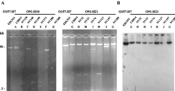
Plasmid pO157, the 90-kb virulence plasmid of strain EDL933, an isolate from an outbreak in Michigan that occurred in 1982, was used as a control. The plasmid profiles obtained for the O91:H10 and O91:H21 isolates are shown in Fig. 3A. All isolates carried one to four plasmids whose sizes ranged from
FIG. 3. Representative plasmid profiles of the STEC strains. (A) Plasmid DNA of the O91:H10 and O91:H21 STEC strains. The profiles of OX3 isolates are not shown; these isolates produced plasmid profile G similar to the profile of O91:H21 strain CH014. The positions of molecular size standards are indicated on the left. (B) Southern blot hybridization of plasmid DNA of the O91:H21 strains with a specific ehxA probe. The strains and profiles are described in Table 3.
TABLE 3. Comparison of ribotype patterns stx2-RFLP profiles, stx2types and plasmid profiles of the STEC strains
Serotype Strain Origina Ribotype
patternb
stx2-RFLP profilec
stx2typed Plasmidprofilee
HindIII EcoRI
O91:H10 CH013 HUS A1-C 26, 7 10, 7.5 2, 2vh-a, 2vh-b A
NV148 Cattle A1-C 26 12 2vh-a C NV271 Cattle A1-C 26 12 2vh-a E NV308 Cattle A1-C 26 15 2vh-b D NV130 Cattle A1-C 26, 20 15, 10 2vh-a, 2vh-b B NV199 Cattle A1-C 26, 20 15, 12 2vh-a, 2vh-b D NV280 Cattle A2-C 26, 11 12 2vh-a F
O91:H21 CH014 HUS B1-D1 26, 20 10, 7.5 2, 2vh-a, 2vh-b G
NV127 Cattle B1-D1 26, 20 10, 7.5 2vh-a, 2vh-b H NV32 Beef B1-D1 5.5 4.5 2vh-b H NV33 Beef B1-D1 5.5 4.5 2vh-b H NV200 Cattle B1-D1 5.5 4.5 2, 2vh-b G NV197 Cattle B1-D1 20, 9, 5.5 12, 5, 4.5 2, 2vh-b J NV74 Cheese B2-E 5.5 4.5 2vh-b I OX3:H⫺ CH016 HUS B 3-D2 12 15 2vh-a G NV141 Cattle B3-D2 30, 26 12, 4.5 2vh-b G OX3:H21 NV234 Cattle B3-D2 3.5 13 2vh-b G NV260 Cattle B3-D2 3.5 13 2vh-a G NV255 Cattle B3-D2 3.5 12 2vh-b G NV313 Cattle B3-D2 4, 3.5 12, 4.5 2vh-b G NV269 Cattle B3-D2 5.5 13 2vh-a G
aHUS, isolate obtained from HUS patient; cattle, isolate obtained from bovine feces; beef, isolate obtained from beef sample; cheese, isolate obtained from cheese
sample.
bThe letters differentiate groups of isolates, and the subscript numbers indicate minor differences between isolates of the same group. cThe numbers indicate the sizes (in kilobases) of the different stx
2fragments obtained.
d2, stx
2; 2vh-a, stx2vh-a; 2vh-b, stx2vh-b
eThe letters differentiate isolates on the basis of the number and size of the plasmids.
on October 18, 2017 by INRA-MME CORNIC
http://aem.asm.org/
approximately 3 to more than 90 kb. Ten different plasmid profiles, profiles A to J, were obtained for the 21 isolates tested (Table 3). Endonuclease restriction analysis of the plasmid content with EcoRI showed that strains with the same plasmid profile had the same cleavage pattern (data not shown).
Six plasmid profiles, profiles A to F, were obtained for the seven O91:H10 E. coli strains. Only two isolates from cattle (NV199 and NV308) produced the same profile, profile D. For the O91:H21 strains, we identified four distinct plasmid pat-terns. All of the strains contained at least one large plasmid (more than 50 kb long), and sometimes the strains contained additional small plasmids (Fig. 3A). Profile G was obtained for the CH014 HUS strain and one bovine isolate (NV200), profile H was obtained for one bovine isolate (NV127) and two beef isolates (NV32 and NV33), profile I was characteristic of the NV74 cheese isolate, and profile J was characteristic of O91: H21 isolate NV197. In contrast, the two OX3:H⫺ and five OX3:H21 E. coli strains produced the same plasmid profile, profile G, which was further evidence of the close relationship of these strains.
Analysis of plasmid-encoded determinants.Of all the strains analyzed in this study, only the serotype O91:H21 strains con-tained the ehxA gene (Table 2) (30), which was originally found on large plasmid pO157 of O157:H7 E. coli strains (7). All seven O91:H21 strains contained at least one such large plas-mid (Fig. 3A), which was hybridized with a probe complemen-tary to the ehxA sequence. The ehxA gene was present on one of the large plasmids in all of the O91:H21 STEC strains (Fig. 3B). Since the espP gene is also known to be located on pO157, we checked for its presence by performing colony blot ization with an espP-specific probe. The probe did not hybrid-ize with any of the O91:H21 STEC strains. These results
un-derline the heterogeneity of the STEC plasmids. Most strains had a unique combination of characteristics; the only excep-tions were two O91:H21 beef isolates (NV32 and NV33), which were identical (Table 3).
PFGE analysis.Numerous reports have shown that PFGE typing is a highly discriminatory and reproducible method (2, 6, 26, 32, 38), and we decided to compare this method with ribotyping and the molecular techniques used in this study. To determine clonal relatedness among human, bovine, and food isolates belonging to the same serotypes (O91:H10, O91:H21, OX3:H⫺, and OX3:H21), we analyzed their XbaI patterns by PFGE. However, several efforts to obtain intact DNA from the OX3 isolates were unsuccessful, and DNA degradation was always observed, which prevented pattern analysis. Thus, 14 O91 isolates (7 O91:H10 and 7 O91:H21 isolates) and two O157:H7 strains, used as controls, were analyzed (Fig. 4). PFGE produced 11 to 19 fragments ranging in size from ap-proximately 50 to 580 kb. A dendrogram analysis of PFGE patterns (Fig. 4) revealed that the isolates were distributed among two main branches of the phylogenetic tree. One clus-ter included all the O91:H21 isolates except the NV74 strain from cheese. In this cluster, the PFGE patterns of strains from cattle and beef were more than 75% similar whereas the HUS isolate was more distantly related (63% similarity). Only two O91:H21 beef isolates (NV32 and NV33) produced closely related restriction patterns, and there was just one band dif-ference (38). O91:H21 cattle isolate NV200 may be related to isolates NV32 and NV33 since there were only five and four band differences, respectively, which is consistent with two independent genetic events (38). The other O91:H21 isolates were not genetically related according to the criteria of Teno-FIG. 4. Dendrogram showing the clonal relationships of bovine, food, and HUS non-O157:H7 isolates, as determined by macrorestriction fragment analysis by PFGE, after digestion of genomic DNA by XbaI. The tree was constructed by using the unweighted pair group arithmetic average method. Strain characteristics are described in Table 3.
VOL. 67, 2001 STEC CHARACTERIZATION BY MOLECULAR SUBTYPING 2465
on October 18, 2017 by INRA-MME CORNIC
http://aem.asm.org/
ver et al. (38), since their PFGE patterns had more than five band differences.
Two main subgroups were identified in the second cluster. The first subgroup included O91:H10 isolates from cattle and patients with HUS, which were more than 76% similar. Inter-estingly, HUS isolate CH013 and cattle isolates NV148 and NV271 may be related since there were only three and five band differences, respectively. The second subgroup contained the two O157:H7 isolates included in this study (cattle isolate NV95 and reference strain EDL933) and one O91:H10 isolate from cattle, which were more than 78% similar. The O91:H21 isolate from cheese (NV74) was distantly related to the second cluster, with only 63.7% similarity.
DISCUSSION
In this study, we used a collection of 25 STEC isolates from sporadic cases of HUS, cattle, and food products, obtained in the same geographical area over a short period of time, to perform a detailed analysis of the molecular epidemiology of non-O157:H7 STEC. We compared different typing methods; some of these methods were used to explore potentially mobile elements like the stx genes or plasmids (stx2-RFLP analysis, stx2 gene variant analysis, and plasmid analysis), and others were used to study the whole genome (ribotyping and PFGE).
Identical or very similar ribotype profiles were obtained for members of each serotype. Using two enzymes, we identified eight different profiles for 25 isolates belonging to four sero-types. A total of 19 of 25 isolates could not be differentiated, including six of seven O91:H10 isolates, six of seven O91:H21 isolates, and 7 of 11 OX3:H21/H⫺isolates. This indicates that ribotyping, which can be a very useful tool for epidemiologi-cal investigation, was not able to discriminate between STEC isolates belonging to the same serotype. We therefore used
stx2-RFLP analysis, a sensitive and specific method for differ-entiation of STEC strains, which has also been used in epide-miological investigations of outbreaks and sporadic cases of infection (16, 33). With this technique, 10 of 21 isolates could not be differentiated; two O91:H10 isolates had identical pro-files, as did two O91:H21 isolates, four other O91:H21 isolates and two OX3:H21 isolates (Table 3). When this technique was combined with stx2variant analysis, only 5 of 21 isolates could not be differentiated (O91:H10 isolates NV148 and NV271 and O91:H21 isolates NV32, NV33, and NV74 [NV74 produced a distinct ribotype pattern]). Plasmid analysis revealed further heterogeneity of the STEC isolates, except for those belonging to the OX3 serogroup (all of which harbored a⬎90-kb plas-mid). Taken together, the data obtained with the stx2-RFLP,
stx2variant, and plasmid profile analyses were very discrimina-tory, since only 2 of 21 isolates (the two O91:H21 beef isolates, NV32 and NV33) could not be differentiated. We then com-pared the results with those obtained by PFGE. This technique is probably the most powerful tool available for strain differ-entiation and has been used for a broad range of bacterial pathogens (4, 17, 38). It has also been used in investigations of several outbreaks of E. coli O157 infection and has been shown to discriminate between strains belonging to the same ribotype (2, 6, 26, 31, 32). In our study, PFGE had the greatest discrim-inatory power for non-O157:H7 STEC isolates, since each iso-late produced a unique PFGE pattern. Interestingly, NV32 and
NV33, the two O91:H21 beef isolates that could not be distin-guished by the stx2and plasmid analyses, were shown to be the most closely related isolates by PFGE, with⬎96% homology, and one of these isolates may have been derived from the other by a single genetic event (38). They were isolated from two beef samples purchased from the same butcher’s shop 2 days apart and are probably subtypes of the same strain, which confirms the discriminatory power of PFGE. Overall, our re-sults indicate that the combination of stx2-RFLP, stx2variant, and plasmid profile analyses was as powerful as PFGE for molecular investigation of STEC diversity.
Sequences that are homologous to the stx2 probe are as-sumed to possess neither EcoRI nor HindIII restriction sites; hence, the smallest number of stx2 genes in the genome was estimated for each strain. Since the HUS-associated strains were more cytotoxic than their bovine counterparts that be-longed to the same serotype (30), we tried to establish the relationship between the number of stx2genes and stx2variants and cytotoxicity. We were unable to establish any correlation between the number of stx2 genes and cytotoxicity for Vero cells, which was determined in a previous study, except for the two HUS-associated isolates, CH013 and CH014, both of which contained a combination of three variants and were the most cytotoxic isolates among the 25 isolates tested. Several factors may contribute to the degree of cytotoxicity of STEC strains, including not only the number of stx2genes present in the genome but also the regulation of expression of these genes, which are carried by the genomes of different phages (40). Further studies at the RNA and DNA levels involving sequence analysis of the regulatory stx2-flanking regions will be necessary to compare the STEC isolates. Most serotypes were not linked to a specific stx genotype, and different combina-tions of stx genes were found in strains belonging to the same serotype. No correlation could be established between the stx2 variant and stx2-RFLP band size or any other characteristic of the strains (serotype, origin, or level of cytotoxicity for Vero cells). Finally, the number of EcoRI and HindIII fragments that hybridized with the stx2gene probe was greater than the number of stx2 variants in three strains (O91:H21 isolate NV197 and OX3 isolates NV141 and NV313 [Table 3]), which suggests that several copies of the stx2gene may be present.
As STEC isolates belonging to different serotypes often har-bor one or more small or large plasmids, plasmid profile anal-ysis was used in this study as a typing method. The plasmid profiles of the O91:H10 and O91:H21 serotypes varied widely (nine patterns were identified for 14 isolates). The differences were not correlated with the origin, with the serotype, or with other molecular characteristics of the strains. The presence of an approximately 90-kb plasmid carrying several genes encod-ing functions associated with virulence is a characteristic at-tribute of many STEC strains (8). All of our strains carried similarly large plasmids, but the plasmids were heterogeneous in terms of their virulence gene contents. The variations were serotype related. All of the O91:H21 strains contained large plasmids encoding the ehxA gene but not the espP gene. None of the O91:H10 or OX3:H⫺large plasmids contained the ehxA or espP gene. In contrast, 90-kb plasmid pO157 of strain EDL933 (O157:H7) contained both genes (14). Other studies have shown that STEC isolates have different plasmid contents based on serotype (12, 35), and our data confirmed the high
on October 18, 2017 by INRA-MME CORNIC
http://aem.asm.org/
level of plasmid diversity among STEC isolates, even within a single serotype.
One of the aims of our study was to determine whether the non-O157:H7 STEC strains isolated from HUS patients were related to those isolated from cattle and food samples in the same geographical area. A close relationship among strains of human, animal, and food origins was suggested by previous serotyping and virulence factor pattern analyses. Most of the strains belonging to the same serotype produced identical ri-botype patterns, but both PFGE and the combination of stx2 -RFLP, stx2 variant, plasmid profile analyses showed that the strains were clearly different. HUS-associated isolate CH014 was distantly related to the O91:H21 isolates from cattle and food, with only 63.3% similarity as determined by PFGE, al-though it differed from bovine isolate NV127 only by the stx2 variants and by the absence of a small plasmid (Table 3 and Fig. 3). The other HUS-associated isolate, CH013, was closely related to O91:H10 cattle isolate NV148 as determined by PFGE (88% similarity), indicating that cattle may be a reser-voir for pathogenic STEC in France. However, CH013 differed from NV148 by additional stx2 and stx2vh-b genes, stx2-RFLP pattern, and plasmids, suggesting that such pathogenic strains may have evolved from bovine strains by acquisition of addi-tional virulence factors. Finally, the data obtained by examin-ing mobile elements (stx2-RFLP, stx2variant, and plasmid anal-yses) were found to be complementary to those provided by PFGE.
With regard to clonal relatedness, the two OX3:H⫺strains (one from a HUS patient and one from cattle) and five of the nine OX3:H21 isolates had similar ribotypes and identical plas-mid profiles. In contrast, 13 OX3:H2 STEC strains obtained from bovine and food sources during the same survey (30) had different virulence profiles and different ribotype patterns (data not shown). We presumed that the OX3:H⫺strain obtained from a HUS patient was a nonmotile variant of an OX3:H21 strain. OX3:H21 strains have been associated previously with severe human diseases (22). The HUS OX3:H⫺isolate differed from its OX3:H⫺and OX3:H21 environmental counterparts only by the stx2type and stx2-RFLP profile. However, we were not able to further explore the clonal relatedness between these OX3 isolates by PFGE since DNA degradation made typing impos-sible. The high nuclease activity observed in the OX3 isolates, by limiting the spread of exogenous plasmids, could account for the fact that only one plasmid profile was obtained.
Several studies have shown that there is significant genomic diversity and great variability in virulence among O157:H7 isolates that have the same known virulence determinants (5, 24). A comparison of human and bovine isolates demonstrated that there are two distinct lineages of E. coli O157:H7 and suggested that one of these lineages may be less virulent for humans or may not be efficiently transmitted to humans from bovine sources (24). We postulated that such pathogenic lin-eages might exist for non-O157:H7 STEC. However, we could not identify any characteristic that was specific to HUS-asso-ciated isolates. Further studies are needed to identify the spe-cific properties of the pathogenic clones that distinguish them from their nonpathogenic counterparts.
In conclusion, several powerful molecular techniques showed that there was great diversity among non-O157:H7 STEC strains isolated in central France. The close relationship among
strains of human, animal, or food origin belonging to serotypes O91:H10, O91:H21, OX3:H⫺, and OX3:H21 was initially sug-gested by serotyping and virulence factor pattern analysis. Most of the strains belonging to the same serotype produced identical ribotype patterns but unique typing patterns when they were subjected to PFGE and analysis of mobile elements. The non-O157:H7 STEC strains isolated from HUS patients were related to, but not identical to, those isolated from cattle and food samples in the same geographical area. Overall, our findings indicate that the combination of stx2-RFLP, stx2 vari-ant, and plasmid profile analyses is as powerful as PFGE for molecular investigation of STEC diversity.
ACKNOWLEDGMENTS
We thank C. P. Vivares from the Laboratoire Parasitologie Mole´cu-laire et CelluMole´cu-laire, UMR CNRS 6023, Universite´ Blaise Pascal, for providing the Gene Navigator apparatus and advice concerning PFGE experiments. We also thank Liliane Millet from the Laboratoire UR545 Recherche Fromage`res INRA Aurillac, for her help with the profile comparison in which the GelCompar software was used and Katy Durieux for technical assistance with ribotyping.
This study was supported in part by the Ministe`re de l’Enseignement supe´rieur et de la Recherche (grant EA2148) and by the Ministe`re de l’Ame´nagement du Territoire et de l’Environnement (Programme En-vironnement Sante´ EN 98-17).
REFERENCES
1. Allison, L., A. Stirrat, and F. M. Thomson-Carter. 1998. Genetic heteroge-neity of Escherichia coli O157:H7 in Scotland and its utility in strain subtyp-ing. Eur. J. Clin. Microbiol. Infect. Dis. 17:844–848.
2. Allison, L. J., P. E. Carter, and F. M. Thomson-Carter. 2000. Characteriza-tion of a recurrent clonal type of Escherichia coli O157:H7 causing major outbreaks of infection in Scotland. J. Clin. Microbiol. 38:1632–1635. 3. Alos, J.-I., T. Lambert, and P. Courvalin. 1993. Comparison of two
molec-ular methods for tracing nosocomial transmission of Escherichia coli K1 in a neonatal unit. J. Clin. Microbiol. 31:1704–1709.
4. Arbeit, R. D., M. Arthur, R. Dunn, C. Kim, R. K. Selander, and R. Goldstein. 1990. Resolution of recent evolutionary divergence among Escherichia coli from related lineages: the application of pulsed field electrophoresis to molecular epidemiology. J. Infect. Dis. 161:230–235.
5. Baker, D. R., R. A. Moxley, and D. H. Francis. 1997. Variation in virulence in the gnotobiotic pig model of O157:H7 Escherichia coli strains of bovine and human origin. Adv. Exp. Med. Biol. 412:53–58.
6. Barrett, T. J., H. Lior, J. H. Green, R. Khakhira, J. G. Wells, B. P. Bell, K. D.
Greene, J. Lewis, and P. M. Griffin.1994. Laboratory investigation of a multistate food-borne outbreak of Escherichia coli O157:H7 by using pulsed-field gel electrophoresis and phage typing. J. Clin. Microbiol. 32:3013–3017. 7. Bauer, M., and R. A. Welch. 1996. Characterization of an RTX toxin from enterohemorrhagic Escherichia coli O157:H7. Infect. Immun. 64:167–175. 8. Beutin, L., D. Geier, S. Zimmermann, and H. Karch. 1995. Virulence
mark-ers of Shiga-like toxin-producing Escherichia coli strains originating from healthy domestic animals of different species. J. Clin. Microbiol. 33:631–635. 9. Bohm, H., and H. Karch. 1992. DNA fingerprinting of Escherichia coli O157:H7 strains by pulsed-field gel electrophoresis. J. Clin. Microbiol. 30: 2169–2172.
10. Bonnet, R., B. Souweine, G. Gauthier, C. Rich, V. Livrelli, J. Sirot, B. Joly,
and C. Forestier.1998. Non-O157 Stx2-producing Escherichia coli strains associated with sporadic cases of hemolytic-uremic syndrome in adults. J. Clin. Microbiol. 36:1777–1780.
11. Brosius, J., A. Ullrich, M. A. Raker, A. Gray, T. J. Dull, R. R. Gutell, and
H. F. Noller.1981. Construction and fine mapping of recombinant plasmids containing the rrnB ribosomal RNA operon of E. coli. Plasmid 6:112–118. 12. Brunder, W., H. Schmidt, M. Frosch, and H. Karch. 1999. The large
plas-mids of Shiga-toxin-producing Escherichia coli (STEC) are highly variable genetic elements. Microbiology 145:1005–1014.
13. Brunder, W., H. Schmidt, and H. Karch. 1997. EspP, a novel extracellular serine protease of enterohaemorrhagic Escherichia coli O157:H7, cleaves human coagulation factor V. Mol. Microbiol. 24:767–778.
14. Burland, V., Y. Shao, N. T. Perna, G. Plunkett, H. J. Sofia, and F. R.
Blattner. 1998. The complete DNA sequence and analysis of the large virulence plasmid of Escherichia coli O157:H7. Nucleic Acids Res. 26:4196– 4204.
15. Cebula, T. A., W. L. Payne, and P. Feng. 1995. Simultaneous identification of strains of Escherichia coli serotype O157:H7 and their Shiga-like toxin type
VOL. 67, 2001 STEC CHARACTERIZATION BY MOLECULAR SUBTYPING 2467
on October 18, 2017 by INRA-MME CORNIC
http://aem.asm.org/
by mismatch amplification mutation assay-multiplex PCR. J. Clin. Microbiol.
33:248–250.
16. Grimm, L. M., M. Goldoft, J. Kobayashi, J. H. Lewis, D. Alfi, A. M.
Perdi-chizzi, P. I. Tarr, J. E. Ongerth, S. L. Moseley, and M. Samadpour.1995. Molecular epidemiology of a fast-food restaurant-associated outbreak of
Escherichia coli O157:H7 in Washington State. J. Clin. Microbiol. 33:2155–
2158.
17. Grundmann, H., C. Schneider, D. Hartung, F. D. Daschner, and T. L. Pitt. 1995. Discriminatory power of three DNA-based typing techniques for
Pseudomonas aeruginosa. J. Clin. Microbiol. 33:528–534.
18. Hall, R. H., and J. G. Xu. December 1995. A new and distinctive DNA sequence of E. coli O157:H7 and its use for the rapid, sensitive and specific detection of O157:H7 and other enterohemorrhagic E. coli. International patent WO 95/34682.
19. Johnson, W. M., D. R. Pollard, H. Lior, S. D. Tyler, and K. R. Rozee. 1990. Differentiation of genes coding for Escherichia coli verotoxin 2 and the verotoxin associated with porcine edema disease (VTe) by the polymerase chain reaction. J. Clin. Microbiol. 28:2351–2353.
20. Kado, C. I., and S. T. Liu. 1981. Rapid procedure for detection and isolation of large and small plasmids. J. Bacteriol. 145:1365–1373.
21. Karmali, M. A., M. Petric, C. Lim, P. C. Fleming, G. S. Arbus, and H. Lior. 1985. The association between idiopathic hemolytic-uremic syndrome and infection by verotoxin-producing Escherichia coli. J. Infect. Dis. 151:775–782. 22. Keskima¨ki, M., R. Ika¨heimo, P. Ka¨rkka¨inen, F. Scheutz, Y. Ratiner, R.
Puohiniemi, and A. Siitonen.1997. Shiga toxin-producing Escherichia coli serotype OX3:H21 as a cause of hemolytic-uremic syndrome. Clin. Infect. Dis. 24:1278–1279.
23. Keskima¨ki, M., M. Saari, T. Heiskanen, and A. Siitonen. 1998. Shiga toxin-producing Escherichia coli in Finland from 1990 through 1997: prevalence and characteristics of isolates. J. Clin. Microbiol. 36:3641–3646.
24. Kim, J., J. Nietfeldt, and A. K. Benson. 1999. Octamer-based genome scan-ning distinguishes a unique subpopulation of Escherichia coli O157:H7 strains in cattle. Proc. Natl. Acad. Sci. USA 96:13288–13293.
25. Maas, R. 1983. An improved colony hybridization method with significantly increased sensitivity for detection of single genes. Plasmid 10:296–298. 26. Martin, I. E., S. D. Tyler, K. D. Tyler, R. Khakhira, and W. M. Johnson.
1996. Evaluation of ribotyping as epidemiologic tool for typing Escherichia
coli serogroup O157 isolates. J. Clin. Microbiol. 34:720–723.
27. Oelschlaeger, T. A., and J. Hacker. 1999. Pathogenicity islands and their role in virulence of bacteria, p. 73–85. In Verocytotoxigenic E. coli in Europe. The National Food Centre, Lie`ge, Belgium.
28. Picard-Pasquier, N., M. Ouagued, B. Picard, P. Goullet, and R.
Krish-namoorthy.1989. A simple, sensitive method of analyzing bacterial ribo-somal DNA polymorphism. Electrophoresis 10:186–189.
29. Pierard, D., G. Muyldermans, L. Moriau, D. Stevens, and S. Lauwers. 1998. Identification of new verocytotxin type 2 variant B-subunit genes in human and animal Escherichia coli isolates. J. Clin. Microbiol. 36:3317–3322.
30. Pradel, N., V. Livrelli, C. D. Champs, J.-B. Palcoux, A. Reynaud, F. Scheutz,
J. Sirot, B. Joly, and C. Forestier.2000. Prevalence and characterization of Shiga toxin-producing Escherichia coli isolated from cattle, food, and chil-dren during a one-year prospective study in France. J. Clin. Microbiol.
38:1023–1031.
31. Preston, M. A., W. Johnson, R. Khakhria, and A. Borczyk. 2000. Epidemi-ologic subtyping of Escherichia coli serogroup O157 strains isolated in On-tario by phage typing and pulsed-field gel electrophoresis. J. Clin. Microbiol.
38:2366–2368.
32. Rios, M., V. Prado, M. Trucksis, C. Arellano, and C. Borie. 1999. Clonal diversity of Chilean isolates of enterohemorrhagic Escherichia coli from patients with hemolytic-uremic syndrome, asymptomatic subjects, animal reservoirs, and food products. J. Clin. Microbiol. 37:778–781.
33. Samadpour, M. 1995. Molecular epidemiology of Escherichia coli O157:H7 by restriction fragment length polymorphism using Shiga-like toxin genes. J. Clin. Microbiol. 33:2150–2154.
34. Schmidt, H., L. Beutin, and H. Karch. 1995. Molecular analysis of the plasmid-encoded hemolysin of Escherichia coli O157:H7 strain EDL 933. Infect. Immun. 63:1055–1061.
35. Schmidt, H., C. Geitz, P. I. Tarr, M. Frosch, and H. Karch. 1999. Non-O157:H7 pathogenic Shiga toxin-producing Escherichia coli: phenotypic and genetic profiling of virulence traits and evidence for clonality. J. Infect. Dis.
179:115–123.
36. Schmidt, H., B. Ko¨hler, A. Unkmeir, M. Bielaszewska, and H. Karch. 1999. The role of Stx-encoding bacteriophages in pathogenicity and virulence of Shiga toxin-producing Escherichia coli (STEC), p. 11–17. In Verocytotoxi-genic E. coli in Europe. The National Food Centre. Lie`ge, Belgium. 37. Schmidt, H., J. Scheef, S. Morabito, A. Caprioli, L. H. Wieler, and H. Karch.
2000. A new Shiga toxin 2 variant (Stx2f) from Escherichia coli isolated from pigeons. Appl. Environ. Microbiol. 66:1205–1208.
38. Tenover, F. C., R. D. Arbeit, R. V. Goering, P. A. Mickelsen, B. E. Murray,
D. H. Persing, and B. Swaminathan.1995. Interpreting chromosomal DNA restriction patterns produced by pulsed-field gel electrophoresis: criteria for bacterial strain typing. J. Clin. Microbiol. 33:2233–2239.
39. Tyler, S. D., W. M. Johnson, H. Lior, G. Wang, and K. R. Rozee. 1991. Identification of verotoxin type 2 variant subunit B genes in Escherichia coli by the polymerase chain reaction and restriction fragment length polymor-phism analysis. J. Clin. Microbiol. 29:1339–1343.
40. Unkmeir, A., and H. Schmidt. 2000. Structural analysis of phage-borne stx genes and their flanking sequences in Shiga toxin-producing Escherichia coli and Shigella dysenteriae type 1 strains. Infect. Immun. 68:4856–4864. 41. Yu, J., and J. B. Kaper. 1992. Cloning and characterization of the eae gene
of enterohemorrhagic Escherichia coli O157:H7. Mol. Microbiol. 6:411–417. 42. Zhang, W.-L., M. Bielaszewska, A. Liesegang, H. Tscha¨pe, H. Schmidt, M.
Bitzan, and H. Karch.2000. Molecular characteristics and epidemiological significance of Shiga toxin-producing Escherichia coli O26 strains. J. Clin. Microbiol. 38:2134–2140.